Olfactory coding in vertebrates:
a novel tuning mechanism for receptor affinity and evolution of the olfactory receptor repertoire
Inaugural-Dissertation
zur
Erlangung des Doktorgrades
der Mathematisch-Naturwissenschaftlichen Fakultät
der Universität zu Köln
vorgelegt von
Kanika Sharma aus Jalandhar, India
Köln, 2018
2 Berichterstatter: Prof. Dr. Sigrun I. Korsching
Prof. Dr. Arnd Baumann
Tag der mündlichen Prüfung: 26.10.2018
3
ABSTRACT
Information about our environment is to a large extent carried by the chemical senses, and in particular the olfactory sense. Vertebrates perceive thousands of diverse odor molecules with a supply of a wide range of essential information ranging from localising prey or food, avoiding predators, mating behaviour, to social communication. Because olfactory receptor proteins play such an essential role in the specific recognition of diverse stimuli, understanding how they interact with and transduce their cognate ligands is a high priority. This constitutes one of the most complex ligand/receptor binding problems in biology due to the sheer quantity of potential odor molecules facing a limited albeit huge number of different olfactory receptors.
Most olfactory receptors are G-protein coupled receptors and form large gene families. One type of olfactory receptors is the trace amine-associated receptor family (TAAR). TAARs generally recognize amines and one particular member of the zebrafish TAAR family, TAAR13c, is a high affinity receptor for the death-associated odor cadaverine, which induces aversive behavior.
Here we have modeled the cadaverine/TAAR13c interaction by multistep docking. By exchanging predicted binding residues via site-directed mutagenesis, and measuring the activity of the mutant receptors, we confirmed a binding site for cadaverine at the external surface of the receptor, in addition to an internal binding site, whose mutation resulted in complete loss of activity. Elimination of the external binding site generated supersensitive receptors which suggests this site to act as a gate, limiting access of the ligand to the internal binding site and thereby downregulating the affinity of the native receptor. Potentially related mechanisms have been described for non-olfactory G-protein coupled receptors.
The topology of TAAR-expressing neurons in the teleost olfactory epithelium has not been described yet. We have investigated representative taar genes from three classes to test the principle of partial spatial segregation known from other olfactory receptor families for the TAAR family. We report that expression of taar genes is intermingled with expression zones of odorant receptor genes, which in fish share a single sensory surface with TAARs. Individual taar genes show distinct, albeit broadly overlapping expression zones.
In the third part of my thesis I investigated the genome of a cartilaginous fish,
Scyliorhinus canicula, commonly known as small spotted catshark in order to
delineate its chemosensory receptor repertoire: OR, V1R/V2R, TAAR, and
T1R/T2R. This is the first repertoire described for a true shark, an important
intermediate in the evolution of vertebrates. In contrast to bony vertebrates, but
very similar to a chimera (elephant shark), the olfactory receptor repertoire of
catshark is dominated by the V2R family.
4
Zusammenfassung
Informationen über unsere Umwelt werden zu einem großen Teil von den chemischen Sinnen getragen, insbesondere vom Geruchssinn. Wirbeltiere nehmen tausende verschiedene Geruchsmoleküle wahr, die eine breite Palette von essentiellen Informationen liefern, die von der Lokalisierung von Beutetieren, der Vermeidung von Raubtieren, dem Paarungsverhalten bis hin zur innerartlichen Kommunikation reichen. Da olfaktorische Rezeptorproteine eine so wichtige Rolle bei der spezifischen Erkennung verschiedener Stimuli spielen, hat das Verständnis, wie sie mit ihren verwandten Liganden interagieren eine hohe Priorität. Dies stellt eines der komplexesten Ligand / Rezeptor-Bindungsprobleme in der Biologie dar, und zwar aufgrund der schieren Menge potentieller Geruchsmoleküle, die einer begrenzten, wenn auch sehr großen Anzahl verschiedener Geruchsrezeptoren gegenüberstehen.
Die meisten Geruchsrezeptoren sind G-Protein-gekoppelte Rezeptoren und bilden große Genfamilien. Eine Art von Geruchsrezeptoren sind die Trace-Amin- assoziierten Rezeptoren (TAAR). TAARs erkennen im Allgemeinen Amine und ein spezielles Mitglied der Zebrafisch-TAAR-Familie, TAAR13c, ist ein hochaffiner Rezeptor für den Verwesungsgeruch Cadaverin, der ein aversives Verhalten hervorruft.
Hier haben wir die Cadaverin / TAAR13c-Interaktion durch mehrstufiges Docking modelliert. Durch zielgerichtete Mutagenese der vorhergesagten Bindungsstellen und Aktivitätsmessung der mutierten Rezeptoren bestätigten wir eine Bindungsstelle für Cadaverin an der äußeren Oberfläche des Rezeptors, zusätzlich zu einer internen Bindungsstelle, deren Mutation zu einem vollständigen Verlust der Aktivität führte.
Die Eliminierung der externen Bindungsstelle erzeugte supersensitive Rezeptoren, was darauf hindeutet, dass diese Stelle als ein Gate wirkt, welches den Zugang des Liganden zur inneren Bindungsstelle einschränkt und dadurch die Affinität des nativen Rezeptors herabreguliert. Potentiell ähnliche Mechanismen sind für nicht- olfaktorische G Protein-gekoppelte Rezeptoren beschrieben worden.
Die Topologie der TAAR-exprimierenden Neuronen im olfaktorischen Epithel des Zebrafisches wurde bisher nicht beschrieben. Wir haben repräsentative taar Gene aus 3 Klassen untersucht, um das von anderen olfaktorischen Genfamilien bekannte Prinzip der räumlichen Segregierung für die TAAR-Familie zu überprüfen.
Verschiedene taar Gene werden in breiten, aber klar voneinander verschiedenen Expressionszonen exprimiert, wobei die Taar-exprimierenden Neurone integriert sind in die Expressionszonen anderer Duftstoffrezeptorgene, da bei Fischen im Gegensatz zu Tetrapoden eine einzige sensorische Oberfläche vorliegt.
In einem dritten Teil meiner Arbeit untersuchte ich das Genom eines Knorpelfisches,
Scyliorhinus canicula, gemeinhin als Kleingefleckter Katzenhai bekannt, um das
Repertoire an chemosensorischen Rezeptoren (OR, V1R / V2R, TAAR und T1R /
T2R) zu identifizieren. Dies ist die erste derartige Untersuchung für einen echten Hai,
eine wichtige Zwischenstufe in der Evolution der Wirbeltiere. Im Gegensatz zu
Knochenfischen, aber ähnlich der Situation bei einer Chimäre (elephant shark) wird
das chemosensorische Genrepertoire der Haifische durch die V2R Familie dominiert.
Contents
1. Acknowledgement ... 7
2. Introduction... 9
2.1 General anatomy and function of the olfactory organ is conserved from fish to mammals……9
2.1.1 The rodent olfactory system ... 10
2.1.2 The teleost fish olfactory system ... 11
2.1.3 Insect olfactory system ... 11
2.2 Olfactory receptor gene family repertoire ... 12
2.2.1 Odorant Receptors (ORs)... 12
2.2.2 Trace Amine-Associated Receptor Family (TAARs) ... 13
2.2.3 Vomeronasal Receptors ... 14
2.2.4 Other receptor types ... 15
2.3 Olfactory receptor structure prediction ... 15
3. Aims of the study ... 17
4. Publications of the Dissertation ... 18
4.1.Elimination of a ligand gating site generates a supersensitive olfactory receptor ... 19
4.2.Full rescue of aninactive lfavory receptor mutant by elimination of an allosteric ligand-gating site ... 32
4.3. The Chemosensory Receptor Repertoire of a True Shark Is Dominated by a Single Olfactory Receptor Family ... 44
4.4. Manuscript in preparation ... 66
Spatial topology of zebrafish TAAR expressing neurons is broadly overlapping but distinct ... 67
4.4.1 Abstract ... 68
4.4.2 Background ... 68
4.4.3 Results and discussion ... 68
4.4.3.1 Relative Radius ... 70
4.4.3.2 Relative Height ... 72
4.4.4 Materials and Methods ... 74
5. Discussion ... 76
Evolution of olfactory receptors: from phylogeny to function ... 76
6. References... 81
7. Summary ... 87
8. APPENDIX ... 89
9. AUTHOR CONTRIBUTIONS ... 90
10. ERKLÄRUNG (DECLARATION) ... 91
11. Curriculum vitae... 92
6
For the love of science
and
My parents, who tried
7
1. Acknowledgement
At the beginning of the thesis, I would like to take some time to thank all the people without whom these projects would never have been possible. Although it is just my name on the cover, many people have contributed to the research in their own particular way and for that I want to give them special thanks.
First of all, I am deeply indebted to my adviser Prof. Dr. Sigrun Korsching for her fundamental role in my doctoral work. Thank you for believing in my abilities and being such a big inspiration. Sigrun provided me with every bit of guidance, assistance, and expertise that I needed during my first few months; then, when I felt ready to venture into research on my own and branch out into new research areas, she gave me the freedom to do whatever I wanted, at the same time continuing to contribute valuable feedback, advice, and encouragement. I cannot express enough gratitude towards her for having confidence in my abilities as someone from computational background wanting to learn wet biology. In addition to our academic collaboration, I greatly value the close personal rapport that Sigrun and I have developed over the years. I am always going to remember her “don’t do as I do, do as I say” phrase and all the examples of logical fallacies. I am also thankful for the excellent example she has provided as a successful woman biologist, an incredible professor and a great mentor. I quite simply cannot imagine a better adviser.
I would also like to thank Prof. Dr. Arnd Baumann for all the guidance and successful collaborations. Many thanks to Prof. Dr. Kay Hofmann and Prof. Dr. Arnd Baumann for accepting to be in my thesis committee, and for the interest in my work. Many thanks to Sabine for carrying out the work that required a lot of patience and skills.
Being a part of graduate school has been an incredible experience. I would like to extend a big thanks to Dr. Isabel for showing me the right way when I need to see and being an excellent advisor during the tough times. Also, Kathy for taking such good care of me during all these years. You were the first person I would run to for all complications I faced in Germany, and still do.
Because of the research environment sustained by Sigrun, I have crossed paths with
many graduate students and postdocs who have influenced and enhanced my
research. I am indebted to all the present and former Olfis for providing a wonderful
working environment. Thank you Ivan and Gaurav for introducing me to molecular
biology. It was endearing experience working with you both. Vladimir (soldier) who
was a great friend to share stories and experiences. Daniel has been a great co-
worker to talk to. Thank you for all the short and long conversations we had during
this time. You have always been keen to extend your help in whichever way possible
keeping the sense of humour intact. Thanks Milan for your scientific feedback during
our talks. Manish has been a great friend throughout this time in the lab and
probably the only one who listened to all my journal clubs with full attention. Thank
you Mehmet for always ordering equipment, kits and taking out time to look out for all
the solutions, chargers, screens and whatever I asked you for. Adnan has led me to
the current state of my entire work. He provided me the benefits of his own work
experience and during the most difficult times when writing this thesis and papers, he
gave me the moral support and the freedom I needed to move on. He has been my
8
support system throughout. I would also like to thank Venkatesh and Shahrzad for being great lab members. I had a great scientific group in PhD and I want to thank all of you for being incredible folks outside lab. Discussing science on lab retreats, learning skiing (thanks Daniel), cooking together and not to forget, theist vs. atheist conversations during retreats were incredible fun.
As long as I’m writing names down, I cannot neglect that of Heena, my best, most constant friend since high school. She’s provided me a lot of support and encouragement over the years; a great girl and I don’t want to miss an opportunity to get that in the permanent record.
All this has been possible because of my parents, who stood against everything to
see me here. Thank you ma for your unconditional love and support. Thank you papa
for being such a great role model. I can’t thank you enough for going through all the
hardships just to see us in better schools. I can’t even begin to thank you both for all
of your sacrifices as parents. Finally my sister, Kritika, who is everything to me, apart
from being a very annoying sibling.
9
2. Introduction
Chemoreception - the ability to perceive chemicals through adequate receptor molecules or mechanisms – enables a motile organism to move towards an energy source and is present from bacteria to higher eukaryotes. The ability to find and utilize energy sources is crucial for all living organisms. In humans, many times a given smell redirects us to a certain episode of our childhood. In the animal world the olfactory sense is one of the primary tools used to make sense of the environment.
Animals in their natural environment are surrounded by odors which are a rich source of information for mate choice, mother–infant recognition and signalling between members of a group. Potentially millions of structurally diverse odor molecules are perceived and discriminated by vertebrates which supply them with a wide range of information, ranging from predator and prey localization to mating behaviour, underlining the importance of the olfactory sense to the survival of the species.
The molecular understanding of olfaction reached a breakthrough in 1991 with the significant discovery of a large, multigene family of olfactory receptors in rat by Linda Buck and Richard Axel (Buck and Axel 1991) which was recognized in 2004 by the award of Nobel Prize in Physiology or Medicine. One of the major questions currently under investigation in the field concerns the evolution of olfactory receptors and their function. Novel techniques like next generation sequencing led to the publication of hundreds of genomes allowing large-scale studying of receptor evolution covering entire branches of the tree of life, including early-derived as well as evolutionary young species. This could lead us to understanding the evolutionary origin of olfaction as a specialized chemosensory sense.
2.1 General anatomy and function of the olfactory organ is conserved from fish to mammals
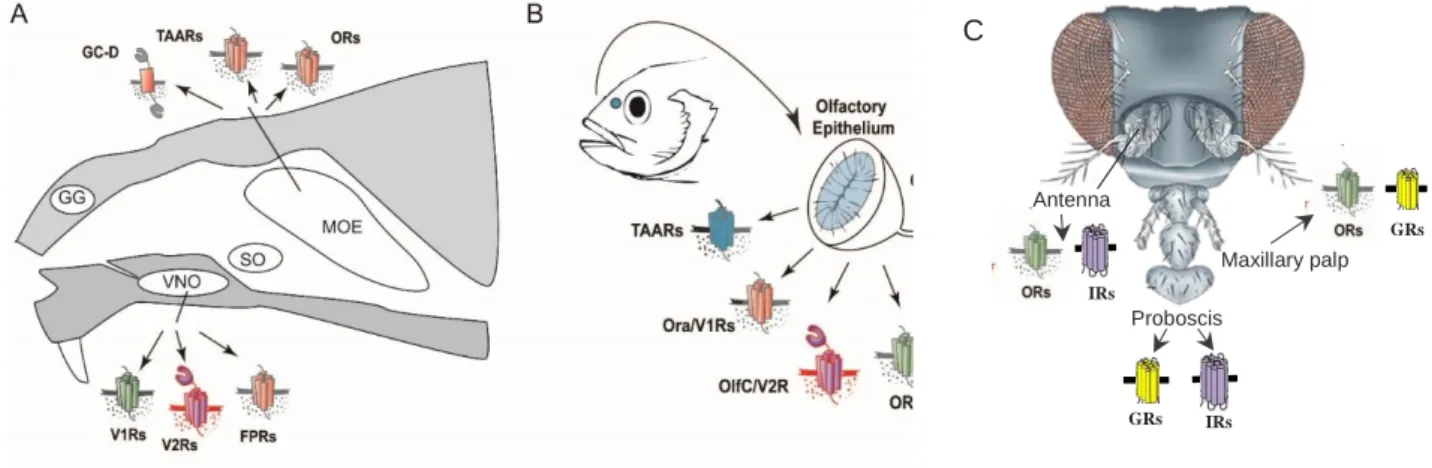
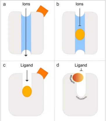
Chemosensory systems develop very early in evolution, as even bacteria can respond to chemical change. This type to reception is universal and found universally. Most olfactory stimuli do not consist of a single compound, but are complex mixtures of different active compounds. The smell of a rose for example is made up by a mixture of 275 different components (Ohloff, 1994). Considering the vast array of different chemicals, it is not surprising that different organisms use a large repertoire of distinct receptors, signalling pathways and anatomically segregated subsystems to sample their environment (Fig 1).
In both mammals as well as fish, the olfactory system harbours specialized sensory
neurons located in epithelial sensory surfaces. These neurons, referred to as
olfactory sensory neurons (OSNs), choose to express a single olfactory receptor,
from a variety of olfactory receptor families. The “one neuron - one receptor” rule
represents the first principle of organization in olfactory systems (Serizawa,
Miyamichi et al. 2004). This rule seems to be valid for mature OSNs of mouse
(Serizawa, Ishii et al. 2000, Serizawa, Miyamichi et al. 2004) as well as zebrafish
(Barth, Dugas et al. 1997) although there are reported exceptions (Mombaerts 2004,
Sato, Miyasaka et al. 2007, Hanchate, Kondoh et al. 2015). The total number of
different olfactory receptors expressed in an animal´s OSNs constitutes their receptor
10
repertoire. The axons of olfactory sensory neurons project to the olfactory bulb (OB), where axons from neurons expressing the same receptor converge onto glomeruli.
The principle of axonal convergence, “one receptor - one glomerulus”, represents the second principle of olfactory system organization. It has been described in the mammalian olfactory system (Takeuchi and Sakano 2014) as well as in fish (Ahuja, Ivandic et al. 2013, Ahuja and Korsching 2014). Odorants bind to their respective receptors, creating an action potential that is carried to the olfactory bulb glomeruli.
The glomeruli are innervated by specialized interneurons, i.e. mitral cells, and the signal is transported to higher brain areas, where odor evaluation takes place and adequate behavioral responses are generated.
The principal structure of the olfactory system with the basic building blocks (receptors, OSNs, glomeruli, and central nervous system) is conserved from insects to mammals. In mammals, reptiles, and amphibians the olfactory system underwent a segregation and comprises an additional olfactory surface, the vomeronasal organ (VNO). Sensory neurons in the VNO have different properties and primarily express receptors for the detection of pheromones (Karlson and Luscher 1959, Dulac and Axel 1995). The ontogenetic development of the additional olfactory subsystem has been described in an metamorphic amphibian (Xenopus laevis), whose olfactory receptor repertoires in the different subsystems also allow implications on the transition of the olfactory system from water to land living animals (Gliem, Syed et al.
2013, Syed, Sansone et al. 2017). In fish, which only have one olfactory organ, the olfactory epithelium, a partial segregation within the sensory surface can be observed. Pheromone and amino acid-sensing OSNs are present in the apical layer, while the OSNs harbouring the classical olfactory receptors and a family of amine sensing receptors are present in the basal layer of the epithelium. These cell populations also have distinct projection targets in the olfactory bulb (Sato, Miyasaka et al. 2005, Braubach, Fine et al. 2012).
2.1.1 The rodent olfactory system
The olfactory system functions as an elaborate molecular and cellular machinery for detection and discrimination of a vast number of chemical compounds in the environment (Dulac and Axel 1995). To manage these complex and diverse varieties of functions, rodents and tetrapods in general have a bipartite anatomically segregated olfactory system divided into a main olfactory epithelium (MOE) and a vomeronasal organ (VNO). Apart from these, rodents also possess two other subsystems i.e. the septal organ (SO) and the Grueneberg ganglion (GG) (Figure 1A) (Fleischer, Schwarzenbacher et al. 2006).
The MOE consists of ciliated neurons expressing an enormous repertoire of olfactory
receptors (odorant receptors, ORs) (Mombaerts, Wang et al. 1996). MOE also
expresses other receptors called trace amine-associated receptors (TAARs) (Zou
and Buck 2006), and membrane guanylyl cyclase receptor (GC-D) (Fulle, Vassar et
al. 1995, Lindemann, Ebeling et al. 2005). The VNO contains microvillous neurons
expressing two families of vomeronasal receptors (V1Rs and V2Rs) and formyl
peptide receptors (FPRs) believed to detect pheromones (Buck 2000, Riviere, Challet
et al. 2009) although it does not have a monopoly in this regard. The main olfactory
11
system can also mediate pheromone responses, for instance in the rabbit (Hudson and Distel 1986), pig (Dorries, Adkins-Regan et al. 1997) and mice (Keller, Douhard et al. 2006). Information from the VNO is transmitted to the accessory olfactory bulb (AOB), which further projects towards amygdala and hypothalamus that are involved in aggression and mating behavior (Hasen and Gammie 2009).
Figure 1: Schematic diagram of olfactory systems in moue, zebrafish and drosophila (A): Main olfactory epithelium (MOE) showing TAARs, Guanylyl cyclase-D and ORs receptors, Grueneberg ganglion (GG), vomeronasal organ (VNO) expressing V2Rs, V1Rs, and FPRs, septal organ of masera (SO). (B): Zebrafish olfactory system. Scheme showing olfactory epithelium expressing TAARs, ORA/V1Rs, OlfC/V2Rs and ORs. (A) (B) Modified from (Luis Saraiva doctoral thesis (Saraiva & Korsching 2007) (C) Modified from (Kaupp 2010)
2.1.2 The teleost fish olfactory system
In contrast to the mammalian olfactory system, teleosts have single sensory surface which called OE for olfactory epithelium (Figure 1B). In many teleost fish species, the OE is rosette-shaped, with an inner region containing sensory surface and non- sensory region on the periphery (Figure 1B). There are four types of OSNs present in the sensory region of the OE. These OSNs (ciliated, microvillous, crypt and kappe (only shown in zebrafish so far)) project their axons in the olfactory bulb (OB) (Traynelis, Wollmuth et al. 2010) (Hansen and Zielinski 2005) (Ahuja and Korsching 2014) labelled with their specific markers, OMP, S100, TRPC2 and Go respectively (Germana, Montalbano et al. 2004, Sato, Miyasaka et al. 2005). Microvillous neurons express V2R/OlfC receptors, ciliated neurons express large families of OR and TAAR genes, and crypt neurons express a single V1R-related ORA receptor (Hansen and Zielinski 2005, Alioto and Ngai 2006, Hussain, Saraiva et al. 2009, Oka, Saraiva et al.
2012).
2.1.3 Insect olfactory system
Olfaction is the most important of the senses for insects, critical for feeding, mate recognition and predator avoidance (Hansson and Stensmyr 2011, Gadenne,
GRs IRs Antenna
Maxillary palp Proboscis
GRs
IRs
C
12
Barrozo et al. 2016). Insects possess antennal structures, the functional equivalents of the human nose, for receiving odours. Apart from antennae, insects also detect odours with their maxillary palps and/or labial palps (Hansson and Stensmyr 2011).
The distal segment of the antennae is covered with olfactory sensilla which encapsulate and protect the sensitive dendrites of the olfactory sensory neurons (OSNs) (Zacharuk, 1980) (Figure 1C). Odor molecules diffuse through pores in the sensilla walls, enter the sensillum lymph where they interact with odorant- binding proteins (OBPs) and are then transferred through the aqueous medium towards the dendrites of OSNs (Leal 2013). To recognize olfactory signals, insects use several families of receptor proteins, the ionotropic receptors (IRs) which are ligand-gated ion channels sensitive to acid and amine odours, carbon dioxide- sensing gustatory receptors (GRs) and, large class of odorant receptors (ORs) which are also ligand-gated ion channels and are related to GRs (Suh, Bohbot et al. 2014, Wicher 2015).
2.2 Olfactory receptor gene family repertoire
The molecular identity of the olfactory receptors came to light in 1991 with the pioneer work of Linda buck and Richard Axel leading to discovery of large and diverse family of G protein coupled receptors (GPCRs) proposed to function as olfactory receptors (Buck and Axel 1991). The repertoire of vertebrate olfactory receptors currently consists of 6 gene families the odorant receptors (ORS), vomeronasal receptors (V1R/ORA and V2R/OlfC), trace amine-associated receptors (TAAR), guanylate cyclase (GC-D), recently identified non-GPCR receptor family MS4A, which is coexpressed with the guanylate cyclase GC-D, and the recently characterised formyl peptide-like receptors (FPR) (Buck 2004, Fleischer, Schwarzenbacher et al. 2006). Insects also possess several dedicated families of olfactory receptors ORs, IRs, and GRs. Where vertebrate ORs are seven transmembrane helix (TMH) G protein-coupled receptors (GPCRs) (Kato and Touhara 2009), the insect ORs are also seven TMH proteins; however, membrane topology analysis of the insect OR subunits both in vivo and expressed in cell lines revealed that they have the opposite orientation in the membrane, with an intracellular N-terminus and an extracellular C-terminus (Benton, Sachse et al. 2006, Lundin, Kall et al. 2007, Tsitoura, Andronopoulou et al. 2010). Also, insect OR- mediated olfaction requires the co-expression of two OR genes in each OSN: a co- receptor Orco, previously known as Or83b (Vosshall and Hansson 2011) which is broadly expressed across OSNs (Larsson, Domingos et al. 2004),and an odorant- binding subunit (OrX) that is expressed in a specific subset of OSNs (Carey, Wang et al. 2010).
2.2.1 Odorant Receptors (ORs)
Vertebrate ORs are G protein–coupled receptors (GPCRs) having seven
transmembrane α-helical regions and can be classified into five groups by sequence
similarities (Fredriksson, Lagerstrom et al. 2003), and ORs belong to the largest
group of them, the rhodopsin-like class A GPCR superfamily. Insects also have OR
13
genes in their genomes, but insect and vertebrate OR genes share no sequence similarity (Hansson and Stensmyr 2011). Repertoires of ORs studied in humans (Glusman, Yanai et al. 2001), mice (Young, Friedman et al. 2002), dogs (Olender, Fuchs et al. 2004), and other mammals (Niimura and Nei 2007) revealed that the number of ORs in mammals varies from <400 in higher primates to around 1200 in rats or opossums (Niimura and Nei 2007). Despite a significant fraction of the genome dedicated to ORs, humans (and higher primates) have undergone extensive psudogenisation (Niimura, Matsui et al. 2018), possibly reflecting the relatively reduced importance of olfaction in those species. The teleost fishes have a much smaller number of ORs than mammals (Alioto and Ngai 2005, Niimura and Nei 2005). ORs are present in clusters in vertebrate genomes (Niimura and Nei 2003).
The evolutionary dynamic nature of this family is characterized by rapid expansion, gene duplication, extensive gene loss via pseudogenization, and diversifying selection (Young and Trask 2002, Alioto and Ngai 2005). Vertebrates can detect and discriminate higher number of different volatile chemicals than the number of ORs encoded in the genome. This perception is possibly achieved through a mechanism called ‘combinatorial receptor code’ i.e. an odour molecule can be recognized by more than one ORs, and one olfactory receptor can recognize several odour molecules (Friedrich and Korsching 1997) (Malnic, Hirono et al. 1999). The evolutionary origin of OR gene family was elucidated by comparing teleosts, amphibian, and mammalian ORs and appears that they were already present in the common ancestor of all teleosts and tetrapods (Alioto and Ngai 2005, Niimura and Nei 2005). Some of the OR genes even go back to the common ancestor of jawed and jawless fish (Freitag et al., 1999) (Grus and Zhang 2009). OR gene family in zebrafish contains about 150 genes (Alioto and Ngai 2005, Korsching 2009) as compared to up to two thousand genes in mammals (Fleischer, Breer et al. 2009, Korsching 2009).
2.2.2 Trace Amine-Associated Receptor Family (TAARs)
TAARs belong to the class A (rhodopsin-like) GPCRs and share homology with other biogenic amine receptors such as dopamine and serotonin receptors which recognize amines through a key salt bridge involving three conserved transmembrane aspartic acid residues (Shi and Javitch 2002). Initially, TAARs were considered as neurotransmitter receptors as well, however, recently in mammals, they were reported to be expressed in olfactory sensory neurons (Liberles and Buck 2006).
Thus, TAARs joined GPCR family that serve as olfactory receptors. TAARs are an important class mediating fear and/or aversive responses in mammals and fish (Dewan, Pacifico et al. 2013) (Hussain, Saraiva et al. 2013, Takahashi 2014).
Among the structurally diverse ligands for mammalian TAARs are predator odors, as well as biogenic amines and amines of other sources (Ferrero, Lemon et al.
2011, Ferrero, Wacker et al. 2012, Pacifico, Dewan et al. 2012). The zebrafish
TAAR gene repertoire is the only family which is much larger than the mammalian
repertoire with 112 TAARs in zebrafish and only 15 characterized TAARs in mice
(Hussain, Saraiva et al. 2009). TAAR genes can be classified into 3 classes, with the
third and youngest class emerging in teleost fish (Hussain, Saraiva et al. 2009). This
class is also characterized by the complete loss of the aminergic ligand-binding motif
14
which is stringently conserved in the other 2 classes (Hussain, Saraiva et al. 2009), however may still be able to detect amines by non-classical monoamine recognition (Li, Tachie-Baffour et al. 2015). The third class is the largest clade in teleost fish TAARs forming three-fourths of all teleost TAAR genes (Hussain, Saraiva et al.
2009). Except TAAR1 which is not expressed in zebrafish OE, all other TAARs are assumed to function as olfactory receptors, based on studies in rodent, primate, and fish where they are involved primarily in detecting social or alarm cues using volatile amines as ligands (Hussain, Saraiva et al. 2009, Pacifico, Dewan et al. 2012, Li, Tachie-Baffour et al. 2015). OSNs expressing TAARs co-express Golf, the G protein to which also odorant receptors couple (Liberles and Buck 2006). Luike ORs, TAAR repertoire has very dynamic evolution and has undergone expansion, contraction, and mutations across the phylogeny allowing recognition of diverse sets of amines (Hussain, Saraiva et al. 2009, Li, Tachie-Baffour et al. 2015).
Recently the first teleost TAAR, TAAR13c was deorphanised in Korsching lab (Hussain, Saraiva et al. 2013). In zebrafish, TAAR13c was shown to give high-affinity response to its natural ligand, cadaverine (1, 5-diaminopentane, a major product of fish tissue decay) and other related aliphatic diamines with odd carbon chain lengths (Hussain, Saraiva et al. 2013). Most mammalian TAARs, and some from teleosts retain the negatively charged Asp3.32, which participates in volatile amine recognition (Li, Tachie-Baffour et al. 2015). Among these, a small group of TAARs contain a second aspartate at position 5.42 participating in ligand recognition. In my PhD time I explored the impact of these two negative charges in the binding of ligands using computational bioinformatics and confirmed the results with calcium imaging.
2.2.3 Vomeronasal Receptors
Tetrapod vomeronasal receptors type 2 (V2Rs) were independently described as putative pheromone receptors by three different groups only two years after the discovery of V1Rs (Herrada and Dulac, 1997, Matsunami and Buck, 1997, Ryba and Tirindelli, 1997). V2Rs belong to the class C of GPCRs, and are closely related to the mammalian metabotropic glutamate receptors. This is also the reason why the teleost V2R-corresponding receptor gene family has been termed OlfC. In rodents, there are 3 families of GPCR, vomeronasal receptor type1 and type 2 (V1R, V2R) and formyl-peptide receptors (FPRs), all of which are expressed in the sensory neurons of the accessory olfactory organ named vomeronasal epithelium (VNO) (Herrada and Dulac 1997, Matsunami and Buck 1997, Dulac 2000, Riviere, Challet et al. 2009). The teleost odorant receptors A (ORA) family is related to V1R family in mammals (Pfister and Rodriguez 2005, Saraiva and Korsching 2007, Behrens, Frank et al. 2014). ORA receptors exhibit high sequence diversity and in teleost ORA receptor family is relatively small with typically 6 members, compared to over 100 genes in the rodent rodents. Moreover there are very few gene birth and death events in the ORA family, compared to the rapidly evolving V1R family (Zapilko and Korsching 2016). In contrast to all other olfactory receptors of the GPCR type, the teleost OlfC is related to mammalian V2R family and belongs to class C GPCRs.
They have a large (70 kDa) N-terminal extracellular domain (Pin, Galvez et al. 2003).
Zebrafish has about 60 V2R genes (Ahuja, Reichel et al. 2018) while no intact V2R
15
genes are present in humans (Shi and Zhang 2009) and have been proposed to recognize mainly amino acids (Luu, Acher et al. 2004). Mammalian V2Rs may also recognize small peptides that serve as ligands for major histocompatibility complex (MHC) molecules (Leinders-Zufall, Brennan et al. 2004, Leinders-Zufall, Ishii et al.
2014).
2.2.4 Other receptor types
In recent work an adenosine-sensing GPCR has been identified in zebrafish (Wakisaka et al., 2017). This receptor, termed A2c, is expressed in a specialized type of OSNs and seems to be involved in food-finding behaviour. MS4A receptors have been identified as a non-GPCR family of olfactory receptors in mice, which recognize pheromones and fatty acids (Greer et al., 2016). In mice MS4As are expressed in a special type of OSNs projecting to the necklace glomeruli. These four transmembrane spanning receptors, which are also present in the zebrafish genome (Zuccolo et al., 2010), are coexpressed with guanylate cyclase-D and, as they do not define as GPCRs, rely on an alternative signal transduction pathway using cGMP.
Interestingly, several MS4A receptors are co-expressed in single OSNs, indicating a novel mechanism for olfactory detection and encoding.
2.3 Olfactory receptor structure prediction
The interaction of odors with their receptors is one of the most complex ligand- receptor binding problems in biology due to the large quantity of potential odor molecules facing a limited albeit huge number of different olfactory receptors.
Because olfactory receptor proteins play such an essential role in the specific recognition of diverse stimuli, understanding how they interact with and transduce their cognate ligands is a high priority. However, only in very few cases we possess a molecular understanding of the binding interaction between a receptor and its odorant. Until now crystal structures are not available for any olfactory receptor, and therefore the prediction of olfactory receptor structures has relied on computational studies using established templates such as the beta-adrenergic receptor (β- AR)1(Cherezov, Rosenbaum et al. 2007) and rhodopsin (Palczewski, Kumasaka et al. 2000). Molecular dynamics techniques have been used to study ligand-GPCR interactions in opsins (Lemaitre, Yeagle et al. 2005), cholecystokinin-1 receptor (Henin, Maigret et al. 2006), β
2-adrenergic receptor (Huber, Menon et al. 2008, Niesen, Bhattacharya et al. 2011), and opioid receptor models (Zhang, Sham et al.
2005).
In my PhD studies, I aimed to understand the interaction of TAAR13c with its native
ligand cadaverine i.e. the molecular basis at the very beginning of this neural circuit. I
chose TAAR13c because there is a good evidence that activation of this single
receptor can result in generating a behaviour response in zebrafish therefore
constitutes a molecular basis of this particular neuronal circuit. Also, TAAR13s is
specifically activated by diamines, with pronounced selectivity for odd chains of
medium length. We modelled the cadaverine/TAAR13c interaction, exchanged
predicted binding residues by site-directed mutagenesis, and measured the activity of
16
the mutant receptors. We observed two binding sites for cadaverine; one at the external surface, and an internal binding site, whose mutation resulted in complete loss of activity. In stark contrast, elimination of the external binding site generated supersensitive receptors. Receptor modeling suggested this site to act as a gate, limiting access of the ligand to the internal binding site and thereby downregulating the affinity of the native receptor. This constitutes a novel mechanism to fine-tune physiological sensitivity to socially relevant odors. We used a multistep docking algorithm which suggested a plausible path for cadaverine from the external to the internal binding site. Furthermore we combined a gain-of-function gating site mutation and a loss-of-function internal binding site mutation in one recombinant receptor. This receptor had almost wildtype ligand affinities, consistent with modeling results that showed localized effects for each mutation.
2.4 Topology for zebrafish TAAR expressing OSNs
Olfaction is different from the majority of the other senses in that the sensory surface does not map the parameter to be represented. Instead, neurons expressing the same sensory receptor, are scattered within the sensory surface (Ressler, Sullivan et al. 1993, Weth, Nadler et al. 1996). However, analysis of expression patterns of zebrafish and rodent odorant receptors has shown that different ORs segregate into distinct spatial subdomains within a common sensory surface and the expression is not completely random (Miyamichi, Serizawa et al. 2005). The borders between subdomains in some cases appear to be sharp, e.g. between zone I and II in the mammalian OE (Ressler, Sullivan et al. 1993, Vassar, Ngai et al. 1993, Strotmann, Wanner et al. 1994), but in many cases the expression zones of different genes overlap widely (Weth, Nadler et al. 1996, Miyamichi, Serizawa et al. 2005).
In the fish olfactory system, a single sensory surface holds all four olfactory receptor gene families. Zebrafish Ors are found in distinct if broadly overlapping expression zones that seem to cover the entire sensory region of the OE (Weth, Nadler et al.
1996). Recently, our group showed that the expression of a major olfactory receptor family, the V2R-related OlfCs follows a similar patterns and is intermingled with OR- representing OSNs (Ahuja, Reichel et al. 2018). However, nothing was known about the topology of TAAR-expressing neurons in the zebrafish OE.
During my thesis, I investigated the expression pattern of five representative taar
genes (TAAR10 from class 1, TAAR12f and TAAR13c from class 2, TAAR15a and
TAAR19l from class 3) in olfactory epithelia of adult zebrafish. The results show that
taar genes follow the same expression logic as the OR family, expression is
scattered and non-random with broad expression zones.
17
3. Aims of the study
Chemical senses are essential in enabling organisms to detect food, predators, find suitable mates and analyse food quality. Their importance can be gleaned from the continuous presence of a large receptor repertoire, which comes at considerable metabolic cost. Due to a large number of cognate olfactory receptors and even larger multitude of odor molecules, olfaction poses one of the most complex ligand- receptor matching problems in biology. Moreover, investigating receptor/ligand interaction in selected cases will generate a deeper understanding of olfactory receptor function.
In this study I have modelled the TAAR13c and predicted the interaction with its natural ligand, cadaverine, followed by exchanging predicted binding residues by site-directed mutagenesis, and measured the activity of the mutant receptors. In the course of this study, I identified an external binding site of cadaverine which acts as a gate. This constitutes a novel molecular mechanism for regulating ligand access to the activating binding site described for the first time in an olfactory receptor of any species so far.
I have also investigated representative taar genes from 3 classes to test whether the principle of spatial segregation observed for odorant receptors and OlfC genes extends to TAAR family. Furthermore I thought to examine, how expression of taar genes is integrated into expression zones of odorant receptor genes, which in fish share a single sensory surface with TAARs.
In my third, part I have delineated the chemosensory receptor repertoire OR,
V1R/V2R, TAAR, and T1R/T2R of a cartilaginous fish, Scyliorhinus canicula
commonly known as small spotted catshark. This is the first repertoire described
for a true shark, an important intermediate in the evolution of vertebrates.
18
4. Publications of the Dissertation
19
4.1. Publication 1
Kanika Sharma, Gaurav Ahuja, Ashiq Hussain, Sabine Balfanz, Arnd Baumann and Sigrun I. Korsching “Elimination of a ligand gating site generates a supersensitive olfactory receptor." Sci Rep 6, 28359 (2016).
doi: 10.1038/srep28359
https://www.nature.com/articles/srep28359
www.nature.com/scientificreports
Elimination of a ligand gating site generates a supersensitive olfactory receptor
Kanika Sharma1, Gaurav Ahuja1,*, Ashiq Hussain1,†,*, Sabine Balfanz2,*, Arnd Baumann2 &
Sigrun I. Korsching1
Olfaction poses one of the most complex ligand-receptor matching problems in biology due to the unparalleled multitude of odor molecules facing a large number of cognate olfactory receptors. We have recently deorphanized an olfactory receptor, TAAR13c, as a specific receptor for the death- associated odor cadaverine. Here we have modeled the cadaverine/TAAR13c interaction, exchanged predicted binding residues by site-directed mutagenesis, and measured the activity of the mutant receptors. Unexpectedly we observed a binding site for cadaverine at the external surface of the receptor, in addition to an internal binding site, whose mutation resulted in complete loss of activity. In stark contrast, elimination of the external binding site generated supersensitive receptors. Modeling suggests this site to act as a gate, limiting access of the ligand to the internal binding site and thereby downregulating the affinity of the native receptor. This constitutes a novel mechanism to fine-tune physiological sensitivity to socially relevant odors.
The interaction of odors with their cognate receptors constitutes one of the most complex ligand/receptor binding problems in biology due to the sheer quantity of potential odor molecules facing a limited albeit huge number of different olfactory receptors which in some species comprise close to 10% of all proteins1,2. The tuning width of these receptors is extremely variable, with odor spectra ranging from exceedingly broad3,4 to monospecific5. In some cases a single functional group of the ligand dominates the specificity of the ligand/receptor interaction, in other cases an ensemble of chemical features is recognized6–8. However, only in very few cases do we possess a molecular understanding of the binding interaction between an odorant and its receptor9. So far crystal structures are not available for any olfactory receptor, and thus prediction of olfactory receptor structures has relied on mod- eling studies using established templates such as the beta-adrenergic receptor (β -AR)10 and rhodopsin11, further supported by site-directed mutagenesis and subsequent functional analysis of mutant receptors12.
We have used a similar approach to unravel the ligand interaction of a zebrafish olfactory receptor specific for aliphatic diamines, TAAR13c6. The trace amine associated receptor (TAAR) family is the only olfactory receptor family that is much larger in teleost fish compared to tetrapods, suggesting an essential role for TAARs in fish13. Zebrafish possess 112 taar genes, compared to only 15 in mouse and even less in the amphibian and avian lineages6. Since zebrafish serve as a model system for vertebrates, and their olfactory system is qualitatively similar to that of vertebrates including mammals14, zebrafish are well suited to gain deeper insight into vertebrate olfactory receptor properties.
We have recently shown TAAR13c to be a highly sensitive and specific receptor for the death-associated odor cadaverine6, which emanates from carrion via bacterial decarboxylation of lysine. Cadaverine is strongly repul- sive for humans and, interestingly, it also elicits strong innate aversive behavior in zebrafish6. At low concentra- tions of cadaverine mostly TAAR13c-expressing neurons get activated suggesting that a single olfactory receptor might suffice to generate a powerful odor-driven behavior6. Here we aimed to understand the molecular basis at the very beginning of this neural circuit, i.e. the interaction of TAAR13c with its native ligand cadaverine.
We have performed thorough modeling of the TAAR13c receptor to identify potential binding site residues, and found all of them clustering in the upper third of the transmembrane domains of TAAR13c. We mutated several of these candidate residues and compared the activation of mutant to wildtype receptors in a heterologous
1Institute of Genetics, Biocenter, University at Cologne, Zülpicherstrasse 47a, 50674 Cologne, Germany. 2Institute of Complex Systems (ICS-4), Research Center Jülich, 52428 Jülich, Germany. †Present address: Max Planck Institute for Neurobiology, Am Klopferspitz 18A, 82152 Martinsried, Germany. *These authors contributed equally to this work.
Correspondence and requests for materials should be addressed to S.I.K. (email: sigrun.korsching@uni-koeln.de) Received: 05 April 2016
accepted: 01 June 2016 Published: 21 June 2016
OPEN
www.nature.com/scientificreports/
2
Scientific RepoRts | 6:28359 | DOI: 10.1038/srep28359
cell expression system. Two aspartates, Asp1123.32 and Asp2025.42, buried in the plane of the membrane, were identified as essential components of an internal binding site for cadaverine. Another aspartate, Asp2796.58, was found to constitute an essential residue of a second binding site located at the extracellular surface of the receptor.
Both conservative and non-conservative substitutions of Asp2796.58 generated supersensitive receptors. Based on our modeling data we suggest the external binding site to act as a gate, which cadaverine has to pass on its way to the internal binding site. As long as the external binding site is occupied, the gate is closed, and thus limits the free access of cadaverine to the internal binding site. This constitutes a novel molecular mechanism for regulating ligand access to the activating binding site. To the best of our knowledge, such a gating mechanism has not been suggested for any olfactory receptor of any species so far.
Results
Modeling of TAAR13c predicts the expected 7TM structure and two additional small helices in ECL2 and C-terminus. We have modeled the TAAR13c structure as a prerequisite to gain structural insights into the molecular architecture and functional constraints of its binding pocket. The homology model of TAAR13c was based on X-ray crystal structures of six templates (see Mat. & Meth and Supplementary Data 2).
The TAAR13c primary structure shares a maximal identity of 33% to its closest homolog, the β1 adrenergic recep- tor (β1AR; Protein Data Bank Entry 4AMJ)15.
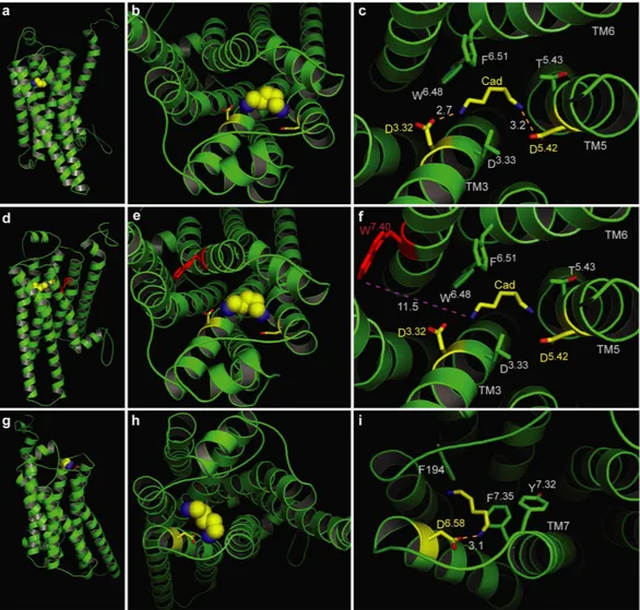
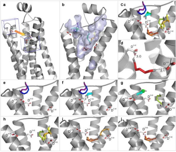
The homology model of TAAR13c (Fig. 1a) revealed the canonical bundle of seven transmembrane (TM) α -helices followed by an eighth intracellular helix (H8) running parallel to the membrane axis. In rhodopsin, an interaction between H8 and TM7 keeps the receptor in a prereceptive state16, but no such interaction was seen in the TAAR13c model. Interestingly, in the model based on β1AR we also observed a short α -helix in extracellular loop 2 (ECL2) of TAAR13c, which is absent in most class A GPCRs, but present in the β -adrenergic receptors17. Furthermore, a disulfide bridge is predicted between Cys1053.25 at the extracellular end of TM3 and Cys190 in ECL2. This disulphide bond provides conformational restraint and is important for effectively tethering ECL2 to the helical bundle18. The highly conserved landmark motif DRH/Y, here DRH, is located at the cytoplasmic end Figure 1. Homology modeling of TAAR13c predicts 7 TM, two additional short helices, and potential binding residues clustered in the upper third of the TM region. (a) Cartoon representation of the TAAR13c model based on comparison with six crystal structures shows the expected seven transmembrane domains and two short extra helices. The planks representation to the right shows a short α -helix to be located in ECL2 and an intracellular eighth helix, H8 located parallel to the membrane plane. (b) Ligand binding residues (given as residue number in TAAR13c) as predicted by sequence profile comparison with binding sites of PDB templates 3pdsA (FAUC-50-β 2 adrenoceptor complex) and 1F88 (bovine rhodopsin); blue, residues only reported in one of the models; green, residues predicted in both models. (c) Predicted binding residues listed in panel (b) shown as spheres in the TAAR13c structure, color code as before. Note the presence of two ‘green’ columns.
www.nature.com/scientificreports/
of TM3 as expected17. This motif stabilizes the inactive state in some receptors, and governs G-protein coupling in other receptors19. We also observed the ‘ionic lock’ between the DRH motif and a glutamate residue at the cytosolic surface of TM6. Due to a salt bridge that is formed between Arg1303.50 of the DRH motif and Glu2516.34 in TM6, the third and sixth TM helices are connected, a feature which is conserved among all family A GPCRs17. Docking of cadaverine validates a binding pocket in the outer-third of the TM domain. We then used COACH20 to predict putative cadaverine binding residues by sequence profile comparison of TAAR13c with binding sites of several PDB structures, with best fits found for 3pdsA (FAUC50/β 2 adrenoceptor complex) and 1F88 (bovine rhodopsin) in the TAAR13c homology model (Fig. 1b). COACH is a meta-server approach to generate complementary ligand binding site predictions using comparative methods, which recognize ligand-binding tem- plates from BioLiP protein function database by binding-specific substructure and sequence profile comparisons21. Initially 30 such residues were found. They all clustered in the upper third of the TM domain suggesting that the putative binding pocket is located within this region (Fig. 1c). The classical amine-binding motif of aminergic receptors consists of Asp1123.32 and Trp2967.40, both of which were also predicted as binding partners of cadav- erine22. In close proximity to these residues and at the same plane of the membrane another aspartate residue, Asp2025.42, was predicted as a binding partner. We hypothesized that this residue might be involved in bind- ing to the second amino group of cadaverine and examined the region surrounding Asp1123.32, Asp2025.42 and Trp2967.40 (i.e. the upper one-third of TM3, 5, and 6) by computational docking of cadaverine.
The docking results confirmed the involvement of Asp1123.32 and Asp2025.42 in ligand binding. Our results suggest that one amino group of cadaverine (protonated at physiological pH) forms a salt bridge with Asp1123.32 at a distance of 2.7 Å (Fig. 2c, Table 1), well within the range given for salt bridges 1.75–4.0 Å23. This Asp1123.32 is sta- bilized by a hydrogen bond to the hydroxyl group of Tyr299. A similar salt bridge has been described for β 1- and β 2ARs24. The second protonated amino group of cadaverine was docked 3.2 Å away from Asp2025.42 allowing formation of another salt bridge (Fig. 2c). This particular residue is known to undergo binding interactions with ligands in many other GPCRs, forming hydrogen bonds, van der Waals interactions and salt bridges25. Further binding residues validated by docking were Leu1133.33, Thr2035.43, Trp2696.48, and Phe2726.51, all situ- ated within 5 Å distance from cadaverine and thus well within the range of van der Waals interactions (3–6 Å26) (Supplementary Table 1). Thus, these residues are likely candidates for stabilizing the hydrophobic backbone of cadaverine. In addition to cadaverine, diaminoheptane, which has a very similar affinity to TAAR13c6, forms the same two salt bridges as cadaverine (2.9 Å distance to Asp1123.32 and 2.4 Å to Asp2025.42).
Docking did not confirm Trp2967.40 as a binding residue in TAAR13c. Notably, Trp2967.40 is 11.5 Å away from the docked cadaverine, and furthermore the residue is located on the distal side of TM7 relative to cadaverine, excluding a van der Waals interaction (Fig. 2f). This was unexpected, because in class A GPCRs this tryptophan is highly conserved, and serves to stabilize the hydrophobic backbone of amines as part of the amine-binding motif22.
In addition to cadaverine, TAAR13c is activated by putrescine, a smaller diamine, albeit with much lower affinity6. Docking of putrescine into the TAAR13c model revealed the same salt bridge with Asp1123.32 as for cadaverine, albeit at a slightly larger distance of 3.0 Å (Table 1). However, Asp2025.42 is not able to form the sec- ond salt bridge, because the distance of 5.5 Å between the amino group of putrescine and the carboxylic group of Asp2025.42 (Table 1) is too large for a typical salt bridge and only allows a rather weak binding interaction27. This finding would explain the decrease in affinity of TAAR13c for putrescine compared to cadaverine. Nevertheless the weak interaction between Asp2025.42 and the second amino group of putrescine seems to be relevant because the corresponding monoamine (butylamine) is not able to activate TAAR13c at all6.
Taken together, these data strongly suggest that TAAR13c activation relies on the interaction of two amino groups provided by the ligand with two negatively charged residues in the binding cavity of the receptor, and that stabilization of the ligand backbone is not achieved by the canonical tryptophan, here Trp2967.40, in TM7 of the DW motif.
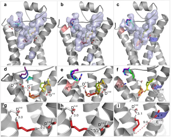
Docking of cadaverine suggests a second binding site for diamines. In addition to the above-mentioned residues, we identified another docking site on the extracellular surface of the receptor (Fig. 2g,h). The main binding residue of this second site is Asp2796.58, which forms a salt bridge with one amino group of cadaverine (Fig. 2i) and putrescine, each at a distance of 3.1 Å (Table 1). Surrounding apolar residues Phe194 in ECL2 and Phe2917.35 in TM7 are also predicted as interaction partners, and presumably serve to sta- bilize the ligand’s apolar backbone (Supplementary Table 1). No residue coordinating the second amino group was detected in this docking site, suggesting that it might not discriminate for chain length. Thus, this site dif- fers in two properties from the internal docking site: (i) it does not noticeably distinguish between cadaverine and putrescine, and (ii) it does not require a second amino group to bind the ligand. Since TAAR13c activation strongly depends on the ligand’s chain length and absolutely requires the second positive charge of the ligand6, this second docking site containing Asp2796.58 is rather unlikely to serve as the ligand binding site that activates the receptor. In order to provide independent experimental proof for the predicted docking residues, we gener- ated a series of receptor mutants by site-directed mutagenesis and studied receptor activity after heterologous expression in mammalian cells.
Mutation of the aminergic DW motif shows only the aspartate as required for ligand binding.
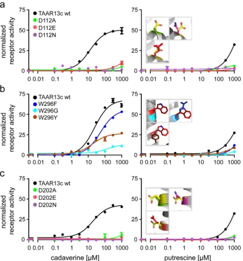
As described above, docking predicted only the Asp1123.32 but not the Trp2967.40 residue of the conserved DW motif to interact with cadaverine (Fig. 2) and putrescine. A series of substitutions were generated for both resi- dues to examine their effects on receptor activity. For the aspartate we chose D112E as a conservative exchange, since the charge is kept and only its position is slightly changed due to the longer side chain in glutamate. Other mutations employed were D112N, which eliminates the charge, but keeps the polarity, and finally D112A, which
www.nature.com/scientificreports/
4
Scientific RepoRts | 6:28359 | DOI: 10.1038/srep28359
removes both the charge and polarity. Mutant receptors were stably transfected into HEK293 cells that consti- tutively express the A2 subunit of an olfactory cyclic nucleotide-gated (CNG) ion channel (see Mat. & Meth.).
Activation of TAAR receptors and subsequent cAMP production could be monitored in these cell lines as ele- vated Ca2+ levels due to Ca2+ influx through cAMP-dependent opening of the CNG channels. We observed that even the most conservative exchange, D112E, reduced TAAR13c activation drastically, and shifted the dose response curve for cadaverine more than two orders of magnitude to higher concentrations (Fig. 3a, Table 1).
Mutation to either asparagine or alanine completely abolished cadaverine-evoked activity. Consistent with these experimental results, docking simulations with cadaverine showed the absence of the wildtype salt bridge in all three mutants (Table 1). Taken together we conclude that Asp1123.32 is a pivotal part of the binding site leading to activation of the TAAR13c receptor by cadaverine.
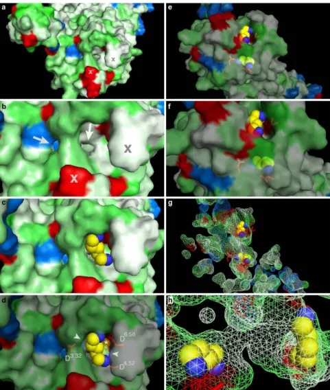
Figure 2. Two binding sites for cadaverine predicted by docking to wildtype TAAR13c. TAAR13c structure (green) is shown with cadaverine (cad; yellow, backbone; blue, amino groups) and coordinating aspartate residues (yellow, backbone; red, carboxyl group). (a) Sideview of TAAR13c shows spatial position of cadaverine docked to the internal binding site, located in the external third of the TM region. (b) Enlarged view from the extracellular surface onto the same binding site shown in panel (a). (c) Enlargement from panel (b) (same view) showing cadaverine, the major interacting residues and the distances in Å from the carboxyl groups of aspartates D3.32 and D5.42 to the amino groups of cadaverine. Salt bridges are visualized as orange dashed lines.
(d) Side view (turned about 90° compared to panel (a) shows the side chain of W7.40 located away from the predicted binding site. (e) View from the extracellular surface, enlarged, same orientation as panel (b). W7.40 is positioned on the distal side of its TM relative to the binding site. (f) Enlargement from panel (e) (same view) showing cadaverine, the two interacting aspartate residues and the large distance to the side chain of W7.40 (purple dashed line), which is thus unlikely to participate in binding interactions. (g) Side view (turned about 90°
compared to panel (a) showing cadaverine bound at an additional binding site on the external surface. (h) View onto the extracellular surface, enlarged, orientation turned 180° relative to panel (b). A single aspartate (D6.58) coordinates cadaverine. (i) Enlargement from panel (h) (similar view) showing cadaverine, the major
interacting residues and the distance from the carboxyl group of D6.58 to the amino group of cadaverine. The salt bridge is visualized as orange dashed line.
www.nature.com/scientificreports/
Similar effects were observed for the mutant receptors when putrescine was applied as a ligand (Fig. 3a, Table 1), and again, even the D112E variant displayed a drastic reduction of the putrescine response. These results confirm the involvement of Asp1123.32 also for the predicted interaction with putrescine. We were intrigued that binding to putrescine, which is one methylene group shorter than cadaverine, could not be improved by the longer side chain of glutamate28, and therefore performed docking simulations with the D112E mutant and putrescine. Indeed, the distance between the charged glutamate side chain and the amino group of putrescine was predicted as 9.8 Å (Table 1), well beyond the range of a salt bridge27, and thus consistent with the drastic reduction in affinity observed for the D112E receptor mutant. In fact, docking of putrescine showed for all three mutants that at most one amino group is able to form a typical salt bridge, whereas the other amino group is too far away for a binding interaction (Table 1). Taken together, experimental and modeling results obtained with Asp1123.32 mutants and putrescine again confirmed the involvement of Asp1123.32 in ligand binding as predicted by mode- ling and docking the wildtype receptor (see above).
The tryptophan residue Trp2967.40 of the DW motif13 was not predicted to be part of the binding site (Fig. 2f).
We therefore examined whether replacement of this residue might affect receptor activity. Three mutants were generated, either exchanging the tryptophan for phenylalanine (less bulky), tyrosine (switch to polar residue) or glycine (no side chain interaction possible). Stably transfected cell lines were established with all mutants, and receptor activity was examined with a series of cadaverine and putrescine concentrations (Fig. 3b).
Cadaverine and putrescine were able to activate all three mutants, with similar affinity as wildtype, even for the drastic W296G exchange (Fig. 3b, Table 1), in sharp contrast to the almost complete loss of activity in all Asp1123.32 mutants. Furthermore, the efficacy of W296F was similar to wildtype TAAR13c. A slight reduction of efficacy was observed for the W296Y mutant, which might be caused by a different interaction of the more hydro- philic tyrosine with neighboring side chains compared to the tryptophan in wildtype TAAR13c. Efficacy was strongly reduced in the W296G mutant, conceivably due to a loss of structural stability by insertion of the highly flexible glycine into a transmembrane domain. Taken together, binding experiments for all three mutants, W296F, W296Y, and W296G, show little loss in affinity and (with one exception) efficacy, confirming that Trp2967.40 is irrelevant for the binding interaction to cadaverine and putrescine, as predicted by the theoretical model.
A second aspartate, Asp2025.42 in TM5 is required for activation of TAAR13c by cadaverine and putrescine. Docking studies suggested an interaction of Asp2025.42 with the second amino group of cadav- erine (Fig. 2c). We followed a similar strategy as before and generated cell lines expressing the following receptor mutants: D202E, D202N, and D202A. Functional testing with cadaverine revealed that even the most subtle replacement, the D202E substitution, eliminated the activity of the receptor completely (Fig. 3c). The same results were obtained for the other substitutions of D202 to either asparagine or alanine (Fig. 3c). Docking simulations for cadaverine with the mutants showed a large distance of about 10 Å between the mutated residues and the sec- ond amino group of cadaverine, consistent with a loss of the second salt bridge present in the wildtype receptor (Table 1). Hence we conclude that Asp2025.42 is another pivotal residue in the cadaverine binding site and partic- ipates in the activation of the TAAR13c receptor.
As observed for cadaverine, all three receptor variants remained quiescent when cell lines were treated with putrescine (Fig. 3, Table 1). Docking data showed that the distance between the glutamate and the second amino group of putrescine was about twice as large as in the wildtype receptor (Table 1), consistent with the inability of putrescine to activate the D202E mutant receptor. A similar result was obtained for the other two mutations (Table 1). Thus, all experiments performed with Asp2025.42 mutants are consistent with docking results for these mutants, and confirm the prediction of Asp2025.42 as an essential component of the diamine binding site that can activate TAAR13c.
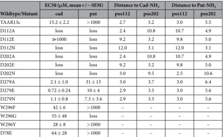
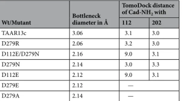
Wildtype/Mutant
EC50 (μM, mean+/−SEM) Distance to Cad-NH2 Distance to Put-NH2
cad put pos112 pos202 pos112 pos202
TAAR13c 15.2 ± 2.2 > 1000 2.7 3.2 3.0 5.5
D112A loss loss 2.4 10.8 10.7 4.9
D112E ≫1000 loss 9.2 3.2 9.8 5.0
D112N loss loss 12.0 3.1 12.0 3.1
D202A loss loss 2.4 10.8 10.7 4.9
D202E loss loss 9.2 3.2 9.8 5.0
D202N loss loss 3.0 9.5 2.5 10.6
D279A 2.1 ± 1.0 31 ± 13 3.0 3.7 3.0 6.4
D279E 0.72 ± 0.24 10 ± 4 2.9 3.5 3.0 5.6
D279N 1.1 ± 0.8 7.3 ± 3.6 2.9 3.5 3.0 5.6
W296F 42 ± 6 > 1000 – – – –
W296G 55 ± 48 loss – – – –
W296Y 28 ± 8 > 1000 – – – –
D78E 64 ± 28 > 1000 – – – –
Table 1. EC50 and contact distances for cadaverine and putrescine amino groups with Asp1123.32 and Asp2025.42 for mutant and wildtype TAAR13c.