hydrodynamic
scanning electrochemical microscopy
Dissertation
zur Erlangung des Doktorgrades der Naturwissenschaften (Dr. rer. nat.) der Fakultät für Chemie und Pharmazie
der Universität Regensburg vorgelegt von
Timo Raith aus Untertraubenbach
Fakultät IV - Chemie und Pharmazie - der Universität Regensburg.
Die Arbeit wurde angeleitet von Prof. Dr. Frank-Michael Matysik.
Promotionsgesuch eingereicht am: 16.01.2020 Tag der mündlichen Prüfung: 26.06.2020
Prüfungsausschuss:
Vorsitzender: Prof. Dr. Rainer Müller
Erstgutachter: Prof. Dr. Frank-Michael Matysik
Zweitgutachter: Prof. Dr. Gerd-Uwe Flechsig
Drittprüfer: PD Dr. Hans-Heiner Gorris
An dieser Stelle möchte ich mich bei allen Personen bedanken, die mich auf dem Weg zur Promotion begleitet und unterstützt haben.
Mein Dank gilt hierbei in gleichem Maße:
Herrn Prof. Dr. Frank-Michael Matysik für die Anleitung der Arbeit, sowie die stetige Unterstützung und wertvolle Diskussionen.
Herrn Prof. Dr. Gerd-Uwe Flechsig für die Tätigkeit als Zweitgutachter.
Herrn PD Dr. Hans-Heiner Gorris für die Tätigkeit als Drittprüfer.
Herrn Prof. Dr. Rainer Müller für den Vorsitz des Prüfungsausschusses.
Der Studienstiftung des Deutschen Volkes für die finanzielle Unterstützung.
Meinen Arbeitskollegen für sämtliche Diskussionen und die einzigartige Arbeitsatmosphäre.
Insbesondere Dr. Christian Iffelsberger, Dr. Patrick Hanekamp und Stefan Wert – unser Team SECM.
Insbesondere Thomas Herl, der mich seit meiner Masterarbeit begleitet und durch wertvolle Diskussionen sowie graphische Zuarbeit unterstützt hat.
Meinen Freunden, insbesondere Markus Tautz, für die Unterstützung und den stetigen Ansporn.
Meiner Familie Gabriele Raith, Werner Raith, Fabian Raith, Florian Raith und Nina Feirer für den stetigen Rückhalt.
Table of contents
Table of contents ... I List of publications ... III Conference contributions ... IX Supervised students ... XI Supervised lab courses ... XII Awards & honors ... XIII Declaration of collaboration ... XIV List of abbreviations ... XV
1. Introduction ... 1
2. Theoretical background ... 8
2.1 Voltammetry ... 8
2.1.1 Introduction to voltammetry ... 8
2.1.2 Voltammetric behavior of macroelectrodes and ultramicroelectrodes ... 9
2.1.3 Cyclic voltammetry and amperometry ... 13
2.1.4 Mass transfer and hydrodynamic effects ... 15
2.2 Scanning electrochemical microscopy ... 18
2.2.1 Setup of the scanning electrochemical microscope ... 18
2.2.2 Probes for scanning electrochemical microscopy ... 19
2.2.3 Operational modes of scanning electrochemical microscopy ... 20
2.2.4 Forced convection in scanning electrochemical microscopy ... 25
3. Experimental ... 32
3.1 Software, instrumentation, materials, chemicals ... 32
3.2 Electrochemical mediators ... 34
3.3 Fabrication of ultramicroelectrodes ... 35
3.4 Scanning electrochemical microscopy setup ... 36
4. Results and discussion ... 41
4.1 Development and characterization of electrochemical flow cells for hydrodynamic scanning electrochemical microscopy ... 41
4.1.1 Introduction ... 42
4.1.2 Results and discussion ... 43
4.1.3 Conclusion ... 49
4.1.4 Experimental ... 49
4.2 Impacts of forced convection generated via high precision stirring on scanning electrochemical microscopy experiments in feedback mode ... 56
4.2.1 Introduction ... 57
4.2.2 Experimental ... 59
4.2.3 Results and discussion ... 62
4.2.4 Conclusion ... 69
4.2.5 Supporting information ... 71
4.3 Enhanced resolution of generator-collector studies of enzymatic structures by means of hydrodynamic scanning electrochemical microscopy ... 76
4.3.1 Introduction ... 77
4.3.2 Experimental ... 78
4.3.3 Results and discussion ... 82
4.3.4 Conclusion ... 87
5. Summary ... 93
6. Zusammenfassung in deutscher Sprache ... 95
List of publications
Peer-reviewed articles
2018 Development and characterization of electrochemical flow cells for hydrodynamic scanning electrochemical microscopy
Timo Raith, Stefan Wert, Christian Iffelsberger, Frank-Michael Matysik Monatshefte für Chemie - Chemical Monthly 149 (2018) 1671–1677
Abstract
In the frame of this contribution, two electrochemical flow cells developed for scanning electrochemical microscopy (SECM) are presented. Forced convection was generated by a flow of the mediator solution through the flow cells. A description of the mandatory design aspects of the experimental flow cell setups is included. Using a macroscopic working electrode as a substrate electrode, forced convection leads to the formation of a stable diffusion layer during amperometric experiments in contrast to a growing diffusion layer in quiescent solution. To characterize the effects of forced convection, the diffusion layer around a platinum substrate electrode integrated into the cells was investigated utilizing chronoamperometric measurements and hydrodynamic SECM imaging in amperometric substrate generation-tip collection (SG/TC) mode. Both methods proved the stability and the time- independency of the diffusion layer. Mathematical simulations using COMSOL Multiphysics were computed to investigate the flow profile generated by the flowing mediator solution in the relevant region close to the substrate electrode.
In summary, two different electrochemical flow cells for SECM were developed and characterized. Both cell designs enabled steady-state diffusion layer characteristics at a macroscopic substrate electrode offering interesting possibilities such as time-independent measurements in the context of the SG/TC mode.
2018 Detection and imaging of reactive oxygen species associated with the electrochemical oxygen evolution by hydrodynamic scanning electrochemical microscopy
Christian Iffelsberger, Timo Raith, Preety Vatsyayan, Vlastimil Vyskočil, Frank- Michael Matysik
Electrochimica Acta 281 (2018) 494-501
Abstract
Hydrodynamic scanning electrochemical microscopy (SECM) was applied for the characterization of Pt and boron-doped diamond (BDD) macroelectrodes operated in a potential region producing reactive oxygen species (ROS) during oxygen evolution reaction (OER). Forced convection introduced by highprecision stirring enabled the formation of a stable diffusion layer of electrochemically produced species and tip-substrate voltammetry was used for the detection of different ROS species produced during OER at BDD.
Hydrodynamic SECM imaging in substrate generation/tip collection mode revealed local differences in the production of the ROS species across the BDD electrode surface.
2018 New Electrochemical Flow-Cell Configuration Integrated into a Three- Dimensional Microfluidic Platform: Improving Analytical Application in the Presence of Air Bubbles
Magno Aparecido Gonçalves Trindade, Cauê Alves Martins, Lucio Angnes, Thomas Herl, Timo Raith, Frank-Michael Matysik
Analytical Chemistry 90 (2018) 10917-10926
Abstract
A newly configured electrochemical flow cell to be used for (end-channel) amperometric detection in a microfluidic device is presented. The design was assembled to place the reference electrode in a separated compartment, isolated from the flow in the microchannel, while the working and counter electrodes remain in direct contact with both compartments. Moreover, a three- dimensional coil-shaped microfluidic device was fabricated using a nonconventional protocol. Both devices working in association enabled us to solve the drawback caused by the discrete injection when the automatic micropipette was used. The high performance of the proposed electrochemical flow cell was demonstrated after in situ modifying the surface of the platinum working electrode with surfactant (e.g., using Tween 20 at 0.10%). As the reference electrode remained out of contact with the flowing solution, there was no trouble by air bubble formation (generated by accidental insertion or by presence of surfactants) throughout the measurements. This device was characterized regarding its analytical performance by evaluating the amperometric detection of acetaminophen, enabling determination from 6.60 to 66.0 μmol L−1. This issue is important since at high concentration (e.g., as assessed in clinical analysis) the acetaminophen is known to passivate the working electrode surfaces by electrogenerated products, impairing the accuracy of the electrochemical measurements.
2019 Impacts of Forced Convection Generated via High Precision Stirring on Scanning Electrochemical Microscopy Experiments in Feedback Mode Timo Raith, Christian Iffelsberger, Preety Vatsyayan, Frank-Michael Matysik Electroanalysis 31 (2019) 273-281
Abstract
In this study, the effects of forced convection on scanning electrochemical microscopy (SECM) experiments in feedback mode using ferrocenemethanol as redox mediator are presented. Forced convection, which enhances the mass transfer inside the system, was generated via an electrical high precision stirrer integrated into the SECM setup. A thin-film interdigitated array electrode serving as model substrate was investigated with probe scan curves in z-direction and SECM imaging in constant-height mode utilizing ultramicroelectrodes (UME) with diameters (dprobe) of 25 µm and 12.5 µm. It was found that forced convection increased the overall current during SECM imaging without distorting distinctive features of the imaged structure when working with a 25 µm UME at substrate- to-tip distances of 14 µm and 11 µm. Furthermore, the electrochemical contrast was improved under hydrodynamic conditions for a substrate-to-tip distance of 11 µm and scan rates of 5 µm s-1, 10 µm s-1, 20 µm s-1 and 40 µm s-1. When further decreasing the gap between the UME and the substrate to 9 µm, almost no effects of the forced convection were observed. Consequently, for a 25 µm UME, forced convection led to higher currents and improved performance during SECM experiments in feedback mode at substrate-to-tip distances of 14 µm and 11 µm, whereas no effects were observed for a 12.5 µm UME at a distance of 8 µm.
2019 Material contrast studies of conductive thin films on semiconductor substrates using scanning electrochemical microscopy
Patrick Hanekamp, Timo Raith, Christian Iffelsberger, Tobias Zankl, Werner Robl, Frank-Michael Matysik
Journal of Applied Electrochemistry 49 (2019) 455-463
Abstract
In this paper, a mediator-free scanning electrochemical microscopy (SECM) imaging concept is presented, which is capable of generating high electrochemical contrast and high spatial resolution between two conductive materials. The methodical approach is based on the hydrogen evolution reaction which shows potential dependent material selectivity. Various conductive thin films deposited on silicon substrates were studied. The investigated materials included copper, ruthenium, platinum, tantalum nitride, and titanium nitride. The hydrogen evolution was studied with chronoamperometry (Esubstrate = 1 V vs.
Ag/AgCl/3 M KCl) to characterize the material selectivity of this reaction for the above-listed thin films. SECM imaging in the substrate generation-tip collection (SG/TC) mode was carried out and applied to study the boundary regions of thin copper films in combination with the aforementioned thin film materials. In addition, the spatial resolution of hydrogen based SG/TC SECM imaging was characterized using lithographically fabricated platinum/copper structures as test substrates. For comparison, the common feedback mode was also applied for SECM imaging of the conducting thin film combinations. It was found that only the hydrogen based SG/TC mode enabled SECM imaging with clear material contrast between different conductive materials which was not possible in the feedback mode.
2020 Enhanced resolution of generator-collector studies of enzymatic structures by means of hydrodynamic scanning electrochemical microscopy
Timo Raith, Anna Kröninger, Matthias J. Mickert, Hans H. Gorris and Frank-Michael Matysik
Talanta DOI: 10.1016/j.talanta.2020.120844
Abstract
In this report, the effects of forced convection on scanning electrochemical microscopy (SECM) studies of enzymes in the context of the generator-collector mode (G/C mode) were investigated. Forced convection was generated via an electrical high precision stirrer integrated into the electrochemical cell. Circular spots of glucose oxidase were immobilized on a gold support serving as model substrate. The diffusion layer of enzymatically generated H2O2 was characterized recording probe scan curves (PSCs) in z-direction. Furthermore, the enzyme-modified surfaces were investigated via constant-height SECM imaging in feedback mode and in G/C mode. For methodical comparison all sets of experiments were performed in quiescent solution (conventional approach) and with forced convection, respectively. In contrast to a growing diffusion layer without forced convection by applying forced convection, a constant diffusion layer of produced H2O2 was observed. Hence, via hydrodynamic SECM time- independent SECM images within a reasonable time scale of SECM measurements in G/C mode were enabled and their resolution was enhanced.
Non-peer-reviewed articles
2017 Trends in der elektrochemischen Rastermikroskopie
Christian Iffelsberger, Timo J. Raith, Patrick J. Hanekamp, Preety Vatsayayan und Frank- Michael Matysik
chrom+food FORUM 4 (2017) 20-22
Conference contributions
Oral Presentations
2017 11. Interdisziplinäres Doktorandenseminar, 12.-14.03.2017, Berlin, Germany
Forced convection in scanning electrochemical microscopy
2017 13th International Students Conference Modern Analytical Chemistry, 21.22.-09.2017, Prague, Czech Republic
Forced convection in scanning electrochemical microscopy introduced by an electrochemical flow cell
2018 1st Cross-Border Seminar on Electroanalytical Chemistry, 04.-06.04.2018, Furth im Wald, Germany
Scanning electrochemical microscopy with forced convection
2018 17th International Conference on Electroanalysis (ESEAC), 03.-07.06.2018, Rhodes Island, Greece
Forced convection in scanning electrochemical microscopy
2018 Electrochemistry 2018, 24.-26.09.2018, Ulm, Germany
Methodical developments of hydrodynamic scanning electrochemical microscopy
2019 ANAKON 2019, 25.-28.03.2019, Münster, Germany
Generation of forced convection in the context of scanning electrochemical microscopy
2019 2nd Cross-Border Seminar on Electroanalytical Chemistry, 10.-12.04.2019, České Budějovice, Czech Republic
Impacts of Forced Convection Generated via High Precision Stirring on Scanning Electrochemical Microscopy Experiments in Feedback Mode
2019 7th Regional Symposium on Electrochemistry for South-East Europe, 25.- 30.05.2019, Split, Croatia
Hydrodynamic scanning electrochemical microscopy
Poster Presentations
2017 ANAKON 2017, 03.-06.04.2017, Tübingen, Germany
Forced convection in scanning electrochemical microscopy introduced by an electrochemical flow cell
2018 International conference on ion analysis, 09.-13.09.2018, Berlin, Germany Formation of a stable diffusion layer of an electrochemically generated ion via forced convection in the context of scanning electrochemical microscopy
2019 ANAKON 2019, 25.-28.03.2019, Münster, Germany
Avoiding transient signals via forced convection in the generator/collector mode of scanning electrochemical microscopy
Supervised students
2017 Untersuchung zur hydrodynamischen elektrochemischen Rastermikroskopie
Michael Biendl - bachelor thesis in cooperation with Ostbayerische Technische Hochschule Regensburg
2017-2018 Development and optimization of an electrochemical flow cell configuration for hydrodynamic scanning electrochemical microscopy Stefan Wert - master thesis
2018 Charakterisierung verschiedener Messparameter in der elektrochemischen Rastermikroskopie
Jonas Fuchs - bachelor thesis
2019 Scanning electrochemical microscopy studies of enzymes Anna Kröninger – research internship
Supervised lab courses
2017 - 2019 „Praktikum Chemie wässriger Lösungen Analytischer Teil“ for chemistry students of the 2nd semester
2017 „Praktikum Pharmazeutische Bioanalytik“ for chemistry students of the 5th semester
2017 „Praktikum Chemie in der Medizin“ for medicine students of the 1st semester
2018 „Chemiecamp“ for pupils
2018/2019 „Bioanalytical Lab“ for chemistry, biochemistry and medical chemistry students of the 8th semester
Awards & honors
2017 – 2019 Funding of doctoral studies by Studienstiftung des deutschen Volkes
2017 Price for the second best presentation awarded by Gesellschaft Deutscher Chemiker in the frame of the 11. Interdisziplinären Doktorandenseminar in Berlin, Germany
2018 Price for the second best presentation awarded by Gesellschaft Deutscher Chemiker in the frame of the 1st Cross-Border Seminar on Electroanalytical Chemistry in Furth im Wald, Germany
2018 Funding of conference participation at 17th International Conference on Electroanalysis (ESEAC) on Rhodes Island, Greece by Internationales Promotionsprogramm der Universität Regensburg
2019 Price for the third best presentation awarded by Gesellschaft Deutscher Chemiker in the frame of the 2nd Cross-Border Seminar on Electroanalytical Chemistry in České Budějovice, Czech Republic
2019 Opening keynote lecture of the Electroanalytical Chemistry session in the frame of the ANAKON 2019 in Münster, Germany
Declaration of collaboration
The research presented within this thesis was partly obtained in cooperation with other scientists. In accordance with § 8 Abs. 1 Satz 2 Punkt 7 of the Ordnung zum Erwerb des akademischen Grades eines Doktors der Naturwissenschaften (Dr. rer. nat.) an der Universität Regensburg vom 18. Juni 2009, these collaborations are described within this section.
4.1. Development and characterization of electrochemical flow cells for hydrodynamic scanning electrochemical microscopy
Conceptual design and writing was done by the author. The first generation cell prototype was developed by the author. Following the ideas of the author, the second generation cell prototype was developed during the master thesis of Stefan Wert, which was supervised by the author. Hence, all experiments concerning the first prototype were conducted by the author, whereas all experiments concerning the second prototype were conducted by Stefan Wert.
The artwork was partly established in collaboration with Thomas Herl. Dr. Christian Iffelsberger was involved in discussions. The project was supervised by Prof. Dr. Frank-Michael Matysik.
4.2. Impacts of Forced Convection Generated via High Precision Stirring on Scanning Electrochemical Microscopy Experiments in Feedback Mode
Conceptual design and writing was done by the author. The experimental work was performed by the author. Dr. Christian Iffelsberger and Dr. Preety Vatsyayan were involved in discussions.
The artwork was partly established in collaboration with Thomas Herl. The project was supervised by Prof. Dr. Frank-Michael Matysik.
4.3. Enhanced resolution of generator-collector studies of enzymatic structures by means of hydrodynamic scanning electrochemical microscopy
Conceptual design and writing was done by the author. SECM measurements were performed by the author and Anna Kröninger during her research internship, which was supervised by the author. The immobilization of the enzymes was performed in collaboration with Matthias Mickert and PD Dr. Hans-Heiner Gorris. The artwork was partly established in collaboration with Thomas Herl. The project was supervised by Prof. Dr. Frank-Michael Matysik.
List of abbreviations
In the following, a general overview in alphabetical order on commonly used abbreviations is given. Any specialized expressions are described in the individual chapters within the results section.
Abbreviation Full name Unit
AFM atomic force microscopy
c concentration mol l-1
CE counter electrode
D diffusion coefficient cm2 s-1
dinner inner diameter m
douter outer diameter m
E potential V
e- electron
EDC N-(3-dimethylaminopropyl)-N’- ethylcarbodiimide hydrochloride
F faraday constant A s mol-1
FcMeOH ferrocenemethanol
frot stirrer rotation frequency s-1
HPLC high performance liquid chromatography
I or i current A
J flux mol cm-2 s-1
L normalized distance
NHS N-hydroxysuccinimide
PEEK polyether ether ketone
PSC probe scan curve
R
universal gas constant and
resistance
J mol-1 K-1
Ω
r radius m
RE reference electrode
RG RG value
SECM
scanning electrochemical microscopy or scanning electrochemical microscope SG/TC substrate generation-tip collection
SICM scanning ion conductance microscopy
SPM scanning probe microscopy
T temperature K
t time s
TG/SC tip generation-substrate collection
UME ultramicroelectrode
WE working electrode
δ diffusion layer thickness μm
Φ electric field potential V
1. Introduction
In the late 1980s, a new representative of the scanning probe techniques was introduced to the science community: scanning electrochemical microscopy (SECM) [1,2]. Numerous developments regarding different measuring modes [2–8], instrumental aspects [9–15] and target substrates [16–23] were established which led to the SECM evolving into a powerful and versatile technique with a vast field of applications ranging from corrosion science [24,25] to biological systems [26–30]. Around 15 years after its introduction, developments regarding the technique itself slowly started to remain static and a shift of research focus to coupling of the SECM with other methods such as scanning ion conductance microscopy (SICM) [31–33], atomic force microscopy (AFM) [34–36] or fluorescence microscopy [37,38] was observable.
Nevertheless, refining and improving an original technique within fundamental research always remains equally important.
One new development concerning this topic, as introduced by the Matysik group, was the integration of an electrical high precision stirrer into the electrochemical SECM cell and thus the generation of forced convection within the SECM system. Generation of hydrodynamics via forced convection in form of flow systems, rotating electrodes or sonication is a well- established approach in electroanalytical chemistry to enhance the mass transfer inside the system and generally speaking the performance of the measurement technique. Nevertheless, in literature only a few publications can be found concerning the topic of forced convection in SECM [39–44]. What all of these publications have in common is that the convection was generated intrinsically via fast scan movement of the probe and usually it was suggested that this has to be avoided.
Until the beginning of 2017, no publication treating the external generation of forced convection within the SECM system could be found which seems surprising keeping in mind the fact that SECM belongs to electroanalytical chemistry and forced convection plays such a traditional and important role within this field. In 2017, a pioneering study published by the Matysik group [45] showed that forced convection can be generated within the SECM system in a controlled manner. Its effects were characterized and advantages of the newly established hydrodynamic SECM system concerning measurements in amperometric substrate generation-tip collection (SG/TC) mode were presented.
The objective of this thesis was to expand this new research field of forced convection in scanning electrochemical microscopy based on three categories:
o Methodical developments to diversify the possibilities of generating forced convection within the SECM system
o Fundamental research to characterize the effects of forced convection concerning different SECM operating modes
o Transfer of the hydrodynamic SECM system from fundamental research to the field of applications
References
[1] A.J. Bard, F.R.F. Fan, J. Kwak, O. Lev, Scanning Electrochemical Microscopy.
Introduction and Principles, Anal. Chem. 61 (1989) 132–138.
doi:10.1021/ac00177a011.
[2] R.C. Engstrom, C.M. Pharr, Scanning electrochemical microscopy, Anal. Chem. 61 (1989) 1099A-1104A. doi:10.1021/ac00194a002.
[3] A.J. Bard, F.F. Fan, J. Kwak, O. Lev, Scanning Electrochemical Microscopy.
Introduction and Principles, 138 (1989) 132–138.
[4] F. Zhou, P.R. Unwin, A.J. Bard, Scanning electrochemical microscopy. 16. Study of second-order homogeneous chemical reactions via the feedback and generation/collection modes, J. Phys. Chem. 96 (1992) 4917–4924.
doi:10.1021/j100191a036.
[5] K. Eckhard, X. Chen, F. Turcu, W. Schuhmann, Redox competition mode of scanning electrochemical microscopy (RC-SECM) for visualisation of local catalytic activity, Phys.
Chem. Chem. Phys. 8 (2006) 5359–5365. doi:10.1039/b609511a.
[6] S. Meltzer, D. Mandler, Microwriting of Gold Patterns with the Scanning Electrochemical Microscope, J. Electrochem. Soc. 142 (1995) 82–85. doi:10.1149/1.2044252.
[7] Y.M. Wuu, F.R.F. Fan, A.J. Bard, High Resolution Deposition of Polyaniline on Pt with the Scanning Electrochemical Microscope, J. Electrochem. Soc. 136 (1989) 885–886.
doi:10.1149/1.2096765.
[8] E.E.D.M. El-Giar, D.O. Wipf, Microparticle-based iridium oxide ultramicroelectrodes for pH sensing and imaging, J. Electroanal. Chem. 609 (2007) 147–154.
doi:10.1016/j.jelechem.2007.06.022.
[9] C.G. Zoski, Ultramicroelectrodes: Design, fabrication, and characterization, Electroanalysis. 14 (2002) 1041–1051. doi:10.1002/1521- 4109(200208)14:15/16<1041::AID-ELAN1041>3.0.CO;2-8.
[10] D. Schäfer, A. Puschhof, W. Schuhmann, Scanning electrochemical microscopy at variable temperatures, Phys. Chem. Chem. Phys. 15 (2013) 5215–5223.
doi:10.1039/c3cp43520b.
[11] M.A. O’Connell, A.J. Wain, Combined electrochemical-topographical imaging: A critical review, Anal. Methods. 7 (2015) 6983–6999. doi:10.1039/c5ay00557d.
[12] K. McKelvey, M.A. Edwards, P.R. Unwin, Intermittent contact-scanning electrochemical microscopy (IC-SECM): A new approach for tip positioning and simultaneous imaging of interfacial topography and activity, Anal. Chem. 82 (2010) 6334–6337.
doi:10.1021/ac101099e.
[13] M. Gȩbala, W. Schuhmann, F. La Mantia, A new AC-SECM mode: On the way to high- resolution local impedance measurements in SECM, Electrochem. Commun. 13 (2011) 689–693. doi:10.1016/j.elecom.2011.04.010.
[14] C. Cougnon, K. Bauer-Espindola, D.S. Fabre, J. Mauzeroll, Development of a phase- controlled constant-distance scanning electrochemical microscope, Anal. Chem. 81 (2009) 3654–3659. doi:10.1021/ac802211u.
[15] C. Kranz, Recent advancements in nanoelectrodes and nanopipettes used in combined scanning electrochemical microscopy techniques., Analyst. 139 (2014) 336–52.
doi:10.1039/c3an01651j.
[16] C. Lee, J. Kwak, A.J. Bard, Application of scanning electrochemical microscopy to biological samples, Proc. Natl. Acad. Sci. U. S. A. 87 (1990) 1740–1743.
doi:10.1073/pnas.87.5.1740.
[17] L. Li, C. Bu, Y. Zhang, J. Du, X. Lu, X. Liu, Composite system based on biomolecules- functionalized multiwalled carbon nanotube and ionic liquid: Electrochemistry and electrocatalysis of tryptophane, Electrochim. Acta. 58 (2011) 105–111.
doi:10.1016/j.electacta.2011.08.097.
[18] V. Alizadeh, M.F. Mousavi, M.A. Mehrgardi, S.H. Kazemi, H. Sharghi, Electron transfer kinetics of cytochrome c immobilized on a phenolic terminated thiol self assembled monolayer determined by scanning electrochemical microscopy, Electrochim. Acta. 56 (2011) 6224–6229. doi:10.1016/j.electacta.2011.03.029.
[19] P. Bertoncello, Advances on scanning electrochemical microscopy (SECM) for energy, Energy Environ. Sci. 3 (2010) 1620–1633. doi:10.1039/c0ee00046a.
[20] T. Yasukawa, T. Kaya, T. Matsue, Characterization and imaging of single cells with scanning electrochemical microscopy, Electroanalysis. 12 (2000) 653–659.
doi:10.1002/1521-4109(200005)12:9<653::AID-ELAN653>3.0.CO;2-S.
[21] K. Yamashita, M. Takagi, K. Uchida, H. Kondo, S. Takenaka, Visualization of DNA microarrays by scanning electrochemical microscopy (SECM), Analyst. 126 (2001) 1210–1211. doi:10.1039/b105097b.
[22] D.T. Pierce, P.R. Unwin, A.J. Bard, Scanning electrochemical microscopy. 17. Studies of enzyme-mediator kinetics for membrane- and surface-immobilized glucose oxidase, Anal. Chem. 64 (1992) 1795–1804. doi:10.1021/ac00041a011.
[23] D. Mandler, A.J. Bard, Scanning Electrochemical Microscopy: The Application of the Feedback Mode for High Resolution Copper Etching, J. Electrochem. Soc. 136 (1989) 3143–3144. doi:10.1149/1.2096416.
[24] L. Niu, Y. Yin, W. Guo, M. Lu, R. Qin, S. Chen, Application of scanning electrochemical microscope in the study of corrosion of metals, J. Mater. Sci. 44 (2009) 4511–4521.
doi:10.1007/s10853-009-3654-x.
[25] P. Dauphin-Ducharme, J. Mauzeroll, Surface Analytical Methods Applied to Magnesium Corrosion, Anal. Chem. 87 (2015) 7499–7509. doi:10.1021/ac504576g.
[26] A.J. Bard, X. Li, W. Zhan, Chemically imaging living cells by scanning electrochemical microscopy, Biosens. Bioelectron. 22 (2006) 461–472. doi:10.1016/j.bios.2006.04.028.
[27] G. Wittstock, Modification and characterization of artificially patterned enzymatically active surfaces by scanning electrochemical microscopy, Fresenius. J. Anal. Chem. 370 (2002) 303–315. doi:10.1007/s002160100795.
[28] A. Hamzehloei, S. Zahra Bathaie, M.F. Mousavi, Probing redox reaction of azurin protein immobilized on hydroxyl-terminated self-assembled monolayers with different lengths, J. Electroanal. Chem. 755 (2015) 27–38. doi:10.1016/j.jelechem.2015.07.005.
[29] M.E. Abdelhamid, S. Piantavigna, A.M. Bond, B. Graham, L. Spiccia, L.L. Martin, A.P.
O’Mullane, An SECM study on the influence of cationic, membrane-active peptides on a gold-supported self-assembled monolayer, Electrochem. Commun. 51 (2015) 11–14.
doi:10.1016/j.elecom.2014.11.018.
[30] A.J. Bard, F.R.F. Fan, D.T. Pierce, P.R. Unwin, D.O. Wipf, F. Zhou, Chemical imaging of surfaces with the scanning electrochemical microscope, Science (80). 254 (1991) 68–74. doi:10.1126/science.254.5028.68.
[31] Y. Takahashi, A.I. Shevchuk, P. Novak, Y. Murakami, H. Shiku, Y.E. Korchev, T.
Matsue, Simultaneous noncontact topography and electrochemical imaging by SECM/SICM featuring ion current feedback regulation, J. Am. Chem. Soc. 132 (2010) 10118–10126. doi:10.1021/ja1029478.
[32] D.J. Comstock, J.W. Elam, M.J. Pellin, M.C. Hersam, Integrated ultramicroelectrode-
[33] C.A. Morris, C.C. Chen, L.A. Baker, Transport of redox probes through single pores measured by scanning electrochemical-scanning ion conductance microscopy (SECM- SICM), Analyst. 137 (2012) 2933–2938. doi:10.1039/c2an16178h.
[34] K. Leonhardt, A. Avdic, A. Lugstein, I. Pobelov, T. Wandlowski, M. Wu, B. Gollas, G.
Denuault, Atomic force microscopy-scanning electrochemical microscopy: Influence of tip geometry and insulation defects on diffusion controlled currents at conical electrodes, Anal. Chem. 83 (2011) 2971–2977. doi:10.1021/ac103083y.
[35] M. Salomo, S.E. Pust, G. Wittstock, E. Oesterschulze, Integrated cantilever probes for SECM/AFM characterization of surfaces, Microelectron. Eng. 87 (2010) 1537–1539.
doi:10.1016/j.mee.2009.11.032.
[36] J. Wiedemair, J.S. Moon, F. Reinauer, B. Mizaikoff, C. Kranz, Ion beam induced deposition of platinum carbon composite electrodes for combined atomic force microscopy-scanning electrochemical microscopy, Electrochem. Commun. 12 (2010) 989–991. doi:10.1016/j.elecom.2010.05.008.
[37] S.E. Salamifar, R.Y. Lai, Use of combined scanning electrochemical and fluorescence microscopy for detection of reactive oxygen species in prostate cancer cells, Anal.
Chem. 85 (2013) 9417–9421. doi:10.1021/ac402367f.
[38] C. Renault, K. Marchuk, H.S. Ahn, E.J. Titus, J. Kim, K.A. Willets, A.J. Bard, Observation of nanometer-sized electro-active defects in insulating layers by fluorescence microscopy and electrochemistry, Anal. Chem. 87 (2015) 5730–5737.
doi:10.1021/acs.analchem.5b00898.
[39] P.A. Kottke, A.G. Fedorov, Advective and transient effects in combined AFM/SECM operation, J. Electroanal. Chem. 583 (2005) 221-231.
doi:10.1016/j.jelechem.2005.06.017.
[40] R. Cornut, S. Poirier, J. Mauzeroll, Forced convection during feedback approach curve measurements in scanning electrochemical microscopy: Maximal displacement velocity with a microdisk, Anal. Chem. 84 (2012) 3531–3537. doi:10.1021/ac203047d.
[41] C. a Nkuku, R.J. LeSuer, Electrochemistry in deep eutectic solvents., J. Phys. Chem.
B. 111 (2007) 13271–13277. doi:10.1021/jp075794j.
[42] S. Kuss, D. Trinh, J. Mauzeroll, High-Speed Scanning Electrochemical Microscopy Method for Substrate Kinetic Determination: Application to Live Cell Imaging in Human Cancer, Anal. Chem. 87 (2015) 8102–8106. doi:10.1021/acs.analchem.5b01269.
[43] S. Kuss, C. Kuss, D. Trinh, S.B. Schougaard, J. Mauzeroll, Forced convection during scanning electrochemical microscopy imaging over living cells: Effect of topographies and kinetics on the microelectrode current, Electrochim. Acta. 110 (2013) 42–48.
doi:10.1016/j.electacta.2013.03.149.
[44] M.A. Edwards, A.L. Whitworth, P.R. Unwin, Quantitative analysis and application of tip position modulation-scanning electrochemical microscopy, Anal. Chem. 83 (2011) 1977–1984. doi:10.1021/ac102680v.
[45] C. Iffelsberger, P. Vatsyayan, F. Matysik, Scanning Electrochemical Microscopy with Forced Convection Introduced by High-Precision Stirring, Anal. Chem. 89 (2017) 1658–
1664. doi:10.1021/acs.analchem.6b03764.
2. Theoretical background
2.1 Voltammetry
2.1.1 Introduction to voltammetry
Voltammetry is based on the measurement of the current I flowing through an electrochemical cell in dependence of the potential E. As presented schematically in Figure 2.1, voltammetric measurements are usually performed in a three-electrode setup [1,2].
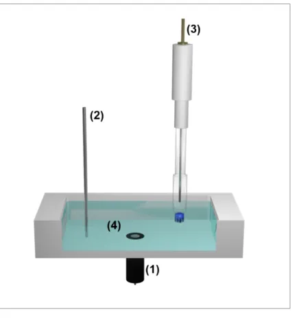
Figure 2.1: Schematic representation of the experimental setup for electrochemical experiments. Working electrode WE (1), counter electrode CE (2) and reference electrode RE (3) are immersed into the electrolyte solution (4) containing a redox active species. All electrodes are connected to a potentiostat for potential control and current read-out.
A reference electrode (RE), a counter electrode (CE) and a working electrode (WE) are immersed into the electrolyte solution usually containing an electrochemically active species.
All electrodes are connected to a potentiostat enabling potential control and current read-out.
The potential applied to the working electrode is adjusted versus the reference electrode.
Measuring this reference potential should be currentless as otherwise part of the applied potential is lost due to the resistance Rsol of the solution. Considering this so-called ohmic
potential drop, the potential Eeff which is effectively applied to the working electrode is differing from the initial potential E according to:
𝐸!"" = 𝐸 − 𝐸#$% = 𝐸 − 𝑅#$%∙ 𝐼 (1)
This potential Eeff leads to a current flow between working electrode and counter electrode.
Two contributions add up to the measured current I:
𝐼 = 𝐼&+ 𝐼' (2)
The capacitive current IC results from the charging process of the electrical double layer formed at the electrode when a potential is applied, whereas the Faradaic current IF results from the conversion of a redox active species present in the electrolyte solution. Consequently, only the Faradaic current delivers qualitative and quantitative information about the investigated system meaning that a large ratio IF/IC is desired. As both currents are superimposed, pulsed techniques or very small electrodes can be applied to increase this ratio [3-5]. These so-called ultramicroelectrodes (UMEs) exhibit a negligible contribution of the capacitive current IC to the overall current leading to large IF/IC ratios and thus increased accessibility of analytical information. Furthermore, since UMEs exhibit a high current density but low currents in the range of nanoampere or even less, the ohmic drop can be neglected. According to equation 1, the effectively applied potential Eeff equals the initial potential E in this case. Hence, utilizing UMEs electrochemical systems can be operated in a two-electrode setup without a reference electrode. Nevertheless, the voltammetric behavior of UMEs differs strongly from conventional macroscopic electrodes.
2.1.2 Voltammetric behavior of macroelectrodes and ultramicroelectrodes
The electrochemical conversion of a redox active species at the working electrode resulting in the Faradaic current IF is composed of three major steps. The mass transfer of the species from the bulk solution to the electrode surface, the transfer of electrons between electrode and species and the mass transfer away from the electrode. Comparing these steps, electrochemical reactions are reversible if the electron transfer is faster than the mass transfer and irreversible vice versa [6].
Planck equation with the three terms on the right representing the contributions of diffusion, migration and convection [1,6].
𝐽((𝑥) = −𝐷(∙𝜕𝑐((𝑥)
𝜕𝑥 −𝑧 ∙ 𝐹
𝑅𝑇 ∙ 𝐷(∙𝑐(𝜕𝛷(𝑥)
𝜕𝑥 + 𝑐(∙ υ(x) (3)
Di describes the diffusion coefficient of the species i, ci its concentration, z the number of transferred electrons, R the universal gas constant, T the temperature, ϕ the electric field potential and υ the velocity. The concentration gradient of the electroactive species that is formed when the electrochemical reaction is started and the species is consumed at the electrode is described by the change in concentration along the x-dimension ∂ci/∂x.
Usually, when designing an electrochemical experiment, migration and convection are suppressed by the addition of an inert supporting electrolyte and the absence of a convective source. In this case, the diffusion resulting from the concentration gradient is the predominant contribution to the mass transfer of the analyte species leading to a simplified form of equation 3 known as Fick’s first law.
−𝐽((𝑥) = 𝐷(∙𝜕𝑐((𝑥)
𝜕𝑥 (4)
Diffusion in turn is composed of linear and radial diffusion. As presented in Figure 2.2, the ratio of these contributions is dependent on the geometry and the size of the electrode where the electrochemical reaction takes place.
Figure 2.2: Schematic representation of a linear diffusion field occurring during electrochemical reactions at planar macroelectrodes in comparison to a spherical diffusion field occurring at spherical electrodes and ultramicroelectrodesadapted from [7].
Hence, based on the electrode characteristics different forms of Fick’s second law which describes the variation of concentration with time due to diffusion have to be considered. For
planar macroelectrodes the flux towards the surface is controlled by linear diffusion as the radial diffusion on the edges of the electrodes is negligible:
𝜕𝑐((𝑥, 𝑡)
𝜕𝑡 = 𝐷(∙ 8𝜕*𝑐((𝑥, 𝑡)
𝜕𝑥* 9 (5)
Solving equation 5 delivers the Cottrell equation for the diffusion-limited current Iplanar measured at a planar macroelectrode in presence of a redox active species with initial concentration ci0
when a potential is applied. The other parameters represent the electrode area A and the the number of transferred electrons z.
𝐼+%,-,.(𝑡) = 𝑧 ∙ 𝐹 ∙ 𝐴 ∙ ; 𝐷(
𝜋 ∙ 𝑡∙ 𝑐(/ (6)
If the electrode is rather spherical than planar, radial diffusion is the predominant contribution leading to a spherical diffusion field and thus a different form of Fick’s second law [4,8]:
𝜕𝑐((𝑥, 𝑡)
𝜕𝑡 = 𝐷(∙ 8𝜕*𝑐((𝑥, 𝑡)
𝜕𝑟* +2
𝑟∙𝜕𝑐((𝑥, 𝑡)
𝜕𝑟 9 (7)
Solving equation 7 delivers the equation for the current Ispherical measured at a spherical electrode.
𝐼#+0!.(1,%(𝑡) = 𝑧 ∙ 𝐹 ∙ 𝐴 ∙ 𝐷(8 1
A𝐷(∙ 𝜋 ∙ 𝑡+ 1
𝑟!9 ∙ 𝑐(/ (8)
In comparison to planar macroelectrodes, equation 8 contains a time-independent component 1/re with re being the radius of the electrode. Increasing the electrode radius, the time- independent component becomes negligible leading to a transition of equation 8 into the Cottrell equation for planar electrodes.
Decreasing the radius, however, UMEs are obtained exhibiting at least one dimension smaller than 25 µm. For a disk UME representing the most important practical UME geometry Fick’s second law for two dimensions is given by
𝜕𝑐((𝑟, 𝑧, 𝑡)
𝜕𝑡 = 𝐷(∙ 8𝜕*𝑐((𝑟, 𝑧, 𝑡)
𝜕𝑟* +1
𝑟∙𝜕𝑐((𝑟, 𝑧, 𝑡)
𝜕𝑟 +𝜕*𝑐((𝑟, 𝑧, 𝑡)
𝜕𝑧* 9 (9)
where r describes the radial position and z the linear displacement normal to the plane of the electrode. Solving Fick’s second law and applying further simplifications delivers the equation for the steady-state current IUME measured late in the experiment at a disk UME with an insulating mantle of infinite thickness [4,9].
𝐼234= 4 ∙ 𝑧 ∙ 𝐹 ∙ 𝐷(∙ 𝑐(/∙ 𝑟 (10)
Disk UMEs exhibit a time-independent current corresponding to a hemispherical diffusion field with a constant diffusion layer. In comparison to an insulating mantle of infinite thickness, an additional geometrical factor β(RG) has to be considered if the mantle is finite:
𝐼234 = 4 ∙ 𝑧 ∙ 𝐹 ∙ 𝐷(∙ 𝑐(/∙ 𝑟 ∙ 𝛽(𝑅𝐺) (11)
This factor β(RG) describes the flux from behind the electrode surface and is defined as
𝛽(𝑅𝐺) = 1 + 0.23
(𝑅𝐺5− 0.81)/.57 (12)
with RG being defined as the ratio between radius of the insulating mantle rg and radius of the active electrode area r.
Consequently, different equations have to be considered due to the different diffusional contributions resulting in different behavior of macroelectrodes and UMEs during electrochemical experiments such as cyclic voltammetry or amperometry [7,10].
2.1.3 Cyclic voltammetry and amperometry
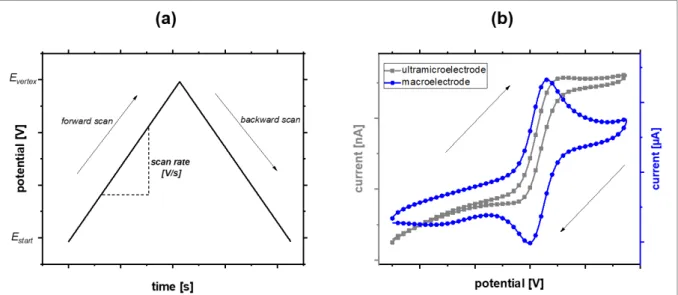
Cyclic voltammetry (CV) is a frequently applied voltammetric measurement technique to obtain information about the electrochemical properties of an analyte [6]. Figure 2.3 delivers an overview of the technique’s most important characteristics.
Figure 2.3: (a) Time-dependent potential change during cyclic voltammetry. (b) Typical current response of a macroelectrode and an UME during cyclic voltammetry in presence of a reversibly convertible redox active species exhibiting a one electron transfer.
As can be seen in Figure 2.3 (a), the starting potential Estart applied to the working electrode is linearly increased during the forward scan and decreased again during the backward scan upon reaching a certain turning point Evertex. This so-called scan cycle can be repeated arbitrarily. The current I is recorded throughout the cycle delivering the typical current-potential (I-E) curves known as cyclic voltammograms (also abbreviated as CV).
The shape of these curves strongly depends on both the characteristics of the analyte as well as the dimensions of the working electrode. Figure 2.3 (b) compares I-E curves obtained for a diffusion limited reaction during cyclic voltammetry in presence of a reversibly oxidizable and reducible species exhibiting a one electron transfer utilizing a macroelectrode and an UME. A peak-shaped curve is observed for a macroelectrode due to the current decrease according to the Cottrell equation (equation 6). Furthermore, forward and backward scans show hysteresis due to the rather high contribution of capacitive currents. An s-shaped curve exhibiting a steady-state current with very small hysteresis is observed for an UME due to the lack of a time-dependent component in the valid equation (equation 10) and the small contribution of capacitive currents.
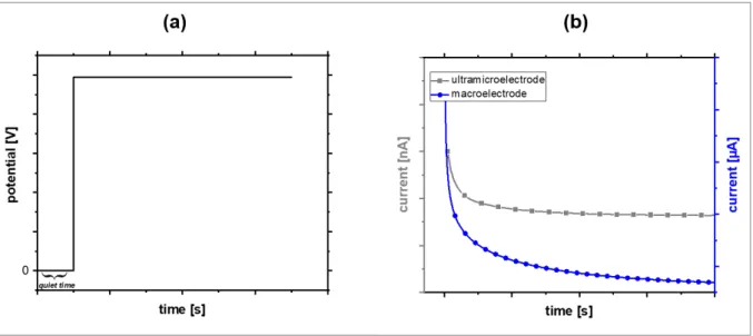
Similar differences can be observed during amperometric measurements [6]. In comparison to cyclic voltammetry, the potential applied to the working electrode is kept constant during amperometry as can be seen in Figure 2.4 (a).
Figure 2.4: (a) Time-dependent potential change during chronoamperometry. (b) Typical current response of a macroelectrode and an UME during chronoamperometry in presence of a redox active species.
Figure 2.4 (b) compares the time-dependent current for a diffusion limited reaction recorded with a macroelectrode and an UME when a constant potential is applied. Similar to cyclic voltammetry, a constant current is observed for the UME whereas the current decreases for the macroelectrode due to its Cottrellian behavior.
Consequently, the different behavior of macroelectrodes and UMEs during cyclic voltammetry and amperometry can be attributed to the different sizes of the electrodes and thus the different diffusional characteristics as described in section 2.1.2. Nevertheless, the voltammetric behavior of the electrodes in dependence of their size is only valid if migration and convection are excluded and mass transfer is governed solely by diffusion.
2.1.4 Mass transfer and hydrodynamic effects
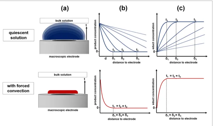
When forced convection is generated within an electrochemical system rather than being excluded, hydrodynamic effects on the mass transfer have to be considered [1,6,10]. For instance, forced convection can be generated by stirring the electrolyte solution [11], by flow systems [10,12], by sonication [13] or by rotating electrodes [14,15]. Figure 2.5 schematically sums up the processes at a macroscopic electrode during electrochemical reactions in quiescent solution and with forced convection.
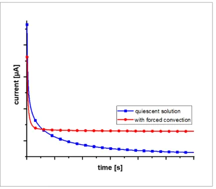
Figure 2.5: Schematic representation of the time-dependency t of the diffusion layer δ of the product species generated during an electrochemical reaction at a macroelectrode (a) and the corresponding change of the product concentration profile (b). The time-dependency t of the concentration profile δ of the species that is consumed during the electrochemical reaction at the electrode is shown in (c). All characteristics are depicted in quiescent solution (top) and with forced convection (bottom)adapted from [2].
As depicted in part (a) of Figure 2.5 a time-dependent growing diffusion layer of the product species generated during an electrochemical reaction is observed in comparison to a stable diffusion layer with forced convection applied to the system. Looking at the concentration profile of the product species in part (b) it is visible that its concentration in the region close to the electrode increases with ongoing reaction time in quiescent solution whereas a constant profile is obtained with forced convection. Analogous, in part (c) the same observations can be made for the concentration profile of the species that is consumed during the electrochemical
currents due to steeper concentration gradients and hence an increased mass transfer. The increase in mass transfer can be adjusted by varying the intensity of the convection. Thus, an additional parameter can be optimized concerning the kinetics of the system that is to be studied.
The theoretical treatment of convective systems involves solving a hydrodynamic problem like determining the flow velocity of the solution as a function of the intensity of the convection before solving the electrochemical one. The simplest treatment is based on a diffusion layer approach meaning that within the diffusion layer close to the electrode surface the convection can be neglected and the mass transfer is governed solely by diffusion. Outside the diffusion layer a constant concentration of all analyte species is to be expected due to the convection.
A more complex approach is the convective-diffusion equation where only the contribution of migration can be excluded from the Nernst-Planck equation (equation 3). Deriving the valid equations of so-called hydrodynamic electrodes requires both modelling as well as simplifying assumptions and strongly depends on the characteristics of the hydrodynamic system [10].
Figure 2.6 representatively sums up the effects of forced convection on an amperometric experiment utilizing a planar macroelectrode.
Figure 2.6: Typical current response of a macroelectrode during chronoamperometry in presence of a redox active species in quiescent solution and with forced convection applied to the system.
A decreasing current is observed in quiescent solution due to the time-dependent growth of the diffusion layers. During this growth the concentration gradient of the species that is consumed flattens causing the mass transfer towards the surface to slow down. The resulting
macroelectrode described in section 2.1.2. With forced convection, however, constant diffusion layers are formed resulting in a steady-state current response similar to UMEs with increased currents due to the increased mass transfer.
Consequently, time-independent diffusion layers of both the product species as well as the species that is consumed are formed during electrochemical reactions with forced convection in contrast to growing diffusion layers without forced convection effecting the electrochemical response of macroelectrodes. Concerning UMEs, the interference of convection is negligible due to their high current density.
2.2 Scanning electrochemical microscopy
Scanning electrochemical microscopy is a versatile analytical method for the characterization of a vast field of substrates ranging from inorganic surfaces to biological samples [9,16-22].
The technique was introduced concurrently by the Bard [23] and Engstrom [24] research groups as a representative of the field of scanning probe microscopy (SPM). In general, in scanning probe microscopy information is generated via processes taking place at a probe while being scanned across a surface. Other representatives include atomic force microscopy and scanning tunneling microscopy. Throughout the years, various instrumental developments and coupling of the SECM with other SPM techniques were established leading to a wide variety of experimental parameters and operational modes. The following sections deliver an overview of the most important aspects fundamental for the research conducted within this thesis.
2.2.1 Setup of the scanning electrochemical microscope
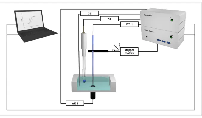
The individual components of the experimental setup for scanning electrochemical microscopy are depicted schematically in Figure 2.7.
Figure 2.7: Schematic representation of the experimental setup for scanning electrochemical microscopy.
Both working electrodes (WE 1, WE 2), as well as reference electrode (RE) and counter electrode (CE) are immersed into the electrolyte solution containing a redox active species. All electrodes are connected to a bipotentiostat. Additionally, WE 1 is connected to stepper motors enabling scan movement of the
![Figure 2.2: Schematic representation of a linear diffusion field occurring during electrochemical reactions at planar macroelectrodes in comparison to a spherical diffusion field occurring at spherical electrodes and ultramicroelectrodes adapted from [7]](https://thumb-eu.123doks.com/thumbv2/1library_info/3738477.1509167/29.892.112.792.763.978/schematic-representation-diffusion-electrochemical-macroelectrodes-comparison-electrodes-ultramicroelectrodes.webp)
![Figure 2.9: Schematic representation of the distance-dependent current response of an UME while approaching a conductor (positive feedback) and an isolator (negative feedback) adapted from [9]](https://thumb-eu.123doks.com/thumbv2/1library_info/3738477.1509167/40.892.191.718.346.960/schematic-representation-distance-dependent-response-approaching-conductor-positive.webp)
![Figure 2.10: Schematic representation of the substrate generation/tip collection mode (a) and the tip generation/substrate collection mode (b) adapted from [34]](https://thumb-eu.123doks.com/thumbv2/1library_info/3738477.1509167/42.892.254.645.415.806/schematic-representation-substrate-generation-collection-generation-substrate-collection.webp)
