synthetic lethal target in head and neck cancer
Sina Heimer
1, Gertrud Knoll
2, Patrick Neubert
2, Karin P. Hammer
3, Stefan Wagner
3, Richard J. Bauer
1,4, Jonathan Jantsch
2and Martin Ehrenschwender
21 Department of Oral and Maxillofacial Surgery, University Hospital Regensburg, Regensburg, Germany 2 Institute of Clinical Microbiology and Hygiene, University Hospital Regensburg, Regensburg, Germany 3 Department of Internal Medicine II, University Hospital Regensburg, Regensburg, Germany
4 Department of Oral and Maxillofacial Surgery, Center for Medical Biotechnology, University Hospital Regensburg, Regensburg, GermanyOpen access funding enabled and organized by Projekt DEAL.
Keywords
BCL-XL; head and neck cancer;
hyperosmotic stress; MCL-1; NOXA Correspondence
M. Ehrenschwender, Institute of Clinical Microbiology and Hygiene, University Hospital Regensburg, Franz-Josef-Strauss- Allee 11, Regensburg 93053, Germany Tel: +49 941 94416440
E-mail: martin.ehrenschwender@ukr.de (Received 16 December 2019, revised 9 June 2020, accepted 20 July 2020) doi:10.1111/febs.15492
Head and neck squamous cell carcinoma (HNSCC) is an aggressive and difficult-to-treat cancer entity. Current therapies ultimately aim to activate the mitochondria-controlled (intrinsic) apoptosis pathway, but complex alterations in intracellular signaling cascades and the extracellular microen- vironment hamper treatment response. On the one hand, proteins of the BCL-2 family set the threshold for cell death induction and prevent acci- dental cellular suicide. On the other hand, controlling a cell’s readiness to die also determines whether malignant cells are sensitive or resistant to anticancer treatments. Here, we show that HNSCC cells upregulate the proapoptotic BH3-only protein NOXA in response to hyperosmotic stress.
Induction of NOXA is sufficient to counteract the antiapoptotic properties of MCL-1 and switches HNSCC cells from dual BCL-XL/MCL-1 protec- tion to exclusive BCL-XL addiction. Hypertonicity-induced functional loss of MCL-1 renders BCL-XL a synthetically lethal target in HNSCC, and inhibition of BCL-XL efficiently kills HNSCC cells that poorly respond to conventional therapies. We identify hypertonicity-induced upregulation of NOXA as link between osmotic pressure in the tumor environment and mitochondrial priming, which could perspectively be exploited to boost effi- cacy of anticancer drugs.
Introduction
Head and neck squamous cell carcinomas (HNSCCs) are among the most common malignancies worldwide [1]. Research efforts during the last decades increased the understanding of this disease but failed to bring up clinically meaningful discoveries. Currently available treatments include surgical eradication, radiotherapy, and chemotherapy but are often ineffective [2]. The mitochondria-controlled (intrinsic) apoptotic pathway largely determines sensitivity or resistance to anti- cancer treatments [3]. Intrinsic apoptosis proceeds by
insertion of the pore-forming proteins BAX and/or BAK in the outer mitochondrial membrane (OMM).
Subsequent release of apoptosis-promoting molecules from the mitochondrial intermembrane space allows formation of the apoptosome, a scaffold that facilitates activation of caspase-9 and (further downstream) acti- vation of caspase-3 and caspase-7, the executioners of apoptosis [4]. To prevent accidental cell death induc- tion, a sophisticated network of pro- and antiapoptotic BCL-2 family proteins imposes a threshold (also
Abbreviations
CI, combination index; EGFR, epidermal growth factor receptor; ER, endoplasmic reticulum; HNSCC, head and neck squamous cell carcinoma; OMM, outer mitochondrial membrane.
1 The FEBS Journal (2020)ª2020 The Authors. TheFEBS Journalpublished by John Wiley & Sons Ltd on behalf of
referred to as ‘mitochondrial priming’) that apoptosis- promoting stimuli must meet for BAX/BAK activation [5]. In cancer cells, mitochondrial priming directly cor- relates with response to chemotherapy [6]. Conse- quently, modulating mitochondrial priming by targeting the safeguards of the OMM (e.g., BCL-2, BCL-XL, and MCL-1) emerged as a novel therapeutic approach [7]. Small molecules that occupy the BH3 domain-binding groove (‘BH3 mimetics’) of antiapop- totic BCL-2 family proteins (e.g., ABT-199 targeting BCL-2, WEHI-539 targeting BCL-XL, or S63845 tar- geting MCL-1) abrogate their protective function and prime mitochondria for death [8 – 10]. Synergistic effects of BH3 mimetics with other treatment modali- ties have been reported in HNSCC [11,12], but thus far failed to demonstrate efficacy in clinical trials [13].
This is not surprising, as most cancer cells rely at least on two antiapoptotic BCL-2 family proteins (e.g., BCL-XL and MCL-1) to ensure OMM integrity [14].
Antiapoptotic BCL-2 family proteins exhibit func- tional redundancy and can play compensatory roles, which allows most cancer cells to survive selective inhi- bition of only one of their mitochondrial safeguards.
Head and neck squamous cell carcinoma often face harsh environmental conditions such as hypoxia and mechanical stress [15]. Adaption to these challenges involves cellular stress responses that ensure survival but can concomitantly alter the cell’s apoptotic thresh- old [16]. For example, accommodation to hyperos- motic stress in the tumor environment also affects levels of BCL-2 family proteins [17]. Hyperosmotic stress (or hypertonicity) occurs when osmolytes that cannot passively diffuse across the plasma membrane (such as NaCl or trehalose) establish an osmotic pres- sure gradient between the intra- and extracellular space. Hypertonicity triggers an adaptive cellular response to compensate for environmental changes, maintain cellular homeostasis, and ensure survival [18].
When osmoadaptive mechanisms fail or hypertonicity persists, a variety of perturbing effects such as cell cycle arrest, DNA damage, inhibition of transcription/
translation, mitochondrial depolarization, and apopto- sis induction can occur [19]. Notably, hypertonicity-in- duced apoptosis is controlled by the mitochondria and regulated by BCL-2 family proteins [20].
In this study, we show that the response of HNSCC cells to hypertonic conditions in the tumor environ- ment transforms dual BCL-XL/MCL-1 dependency into exclusive BCL-XL addiction. Hypertonicity-in- duced upregulation of the proapoptotic BH3-only pro- tein NOXA counteracts the antiapoptotic function of MCL-1 and shifts maintenance of OMM integrity from a cooperative BCL-XL/MCL-1 interplay to
exclusive BCL-XL dependency. Inhibition of BCL-XL under hypertonic conditions is synthetically lethal and efficiently kills cancer cells that poorly respond to epi- dermal growth factor receptor (EGFR) inhibition or radiotherapy. Our results characterize hypertonicity-in- duced upregulation of NOXA as molecular link between osmotic pressure in the tumor environment and mitochondrial priming in HNSCC. Functionally, we show that an adaptive cellular response to changing conditions in the tumor microenvironment can be uti- lized to sensitize HNSCC cells to apoptosis. Our find- ings could be exploitable in future treatment strategies to boost efficacy of anticancer drugs.
Results
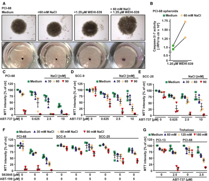
Hypertonicity counteracts MCL-1 in HNSCC cells Previous reports highlighted a key role for both BCL- XL and MCL-1 in HNSCC survival [10,21]. Accord- ingly, the BCL-XL-specific inhibitor WEHI-539 showed little cytotoxicity (Fig. 1A) in various HNSCC cell lines as a single agent, but synergized with the MCL-1-targeting compound S63845 in cell death induction (Fig. 1A and Table 1). In line with dual BCL-XL/MCL-1 dependency, simultaneous knock- down of BCL-XL and MCL-1 displayed cytotoxic effects (Fig. 1B). In HNSCC cell lines exposed to a hypertonic environment, however, exclusive inhibition of BCL-XL was sufficient to trigger cell death, even in the absence of a pharmacological MCL-1 inhibitor (Figs 1C,D,F,H,I and 2B). Calculation of the combi- nation index (CI) confirmed synergistic cell death induction by WEHI-539 and NaCl (Table 1). WEHI- 539-induced loss of viability under hyperosmotic stress showed hallmarks of apoptotic cell death such as increased activity of the effector caspases-3/7 (Fig. 1E, G) and annexin-V/7-AAD positivity (Fig. 2C). Down- regulation of BAX, the main effector protein of the intrinsic apoptosis pathway, or addition of the pan- caspase inhibitor QVD-OPh expectedly reduced cyto- toxicity of WEHI-539 under hypertonic conditions (Fig. 2A,D). Hyperosmotic stress also enhanced sensi- tivity of HNSCC spheroid cell cultures for BCL-XL inhibition (Fig. 3A,B). Besides WEHI-539, other BCL- XL targeting agents such as ABT-737 (targeting BCL- XL, BCL-2, and BCL-W) efficiently triggered cell death under hypertonic conditions (Fig. 3C,D,E,G).
Selective inhibition of BCL-2 (using ABT-199) did not
cooperate with hypertonicity in cell death induction
(Fig. 3F), suggesting that indeed ABT-737-mediated
BCL-XL inhibition is critical. Accordingly, cytotoxic-
ity of the MCL-1 inhibitor S63845 was not enhanced
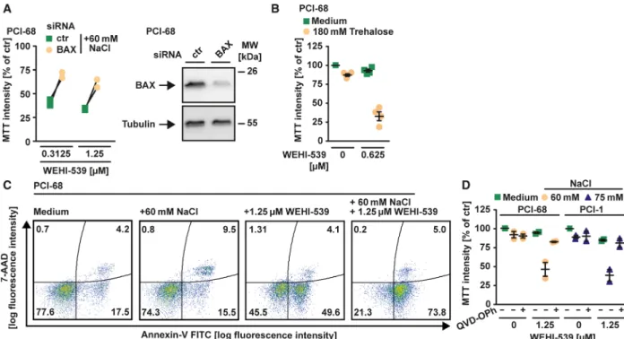
Fig. 1.Hyperosmotic stress shifts BCL-XL/MCL-1-protected HNSCC cells to exclusive BCL-XL addiction. (A) Cells were challenged with the indicated concentrations of the BCL-XL inhibitor WEHI-539 in the presence and absence of the MCL-1 inhibitor S63845. (B) Cells were transfected with siRNA oligonucleotides targeting BCL-XL and MCL-1 or nontargeting control (ctr). After 24 h, cells treated with control siRNA were challenged with WEHI-539 (1.25µM) in the presence and absence of S63845 (1.25µM) for another 18 h. (C, D, F, H, I) Cells were challenged with the indicated concentrations of the BCL-XL inhibitor WEHI-539 in the presence and absence of NaCl. (E, G) Cells were challenged with WEHI-539 (1.25µM) in the presence and absence of the indicated concentrations of NaCl for 18 h. Caspase-3/-7 activity was assessed using the fluorogenic substrate (DEVD)2-R110. For (A, C, D, F, H, I), data points and meanSEM from three independent experiments are shown; for (B), data points and mean from two independent experiments are shown; for (E, G), individual data points of at least two independent experiments are shown. RFU, relative fluorescence units.
under hyperosmotic stress (Fig. 3F). In sum, our data suggested that hyperosmotic stress shifted combined BCL-XL/MCL-1 dependency of HNSCC cells to exclusive BCL-XL addiction by selectively counteract- ing the antiapoptotic function of MCL-1.
Hypertonicity-induced NOXA upregulation shifts dual BCL-XL/MCL-1 dependency toward
exclusive BCL-XL addiction
The hypertonicity-induced loss of MCL-1 protection and the resulting switch from combined BCL-XL/
MCL-1 to exclusive BCL-XL dependency were related neither to reduction of intracellular BCL-XL/MCL-1 levels nor to reduced expression of the corresponding genes (Fig. 4A,B). Notably, interaction with BH3-only proteins such as NOXA can modulate the antiapop- totic function of MCL-1 [5]. Indeed, hypertonicity changed cellular NOXA levels. NOXA was barely detectable under isotonic conditions but increased transiently upon hyperosmotic stress (Fig. 4A,C).
Other apoptosis-promoting BCL-2 family proteins, however, displayed no hypertonicity-induced upregula- tion (Fig. 4A). Downregulation of NOXA was suffi- cient to rescue HNSCC cells challenged with the BCL- XL inhibitor WEHI-539 under hypertonic conditions,
whereas knockdown of BIM had no effect (Fig. 4D).
This indicated functional relevance for hypertonicity- induced NOXA upregulation to shift combined BCL- XL/MCL-1 toward exclusive BCL-XL dependency.
Hypertonicity triggers Ca
2+release from the ER in the absence of ER stress and limits cancer cell proliferation
Hypertonicity is a known trigger of ER stress [22].
This adaptive response to cell-intrinsic and cell-extrin- sic stress has been linked to tumorigenic effects [23], but also to cancer cell killing via Ca
2+-dependent NOXA upregulation [24]. Indeed, hyperosmotic stress promoted a rise in cytoplasmic Ca
2+(Fig. 5A). Hyper- tonicity also increased cytoplasmic Ca
2+levels in cells cultured in a Ca
2+-free Tyrode’s salt solution and was not affected by coincubation with the BCL-XL inhibi- tor WEHI-539 (Fig. 5B). Apparently, hypertonicity triggered release of Ca
2+from intracellular stores such as the ER. Calcium release from this organelle is, among other mechanisms, controlled by ryanodine receptors [25]. Pharmacological inhibition of ryanodine receptors and the selective Ca
2+chelator BAPTA-AM attenuated the hypertonicity-induced rise in intracellu- lar Ca
2+(Fig. 5C). Cyclosporine A, an inhibitor of mitochondrial Ca
2+release, was not effective (Fig. 5D), which strengthened a role for the ER in boosting intracellular Ca
2+levels during hyperosmotic stress. However, we were not able to detect markers of ER stress (such as ATF-4 and ATF-6) in cells chal- lenged with 60 m
MNaCl (Fig. 5E). BAPTA-AM and ryanodine receptor antagonists even aggravated cyto- toxicity of NaCl/WEHI-539 treatment (Fig. 5F,G).
Elevated intracellular Ca
2+levels can also activate p53, a key transcription factor for NOXA [26,27]. Knock- down of p53, however, did not protect PCI-68 cells from NaCl/WEHI-539-induced cell death (Fig. 5H).
Collectively, these data did not indicate critical involvement of elevated cytoplasmic Ca
2+levels, ER stress, and p53 in disruption of antiapoptotic MCL-1 activity and the resulting exclusive BCL-XL addiction.
Notably, Na
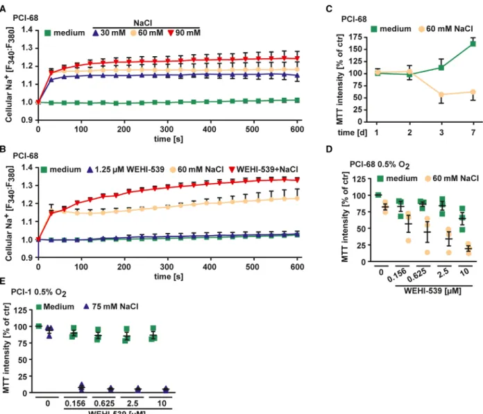
+influx and osmotic stress have previ- ously been linked to enhanced migration and prolifera- tion of cancer cells [28 – 30]. Indeed, NaCl supplementation of the cell culture media increased intracellular Na
+levels in HNSCC cells in a dose-de- pendent manner (Fig. 6A). Combinatorial treatment with WEHI-539/NaCl resulted in more pronounced cytoplasmic Na
+accumulation, while WEHI-539 alone had no effect (Fig. 6B). However, proliferation and migration of HNSCC cells cultured under hyperos- motic conditions were even reduced (Figs 6C and 7A,
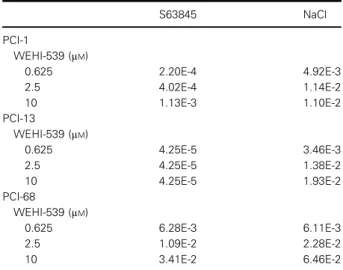
Table 1.Combination index of BCL-XL-targeting BH3 mimetics in the presence of MCL-1 inhibitors or hyperosmotic stress.
Synergistic effects on WEHI-539-mediated cell death by combinatorial treatment with the MCL-1-selective inhibitor S63845 (2.5µM) and NaCl-induced hypertonicity (PCI-1: 75 mM NaCl, PCI- 13: 90 mMNaCl, PCI-68: 60 mMNaCl). Synergism in terms of cell death induction was quantitatively assessed by calculating the CI from mean values from three independent experiments. CI values<1 are considered synergistic, and CI values >1 indicate antagonistic effects.
S63845 NaCl
PCI-1
WEHI-539 (µM)
0.625 2.20E-4 4.92E-3
2.5 4.02E-4 1.14E-2
10 1.13E-3 1.10E-2
PCI-13
WEHI-539 (µM)
0.625 4.25E-5 3.46E-3
2.5 4.25E-5 1.38E-2
10 4.25E-5 1.93E-2
PCI-68
WEHI-539 (µM)
0.625 6.28E-3 6.11E-3
2.5 1.09E-2 2.28E-2
10 3.41E-2 6.46E-2
B). Hyperosmotic stress also efficiently boosted cyto- toxicity of BCL-XL inhibition under low oxygen con- ditions (Fig. 6D,E), which are commonly encountered in HNSCC and associated with poor treatment response [31]. Thus, hypertonic conditions did not enhance proliferation of HNSCC cells but efficiently inhibited MCL-1 even in a hypoxic tumor environ- ment.
BCL-XL inhibition under hyperosmotic stress efficiently kills HNSCC cells that poorly respond to EGFR inhibition or radiotherapy
Treatment options for HNSCC are scarce. Inhibitors of EGFR occupy the tyrosine kinase domain and are the only approved targeted drugs, but often display limited efficacy. Accordingly, erlotinib killed only up to ~ 50% of cells in the HNSCC cell lines tested (Fig. 8A – F). With the notable exception of PCI-1 cells, hyperosmotic stress did expectedly not aggravate cytotoxicity of erlotinib. In contrast, counteracting MCL-1 (either through pharmacological inhibition or
by establishing hypertonic conditions) enabled a full- blown killing of all cell lines upon treatment with the BCL-XL inhibitor WEHI-539 (Fig. 1C,D,F,H,I).
In HNSCC treatment, radiotherapy is still the main- stay and resistance is a major cause of poor survival rates [2]. Exposure to high radiation doses (12 Gy) killed only a fraction of HNSCC cells within 72 h, irrespective whether cells were cultured under iso- or hypertonic conditions (Fig. 8G – I). Targeting BCL-XL under hypertonic conditions, however, was sufficient for near-complete cancer cell elimination within 24 h even in the absence of radiation. These data recapitu- lated the limitations of currently available HNSCC treatments and indicated that approaches targeting antiapoptotic BCL-2 family members could comple- ment existing therapies.
Discussion
Our study highlights that hyperosmotic stress in the tumor environment (a) shifts dual BCL-XL/MCL-1 dependency of HNSCC cells to exclusive BCL-XL
Fig. 2.BCL-XL inhibition under hyperosmotic conditions triggers apoptosis. (A) PCI-68 cells were transfected with siRNA oligonucleotides targeting BAX or nontargeting control. After 48 h, cells were challenged with the indicated concentration of WEHI-539 presence and absence of NaCl for another 18 h. (B) Cells were challenged with WEHI-539 (0.625µM) in the presence and absence of trehalose (180 mM) for 18 h. (C) PCI-68 cells were treated with WEHI-539 (1.25µM) in the presence and absence of NaCl (60 mM). After 8 h, cells were analyzed by flow cytometry for 7-AAD- and annexin-V positivity. (D) Cells were challenged with the indicated concentrations of WEHI-539 in the presence and absence of NaCl and/or QVD-OPh (50µM). For (A), individual data points of two independent experiments are shown; for (B and D), data points and meanSEM from three independent experiments are shown; for (C), data shown are representative of two experiments performed. RFU, relative fluorescence units.
Fig. 3.Hypertonicity counteracts the antiapoptotic function of MCL-1. (A) Spheroids of PCI-68 cells were treated with WEHI-539 (1.25µM) in the presence and absence of NaCl (60 mM). Upper panel: changes in spheroid morphology upon NaCl/WEHI-539 treatment. Scale bar:
100µm. Lower panel: Cells were treated as above, but MTT staining was used to determine cell viability. (B) Spheroid cultures of PCI-68 cells were challenged with WEHI-539 (1.25µM) in the presence and absence of NaCl (60 mM) for 18 h. Caspase-3/-7 activity was assessed using the fluorogenic substrate (DEVD)2-R110. (C–G) Cells were challenged with the indicated concentrations of ABT-737, ABT-199, and S63845 in the presence and absence of the indicated concentrations of the osmotically active solutes NaCl (C–F) or trehalose (G). For (A), data shown are representative of two experiments performed; for (B), individual data points of two independent experiments are shown; for (C–G), data points and meanSEM from three independent experiments are shown. RFU, relative fluorescence units.
Fig. 4.NOXA upregulation is essential for hypertonicity-enforced BCL-XL addiction. (A) Cells were challenged with NaCl (PCI-68: 60 mM, PCI-1: 75 mM) for the indicated periods. After washing and lysis, western blot analysis was performed with antibodies specific for the indicated proteins. (B) PCI-68 cells were challenged with NaCl (60 mM) for the indicated periods. mRNA levels of genes encoding BCL-XL and MCL-1 were analyzed by qPCR. (C) Cells were challenged with the indicated concentrations of NaCl for the indicated periods. mRNA levels ofPMAIP1(encoding NOXA) were analyzed by qPCR. (D) Left panel: Cells were transfected with siRNA oligonucleotides targeting NOXA or BIM or nontargeting control. After 48 h, cells were treated with WEHI-539 (2.5µM) in the presence and absence of the indicated concentrations of NaCl for another 18 h. Right panel: Western blot analysis using specific antibodies for the indicated proteins assessed knockdown efficacy. For (A), data shown are representative of two experiments performed. For (B–D), data points and mean from at least two independent experiments are shown.
addiction in a NOXA-dependent manner; (b) does not promote ER stress, cancer growth, or migration; and (c) grants contextual synthetic lethality to BCL-XL inhibi- tors. The biophysical properties in the microenviron- ment of solid tumors (such as oxygen level, interstitial fluid pressure, and pH) differ greatly from healthy tis- sues and impair treatment response [32]. On the con- trary, components of the tumor environment itself emerged as therapeutic targets in HNSCC [33]. The tumor microenvironment also requires proactive adap- tion of cancer cells to ensure survival. Targeting pro- cesses or consequences of this environment-imposed cellular response are often detrimental and summarized in the concept of contextual synthetic lethality [34].
With this study, we took this approach a step fur- ther. Artificial manipulation of the osmotic pressure in the tumor environment enforces an adaptive cellular response that ‘accidentally’ compromises the cell’s anti- apoptotic safeguard mechanism. The readiness to undergo apoptotic cell death consequently increases:
HNSCC cells are primed for death. Apparently, lower- ing the threshold for cell death induction and exclusive BCL-XL addiction involves hypertonicity-induced upregulation of NOXA (Fig. 4C,D). This is in line with previous studies that reported a decisive role for NOXA in apoptosis induction upon inhibition of BCL-2 family proteins [11,12,35 – 37]. Although a Ca
2+- dependent mechanism of NOXA upregulation has been described [24], our findings suggest a protective rather than a cell death promoting role for the hyper- tonicity-induced rise in cytoplasmic Ca
2+(Fig. 5F).
The intracellular calcium response to hyperosmotic stress is well established in a variety of cell types, including cancer cells [38,39]. Calcium signaling in hypertonic environments can promote both osmoadap- tive processes and apoptosis [40–42]. In cancer cells, elevated intracellular Ca
2+levels boost efficacy of anti- cancer drugs [43]. In our experimental setup, however,
Ca
2+-induced survival pathways seem to be dominant (Fig. 5F).
Our study demonstrates that osmotic pressure (and potentially other biophysical factors) in the tumor microenvironment can change dependencies of cancer cells on antiapoptotic BCL-2 family proteins and therefore modulate the apoptotic threshold. These environment-related changes in mitochondrial priming should be taken into account when predicting sensitiv- ity of cancer cells toward inhibitors of BCL-2 family proteins. Solid tumors, for example, often exhibit ele- vated pressure in the tumor bulk due to fluid pressure and solid stress [44]. Mapping dependencies on BCL-2 family proteins under standardized, nonphysiological conditions in vitro might therefore inaccurately predict susceptibilities/resistances in vivo. Exploring the inter- dependency of biophysical factors in the tumor envi- ronment and death priming of cancer cells could also lay open previously unrecognized therapeutic targets.
Regardless of any properties of the tumor environ- ment, it is important to note that an intact intrinsic apoptosis pathway remains essential for efficient can- cer cell killing with BH3 mimetics.
The cellular response to hyperosmotic stress is com- plex and profoundly alters a cell’s state [19]. Immedi- ate effects occur within minutes and include osmotic efflux of water, cell shrinkage, increased intracellular ionic strength, DNA double-strand brakes, cytoskeletal rearrangements, and elevated levels of reactive oxygen species [45 – 47]. During the process of adaption (lasting hours to days), preexisting ion transport systems increase intracellular concentrations of potassium, sodium, and chloride ions, which ultimately causes water influx and a regulatory volume increase [48].
Multiple pathways signal activation of the transcrip- tion factor nuclear factor of activated T-cell 5 (NFAT5) and ensure increased expression/activity of stress proteins such as p53 and heat-shock proteins [49 – 51]. A cell accommodated to hypertonic stress
Fig. 5.Transition to exclusive BCL-XL dependency is not a consequence of ER stress or cytoplasmic Ca2+increase. (A) PCI-68 cells were challenged with NaCl (60 mM). Intracellular calcium levels were measured using the ratiometric Ca2+indicator Fura-2 in a Ca2+-containing Tyrode’s solution. (B) PCI-68 cells were challenged with WEHI-539 (1.25µM) in the presence and absence of NaCl (60 mM). Intracellular calcium levels were measured as above, but in a Ca2+-free Tyrode’s solution. (C) Cells were treated with NaCl (60 mM) in the presence and absence of the Ca2+ chelating agent BAPTA-AM and the ryanodine receptor antagonist ryanodine. Intracellular calcium levels were measured as above in a Ca2+-free Tyrode’s solution. (D) PCI-68 cells were challenged with NaCl (60 mM) in the presence of cyclosporine A (0.6 and 2.5 mM), which inhibits calcium release from the mitochondria via the permeability transition pore. Intracellular calcium levels were measured using the ratiometric Ca2+indicator Fura-2. (E) Cells were treated for the indicated periods with NaCl (60 mM) or the ER stress- inducing inhibitor tunicamycin (2µgmL 1). After washing and lysis, western blot analysis was performed with antibodies specific for the indicated proteins. (F, G) Cells were challenged with the indicated concentrations of WEHI-539 in the presence and absence of BAPTA-AM and ryanodine for 18 h. (H) PCI-68 cells were transfected with siRNA oligonucleotides targeting p53 or nontargeting control. After 48 h, cells were challenged with the indicated concentration of WEHI-539 in the presence and absence of NaCl for another 18 h. For (A–D and F– H), shown are data points and meanSEM from two or three independent experiments; for (E), data shown are representative of two experiments performed. CyA, cyclosporine A.
displays normal cell volume, ionic strength, cell cycle, and transcription/translation. Nevertheless, initial per- turbations such as DNA damage and increased protein oxidation persist [19]. Even after adaption, the state of a hypertonicity-exposed cell remains strikingly differ- ent from isotonic conditions. Notably, no specific osmosensor has been identified in mammalian cells thus far. Instead, all of the above-mentioned immedi- ate effects of hypertonicity can signal an osmoregula- tory response [19]. Earlier studies already indicated that adaption to hypertonic conditions can alter the
apoptotic threshold of cells. Hyperosmotic stress can repress translation of MCL-1 [52]. This could account for the functional loss of MCL-1 in HNSCC cells under hypertonic conditions (Fig. 1C,D,F,H,I) despite upregulation of MCL-1 mRNA (Fig. 4B). Facilitating the activation of the mitochondria-controlled apoptosis pathway also sensitized cancer cells to death receptor- induced apoptosis [53,54]. Our current study highlights hypertonicity-induced upregulation of NOXA as molecular link between osmotic pressure in the tumor environment and mitochondrial priming in HNSCC.
Fig. 6.Hyperosmotic stress reduces proliferation of HNSCC cells and boosts WEHI-539 cytotoxicity in a hypoxic environment. (A, B) PCI-68 cells were challenged with the indicated concentrations of NaCl in the presence and absence of WEHI-539 (1.25µM). Intracellular sodium levels were measured using the ratiometric Na+indicator SBFI. (C) PCI-68 cells were incubated with NaCl (60 mM) for the indicated periods.
Cell proliferation was determined by MTT staining. (D, E) Cells were cultured under low oxygen conditions (0.5%) and challenged with the indicated concentrations of WEHI-539 in the presence and absence of NaCl (60 mM) for 18 h. For (A, B, D, E), data points and meanSEM from three independent experiments are shown; for (C), meanSEM from three independent experiments is shown.
Nevertheless, the critically involved signaling pathways and/or osmoadaptive processes remain enigmatic.
We are aware that the complexity to establish hyper- tonic conditions in the tumor environment may be a
limiting factor for therapeutic exploitation of our find- ings. However, in solid tumors that are easily reach- able, direct delivery of osmotically active solutes or continuous release from implantable devices could be
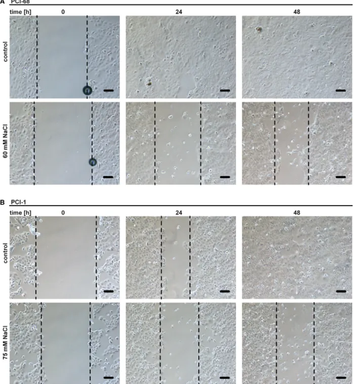
Fig. 7.Migration of HNSCC cells is reduced under hypertonic conditions. (A, B) Wound healing assays of PCI-68 and PCI-1 cells in the presence and absence of NaCl. Phase-contrast images were taken after 0, 24, and 48 h. Scale bar=50µm. The dashed lines indicate the growth front. For (A and B), data shown are representative of two experiments performed.
feasible and boost efficacy of our scarce armamentar- ium currently available for HNSCC treatment.
Materials and methods
Cell lines, antibodies, and reagents
SCC-9 and SCC-25 cells were obtained from LGC Stan- dards GmbH (Wesel, Germany). PCI-1, PCI-13, and PCI- 68 have been described previously
[55]. PCI-1, PCI-13, andPCI-68 cells were maintained in RPMI 1640 medium (PAN-Biotech, Aidenbach, Germany) with 10% (v/v) fetal bovine serum (Sigma, Steinheim, Germany). SCC-9 and SCC-25 cells were grown in a 1 : 1 mixture of Dulbecco’s modified Eagle’s medium (DMEM) and Ham’s F12 med- ium containing 1.2 g L
1sodium bicarbonate, 2.5 m
M L- glutamine, 15 m
MHEPES, and 0.5 m
Msodium pyruvate (Thermo Fisher, Waltham, MA, USA) supplemented with 400 ng mL
1hydrocortisone and 10% (v/v) fetal bovine serum. For spheroid cultures, cells were harvested, washed, and after centrifugation resuspended in DMEM supple- mented with 2% (v/v) fetal bovine serum, 2 m
M L-glutamine, 5 ng mL
1epidermal growth factor, and 0.24% (w/v) methylcellulose (Sigma). Cells were pipetted onto the lid of a petri dish (12.500 cells/drop), the lid was inverted, and the hanging drops were cultured for 2 days (37
°C, 5% CO
2).
The bottom of the petri dish was filled with 5 mL PBS for humidification. Subsequently, spheroids were harvested, seeded in 96-well Nunclon Sphera plates (Thermo Fisher), and challenged with the indicated substances. Irradiation of HNSCC cells was performed using an IBL 437C irradiator (CIS Bio International, Berlin, Germany) for 198 s (12 Gy) according to the manufacturer’s instructions. Oxygen levels were modulated using a Whitley H35 Hypoxystation (Don Whitley Scientific, Shipley, UK). The following antibodies were used: ATF4 (#11815), ATF6 (#65880), BID (#2002), BIM (#2933), BAX (#5023), BAK (#12105), BCL-2 (#2872), BCL-W (#2724) BCL-XL (#2764), p53 (#4866): Cell Signal- ing (Beverly, MA, USA); MCL-1 (ab32087): abcam (Cam- bridge, UK); tubulin (#MS-581): Dunnlab (Asbach, Germany); and NOXA (114C307, #sc-56169): Santa Cruz (Santa Cruz, CA, USA). The following chemicals were used:
MTT (3-[4,5-dimethylthiazol-2-yl]-2,5-diphenyl tetrazolium bromide), BAPTA-AM: Biomol, (Hamburg, Germany);
ABT-737, WEHI-539, QVD-OPh: Hycultec (Beutelsbach, Germany); and ryanodine and cyclosporine A: Santa Cruz;
SBFI and Fura-2: Thermo Fisher.
MTT-based cell viability assay
Cell viability was measured using MTT-based assays and has also been described in earlier studies
[56,57]. Cells were seededin 96-well plates (2
910
4cells/well) and challenged with the indicated concentrations of the indicated substances in dupli- cates (technical replicates). NaCl was added simultaneously.
Unless indicated otherwise, cell viability was determined 18 h after stimulation using MTT staining (2 h at 37
°C). Staining intensity was measured at 595 nm, and the mean was calcu- lated from the technical replicates of each experiment. The mean value of untreated controls was set to 100%. For any other condition, the MTT staining intensity is given relative to the corresponding untreated group (percentage of control).
Data points shown are mean values (calculated from two tech- nical replicates) of independent experiments (n
≥3).
Western blot analysis
Western blot analysis was essentially performed as described previously
[54].siRNA experiments
siRNA oligonucleotides targeting BAX (#L-003308), NOXA (#L-005275), BIM (#L004383), MCL-1 (#L- 004501-00-005), BCL-XL (#L-003458), p53 (#L-003329), and nontargeting control siRNA (#D-001810-10-05) were purchased from Dharmacon (Lafayette, CO, USA). PCI-1 and PCI-68 cells were transfected with 100 n
M(BAX, NOXA, MCL-1, BCL-XL, p53, nontargeting control) and 200 n
M(BIM) siRNA using DharmaFECT 1 transfection reagent (#T-2001-02) according to the manufacturer’s instructions.
Quantitative PCR
Two micrograms of total RNA was transcribed into com- plementary DNA using the high-capacity cDNA reverse transcription kit (Applied Biosystems, Carlsbad, CA, USA). TaqMan gene expression assays were as follows:
BCL2L1 (encoding BCL-XL): Hs00236329_m1; MCL1:
Hs01050896_m1; and PMAIP1 (encoding NOXA):
Hs00560402_m1 (Applied Biosystems). All assays were run
on an ABI Prism 7900 sequence detector (Applied Biosys-
tems). qRT-PCR reactions were performed in quadrupli-
cates for each sample and normalized to the expression of
Fig. 8.Targeting BCL-XL under hyperosmotic stress efficiently eliminates HNSCC cells that poorly respond to radiotherapy or EGFR inhibition. (A–F) Cells were challenged with the EGFR inhibitor erlotinib in the presence and absence of the indicated concentrations of NaCl for 18 h. (G–I) Cells were treated with the indicated concentrations of WEHI-539 in the presence and absence of NaCl (60 or 90 mM) and/or irradiated as indicated (12 Gy). Cell viability was assessed at days 1, 2, and 3 using MTT staining. For (A–F), data points and meanSEM from three independent experiments are shown; for (G–I), data points and mean from three independent experiments are shown.the housekeeping gene HPRT1 (Hs02800695_m1). SDS 2.1 software (Applied Biosystems) calculated the mRNA levels.
Caspase activity assays
Fluorescence-based assessment of caspase activity has also been described in our previous studies
[56,57]. Activity ofcaspase-3 and caspase-7 was measured using the caspase-3/- 7 activity kit (AAT Bioquest, Sunnyvale, CA, USA) according to the manufacturer’s instructions. A Victor 3 Multilabel Reader (PerkinElmer, Waltham, MA, USA) quantified emitted fluorescence.
Flow cytometry
Cell death was assessed by annexin-V and 7-AAD staining and has already been described in our earlier studies
[56,57]. In brief, cells were challenged with the indicatedconcentrations of WEHI-539 in the presence and absence of NaCl for 8 h. Afterward, cells were stained with 7-AAD and annexin-V (4
°C for 15 min in the dark) following the standard procedures and analyzed immediately using a FACSCanto flow cytometer (BD Biosciences, Heidelberg, Germany)
[58].Measurement of intracellular Na
+and Ca
2+concentrations
Cells were seeded on FluoroDish Plates (#FD3510-100;
World Precision Instruments, Sarasota, FL, USA). For Na
+measurements, cells were loaded with 10
µMSBFI (#S1264;
Thermo Fisher) for 90 min at 37
°C. For Ca
2+measure- ments, cells were loaded with 2
µMFura-2 (#F1221; Thermo Fisher) for 15 min at room temperature. Staining was per- formed in calcium-containing Tyrode’s solution (140 m
MNaCl, 4 m
MKCl, 1 m
MMgCl
2, 5 m
MHEPES, 1 m
MCaCl
2, 10 m
Mglucose) or calcium-free Tyrode’s solution (140 m
MNaCl, 4 m
MKCl, 2 m
MMgCl
2, 5 m
MHEPES, 1 m
MCaCl
2, 10 m
Mglucose). After washing with Tyrode’s solution, cells were mounted on the stage of an inverted epifluorescence microscope (IonOptix Cooperation, Westwood, MA, USA).
SBFI and Fura-2 were alternatingly excited at 340 and 380 nm. Emitted fluorescence was passed through a 515-nm long-pass filter, and acquisition with a photomultiplier was performed every 30 s for a duration of 10 min and analyzed using the IonWizard software (IonOptix Cooperation). After background subtraction, fluorescence intensities were normal- ized to F340/F380 at t
=0 min.
Wound healing assay
PCI-1 and PCI-68 cells were seeded in 6-well plates (2
910
5cells/well) and cultured to 90
–100% confluence. The monolayer was ‘scratched’ with a 100
µL pipet tip and
carefully washed with PBS. Subsequently, cells were chal- lenged with the indicated concentrations of NaCl and WEHI- 539. Pictures were taken after 0, 24, and 48 h of incubation.
Calculation of combination index
Calculation of CI has also been described in our previous study
[56]. Briefly, CI values were calculated with the freelyavailable software
COMPUSYNversion 1.0 (ComboSyn Inc., Paramus, NJ, USA) using the median effect/combination index isobologram method
[59]. In this model, CI values<
1 are considered synergistic and CI
>1 indicate antago- nistic effects. Strength of synergism can be further graded:
<