Detection of the intercellular adhesion gene cluster (ica) in clinical Staphylococcus aureus isolates
Nachweis des interzellulären Adhäsions-Gencluster (ica) in klinischen Staphylococcus aureus-Isolaten
Abstract
Staphylococcus aureusis a major hospital and community pathogen having the aptitude to cause a wide variety of infections in men. The
Amirmorteza
Ebrahimzadeh Namvar
1ability of microorganisms to produce biofilm facilitates them to withstand
Babak Asghari
1the host immune response and is recognized as one factor contributing
Fatemeh Ezzatifar
1to chronic or persistent infections. It was demonstrated that the
ica-encoded genes lead to the biosynthesis of polysaccharide intercel-
Gholamreza Azizi
2lular adhesion (PIA) molecules, and may be involved in the accumulation
Abdolaziz Rastegar Lari
1phase of biofilm formation. Different studies have shown the decisive role of theicagene as virulence factors in staphylococcal infections.
This study was carried out to demonstrate the relationship betweenica
gene and production of slime layer inS. aureusstrains. SixtyS. aureus 1 Medical Microbiology Department, Antimicrobial strains were isolated from patients. The isolates were identified morpho-
logically and biochemically following standard laboratory methods. After Resistance Research Center, identification, the staphylococcal isolates were maintained in trypticase Faculty of Medicine, Iran soy broth (TSB), to which 15% glycerol was added, and stored at –20°C. University of Medical
Science, Tehran, Iran Slime formation and biofilm assay was monitored. A PCR assay was
developed to identify the presence oficaD(intercellular adhesion gene) 2 Imam Hassan Mojtaba Hospital, Iran University of gene in all isolates. Thirty-nine slime producing colonies with CRA plates
(65%) formed black colors, the remaining 21 isolates were pink (35%). Medical Sciences, Tehran, In the quantitative biofilm assay 35 (58%) produced biofilm while 25 Iran
(42%) isolates did not exhibit this property. All isolates were positive for detection oficaDgene by PCR method. The interaction oficaAandicaD in the investigated isolates may be important in slime layer formation and biofilm phenomena.
We propose PCR detection of theicagene locus as a rapid and effective method to be used for discrimination between potentially virulent and nonvirulent isolates, with implications for therapeutic and preventive measures pertainin to the management of colonized indwelling cath- eters.
Keywords:Staphylococcus aureus, biofilm, intercellular adhesion gene, PCR detection
Zusammenfassung
Staphylococcus aureusist ein wichtiger nosokomialer und community- assoziierter Krankheitserreger, der verschiedene humane Infektionen verursachen kann. Durch die Fähigkeit von Mikroorganismen zur Bio- filmbildung wird ihre Widerstandsfähigkeit gegenüber der Immunabwehr mit der Folge chronischer oder persistierender Infektionen erhöht. Es wurde nachgewiesen, dass durchica-codierende Gene die Biosynthese des interzellulären Polysaccharid-Adhäsins exprimiert wird, das eine Hauptkomponente für die Akkumulation des Biofilms darstellt. In ver- schiedenen Studien wurde die kritische Rolle derica-Gene als Virulenz- faktor für Staphylokokken-Infektionen nachgewiesen.
Die vorliegende Untersuchung wurde durchgeführt, um den Zusammen- hang zwischen demica- Gen und der Schleimbildung durchS. aureus
1/4 GMS Hygiene and Infection Control 2013, Vol. 8(1), ISSN 2196-5226
Research Article
OPEN ACCESS
zu analysieren. Hierzu wurden 60S. aureusPatientenisolate identifiziert, morphologisch und biochemisch nach Standardmethoden charakterisiert und in Trypticase-Soja-Bouillon (TSB) mit Zusatz von 15% Glycerol bei –20°C aufbewahrt. Die Schleimbildung und Biofilmbildung wurde in einem speziellen Assay detektiert. Mittels eigens entwickelter PCR wurde alle Isolate auf das Vorkommen desicaD(intercellular adhesion gene) untersucht. Von den auf CRA-Platten Schleim produzierenden Kolonien waren 39 (65%) schwarz, die anderen 21 (35%) pinkfarben.
Im quantitativen Biofilmassay bildeten 35 der Isolate (58%) einen Bio- film. Bei all diesen Isolaten wurde dasicaD-Gen nachgewiesen.
Des lässt den Schluss zu, dass der PCR-Nachweis desica-Locus als rasche und effektive Methode zur Unterscheidung zwischen potentiell virulenten and avirulenten Isolaten herangezogen werden kann, um die Therapie und Prävention der Biofilmbildung auf Implantaten zu verbes- sern. Der Synergismus zwischenicaAund icaD in den untersuchten Isolaten scheint bedeutend für die Schleimbildung und Biofilmakkumu- lation sein.
Schlüsselwörter:Staphylococcus aureus, Biofilm, Interzelluläres Adhäsionsgene, PCR Detektion, Adhäsionsfaktor
Introduction
Staphylococcus aureusis an important pathogen causing a wide spectum of infections [1], [2]. A number of studies have been conducted to explain the structures and pathogenic mechanisms by whichS. aureus is able to cause serious infections [3], [4]. The ability ofS. aureus to produce biofilm enables this organisms to withstand the host immune response and is considered to be the cause of chronic or persistent infections, as biofilm cre- ation protects bacteria from opsonophagocytosis and antimicrobial agents [5]. Another concern related to this pathogen is increasing resistance to oxacillin and other antibiotics, but also distribution of multiresistant isolates within the hospital setting [6]. Staphylococcal pathogen- esis is multifactorial, involving a combination of adher- ence and biofilm formation [7]. Complex aggregations of microorganisms can form irreversible attachments to the surfaces and formation of biofilm [8]. Biofilm producing bacteria are the source for persistent or chronic infections [6]. The significance of biofilm production for the virulence ofS. aureuswas supported by a number of clinical and animal studies [9]. Cell aggregation and biofilm accumu- lation are mediated by the products of a gene locus composing of the genesicaADBandC, which encode the essential proteins for the production of polysaccharide intercellular adhesion (PIA) and capsular polysacchar- ide/adhesion (PS/A) inStaphylococcus spp. [10], [11].
It was demonstrated that theica-encoded genes are re- sponsible for the biosynthesis of the PIA, which contains N-acetylglucosamine as a main constituent and in the accumulation phase of biofilm formation, playing a crucial role in invasiveness ofS. aureus [1]. Different studies have shown the decisive role of theicagene as virulence factors in staphylococcal infections [4], [11].
In this study, we developed new primers specific foricaD and determined further the possible relationship between
icaD gene and producing slime layer in clinical isolates ofS. aureusstrains.
Materials and methods
1. Bacterial isolates and phenotypic identification
SixtyS. aureusstrains were isolated from patients and used in this study. Isolates were identified morphologically and biochemically by standard laboratory methods. The coagulase and DNase tests were performed for discrim- ination ofS. aureusfrom coagulase negative staphylo- cocci (CoNS). After identification, the staphylococcal isolates were maintained in trypticase soy broth (TSB), to which 15% glycerol was added, and stored at –20°C.
2. Slime forming colony
This test was used to evaluate slime formation in S. aureusstrains on CRA plates, as previously described [10]. Pink colonies were recorded as nonbiofilm producers while the black colonies were recognized as biofilm pro- ducers.
3. Biofilm assay
S. aureus strains were icubated in (TSB) at 37°C for 24 hours; grown colonies were diluted in 1:200 and incu- bated in microtiter plates. After 24 hours the wells were washed with PBS buffer two up to three times and left in the room temperature for drying. In the next step 0.4%
crystal violet solution was used as stain for 10 min. Finally the absorbance at 490 nm was determined; an OD of 490 nm >0.12 was regarded as a biofilm positive sample.
2/4 GMS Hygiene and Infection Control 2013, Vol. 8(1), ISSN 2196-5226
Namvar et al.: Detection of the intercellular adhesion gene cluster ...
4. Primer design and PCR amplification
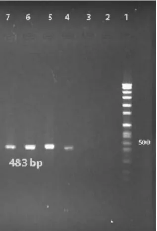
PCR assay was achieved to detect the presence oficaD (intercellular adhesion) gene in all isolates. For preparing DNA, colonies of bacteria were dissolved in 20 µl lysis buffer (0.25% SDS, 0.05N NaOH) and heated at 95°C for 5 min. In the second step, the lysate was centrifuged and diluted using distilled water. A second centrifugation step for 5 min at 16,000 g was performed to remove the cell debris. Supernatants were frozen at –20°C until further use. The primer sequencesF: 5' GAA CCG CTT GCC ATG TGT TG 3' (20 bp) & R: 5' GCT TGA CCA TGT TGC GTA ACC 3' (21 bp)for amplifying a 483 bp oficaDgene was designed from the published GenBank sequences (AF086783) with Alleleid 6 Primer Analysis Software. For recognition oficaD gene each 25 µl PCR mixture con- tained 2.5 µM of MgCI2, 100 µM of each dNTPs, 1U Taq DNA polymerase, and 1 µM of each primer with 200 ng of the DNA sample. PCR amplification was carried out with use of a DNA Thermal cycler (Eppendorf Master cycler personal) with an initial denaturation step (2 min at 95°C) followed by 30 cycles of amplification (denaturation at 95°C for 30 s, annealing at 57.5°C for 30 s and elonga- tion at 72°C for 40 s). Steps 2–4 were repeated 29 more times. Finally, 7 µl of PCR mixture was analyzed by 1.5%
agarose gel electrophoresis. After electrophoresis, gels were seen under UV light. The Gene Ruler TM 100 bp DNA Ladder Plus (Fermentas, Germany) was used as a DNA ladder.
Figure 1: PCR results ofica gene for S. aureus
Results
The investigated 60 strains were isolated from samples of blood catheter (40%), urine catheter (27.5%), sputum (8.3%), wounds (10%), and tracheal tube (14.2%), during the period October 2011 to February 2012. Thirthy-nine slime producing colonies with CRA plates (65%) formed black colors and, the remaining 21 were pink (35%). In the quantitative biofilm assay 35 (58%) produced biofilm, while the remaining 25 (42%) isolated did not exhibit this ability. All of these isolates were positive for detection of icaDgene by PCR method (Figure 1).
Discussion
S. aureusis responsible for infections in humans. It has been demonstrated that strains having an ability to form biofilm cause additionally “chronic polymer-associated”
infection [9], [12], mostly een as implant-associated in- fections. Biofilm support the adhesion and colonization ofS. aureuson surfaces, frequently leading to persistent and difficult to eradicate infections [4], [7]. Indeed, an increasing number of differentS. aureusadhesion mol- ecules is found [13]. Theicaoperon ofS. aureusandS.
epidermidiscontainica (ADBC)that allowStaphylococcus spp. to form slime layers and biofilm. It was shown that the presence oficaAgene in microorganisms yielded from indwelling catheter samples from patients hospitalized for one week was notably frequent. Cramton et al. [14]
demonstrated the presence of icaA gene in S. aureus strains, which was confirmed by Arciola et al. [1] describ- ing 23S. aureusstarins isolated from 14 catheter asso- ciated infections with the ability to form slime layer based on te presence of the geneicaA,but alsoicaD. Indeed, while icaA is required to encode N-acetylglucosaminyl- transferase, coexpression oficaDcan increase the cap- sular polysaccharide phenotypes [15], indicating a signi- ficant role of theicaDlocus as a virulence factor in the pathogenesis ofS. aureusisolated from catheters [13], [16].
Infections associated with the use of invasive medical devices, e.g., catheters, are mainly due to S. aureus, particularly those strains which create an extracellular slime and parts of the biofilm, making clinical treatment extremely challenging. The pocess of biofilm formation needs polysaccharide intercellular adhesion, which is synthesized by the enzymes encoded by the intercellular adhesion cluster (ica). In this study, the percentage of slime producing strains was 58%, while 65% of these had black colonies on CRA plate. However, all isolates contain icaDgene. Our study showed thatS. aureusisolates had no ability to form biofilm unless they were positive for icaDgene. Subsequently, the PCR product was purified from the gel and sequenced (sequence accession no.:
JN226155).
Since the presence of adhesion molecules is required for the establishement of an infection, the presence of ica adhesion genes may explain the role of the various adhe-
3/4 GMS Hygiene and Infection Control 2013, Vol. 8(1), ISSN 2196-5226
Namvar et al.: Detection of the intercellular adhesion gene cluster ...
sion mechanisms in the pathogenesis infection associ- ated with in-dwelling medical devices [17], [18]. It can be concluded that infections caused byicalocus carriing S. aureusstrains can lead to clinically difficult to treat conditions. The detection of the ica locus in clinical S. aureusisolates may improve the clinical decision for treatment and prevention options, and could support development of strategies to interact the bacterial cap- acity to colonize and invade in-dwelling medical devices.
PCR detection of the ica operon may be an effective method to differentiate between virulent and non virulent strains. Finally, the synergistic effect oficaA and icaD genes in the clinicalS. aureusstarines investigated here may be important to further understand slime layer formation and biofilm phenomena.
Notes
Competing interests
The authors declare that they have no competing in- terests.
References
1. Arciola CR, Baldassarri L, Montanaro L. Presence of icaA and icaD genes and slime production in a collection of staphylococcal strains from catheter-associated infections. J Clin Microbiol.
2001 Jun;39(6):2151-6. DOI: 10.1128/JCM.39.6.2151- 2156.2001
2. Gordon RJ, Lowy FD. Pathogenesis of methicillin-resistant Staphylococcus aureus infection. Clin Infect Dis. 2008 Jun 1;46 (Suppl 5):S350-9. DOI: 10.1086/533591
3. O'Neill E, Pozzi C, Houston P, Smyth D, Humphreys H, Robinson DA, O'Gara JP. Association between methicillin susceptibility and biofilm regulation in Staphylococcus aureus isolates from device- related infections. J Clin Microbiol. 2007 May;45(5):1379-88.
DOI: 10.1128/JCM.02280-06
4. Rohde H, Frankenberger S, Zähringer U, Mack D. Structure, function and contribution of polysaccharide intercellular adhesin (PIA) to Staphylococcus epidermidis biofilm formation and pathogenesis of biomaterial-associated infections. Eur J Cell Biol.
2010 Jan;89(1):103-11. DOI: 10.1016/j.ejcb.2009.10.005 5. Foster TJ. Immune evasion by staphylococci. Nat Rev Microbiol.
2005 Dec;3(12):948-58. DOI: 10.1038/nrmicro1289 6. Martín-López JV, Pérez-Roth E, Claverie-Martín F, Díez Gil O,
Batista N, Morales M, Méndez-Alvarez S. Detection of Staphylococcus aureus Clinical Isolates Harboring the ica Gene Cluster Needed for Biofilm Establishment. J Clin Microbiol. 2002 Apr;40(4):1569-70. DOI: 10.1128/JCM.40.4.1569-1570.2002 7. Klug D, Wallet F, Kacet S, Courcol RJ. Involvement of adherence and adhesion Staphylococcus epidermidis genes in pacemaker lead-associated infections. J Clin Microbiol. 2003 Jul;41(7):3348- 50. DOI: 10.1128/JCM.41.7.3348-3350.2003
8. Stobie N, Duffy B, McCormack DE, Colreavy J, Hidalgo M, McHale P, Hinder SJ. Prevention of Staphylococcus epidermidis biofilm formation using a low-temperature processed silver-doped phenyltriethoxysilane sol-gel coating. Biomaterials. 2008 Mar;29(8):963-9. DOI: 10.1016/j.biomaterials.2007.10.057
9. Götz F. Staphylococcus and biofilms. Mol Microbiol. 2002 Mar;43(6):1367-78. DOI: 10.1046/j.1365-2958.2002.02827.x 10. Frank KL, Patel R. Poly-N-acetylglucosamine is not a major
component of the extracellular matrix in biofilms formed by icaADBC-positive Staphylococcus lugdunensis isolates. Infect Immun. 2007 Oct;75(10):4728-42. DOI: 10.1128/IAI.00640-07 11. Hall-Stoodley L, Costerton JW, Stoodley P. Bacterial biofilms: from the natural environment to infectious diseases. Nat Rev Microbiol.
2004 Feb;2(2):95-108. DOI: 10.1038/nrmicro821
12. Götz F. Staphylococci in colonization and disease: prospective targets for drugs and vaccines. Curr Opin Microbiol. 2004 Oct;7(5):477-87. DOI: 10.1016/j.mib.2004.08.014 13. Rooijakkers SH, van Kessel KP, van Strijp JA. Staphylococcal
innate immune evasion. Trends Microbiol. 2005 Dec;13(12):596- 601. DOI: 10.1016/j.tim.2005.10.002
14. Cramton SE, Gerke C, Schnell NF, Nichols WW, Götz F. The intercellular adhesion (ica) locus is present in Staphylococcus aureus and is required for biofilm formation. Infect Immun. 1999 Oct;67(10):5427-33.
15. Satorres SE, Alcaráz LE. Prevalence of icaA and icaD genes in Staphylococcus aureus and Staphylococcus epidermidis strains isolated from patients and hospital staff. Cent Eur J Public Health.
2007 Jun;15(2):87-90.
16. O'Gara JP. ica and beyond: biofilm mechanisms and regulation in Staphylococcus epidermidis and Staphylococcus aureus. FEMS Microbiol Lett. 2007 May;270(2):179-88. DOI: 10.1111/j.1574- 6968.2007.00688.x
17. Merlino J, Watson J, Rose B, Beard-Pegler M, Gottlieb T, Bradbury R, Harbour C. Detection and expression of methicillin/oxacillin resistance in multidrug-resistant and non-multidrug-resistant Staphylococcus aureus in Central Sydney, Australia. J Antimicrob Chemother. 2002 May;49(5):793-801. DOI: 10.1093/jac/dkf021 18. Otto M. Staphylococcal biofilms. Curr Top Microbiol Immunol.
2008;322:207-28. DOI: 10.1007/978-3-540-75418-3_10
Corresponding author:
Abdolaziz Rastegar Lari
Medical Microbiology Department, Antimicrobial Resistance Research Center, Faculty of Medicine, Iran University of Medical Science, Tehran, Iran, Phone:
+98218293181 lari@tums.ac.ir
Please cite as
Namvar AE, Asghari B, Ezzatifar F, Azizi G, Lari AR. Detection of the intercellular adhesion gene cluster (ica) in clinical Staphylococcus aureus isolates. GMS Hyg Infect Control. 2013;8(1):Doc03.
DOI: 10.3205/dgkh000203, URN: urn:nbn:de:0183-dgkh0002035
This article is freely available from
http://www.egms.de/en/journals/dgkh/2013-8/dgkh000203.shtml Published:2013-04-29
Copyright
©2013 Namvar et al. This is an Open Access article distributed under the terms of the Creative Commons Attribution License
(http://creativecommons.org/licenses/by-nc-nd/3.0/deed.en). You are free: to Share — to copy, distribute and transmit the work, provided the original author and source are credited.
4/4 GMS Hygiene and Infection Control 2013, Vol. 8(1), ISSN 2196-5226
Namvar et al.: Detection of the intercellular adhesion gene cluster ...