Vaccine Candidates against Infections caused by Staphylococcus aureus
Inaugural-Dissertation zur Erlangung des Doktorgrades der Mathematisch-Naturwissenschaftlichen Fakultät
der Universität zu Köln
vorgelegt von
Bettina Tosetti
aus Neuss
Tag der mündlichen Prüfung: 12.07.2010
1 Abbreviations 6
2 Introduction 8
2.1 Clinical relevance and pathogenicity ofStaphylococcus aureus . . . 8
2.2 Antibiotic resistance ofS. aureus . . . 10
2.3 Vaccination strategies againstS. aureus . . . 11
2.3.1 Active immunisation . . . 11
2.3.2 Passive immunisation . . . 12
2.3.3 Antibacterial function of immunoglobulins . . . 13
2.4 Aim of the study . . . 14
3 Material and Methods 15 3.1 Material . . . 15
3.1.1 Chemicals and enzymes . . . 15
3.1.2 Bacteria and culture media . . . 15
3.1.3 Buffers and solutions . . . 16
3.1.4 Technical equipment . . . 19
3.1.5 Consumables . . . 20
3.1.6 Kits . . . 20
3.1.7 Oligonucleotides . . . 21
3.1.8 Antibodies . . . 22
3.2 Methods . . . 23
3.2.1 Depletion of specific IgGs from IVIG . . . 23
3.2.2 S. aureusgrowth curves . . . 23
3.2.3 RNA isolation . . . 24
3.2.4 Synthesis and labelling of cDNA . . . 24
3.2.5 Hybridisation of microarrays . . . 25
3.2.6 Analyses of microarrays . . . 25
3.2.7 Quantitative realtime PCR (qPCR) . . . 26
3.2.8 Isolation ofS. aureusanchorless cell wall (ACW) proteins . . . 26
3.2.9 Subtractive proteome analysis (SUPRA) . . . 27
3.2.10 Cloning, expression and purification of vaccine candidates . . . 27
3.2.11 Enrichment of specific IgGs from IVIG . . . 28
3.2.12 Surface localisation of vaccine candidates . . . 29
3.2.13 In vitroopsonophagocytosis ofS. aureusby human neutrophils . . . 29
3.2.14 In vitroopsonophagocytic killing ofS. aureusby human neutrophils . 30 3.2.15 Immunisation of mice . . . 30
3.2.16 Murine model of sepsis . . . 30
3.2.17 Statistical analysis . . . 31
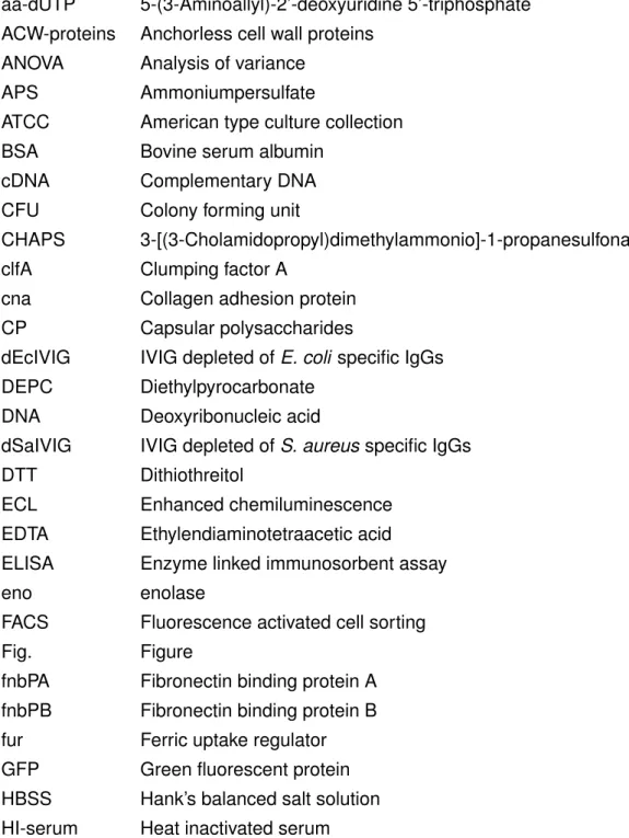
4 Results 32 4.1 Characterisation of the bacteriostatic effect mediated by S. aureus specific IgGs . . . 32
4.1.1 Human serum inhibitsin vitro growth ofS. aureus . . . 32
4.1.2 Intravenous immunoglobulin preparation (IVIG) specifically inhibitsin vitrogrowth ofS. aureus . . . 32
4.1.3 Bacteriostatic effect on S. aureus is mediated by S. aureus specific IgGs . . . 35
4.1.4 Gene expression profiling ofS. aureusover the course of bacteriostasis 37 4.2 Identification and characterisation of potential vaccine candidates against S. aureus . . . 46
4.2.1 Subtractive proteome analysis (SUPRA) of anchorless cell wall (ACW) proteins enables identification of potential vaccine candidates against S. aureus . . . 46
4.2.2 BT1, BT2 and BT3 are localised on the surface ofS. aureus . . . 49
4.2.3 Affinity purified IgGs specific for BT1, BT2 and BT3 trigger opsonophago- cytosis ofS. aureusby human neutrophilsin vitro . . . 49
4.2.4 Affinity purified IgGs specific for BT1 and BT2 mediate killing ofS. aureus by human neutrophilsin vitro . . . 51
4.2.5 Immunisation with recombinant BT1 and BT3 protects mice from death upon lethal challenge withS. aureus . . . 52
5 Discussion 56 5.1 Characterisation of the bacteriostatic effect mediated by S. aureus specific IgGs . . . 56
5.2 Identification and characterisation of potential vaccine candidates against Staphylococcus aureus . . . 59
6 Abstract 64
7 Zusammenfassung 66
8 Bibliography 68
9 Supplement 78
10 Danksagung 86
11 Erklärung 87
12 Lebenslauf 88
aa-dUTP 5-(3-Aminoallyl)-2’-deoxyuridine 5’-triphosphate ACW-proteins Anchorless cell wall proteins
ANOVA Analysis of variance
APS Ammoniumpersulfate
ATCC American type culture collection
BSA Bovine serum albumin
cDNA Complementary DNA
CFU Colony forming unit
CHAPS 3-[(3-Cholamidopropyl)dimethylammonio]-1-propanesulfonate clfA Clumping factor A
cna Collagen adhesion protein CP Capsular polysaccharides
dEcIVIG IVIG depleted ofE. coli specific IgGs DEPC Diethylpyrocarbonate
DNA Deoxyribonucleic acid
dSaIVIG IVIG depleted ofS. aureusspecific IgGs
DTT Dithiothreitol
ECL Enhanced chemiluminescence EDTA Ethylendiaminotetraacetic acid
ELISA Enzyme linked immunosorbent assay
eno enolase
FACS Fluorescence activated cell sorting
Fig. Figure
fnbPA Fibronectin binding protein A fnbPB Fibronectin binding protein B fur Ferric uptake regulator GFP Green fluorescent protein HBSS Hank’s balanced salt solution HI-serum Heat inactivated serum
hp2160 hypothetical protein similar to esterase (spot ID 2160) IEF Isoelectric focussing
IgG Immunoglobulin G
IgM Immunoglobulin M
IPG Immobilised pH gradient Isd Iron surface determinant
IVIG Intravenous immunoglobulin preparation IVIG-DS IVIG dialysed against PBS
kDa Kilo-Dalton
LB Luria Bertani medium
LD50 Dose that kills 50 % of tested animals
LPS Lipopolysaccharide
MALDI-TOF Matrix assisted laser desorption/ionisation time of flight MOI Multiplicity of infection
MSCRAMM Microbial surface component recognising adhesive matrix molecules MWCO Molecular weight cutoff
NHS-activated N-hydroxysuccinimide
OD Optical density
ON Over night
ORF Open reading frame
oxo 3-oxoacyl- (acyl-carrier protein) reductase PBS Phosphate buffered saline
pI Isoelectric point
PMN Polymorphonuclear neutrophils PSI Pounds per square inch
qPCR Quantitative real time PCR
RNA Ribonucleic acid
RT Room temperature
SD Standard deviation
SDS Sodium dodecyl sulfate
SERPA Serological proteome analysis SUPRA Subtractive proteome analysis Tris Tris(hydroxymethyl)aminomethan
2.1 Clinical relevance and pathogenicity of Staphylococcus aureus
Staphlococcus aureus is a facultatively anaerobic gram-positive coccus, belonging to the group of Staphylococcaceae. The coagulase positiveS. aureuscan be found as commen- sal on human skin, with the moist squamous epithelium of the anterior nares as its primary habitat (Roche et al., 2003). Although not showing signs of infection about 20 % of the human population is continuously colonised with S. aureusand another 60 % is transiently colonised. Thus, increasing the risk for invasive infections of endogenous origin (von Eiff et al., 2001).
As nosocomially or community acquired opportunistic pathogen,S. aureuscauses minor infections of the skin, but also serious life-threatening infections like pneumonia, endocardi- tis, sepsis and toxic shock syndrome (Lowy, 1998) in healthy and immunocompromised patients, representing the main causative agent of nosocomial infections (Laupland et al., 2003).
The genome ofS. aureusencodes a broad range of virulence factors, to effectively estab- lish an infection. To date more than 40 different virulence factors playing an important role in the pathogenicity of staphylococcal infections are known (Arvidson and Tegmark, 2001).
Functions of these virulence factors include adherence to host cell structures, evasion from the host immune system and active damage on host cells.
Microbial adhesion to host tissue is essential during the initial steps in the pathogenesis of most infections. Adhesion is mediated by protein adhesins of the MSCRAMM (microbial surface component recognising adhesive matrix molecules) family, which in most cases are covalently linked to the cell wall peptidoglycan (Foster and Hook, 1998). Common to these MSCRAMMs is an N-terminal signal peptide, enabling translocation of the protein across the membrane, and a C-terminal hydrophobic region, harbouring the conserved LPXTG motive essential for covalent linkage to the bacterial cell wall (Kronvall and Jonsson, 1999;
Patti et al., 1994; Schneewind et al., 1995). Most prominent members of this family are the fibronectin binding proteins (FnbpA and B) or clumping factor A (ClfA), a fibrinogen binding protein (Foster and Hook, 1998). So far in total 21 proteins containing the LPXTG motive have been identified inS. aureusby genome screenings (Roche et al., 2003).
Besides covalently linked protein adhesins, also anchorless cell wall proteins (ACW pro- teins) mediating adhesion to extracellular matrix components have been identified. These proteins lack a signal peptide as well as a common sorting signal like LPXTG and are secreted by a so far unknown mechanism. Upon secretion they are suggested to be re- associated to the bacterial cell surface (Chhatwal, 2002). One member of this new group of adhesins is staphylococcal enolase, which apart from its function as glycolytic enzyme mediates binding to laminin and thereby might contribute to tissue invasion and blood dis- semination (Carneiro et al., 2004).
Apart from adhesion, strategies for evasion from the host immune system are crucial to further promote effective colonisation. Protein A, a covalently linked surface protein harbouring the LPXTG motive, mediates immune evasion by binding to the FC portion of immunoglobulin G, thereby hampering efficient opsonisation and subsequent elimina- tion by professional phagocytes (Foster, 2005). Additional components interfering with the immune system are staphylokinase, the capsular polysaccharides and so called super- antigens. By binding to the T-cell receptor and the MHC class II molecule on antigen- presenting cells, superantigens trigger a T-cell activation that results in the release of pro- inflammatory cytokines (Llewelyn and Cohen, 2002). The excessive uncoordinated release of pro-inflammatory cytokines, in particular TNFα, is thought to be responsible for many of the clinical features of toxic shock syndrome (Miethke et al., 1992). In addition, S. aureus avoids the detrimental effects of oxygen free radicals that are formed during the respiratory burst in professional phagocytes by several mechanisms, including the yellow carotenoid pigment as scavenger or expression of two superoxide dismutases, which remove superox- ide radicals (Foster, 2005).
Secreted proteins like the pore-formingα-toxin, hemolysins and other cytolytic toxins (e.g.
leukocydins, Panton-Valentine-leukocydin) lead to the destruction of host cells (Tomita and Kamio, 1997). Besides this, other toxins cause food poisoning (enterotoxins A-O), the toxic shock syndrome (TSST-1) or the staphylococcal scalded skin syndrome, which is triggered by the exfoliative toxins A and B (Ladhani et al., 1999).
Another important feature for the pathogenesis of staphylococcal infections is for exam- ple the availability of iron in the iron-limited host environment. To this endS. aureusevolved strategies to sequester iron by synthesis of high affinity iron chelators, siderophores, and corresponding membrane associated ABC-transporter systems or covalently linked surface proteins (iron-regulated surface determinant; Isd A, B, C, and H) for the sequestration of heme complexed iron (Maresso and Schneewind, 2006). The importance of iron home- ostasis was confirmed by the reduced virulence of a S. aureus mutant lacking the ferric uptake regulator (fur) in a murine skin abscess model of infection (Horsburgh et al., 2001a).
In fact, S. aureus infections always depend on multiple virulence factors and the high re- dundancy in the function of virulence factors is supported by several in vivostudies using mutants lacking for example MSCRAMMs, but not preventing colonisation (Darouiche et al.,
1997; Patel et al., 1987; Peacock et al., 1999).
2.2 Antibiotic resistance of S. aureus
To date antibiotics are the only effective therapy againstS. aureusinfections. However, the steadily increasing incidence of S. aureus strains, resistant to multiple antibiotics aggra- vates the treatment, thereby leading to increased mortality byS. aureusinfections.
Today MRSA is the common denotation for those multiple resistant S. aureus, although it was initially used for ’Methicillin resistant S. aureus’ conferred by an additional penicillin binding protein (PBP2a). The mecA gene encoding PBP2a is located on a DNA element termed SCCmec (staphylococcal cassette chromosome mec), site-specifically integrated into the staphylococccal genome (Beck et al., 1986; Berger-Bachi et al., 1986).
For the treatment of infections caused by MRSA the glycopeptide antibiotic Vancomycin still represents the antibiotic of choice. However, due to the selective pressure during treat- ment, Vancomycin intermediate resistant S. aureus(VISA) evolved, first described in 1997 by Hiramatsu et al. (1997). In contrast to other antibiotic resistances this intermediate resis- tance against Vancomycin is not genetically encoded. In 2002 the first case of a genetically determined Vancomycin resistance (VRSA) was reported (Weigel et al., 2003). In this case the vanA gene, conferring resistance to Vancomycin, was horizontally transferred from a Vancomycin resistantEnterococcus faecalis during co-infection.
Until the late 1990s the predominant pattern of MRSA infections in the USA remained a nosocomially acquired disease, but then MRSA from community-acquired sources (CA- MRSA) without a history of intravenous drug use began to be described more frequently (Corriere and Decker, 2008). In 2006 the overall prevalence of CA-MRSA in the population was 59 %. Due to an almost three times longer stay for inpatients suffering fromS. aureus infection causing nearly thrice the cost compared to non-infected inpatients, infections with MRSA also became an economic problem. Moreover, the treatment of infections caused by MRSA, which accounts for almost 50 % of S. aureus related infections in several US hos- pitals, even doubles the cost caused by infections with a methicillin susceptible S. aureus (MSSA). The mortality rate associated with MRSA infections is with 20.7 % almost thrice as high than observed for infections caused by susceptibleS. aureus (6.7 %) (Corriere and Decker, 2008). Next to this also the prevalence of CA-MRSA is steadily increasing. In the United States a single clone of CA-MRSA (USA-300) has become the most prevalent cause of staphylococcal soft tissue infections acquired in the community (King et al., 2006;
Tenover et al., 2006) and has entered the inpatient setting, causing also invasive diseases (Davis et al., 2006; Gonzalez et al., 2006; Klevens et al., 2007).
The drastic increase in both nosocomially and community acquired MRSA related in- fections and thereby increased mortality rates highlight the pressing need for alternative strategies to prevent and treatS. aureusinfections.
2.3 Vaccination strategies against S. aureus
2.3.1 Active immunisation
Since first vaccination studies employing active immunisation using whole bacteria (Green- berg et al., 1987) or staphylococcal phage lysates (Giese et al., 1996) failed to achieve protective effects, single structures of S. aureus like the capsular polysaccharides (CP) or adhesion molecules of the MSCRAMM family got into focus of vaccine research. These attempts achieved at least a partial protection againstS. aureusinfections.
By prevention of phagocytic activity the CPs represent an important factor in the patho- genicity of S. aureus (Kampen et al., 2005). With over 80 % serotypes 5 and 8 are the most prevalent of the 11 different serotypes amongS. aureus isolates (Sompolinsky et al., 1985; Poutrel et al., 1988). Since purified CP5 and CP8 demonstrated only weak immuno- genicity in mice, purified CPs were conjugated to a non-toxic variant of Exotoxin A (ETA) of Pseudomonas aeruginosato increase immunogenicity. The conjugate induced high titres of CP-specific antibodies and a T-cell dependent immune response (Fattom et al., 1990). Fur- thermore, active immunisation using a bivalent CP5-CP8 conjugate vaccine (StaphVaxTM; Nabi Biopharmaceuticals) led to an increased survival rate in mice (Fattom et al., 2004), but failed to pass the Phase III clinical trial in end stage renal disease patients. A sec- ond Phase III trial in haemodialysis patients also failed, hence halting the development of StaphVax (Garcia-Lara and Foster, 2009). Currently a combined vaccine including CP5, CP8, polysaccharide component 336 and two staphylococcal toxins is under investigation (Pentastaph; http://www.nabi.com).
The polysaccharide intercellular adhesin of S. aureus and S. epidermidis is a poly-N- acetylglucosamine (PNAG) that enables adhesion to inert surfaces and living tissues. Im- munisation using PNAG achieved moderate protection in rabbit models of endocarditis or catheter-associated bacteraemia and a murine model of kidney infection (Maira-Litran et al., 2004).
Also secreted extracellular molecules like TSST-1 have been tested in immunisation studies. For this purpose either a non-toxic mutant or formaldehyde inactivated TSST-1 was used for vaccination of rabbits and achieved a protection from lethal challenge with S. aureus (Hu et al., 2003). However, due to the high variability in the set of toxin genes and their expression between different S. aureus strains, toxins alone are most likely no promising vaccine target.
Due to their function and surface localisation cell wall associated proteins represent promising targets. To date several cell wall anchored proteins harbouring the LPXTG motive have been tested in immunisation studies. For example, improved survival of mice was ob- served upon immunisation with the collagen binding protein (Cna) in a murine model of sep- sis (Nilsson et al., 1998). However, as Cna is not present in allS. aureusstrains, it is not el- igible as single component vaccine. In another attempt immunisation with IsdA, IsdB, SdrD
or SdrE significantly reduced the bacterial load in infected kidneys (Stranger-Jones et al., 2006). Moreover, as observed in the same study immunisation with a multicomponent vac- cine consisting of IsdA, IsdB, SdrD, and SdrE induced opsonophagocytic antibodies against each component in immunised mice, reduced the load ofS. aureusin kidneys and improved survival upon lethal challenge withS. aureus. The iron surface determinant B (IsdB) is also the basis of Merck’s V710 vaccine (Garcia-Lara and Foster, 2009), for which a Phase II clini- cal trial has just been completed (Feb 2010; http://clinicaltrials.gov/ct2/show/NCT00572910).
Immunisation with IsdA and Clumping factor B (ClfB), two major determinants in nasal car- riage, decreased nasal colonisation in a cotton rat and murine model of nasal carriage, respectively (Clarke et al., 2006; Schaffer et al., 2006). Thereby representing a potential option to restrict the spread of CA-MRSA.
Altogether, much effort has been made to characterise potential vaccine candidates in in vivo infection models and some promising results have been achieved. But these re- sults, especially those already tested in clinical trials, strongly suggest that due to the high variability in the spectrum of virulence factors for each strain as well as the redundancy in protein function, a single component vaccine will not be sufficient to confer protection.
The results obtained by Stranger-Jones et al. (2006) point towards a polyvalent vaccine as the most promising strategy to combat S. aureus infections. Thus, the identification and characterisation of eligible candidates is pivotal for the development of a successful multi- component vaccine. In this regard ACW proteins of S. aureus represent a new interesting class of vaccine candidates, since they exhibit both important enzymatic function and sur- face localisation.
2.3.2 Passive immunisation
Since active immunisation will most likely require a booster immunisation to achieve full protection, it is not representing a treatment option for already infected patients or patients short before surgery. In these cases, passive immunisation might represent an interesting alternative. Especially, as it is also applicable in immunocompromised patients, not able to trigger a sufficient humoral response upon active immunisation.
In the pre-antibiotic era the so-called serum therapy represented the first attempt of pas- sive immunisation to overcome infections (Casadevall and Scharff, 1994). Although effec- tive, it caused severe side effects, due to poor purity of the antibody fractions used. Since then antibody purification methods have vastly improved, thereby circumventing adverse effects due to impurities. For example intravenous immunoglobulin preparations (IVIG) de- rived from human plasma are routinely used in immunocompromised patients to provide antibodies against various pathogens and thereby support the immune system.
Several attempts have been made to test for protective effects by passive immunisation against different target molecules. For example a hyperimmune polyclonal immunoglobulin
preparation targeting CP5 and CP8 obtained from volunteers immunised with StaphVaxTM (Altastaph; Nabi Biopharmaceuticals) failed to prove efficacy in two Phase II clinical trials (Benjamin et al., 2006; Rupp et al., 2007).
Biosynexus has developed a humanised mouse chimeric IgG1 monoclonal antibody with in vitro opsonophagocytosis activity to S. epidermidislipoteichoic acid (LTA), a membrane bound glycolipid extending into the cell wall of gram-positive bacteria (Pagibaximab). It conferred around 80 % protection against lethal challenge withS. aureusin rodent models (Weisman, 2007), and Phase I/II clinical trials in low birth weight (LBW) neonates demon- strated that it is non-immunogenic, opsonic against coagulase negativeS. aureus and re- duces the incidence of bacteraemia (Weisman et al., 2009).
Another target under investigation is clumping factor A (ClfA), which belongs to the family of MSCRAMMs. ClfA mediates adhesion by binding to host ligands like fibrinogen and was previously shown to be required for virulence in a murine septic arthritis model (Josefsson et al., 2001). Passive immunisation with either rat or rabbit anti-ClfA polyclonal antibodies conferred protection in murine models of sepsis and arthritis. Veronate® (Inhibitex) is a human immunoglobulin preparation from plasma donors with naturally occurring high titres of IgG to ClfA and to theS. epidermidisfibrinogen binding protein (SdrG). Although protec- tive in animal models (Vernachio et al., 2006), it failed to meet its primary endpoint, a 50 % reduction in late onset S. aureus bacteraemia, in a Phase III clinical trial in LBW neonates (DeJonge et al., 2007).
These results further underline the necessity of the identification ofS. aureusprotein can- didates conferring protection by either active or passive immunisation, as components of a polyvalent vaccine against S. aureus. The major advantage of passive immunisation is the option to combine antibodies against multiple targets exhibiting different functions, e.g. neu- tralisation of bacterial toxins and opsonisation for subsequent elimination by professional phagocytes, thereby further increasing the protective potential of a polyvalent vaccine.
2.3.3 Antibacterial function of immunoglobulins
The main antibacterial functions of immunoglobulins are 1) opsonisation and in turn Fc γ-receptor mediated phagocytosis by professional phagocytes such as neutrophils or ma- crophages, 2) the neutralisation of adhesion to host cell tissue by binding to the respective pathogen associated protein, and 3) neutralisation of bacterial toxins. Besides, immune complexes of antibodies and toxins, which are not sufficient to induce Fc γ-receptor de- pendent uptake by phagocytes, are eliminated by activation of the classical pathway of complement activation. Additionally, antibodies have also been shown to amplify or sup- press the inflammatory response depending on their specificity, isotype and concentration (Casadevall and Pirofski, 2003).
Apart from this indirect antimicrobial activities, there are some examples for direct an-
timicrobial actions mediated by antibodies. It was shown that IgM or IgG specific for Bor- relia burgdorferi surface proteins damage the surface protein coat of the organism, thereby leading to a bactericidal effect in the absence of complement (Connolly and Benach, 2001;
Connolly et al., 2004). Furthermore, an antibody specific forEscherichia coliLPS was found to be bacteriostatic due to interference with the release of an iron chelator (enterochelin), thus preventing iron uptake (Fitzgerald and Rogers, 1980). Similarly, IgM monoclonal an- tibodies specific for surface proteins of Acinetobacter baumanii have been reported to be bactericidal by inhibition of iron uptake (Goel and Kapil, 2001).
In 1971 it was reported that human serum exhibits a bacteriostatic effect on S. aureus (Ehrenkranz et al., 1971). The underlying mechanism is not yet described, but an antibody directly inhibiting the growth S. aureus would be of great benefit in the context of passive immunisation.
2.4 Aim of the study
Due to the worldwide increasing prevalence of S. aureus resistant to multiple antibiotics (MRSA) in both hospital and community as well as the rapid emergence of resistances against new antibiotics, the development of alternative strategies for the treatment and pre- vention of staphylococcal infections is essential. Because S. aureus infections always de- pend on a multitude of virulence factors and there is a high variability in virulence factor genes among S. aureusisolates, single target strategies are less promising. Furthermore, passive immunisation strategies are particularly advantageous, due to their short term ap- plicability and potential composition of immunoglobulins of different isotypes and function- alities.
The aim of this study was to investigate a potential bacteriostatic effect onS. aureuscon- ferred by specific IgGs, using an intravenous immunoglobulin (IVIG) preparation as source of naturally occurring, S. aureus specific IgGs. Due to the enormous potential of a direct bacteriostatic effect elicited by IgGs for passive immunisation, the mechanism underlying bacteriostasis was analysed by gene expression profiling during inhibition of growth.
Additionally, this study aimed to enlarge the pool of potential candidates for a polyvalent vaccine. Thus, immunogenic anchorless cell wall proteins ofS. aureuswere identified and characterised regarding their potential to confer protection against lethal challenge with S. aureusupon active immunisation in a murine model of sepsis.
3.1 Material
3.1.1 Chemicals and enzymes
Chemicals were of research grade and purchased from AppliChem, Merck, Sigma-Aldrich, ROTH or Becton Dickinson GmbH unless stated otherwise. Buffers and solutions were obtained from Bio-Rad or prepared using bi-distilled H2O from an EASYpure UV/UF H2O purification unit (Werner Reinstwassersysteme), degassed and sterilised by autoclaving or filtration through a 0.2 µm membrane, if necessary. Restriction enzymes were obtained from Fermentas and used in recommended buffers. PFU Polymerase was obtained from Promega and recombinant Lysostaphin from Sigma-Aldrich.
3.1.2 Bacteria and culture media
E. Coli Genotype Provider
E. coli K12 XL1Blue
recA1 endA1 gyrA96 thi-1 hsdR17
Stratagene supE44 relA1 lac [F’ proAB lacIqZ∆M15
Tn10 (Tet,r)]
DH5α
F-φ80dlacZ∆M15∆(lacZYA-argF)U169
Invitrogen recA1 endA1 tonA hsdR17(rK-mK+)
phoA supE44λ-thi-1 gyrA96 relA1
BL 21 F- dcm ompT hsdS (rB-mB-) gal Stratagene
S. aureus Resistance Provider
ATCC 29213 MSSA American Type Culture Collection ATCC 29213-GFP Ampicillin/ Inst. for Med. Microbiology,
Chloramphenicol Immunology and Hygiene, S. Leggio
Media / Antibiotics Composition
LB -Medium 1 % Tryptone; 0.5 % Yeast extract; 0.5 % NaCl; pH 7.0 LB-Agar 1 % Tryptone; 0.5 % Yeast extract; 0.5 % NaCl,
15 % Agar; pH 7.0
SOC-Medium
2 % Tryptone, 0.5 % Yeast extract, 0.5 % NaCl, 25 mM KCl,
100 mM MgCl2, 20 mM Glucose; pH 7.0 Ampicillin Stock solution: 50 mg/ml in H2O
Working concentration: 50 µg/ml Chloramphenicol Stock solution: 10 mg/ml in MeOH
Working concentration: 20 µg/ml Kanamycin Stock solution: 50 mg/ml in H2O
Working concentration: 50 µg/ml
3.1.3 Buffers and solutions
Buffers and Solutions Composition cDNA labelling
50 x aa-dNTP Mix 25 mM dATP, 25 mM dGTP, 25 mM dCTP, 15 mM dTTP, 10 mM aminoally-dUTP
1M Phosphate buffer 9.5 ml 1M K2HPO4 , 0.5 ml 1M KH2PO4; pH 8.5-8.7 Phosphate wash buffer 5 mM Phosphate buffer in 80 % Ethanol; pH 8.0 Phosphate elution buffer 4 mM Phosphate buffer; pH 8.0
Dye coupling buffer 100 mM Na2CO3; pH 9.0 (freshly prepared) Cy-Dyes content of one vial reconstituted in 73 µl DMSO, (GE Healthcare) stored dessicated at -20 ° C
Stop solution
100 mM NaOAc; pH 5.2 (Dye coupling)
Microarray hybridisation
20x SSC 3 M NaCl, 0.3 M Natriumcitrate; pH 7.0
Buffers and Solutions Composition
10 % SDS 100 g SDS in 1000 ml A. bidest.
Prehybridisation 3.5 x SSC, 0.1 % SDS, 10 mg/ml BSA in A. bidest
Wash A 1 x SSC, 0.05 % SDS
Wash B 0.06 x SSC
Agarose gels
50 x TAE buffer 40 mM Tris, 20 mM Acetic acid, 2 mM Na2EDTA, pH 8.5
10 x Loading buffer
200 mM Tris-Acetat, 5 mM Na2EDTA,
0,01 % Bromphenolblue, 50 % Glycerin (v/v), pH 8.0 at 37 ° C for 3 h stirring
1 Kb DNA-ladder 100 µl 1 Kb Plus DNA-Ladder (Invitrogen), 400 µl 10 x loading buffer, 500 µl A. bidest 1% Agarosegel 1 g Agarose, 100 ml 1x TAE-Puffer,
1 µl Ethidiumbromid (10 µg/µl) 1D- and 2D-SDS-PAGE
10 x Laemmli buffer 0.06 M Tris-HCl (pH 6.8), 2 % SDS, 25% Glycerol, 0.2 % Bromphenolblue, 10 % 2β-Mercaptoethanol Rehydration solution 8 M Urea, 2 M Thiourea, 40 mM DTT, 1 % CHAPS,
0.5 % Pharmalyte
Equilibration solution 50 mM Tris-HCL (pH 8.8), 8 M Urea, 2 M Thiourea, 30 % Glycerol, 2 % SDS, 0.05 % Bromphenolblue TT buffer 3M Tris-HCl, 0,3 % SDS, 1 mM EDTA; pH 8.5 TE buffer 10 mM Tris/HCl, 1 mM EDTA; pH 8.0
Silver staining
Fixation solution 50 % EtOH, 10 % Acetic acid
Incubation solution 30 % EtOH, 6.8 % NaOAc, 0.5 % Glutaraldehyde, 0.2 % Na2S2O3 x 5 H2O
Staining solution 0.1 % AgNO3, 0.02 % Formaldehyde Developing solution 2.5 % Na2CO3, 0.01 % Formaldehyde Stop solution 5 % Acetic acid
Buffers and Solutions Composition Coomassie staining
Staining solution 2.5 % Serva blue R250, 45 % Ethanol, 15 % Acetic acid De-colourisation solution 45 % Methanol, 15 % Acetic acid
Immunoblotting
TBST buffer (10x) 1.5 M NaCl, 0.5 M Tris, 0.5 % Tween 20, pH 7.4 PBST buffer (10x) 1.4 M NaCl, 27 mM KCl, 100 mM NaH2PO4,
18 mM KH2PO4, 0.2% Tween 20; pH 7.4 Blocking buffer 5 % skim milk powder, 2 % BSA in 1x TBST Transfer buffer 25 mM Tris-HCl, 192 mM Glycine, 20 % Methanol Stripping buffer 62.5 mM Tris-HCl, 2 % SDS,
100 mM 2β-Mercaptoethanol; pH 6.8 Colony Blot
Denaturing solution 0.5 M NaOH, 1.5 M NaCl
Neutralisation solution 1.5 M NaCl, 0.5 M Tris-HCl; pH 7.4 20 x SSC 3 M NaCl, 0.3 M sodium citrate; pH 7.0 Affinity chromatography
Recombinant proteins
10 x PBS 1.4 M NaCl 100 mM Na2PO4, 27 mM KCl, 18 mM KH2PO4; pH 7.4
Resuspension buffer 1 x PBS, 500 mM NaCl, 5 % Glycerol, EDTA-free complete protease inhibitor
Binding buffer 1 x PBS, 500 mM NaCl, 40 mM Imidazole; pH 7.4 Elution buffer 1 x PBS, 500 mM NaCl, 250 mM Imidazole; pH 7.4 Dialysis buffer 1 x PBS, 10 % Glycerol; pH 7.4
Antibody enrichment
Wash A (NHS column) 0.5 M Ethanolamine, 0.5 M NaCl; pH 8.3 Wash B (NHS column) 0.1 M NaOAc, 0.5 M NaCl; pH 4.0 Storage buffer 0.05 M Na2HPO4, 0.1 % NaN3; pH 7.0
1 x PBS 140 mM NaCl, 2.7 mM KCl, 10 mM NaH2PO4, (binding buffer) 1.8 mM KH2PO4; pH 7.4
Buffers and Solutions Composition
Elution buffer 0.1 M Glycine-HCl; pH 2.5 Neutralisation buffer 1 M Tris-HCl; pH 9.0
3.1.4 Technical equipment
Device Specification Provider
Centrifuge Megafuge 1.0 R Thermo Scientific
Colony counter Countermat FLASH IUL
Developer AGFA Curix 60 AGFA
Electrophoresis chamber Criterion Cell Bio-Rad Electrophoresis chamber Ettan Dalt II System GE Healthcare
ELISA-Reader MRX Tc Dynex Technologies
Flow cytometer FACScalibur BD Biosciences
FPLC system ÄKTATM Purifier GE Healthcare
French® Press Cell K20, 20000 PSI Thermo Scientific French pressure cell press French® Press Thermo Scientific
Gel documentation Gel Doc 2000 Bio-Rad
High-speed centrifuge RC 5 C plus Sorvall
IEF Multiphor II GE Healthcare
Incubator (bacteria) Kelvitron t Thermo Scientific
Incubator (cells) Hera Cell 240 Thermo Scientific
Lightcycler® Lightcycler® LC480 Roche
MALDI-TOF MS Reflex IV Bruker Daltonics
Microarray scanner Axon GenePix 4100 Axon Instruments
Microcentrifuge Centrifuge 5417 R Eppendorf
Microscope Axiovert 25 Zeiss
PCR cycler Thermocycler T3 Biometra
Power supply Power Pac 3000 Bio-Rad
Power supply for Multiphor II EPS-3501xL GE Healthcare
Roller-mixer Cat RM5 Zipperer
Spectrophotometer Genesis 20 Thermo Scientific
Spiral plater Eddy Jet IUL
Tank-Blotter Criterion blotter (9.4x15 cm) Bio-Rad
Device Specification Provider Tank-Blotter Trans-Blot cell (16x20 cm) Bio-Rad
Test-Tube-Rotator Model 34528 Snjiders
Thermomixer Comfort Eppendorf
3.1.5 Consumables
Designation Manufacturer
Bottle-Top-Filter PES, 0.45 µm Nalgene Centricon-Plus 70, 30 000 MWCO Millipore Corning hybridisation chambers Corning CriterionTM Empty Cassettes Bio-Rad
Amersham Hyperfilm ECL GE Healthcare
Eddy-Jet tips IUL
HiPrepTM 26/10 Desalting Column GE Healthcare HisTrapTM FF crude Columns GE Healthcare HiTrapTM NHS-activated HP Columns GE Healthcare
IEF-Electrode Strips GE Healthcare
ImmobilineTM Dry Strips (3-10NL/4-7 NL) GE Healthcare LifterslipsTM22 x 22 1 mm Implen
LightCycler® 480 Multiwell Plate 96, white Roche MaxisorpTM flat-bottom 96-well plate Nunc Mueller-Hinton Agarplatten Oxoid
Nitrocellulose Schleicher & Schuell
Sterifix® Injection Filter, 0.2 µm Braun
3.1.6 Kits
Designation Manufacturer
DC-Protein Assay Bio-Rad
AmershamTM ECLTM Detection Reagent GE Healthcare Ettan Dalt II Gel and Buffer Kit GE Healthcare LightCycler® 480 SYBR Green I Master Roche
Designation Manufacturer
QIAprep Spin Miniprep Qiagen
QIAquick PCR Purification Qiagen
RNeasy Mini Kit Qiagen
Superscript® III first strand synthesis system Invitrogen OptiEIATM TMB Substrate Reagent Set BD Biosciences
3.1.7 Oligonucleotides
Desalted oligonucleotides were obtained from Operon and a 100 µM stock-solution ( TE;
pH 8.0) was prepared and stored at -20 ° C. Primers were used in a final concentration of 5 pmol/µl for cloning of the vaccine candidates or 10 pmol/µl for qPCR, respectively.
Gene Name Sequence (5’ to 3’)
BT1 BT1-f CCA TGG CTA AAT CAG TGG CTA
BT1-r AAC CTC GAG CAA CTC TGC GAT TAC
BT2 BT2-f GTT CCA TGG GTC ATC AAG CAG ATG
BT2-r GTG CTC GAG TCT ACT TTG CAA GTA
BT3 BT3-f CCA TGG GAA CAC CAA TTA TAG C
BT3-r GCC CTC GAG TTT TGC ACC TTC TAA qPCR 16SrRNA 16S-f TCG GGG GAC AAA GTG ACA GGT
16S-r AGA GTG CCC AAC TTA ATG ATG GC
qPCR fur fur-f GTT ACC AGA AGT TGA AAA TCG AGT TGA fur-r CTA TCC TTT ACC TTT AGC TTG GCA CG qPCR fhuC fhuC-f ACC TGA AGT AGC AGA TGG CTT AAC TG
fhuC-r CCA ATC AAT TTC TTT CTT ATC CTC AGC qPCR fhuD2 fhuD2-f AGC AAT TGG ACA AGA TGC AAC AGT
fhuD2-r ATG CTT GAT ATA ATA CTT CTC CAC CAC G qPCR ribA ribA-f GAT TGT GGT GCT CAA CTT GAA TCG
ribA-r ATT TGT TTA ACA ATC CTA TGC CAC GAC qPCR catalase cata-f ATA TTC TCT GAA ATA GGT AAG CAA ACC G
cata-r TTA ACG CAA ATC CTC GAA TGT CAC
Gene Name Sequence (5’ to 3’)
qPCR ferritin ferri-f CAG CAC CAA AAA TTG ACT TTT CAA G ferri-r TCT TGA CGA GCG ATT TCA GAT AAG TTA
3.1.8 Antibodies
Antibody Characteristics Provider
Octagam® (IVIG)
Intravenous immunoglobulin preparation Octapharma 1:500
Venimmun® N (IVIG)
Intravenous immunoglobulin preparation Aventis Behring 1:500
Anti-6-His-tag
monoclonal mouse anti 6-His-tag clone 13/45/31 Dianova 1:5000
Anti-human IgG
Polyclonal HRP-goat anti-human IgG Sigma 1:2500
Anti-human FCγ
Polyclonal PE-goat anti-human FCγF(ab)2 Dianova 1:100
Anti-mouse IgG
Polyclonal HRP-goat anti-mouse IgG Sigma 1:5000
3.2 Methods
3.2.1 Depletion of specific IgGs from IVIG
S. aureus ATCC 29213 or E coli K12 XL1blue from an overnight (ON) culture was inocu- lated 1:100 in 400 ml LB and cultured until an optical density of 2.0 at 600 nm (OD600) was reached. Subsequently bacteria were harvested by centrifugation, washed in phosphate- buffered saline (PBS) (pH 7.3) and resuspended in an IVIG preparation (Octagam®, Oc- tapharma) or PBS as control. The suspension was rotated slowly ON at 4 ° C, subsequently bacteria were removed by centrifugation, and the supernatant was sterile filtered through a 0.2 µm membrane filter. Protein concentration of IVIG depleted ofS. aureus (dSaIVIG) or E. coli specific IgGs (dEcIVIG) was determined using the Bio-RAD DC assay according to the manufacturer’s instructions.
3.2.2 S. aureusgrowth curves
S. aureus ATCC 29213 from ON culture was inoculated 1:100 into LB broth and cultured until OD600of 0.3 was reached. Subsequently bacteria were harvested by centrifugation at 4 ° C, 3000 g for 15 minutes. After washing once with cold PBS, the number of S. aureus colony forming units (CFU) was estimated by OD measurement, based on the observation that an OD600 of 3 equates to 1 x 10⁹ CFU/ml. For experimental assays 1 x 10⁴ CFU were inoculated in 500 µl LB broth. 13 mg / ml of either IVIG, BSA, dialysed IVIG (IVIG-DS), dSaIVIG or dEcIVIG adjusted in 500 µl PBS or solely PBS were added (final volume = 1 ml). Where indicated 25 or 50 % of the total volume (1 ml) were substituted by human serum, heat-inactivated (HI) human serum, IVIG or PBS (control). At defined time points (every hour up to 5 hours) samples were taken, diluted 1:2 in 0.1 % Triton X 100, sonicated for 5 minutes and 50 µl of suitable dilutions were plated on Mueller Hinton agar using the Eddy jet spiral plater (IUL Instruments). Plates were incubated at 37 ° C for 16 hours and CFUs were enumerated using the Countermat Flash (IUL Instruments). Experiments were performed using triplicates.
To enable expression profiling during the course of growth inhibition, RNA was isolated fromS. aureuscultured in the presence of IVIG, dSaIVIG or PBS. In a total volume of 10 ml, 1 x 10⁷CFU / ml were co-incubated with either 2.5 mg / ml IVIG, dSaIVIG or PBS substituted with 5 mg / ml Maltose (adjusted to the concentration present in IVIG) in a 1:1 ratio with LB broth. To assess growth of S. aureus under these conditions, triplicate samples for each treatment were included in parallel to the samples for RNA isolation. At defined time points (every 30 minutes up to 120 minutes, or immediately before RNA isolation) a sample was taken, diluted 1:2 in 0.1 % Triton X 100, sonicated for 5 minutes and 50 µl of suitable dilutions were spirally plated on Mueller Hinton agar. Plates were incubated at OD 37 ° C for 16 hours and CFUs were enumerated. RNA samples were immediately harvested by centrifugation
and the pellets washed once with ice-cold TE pH 8.0, and immediately subjected to RNA isolation
3.2.3 RNA isolation
Subsequent to washing with ice cold TE pH 8.0 bacterial pellets were resuspended in 100 µl of 0.5 µg / µl Lysostaphin-TE (pH 8.0) and incubated for 20 minutes at RT, followed by Pro- teinase K treatment for 20 minutes at RT. Subsequently 350 µl RLT buffer containing β -Mercaptoethanol was added and samples stored at -80 ° C until RNA cleanup using the RNeasy® Mini kit (Qiagen) according to the manufacturer’s instructions. To degrade ge- nomic DNA an on column DNA digestion was included as recommended by the manufac- turer. Concentration and purity of RNA diluted in 10 mM Tris pH 7.5 was determined by spectrophotometry. Only RNA samples with an A260/A280ratio of 1.9 to 2.1 were used for expression profiling. Until use RNA was stored at -80 ° C.
3.2.4 Synthesis and labelling of cDNA
For use in quantitative realtime PCR (qPCR) 2 µg total RNA was reverse transcribed into cDNA using the Superscript® III Kit (Invitrogen) using random hexamers according to the manufacturer’s instructions. To account for variability in cDNA synthesis, each target RNA was reverse transcribed in triplicates. Absence of contaminating genomic DNA was con- firmed by analysis of samples without reverse transcriptase (-RT samples).
For subsequent microarray hybridisation samples were indirectly labelled by incorpora- tion of amino-allyl(aa)-dUTPs during cDNA synthesis, using a protocol developed by the in- stitute for genomic research (TIGR) with modifications. 5 µg total RNA were mixed with 6 µg random hexamers adjusted to a volume of 14 µl DEPC-H2O and incubated for 10 minutes at 70 ° C (denaturation), subsequently chilled on ice for one minute and briefly centrifuged.
Then 50 x aa-dNTP mix was added to achieve a final concentration of 0.5 mM for dATP, dCTP and dGTP, or 0.3 mM dTTP and 0.2 mM aa-dUTP, respectively in 30 µl reaction vol- ume. The other components (10 x RT buffer, 25 mM MgCl2, DTT and RNaseOut®) provided by the superscriptIII® Kit were added accordingly. Upon addition of 2 µl reverse transcrip- tase (superscript® III) samples were incubated for 30 minutes at 25 ° C, followed by ON incubation at 42 ° C. Subsequently samples were incubated for 5 minutes at 85 ° C, followed by addition of RNaseH and further incubation for 20 minutes at 37 ° C. Prior to labelling cDNA samples were purified using the QiaQuick® PCR purification kit (Qiagen) according to the manufacturer’s instructions with freshly prepared phosphate wash buffer instead of PE-wash buffer. The cDNA was eluted twice with 30 µl Phosphate elution buffer, followed by drying using the speedvac. Cy-3 and Cy-5 dye esters were reconstituted as recommended by the manufacturer (GE-Healthcare) and 4.5 µl were added to cDNA, resuspended in 4.5 µl freshly prepared 0.1 M Na2CO3 pH 9.0, followed by incubation for 1 hour at RT in the dark.
The Dye-coupling reaction was stopped by addition of 35 µl 100 mM NaOAc pH 5.2. Sam- ples intended for co-hybridisation were mixed and subjected to QiaQuick®PCR purification according to the manufacturer’s instructions. Mixed labelled cDNA was eluted in 30 µl H2O and immediately used for microarray hybridisation.
3.2.5 Hybridisation of microarrays
Microarray slides derived from the Bacterial Microarray Group at Saint Georges (BµG@S) were pre-hybridised in a coplin jar at 65 ° C in 50 ml of prewarmed pre-hybridisation so- lution for 20 minutes, followed by a 1 minute wash in H2O and a final rinse in isopropyl alcohol. Pre-hybridised microarray slides were immediately dried by centrifugation at 425 g for 5 minutes as recommended by BµG@S. Upon elution from QiaQuick columns labelled cDNA was adjusted to a final concentration of 4 x SSC and 0.3 % SDS in a total volume of 45 µl by addition of 20 x SSC and 2 % SDS. Samples were incubated for 2 minutes at 95 ° C, allowed to chill slightly followed by brief centrifugation. Pre-hybridised microarray slides were placed in Corning hybridisation chambers (Corning) and the LifterSlipsTM (Im- plen) were carefully aligned, to cover the spotted area, then the sample was applied and moved by capillary action to the end of the LifterSlipTM. To prevent slides from drying, 15 µl H2O were applied to each of the two wells in the chamber, then chambers were sealed and submerged in a waterbath at 65 ° C for 18 hours. After incubation LifterSlipsTM were re- moved and slides were washed twice in pre-heated wash solution A at 65 ° C for 2 minutes, followed by washing twice in wash solution B under same conditions. Slides were dried by centrifugation and kept dry in the dark until scanning.
Samples derived from co-incubation with IVIG were labelled with Cy5 and were co- hybridised either with Cy3-labelled PBS or dSaIVIG treated samples. Four biological repli- cates for each comparison per time point (t0, t30 and t60) were analysed. To assess Dye- dependent variability, a Dye-swap experiment was performed.
3.2.6 Analyses of microarrays
Microarray slides were scanned using the Axon 4100 Scanner and GenepixPro 6.1 Soft- ware (Molecular Devices). The PMT gain for each dye (Cy3/Cy5) was adjusted to fulfil two criteria: 1) max 0.05 % saturated pixels and 2) an overall intensity ratio of both dyes close to 1. Slides were scanned with a pixel size of 10 µm and averaging the signals upon scanning each line three times. Subsequent to scanning the grid that links spot-location to gene annotation was aligned carefully with the spotted area and the respective spots.
When each spot was correctly aligned, analysis was performed and the raw data were ex- ported to an Acuity (Molecular Devices) database for normalisation and post-analysis. In Acuity all arrays were first normalised using LOWESS normalisation followed by creating data sets for each time point (t0, t30 and t60) and co-hybridisation (IVIG vs. dSaIVIG; IVIG
vs. PBS). Recommended settings for filtering criteria were applied to exclude signals from the dataset, which are not exceeding background or signal to noise ratio levels lower than 3. Genes not showing a fold change of about 1.5 in three of the four arrays in a dataset were excluded from further analysis. The remaining dataset was then statistically analysed using student’s t-test including Benjamini-Hochberg correction (Benjamini and Hochberg, 1995) of resulting p-values. Candidate genes identified with a p-value less than 0.05, were distributed into functional groups following gene annotation and blast analysis against all completely sequenced S. aureusgenomes.
3.2.7 Quantitative realtime PCR (qPCR)
The expression changes of selected genes identified by microarray analysis were validated by qPCR using the LightCycler® LC480 (Roche). As housekeeping gene 16 S rRNA was used. For qPCR 1 µl of a 1:4 dilution of each target-cDNA was used in a total reaction vol- ume of 20 µl, containing primers, 2×LightCycler® 480 SYBR green I mastermix adjusted with nuclease free water. Primers were used at a final concentration of 10 pmol.
qPCR program:
Step of program Temperature Duration Initial denaturing 95 ° C 15 min.
Cyclic denaturing 95 ° C 15 sec.
Annealing 55 ° C 20 sec.
45×
Elongation 72 ° C 20 sec.
Melting curve from 50 ° C to 95 ° C 1 ° C/sec.
Due to differences in the qPCR efficiencies between target and reference (16 S rRNA) genes the ∆ ∆ct method could not be used for analysis of the qPCR data. Therefore, a standard curve was determined for all primer pairs, by measurement of 2 x serial dilutions of control (PBS, t0) cDNA and included in each run, to enable quantification. Values were normalised to 16 S rRNA and mean and SD of triplicates was calculated.
3.2.8 Isolation ofS. aureusanchorless cell wall (ACW) proteins
S. aureusATCC 29213 from ON culture was inoculated into LB broth adjusting to a starting OD600 of 0.05. Bacteria were cultured until early exponential growth phase. ACW proteins were extracted from bacterial pellets using a modified protocol of a previously described method (Antelmann et al., 2002). The pellets were extensively washed, resuspended in 1.5 M LiCl, 25 mM Tris-HCl (pH 7.2) containing complete protease inhibitors, and incubated on ice for 30 min. Subsequent to centrifugation, the supernatants were pooled and preci- pitated with 10 % (w/v) trichloroacetic acid ON at 4 ° C. The precipitate was washed thrice with ice-cold ethanol and dried under vacuum. Extracted proteins were dissolved in 8 M
urea for 2-D gel electrophoresis (2-DE). Protein concentration was determined using the Bio-Rad DC assay in a 1:4 dilution according to the manufacturer’s instructions.
3.2.9 Subtractive proteome analysis (SUPRA)
2-DE was performed according to the method described by Bernardo et al. (2004) using the Multiphor II system according to the manufacturer’s instructions. Proteins dissolved in 8 M urea were separated on 18-cm immobilised pH gradient (IPG) strips using non-linear pH ranges of 3 to 10 and 4 to 7 (GE Healthcare). Isoelectric focusing was performed using 500 µg protein for Coomassie blue-stained gels and Western blots and 100 µg for silver- stained gels and Western blots. S. aureus ACW-proteins were then separated on 12.5 % Tris-glycine-SDS gels (25 cm by 20 cm by 1.0 mm) using the Ettan Dalt II system (GE Healthcare).
For immunoblotting, proteins separated by 2-DE were transferred to nitrocellulose mem- branes using a Trans-Blot cell (Bio-Rad) according to the manufacturer’s instructions. Mem- branes were probed ON at 4 ° C either with IVIG at a dilution of 1:500 or with IVIG de- pleted of S. aureus specific IgGs (dSaIVIG) using an equivalent antibody concentration.
Specific detection of immune complexes was performed using anti-human IgG-HRP con- jugated secondary antibody at a 1:2,500 dilution. After treatment with dSaIVIG the mem- brane was stripped at 50 ° C using stripping buffer containing 62.5 mM Tris-HCl (pH 6.8), 2 % SDS, and 100 mMβ-mercaptoethanol and incubated with IVIG. To assess the repro- ducibility of spot patterns, three separate experiments were performed. Signals of interest were matched with corresponding spots on the Coomassie stained preparative gel, excised, and digested with trypsin, followed by matrix-assisted laser desorption ionisation - time of flight (MALDI-TOF) mass spectrometry performed as previously described (Bernardo et al., 2004). Probability-based scoring (probability based on implementation of the Mowse algo- rithm for assessing peptide and protein matches (Pappin et al., 1993)) was performed by using the equation -10 x log(P), where P is the probability that the observed peptide match is a random event. Scores greater than 56 were considered significant.
3.2.10 Cloning, expression and purification of vaccine candidates
The ORFs encoding BT1, BT2 and BT3, lacking potential secretion signalling sequences, were amplified fromS. aureus ATCC 29213 genomic DNA by PCR using Pfu-Polymerase.
The PCR products were purified using the QiaQuick PCR purification kit according to the manufacturer’s instructions. PCR product and dephosphorylated pET29b vector were re- stricted using NcoI and XhoI in 2x Tango buffer for 2 hours at 37 ° C, subsequently ligated and transformed into E. coli DH5αand selected on Kanamycin-LB plates. Vector pET29b provides a N-terminal S-tag and a C-terminal His-tag in frame with the inserted gene. Cor- rect constructs, identified by colony PCR and restriction analysis, were sequenced and
transformed intoE. coli BL 21 for protein expression.
Clones with the highest efficiency in the expression of the respective fusion proteins were identified by colony blot analysis. Briefly, the colonies from transformation plates after ON incubation were transferred to a fresh plate containing 1 mM IPTG by nitrocellulose filters with colonies side up. After incubation of these plates for 4 hours at 37 ° C, filters were placed colony side up on Whatman paper soaked with 10 % SDS for 10 minutes, then filters were placed on paper soaked with denaturing solution for 5 minutes. Filters were subsequently incubated twice for 5 minutes on neutralising solution, and finally for 15 min- utes on 20 x SSC followed by two 5 minute washes in 1 x TBST. Finally membrane filters were blocked for 30 minutes at RT and subsequently incubated with anti-His primary an- tibody ON at 4 ° C. Prior to and after incubation with HRP-conjugated secondary antibody membranes were washed thrice in 1 x TBST, then treated with ECL reagent. Exposed films were developed, detected colonies aligned with original colonies on transformation plates and selected colonies were used for preparation of glycerol stocks for further use. Expres- sion clones from ON culture in Kanamycin-LB were inoculated 1:100 in 400 ml Kanamycin- LB and cultured until an OD 600 of 0.6. Then IPTG (final concentration 1 mM) was added and incubated for 4 hours. Bacteria were harvested, washed twice in PBS and stored in 1 x PBS buffer containing EDTA-free complete protease inhibitor (Roche), 5 % Glycerol and LysonaseTM at -20 ° C until lysis of the cells by French Press. Upon disruption of the cells by French press with a pressure of 20 000 PSI cell debris was eliminated by centrifugation at 17000 g.
Prior to loading onto equilibrated HisTrapTMFF crude columns (GE Healthcare), lysates were adjusted to a final concentration of 500 mM NaCl and 40 mM Imidazole pH 7.4. Pro- tein was loaded onto the column by ON recirculation at a flowrate of 1 ml per minute at 4 ° C using a peristaltic pump. The loaded column was transferred to the pre-equilibrated FPLC system, washed with binding buffer and fusion protein was eluted with 250 mM imidazole in 1 x PBS containing 500 mM NaCl (elution buffer). To eliminate imidazole, eluates were pooled and dialysed twice ON against 1 x PBS containing 10 % Glycerol. Protein con- centration was determined using the Bio-RAD DC assay according to the manufacturer’s instructions and sterile filtered purified proteins were stored at -20 ° C.
3.2.11 Enrichment of specific IgGs from IVIG
For enrichment ofS. aureusspecific IgGs, 5 to 10 mg of purified recombinant protein (BT1, BT2 or BT3) was covalently linked to N-hydroxysuccinimide-activated Sepharose (NHS;
HiTrapTMNHS activated HP columns) columns according to manufacturer’s instructions (GE Healthcare). Binding of IgGs was performed ON at 4 ° C by slow recirculation of IVIG-DS . Specifically bound IgGs were eluted from the column by pH shifting using 0.1 M glycine-HCl (pH 2.7) with the ÄKTApurifier liquid chromatography system. The pH of the eluted fractions
was neutralised by addition of a sufficient amount of 1 M Tris-HCl (pH 9.0). IgG-containing fractions were pooled and, after buffer exchange into PBS (pH 7.3) using the HiPrepTM 26/10 desalting column, concentrated using Centricon Plus-70 kDa cut off centrifugal filter units (Millipore) stored sterile filtered at 4 ° C .
3.2.12 Surface localisation of vaccine candidates
To assess the surface localisation of the antigens,S. aureus strain ATCC 29213 GFP was inoculated into LB broth and grown at 37 ° C to an OD600 of 0.3. Bacteria were harvested, washed and adjusted to 1 x 10⁸ CFU/ml in PBS containing 5 % bovine serum albumin (BSA) and 1 % normal mouse serum (MS) to block non-specific binding to Protein A. IVIG, dSaIVIG, or enriched specific IgGs (anti-BT1, anti-BT2, and anti-BT3) were added in a con- centration of 5 µg/ml as primary antibodies, incubated for 30 minutes at RT and washed with PBS-0.5 % BSA to remove free antibodies. A 1:100 dilution of a phycoerythrin-conjugated goat anti-human FcγF(ab)2fragment (Dianova) was used as secondary antibody and was incubated for 30 minutes at RT in the dark. After removal of free secondary antibodies, samples were diluted 1:5 in PBS-0.5 % BSA and analysed by flow cytometry using the FACSCalibur system and CELLQuestPro software (BD Biosciences).
3.2.13 In vitro opsonophagocytosis ofS. aureusby human neutrophils
Human polymorphonuclear cells (PMNs) were isolated by dextran sedimentation and Ficoll- Hypaque gradient centrifugation from 50 ml of heparinised blood from a healthy donor ac- cording to standard protocols. Isolated human PMNs were resuspended in HBSS pH 7.3, and number of cells was determined by trypan blue exclusion in a Neubauer chamber. For the opsonophagocytosis assay S. aureus ATCC 29213-GFP was inoculated into LB broth and grown at 37 ° C to an OD 600 of 0.3. Bacteria were harvested, washed and adjusted to 1 x 10⁹ CFU/ml. IVIG, dSaIVIG and specific human IgGs (anti-BT1, anti-BT2, and anti- BT3), enriched from IVIG, were added at a concentration of 100 µg/ml and pre-opsonisation of bacteria was allowed for 20 minutes at 37 ° C. Bacterial uptake in the presence of 2.5 % human serum or HI-serum was used as control. 2.5 x 10⁶ human PMNs and bacteria at a multiplicity of infection (MOI) of 10 were incubated for 5 minutes at 37 ° C with slow rotation upon initial synchronisation of infection by centrifugation at 400 g for 2 minutes. Phagocyto- sis was stopped by centrifugation for 5 minutes at 150 g at 4 ° C, and samples were washed three times, resuspended in HBSS and samples were taken for FACS analysis (1:5 dilution in PBS-0.5 % BSA (pH 7.3); kept on ice until measurement) using the FACSCalibur and Cellquest Pro Software.
3.2.14 In vitro opsonophagocytic killing ofS. aureusby human neutrophils
To assess killing ofS. aureusby neutophils, in contrast to samples intended for analysis of phagocytosis extracellular bacteria were not removed by differential centrifugation. PMNs and samples were isolated and adjusted as described in 3.2.13. Immediately after addition of bacteria (t0) 50 µl of each sample was taken to assess the initial CFU number and subse- quently samples were centrifuged at 400 g for 2 minutes at 25 ° C, to synchronise infection, followed by incubation in the rotating wheel at 37 ° C for 10 minutes (t12). At indicated time points upon incubation at 37 ° C in the rotating wheel (t12, t22, t32 and t92) 50 µl of each sample was taken and diluted 1:50 in PMN lysis buffer(5 mM Na2CO3 pH 11), to enable PMN lysis during incubation for 10 minutes at RT. Subsequently samples were further di- luted 1:10 in PMN lysis buffer and plated on Mueller-Hinton agar using the spiral plater.
Upon incubation at 37 ° C for 16 hours CFU were enumerated using the Countermat Flash (IUL Instruments) and percent killing was calculated by dividing the number of CFUs at the given time point (t12, t22, t32 and t92) by the initial CFU number (t0) of the respective sam- ple multiplied by 100. S. aureusincubated without PMNs were included to assess bacterial growth under experimental conditions. Experiments were performed using triplicates.
3.2.15 Immunisation of mice
Female BALB/c mice (6 to 8 weeks old) were purchased from Charles River Laboratories.
Mice were injected intraperitoneally with a 1:1 emulsion (total volume, 200 µl) containing 80 µg recombinant protein (BT1, BT2, or BT3) and complete Freund’s adjuvant (Sigma) on day 0, followed by subcutaneous administration of two booster doses using an emul- sion containing 40 µg antigen and incomplete Freund’s adjuvant (1:1) on days 31 and 59.
Mice immunised with BSA as non-specific antigen served as controls. For the bivalent immunisation using recombinant BT1 and BT3 50 or 25 µg of each antigen was used for initial or booster immunisation, respectively, to not exceed the total amount of 40 or 20 µg used for single target immunisation. To determine the specific antibody titre, blood samples were taken on days 14, 45, 66 by retro-orbital bleeding under anaesthesia and analysed by ELISA on respective 0.5 µg/ml antigen and totalE. coli lysate as control antigen in a 5-fold serial dilution using the OptiEIATM TMB substrate reagent set (BD Biosciences) according to the manufacturer’s instructions. The titre is given as the dilution corresponding to the half maximal antibody response.
3.2.16 Murine model of sepsis
One week after the last booster immunisation BALB/c mice (n = 11 to 12) were challenged with LD50 of S. aureusATCC 29213 by intravenous (i.v.) injection of 3 x 10⁷ CFU in 300 µl PBS. Mice were monitored daily for clinical signs of infection and mortality for 14 days.
3.2.17 Statistical analysis
Growth curve results were statistically analysed by one-way ANOVA, followed by Bonfer- roni post analysis using GraphPad Prism version 5.03 for Windows (GraphPad Software, www.graphpad.com). Microarray data were analysed by unpaired, two-tailed student’s t-test including Benjamini-Hochberg correction to minimise the false discovery rate (Benjamini and Hochberg, 1995) using Acuity (Molecular Devices). Survival curves (Kaplan-Meier) were analysed by the Gehan-Breslow-Wilcoxon test implemented in Graphpad Prism 5.
4.1 Characterisation of the bacteriostatic effect mediated by S. aureus specific IgGs
4.1.1 Human serum inhibitsin vitro growth ofS. aureus
It was observed previously that human serum exhibits a bacteriostatic effect on growth of S. aureusand that this bacteriostatic effect most likely is caused by antibodies (Ehrenkranz et al., 1971). Since this effect would be highly beneficial regarding the use of antibod- ies in passive immunisation, we aimed to confirm this finding. To this end S. aureus was co-cultured with 25 or 50 % of complete or heat-inactivated (HI) human serum in vitro, to measure the potential bacteriostatic effect in a classical colony forming unit (CFU) enu- meration assay. As shown in Figure 4.1, growth of S. aureus was statistically significantly inhibited in the presence of either 25 % (A) or 50 % (B) human serum compared to control samples, cultured in the presence of an equal amount of PBS or solely LB. Moreover, no difference in the inhibitory capacity of heat-inactivated human serum (HI-serum) compared to complete human serum was detected, hence indicating that heat-stable components, most presumably IgGs, in the serum cause the bacteriostatic effect.
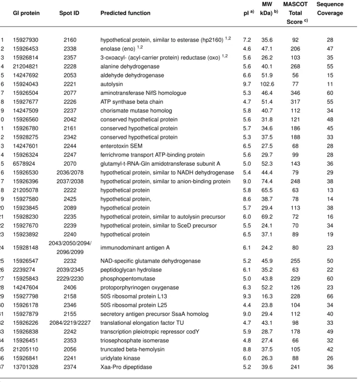
4.1.2 Intravenous immunoglobulin preparation (IVIG) specifically inhibitsin vitro growth ofS. aureus
To determine whether bacteriostasis is mediated byS. aureusspecific IgGs, an IVIG prepa- ration, previously shown to contain opsonising IgGs specific for S. aureus (Glowalla et al., 2009), was tested for effects on bacterial growth. For this purposeS. aureuswas incubated in the presence of either 25 or 50 % IVIG and samples where 25 or 50 % of the culture volume was replaced by PBS served as control.
As shown in Figure 4.2, growth of S. aureus was statistically significantly inhibited for up to 120 minutes upon addition of IVIG. Although the number of CFUs in IVIG-treated samples slightly increased after 120 minutes, the number of CFUs was significantly lower in comparison to the respective PBS control 180 minutes after addition of IgGs.
0 60 120 180 103
104 105 106 LB
25% PBS 25% human serum 25% HI-serum
*** ***
***
t (min)
CFU / ml
0 60 120 180
103 104 105 106 LB
*** ***
***
50% PBS 50% HI-serum 50% human serum
t (min)
CFU / ml
A
B
Figure 4.1:
Effect of human serum on in vitrogrowth of S. aureus. Growth kinetics ofS. aureus in LB (open circles) and upon addition of 25 % (A) or 50 % (B) of human serum (red closed triangles), heat-inactivated human serum (HI-serum; closed triangles) or an equal amount of PBS (open triangles), respectively was assessed using a CFU enumeration assay. At indicated time points 50 µl diluted bacterial culture was spirally plated on Mueller-Hinton agar, incubated for up to 16 h at 37 ° C and subsequently CFU per ml were counted. Shown are mean and SD of triplicates from a representative experiment. Statistical significance between serum treated compared to the respective PBS control sample was determined by one-way ANOVA followed by Bonferroni post analysis. P-values are indicated by asterisks (∗ ∗ ∗= p<0.001).
0 60 120 180 103
104 105 106
LB 25 % PBS 25 % IVIG
*
***
t (min)
CFU / ml
0 60 120 180
103 104 105 106
LB 50 % PBS 50 % IVIG
**
***
t (min)
CFU / ml
A
B
Figure 4.2:
Effect of IVIG onin vitrogrowth ofS. aureus. Growth kinetics ofS. aureusin LB (open circles) and upon addition of 25 % (A) or 50 % (B) IVIG (closed triangles) or an equal amount of PBS (open triangles), respectively was assessed using a CFU enumeration assay as described above.
Shown are mean and SD of triplicates from a representative experiment. Statistical significance between IVIG treated compared to respective PBS control sample was determined by one-way ANOVA followed by Bonferroni post analysis. P-values are indicated by asterisks (∗= p<0.05;
∗∗= p<0.01;∗ ∗ ∗= p<0.001).
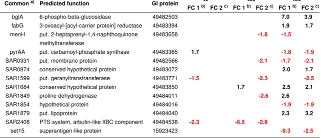
4.1.3 Bacteriostatic effect onS. aureusis mediated byS. aureusspecific IgGs To ensure that the observed inhibitory effect mediated by IVIG is not due to changes in cul- turing conditions by the addition of protein, the experiments were performed using 13 mg / ml BSA or IVIG adjusted in PBS, respectively. This concentration of IVIG equates to 25 % IVIG as used in previously described experiments. In contrast to IVIG treatment the co- incubation of S. aureus with BSA led to significant but only minor inhibition of growth (Fig.
4.3 A), thereby indicating that bacteriostasis is a specific effect mediated by IVIG.
control LB BSA IVIG
0 25 50 75 100 125
***
***
*
120 min CFU / ml (% of control)
control LB IVIG-DS IVIG
0 25 50 75 100 125
*** ***
120 min CFU / ml (% of control)
A
B
Figure 4.3:
Effect of BSA or IVIG-DS onin vitrogrowth ofS. aureus. Growth kinetics ofS. aureusin LB and upon addition of 13 mg / ml BSA (A) or dialysed IVIG (IVIG-DS; B) compared to IVIG in PBS was assessed using a CFU enumeration assay. Samples supplemented with 50 % PBS served as control. Shown are mean and SD of triplicates in percent of PBS control 120 minutes after addition of the respective substances. Statistical significance between indicated samples (capped lines) was determined by one-way ANOVA followed by Bonferroni post analysis. P-values are indicated by asterisks (∗ ∗ ∗= p<0.001).
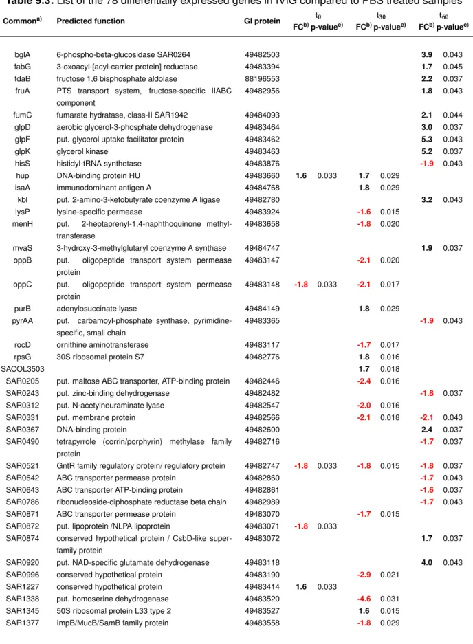
control LB dSaPBS dSaIVIG dEcIVIG IVIG 0
25 50 75 100
125 ***
***
***
120 min CFU / ml (% of control)
Figure 4.4:
Effect of IVIG onin vitrogrowth ofS. aureusupon depletion ofS. aureusorE. coli specific IgGs. Growth kinetics of S. aureus in LB and upon addition of 13 mg / ml IVIG or IVIG upon depletion of S. aureus specific IgGs (dSaIVIG) was assessed using a CFU enumeration assay.
Samples supplemented with 50 % PBS, 50 % PBS co-incubated with S. aureus (dSaPBS) or 13 mg / ml IVIG depleted ofE. coli specific IgGs (dEcIVIG) served as control. Shown are mean and SD of triplicates in percent of the given PBS control 120 minutes after addition of the re- spective substances. Statistical significance between indicated samples (capped lines) was de- termined by one-way ANOVA followed by Bonferroni post analysis. P-values are indicated by asterisks (∗ ∗ ∗= p<0.001).
Furthermore, when IVIG dialysed against PBS (IVIG-DS) was used, the bacteriostatic effect remained (Fig. 4.3 B), indicating that the observed bacteriostatic effect is solely due to the presence of IgGs and independent of the buffer composition of the original IVIG preparation. To elucidate whether S. aureus specific antibodies are responsible for the bacteriostatic effect, IVIG was depleted of S. aureus specific IgGs (dSaIVIG) ON at 4 ° C and upon sterile filtration used as source of non-specific IgGs in a CFU enumeration assay.
To rule out possible side effects caused by the process of depletion, PBS after ON co- incubation with S. aureus (dSaPBS) and IVIG depleted of E. coli specific IgGs (dEcIVIG) were used as additional controls.
When S. aureus was cultured in the presence of dSaIVIG, growth was comparable to bacteria cultured without IgGs (PBS control, LB; Fig. 4.4). In contrast, growth ofS. aureus cultured in the presence of dEcIVIG was inhibited to a comparable extent as bacteria cul- tured in the presence of IVIG. Hence, these results imply that S. aureus specific IgGs are responsible for the bacteriostatic effect on growth ofS. aureus.