UV-C inactivation of Legionella rubrilucens
UV-C-Inaktivierung von Legionella rubrilucens
Abstract
Background:Despite the great health significance ofLegionella, there is only little information on their UV sensitivity. Besides Legionella pneumophilaonlyL. longbeachaehas been investigated so far.
Julian Schmid
1Katharina Hoenes
1Monika Rath
1Methods: In this study L. rubrilucens has been spread on buffered
charcoal yeast extract agar and irradiated with the 254 nm UV-C emis-
Petra Vatter
1sion of a mercury vapor lamp. The disinfection success is measured by
Martin Hessling
1colony counting after incubation and comparison of the number of colonies on irradiated and unirradiated reference agar plates.
Results: The average log-reduction dose is 1.08 mJ/cm2 for free L. rubrilucens, which is at the lower end of the so far published Legion-
1 Ulm University of Applied Sciences, Ulm, Germany ella log-reduction values, but all threeLegionellaspecies show similar
UV-C sensitivities.
Conclusion:The log-reduction dose of legionellae in amoebae has not been investigated, but with the observed high UV-C sensitivity for free Legionella, the idea of a future point-of-use disinfection by small UV-C LEDs in water-taps or shower heads appears to be realistic, even if le- gionellae are more resistant in amoebae.
Keywords:Legionella, UV-C radiation, mercury vapor lamp, log-reduction dose, point-of-use disinfection
Zusammenfassung
Hintergrund:Trotz der großen gesundheitlichen Bedeutung von Legio- nellen gibt es bisher nur wenige Untersuchungen zu ihrer UV-C-Empfind- lichkeit. AußerLegionella pneumophilawurden bisher nur Ergebnisse fürL. longbeachaeveröffentlicht.
Material und Methoden:In der hier vorgestellten Untersuchung wurden Bakterienlösungen mitL. rubrilucensauf gepuffertem Aktivkohle-Hefe- Extrakt verteilt und der 254 nm UV-C-Strahlung einer Quecksilberdampf- lampe ausgesetzt. Der Desinfektionserfolg wird durch die Zählung von Legionellen-Kolonien nach der Bebrütung ermittelt, indem die Anzahl der Kolonien auf bestrahlten Agarplatten mit der Zahl an Kolonien auf unbestrahlten Referenzplatten verglichen wird.
Ergebnisse:Die durchschnittlich notwendige UV-C-Strahlungsdosis für eine Log-Stufen-Reduktion beträgt 1,08 mJ/cm2 für planktonische L. rubrilucens. Dieser Wert liegt im unteren Bereich der 4 bisher veröf- fentlichten Legionellen-Studien und zeigt aber auch, dass alle 3 bisher untersuchtenLegionella-Spezies ähnliche UV-C-Empfindlichkeiten auf- weisen.
Schlussfolgerung: Die UV-C-Empfindlichkeit aller Legionellen-Spezies scheint vergleichbar hoch zu sein. Selbst wenn Legionellen in Amöben etwas unempfindlicher sein sollten, scheint die Idee einer zukünftigen Point-of-Use-Desinfektion sehr realistisch. Kleine UV-C-LEDs in Wasser- hähnen und Duschköpfen könnten Legionellen im schnell vorbeiströ- menden Wasser signifikant inaktivieren. Der Einsatz einer solchen Technik scheint insbesondere in Gesundheitseinrichtungen wie Kran- kenhäusern oder Altenheimen sinnvoll.
Schlüsselwörter:Legionella, UV-C-Strahlung, Quecksilberdampflampe, Log-Stufen-Reduktions-Dosis, Point-of-Use-Desinfektion
Research Article
OPEN ACCESS
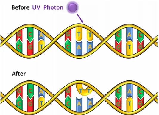
Figure 1: Absorption of a UV-C photon by DNA and formation of a thymine dimer
Introduction
In 2014 about 7,000 cases of detected Legionnaires’
Disease were reported in the European Union [1] and the number of unreported infections is probably much higher.
Legionnaires’ Disease is the most severe form of pneu- monia infections caused by Gram-negativeLegionellaspp.
The most pathogenic Legionella species is Legionella pneumophilathat was identified in the first documented Legionellaoutbreak in Philadelphia in 1976 [2]. Legion- ellae infections often occur by inhaling aerosols derived from contaminated water sources. The reservoirs of these Gram-negative bacteria are natural surface water, and especially drinking and cooling water systems [3]. There are different approaches for clearance of legionellae in these man-made water reservoirs but they usually do not show long lasting effects leading to an omnipresent danger of aLegionellacontamination.
Among the well-known disinfection techniques is the ap- plication of hot water with temperatures of about 70°C, but it is technically difficult to reach these high tempera- tures, especially in long pipe systems with lengths of 10 m and more. Chemical disinfection, e.g. by chlorine com- pounds, shows a reduced disinfection effect due to the fact that legionellae are often incorporated in biofilms and amoebae that protect the bacterium against direct contact with disinfectants [4].
UV-C radiation was successfully applied for water disin- fection already 100 years ago [5]. The radiation is usually generated by mercury vapor lamps, which show strong emission at a wavelength of 254 nm. The UV-C radiation is absorbed by DNA, resulting in the formation of thymine dimers [6] as illustrated in Figure 1. This DNA damage hinders gene expression and DNA replication and should lead to the death of the irradiated microorganisms. Unfor-
tunately UV-C disinfection offers no depot effect. Bacteria that survived the irradiation can proliferate again on their way through the water pipe system. Extremely aggravating is the formation of biofilms on the pipe walls that are capable of repeatedly releasing microorganisms including Legionellathat might get in direct contact with humans without a further disinfection step.
With the recent developments of UV-C LEDs the idea of a point-of-use disinfection with an UV-C LED directly in- stalled in the water-tap or shower head becomes more realistic. These LEDs are very small and offer only low radiation intensities in the range of up to 90 mW [7], which is about a factor of 100 below strong mercury vapor lamps, but do not contain mercury and offer a longer life time. The UV-C LED radiation power might be sufficient forLegionellainactivation, because according to [8], [9], [10], [11] the necessary log-reduction dose for L. pneumophila is somewhere between 0.9 and 3.1 mJ/cm2and 1.4 mJ/cm2forL. longbeachae[9] – a dose that could be reached within 0.03 s if a 90 mW UV-C LED irradiates an area of 1 cm2.
The intention of this paper is to get more information on necessary log-reduction doses for legionellae by perform- ing experiments onL. rubrilucensand compare them with the so far reported log-reduction doses. L. rubrilucens was reported as co-pathogen in a case of Legionnaires’
Disease [12] but its UV-C sensitivity has not been inves- tigated so far.
Materials and methods
The UV-C radiation was generated by a low pressure mercury vapor lamp type TUV 8W FAM/10X25BOX of Philips Lighting Holding B.V. (The Netherlands). It offers
Schmid et al.: UV-C inactivation of Legionella rubrilucens
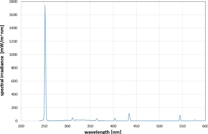
Figure 2: Emission spectrum of the employed low pressure mercury vapor lamp several emission lines in the UV and visible spectral
range, but the emission at 254 nm is by far the strongest as can be seen in Figure 2. In a distance of 15 cm the obtained 254 nm irradiance was 0.47 ± 0.05 mW/cm2 over an area of 10 x 30 cm2. It should be mentioned that the total irradiance, including UV-B, UV-A and visible light is slightly higher with about 0.65 mW/cm2but the disin- fection contribution of these longer wavelength emissions to the disinfection result is negligible with about 3% of the impact of the 254 nm emission, calculated with the spectral antimicrobial action spectra values given in [13]
forEscherichia coli, but it is assumed that other bacteria show a similar behavior.
The bacteria investigated were L. rubrilucens (DSM No. 11884) that were obtained as freeze-dried culture from Deutsche Sammlung von Mikroorganismen und Zellkulturen GmbH (Braunschweig, Germany) and cultiv- ated in buffered yeast extract (BYE) broth [14]. For the UV-C inactivation experiments solid buffered charcoal yeast extract (BCYE) agar [14] was prepared in Petri dishes with a diameter of 90 mm.
Bacterial suspensions of Legionella with an optical density of 0.24 at 600 nm, that contained about 2.4 x 10⁸CFU/ml in preliminary experiments, were further diluted in phosphate-buffered saline to concentrations below 10,000 CFU/ml. 100 µl of this samples were carefully dispensed over the surface of the BCYE agar plate.
The radiation experiments were performed with different exposure times to achieve different irradiation doses. The maximum irradiation time was 12 s, which resulted in doses of up to 5.6 mJ/cm2. Afterwards the agar plates
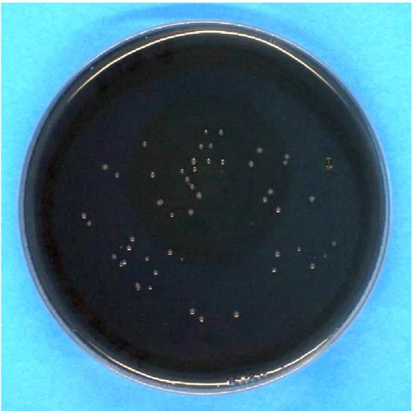
were stored in an incubator at 37°C and high humidity for 4 days before the plates were photographed for better colony counting (see example in Figure 3) and the ob- served colonies on irradiated agar plates were compared to unirradiated reference plates. This procedure was performed three times with three agar plates for each ir- radiation dose and each run.
Results and discussion
The disinfection results are presented in Table 1 and Figure 4. For higher irradiation doses the measured ratios of surviving bacteria follow a straight line in this half logarithmic diagram, which is equivalent to an exponential decrease for increasing UV-C doses. But this straight line doesn’t pass the expected 100%-point (1.0 E+00) for 0 mJ/cm2 irradiation. This well-known phenomenon is called “shoulder effect” [15]. The UV-C sensitivity is lower for lower doses and increases asymptotically for higher doses. Lower sensitivity means a higher necessary irradi- ation dose to reach a log-reduction (log-reduction: de- crease by a factor of ten). The lowest irradiation dose of 1.86 mJ/cm2in Table 1 led to 4.3% or 4.3 E-02 surviving bacteria. This is equivalent to a log-reduction dose of 1.36 mJ/cm2. But if the calculation is based on the highest applied irradiation dose of 5.58 mJ/cm2 and 0.0007% or 7.0 E-06 surviving bacteria the average log- reduction dose forL. rubrilucensbecomes 1.08 mJ/cm2. These results are in good agreement with published L. pneumophilaandL. longbeachaelog-reduction doses of 0.9 and 1.6 mW/cm2measured by [8], [9], [10].
Schmid et al.: UV-C inactivation of Legionella rubrilucens
Figure 3: Example of a BCYE agar plate with visible colonies ofLegionella after incubation
Table 1: Applied UV-C irradiation doses, average bacterial concentrations after irradiation for each run, average reference concentrations for each run and ratio of surviving bacteria (averaged over all three runs)
Schmid et al.: UV-C inactivation of Legionella rubrilucens
Figure 4: Bacterial reduction forL. rubrilucens during UV-C irradiation. Error bars depict the standard deviation of the average of three independent measurements.
It has to be mentioned that the here applied incubation durations of 4 days at 37°C after irradiation were rather short compared to several other published incubation times. Antopol and Ellner incubated their irradiated samples for 4–5 days at 35°C [8], Oguma et al. for 7 days [10] and Cervero-Arago et al. even for 10 days at 37°C [9]. It is theoretically conceivable that legionellae are still in a viable but nonculturable (VBNC) state [16] after 4 days, and over the course of time could recover by dark repair and photo reactivation mechanisms [17] and subsequently form colonies again. This would reduce the real UV sensitivity or increase the log reduction dose.
However, there is no general rule forLegionellaincubation periods. The international DIN-ISO standard “Water quality – Enumeration ofLegionella” [18] gives different incubation times depending on the application. In the case of samples, which are not clear for the presence of legionellae, a 7–10 day incubation should be carried out but for confirmation of legionella suspicion samples should be incubated for a maximum of 5 days. The Aus- tralian Legionella Control Guidelines indicate that the in- cubation period may take up to 10 days, but the average is between 5–7 days [19].
It is to be expected that in the presented experiments, no difference in the number of colonies caused by VBNC legionellae would have been determined between 4 and 10 days of incubation. The first reason for this assumption is the observation that legionellae in the VBNC state seem to need amoebae for their recovery [20] that were not present here in any case. The second reason is the fact that even 10 days is a rather short period compared to the 145 days and more, in which legionellae could stay in a VBNC state according to Alleron et al. [21].
Very important is the fact that legionellae in the VBNC state – at least in an guinea pig animal model – do not lead to infections [20]. Therefore, they probably play a subordinate role during the immediate period after irradi- ation.
So the observed log-reduction dose of about 1.08 mW/cm2forL. rubrilucensshould not be influenced by the shorter incubation time and the result consolidates the impression that differentLegionellaspecies show a similar high sensitivity to UV-C radiation. Legionellae in amoebae required almost twice the UV-C dose of free le- gionellae [9], but this is still a high sensitivity resulting in log-reduction doses that are applicable with UV-C LEDs within fractions of a second. This supports the idea of a future point-of-use water disinfection by small UV-C LEDs that are integrated in water-taps and shower heads and irradiate the pre-passing water at least in healthcare in- stitutions like hospitals or nursing homes.
Notes
Acknowledgement
This work was financially supported by the German Fed- eral Ministry of Economics and Technology within the ZIM project “Clean Spring” (grant number KF2186208CR4).
Competing interests
The authors declare that they have no competing in- terests.
Schmid et al.: UV-C inactivation of Legionella rubrilucens
References
1. European Centre for Disease Prevention and Control.
Legionnaires’ disease in Europe, 2014. Stockholm: ECDC; 2016.
DOI: 10.2900/585125
2. Brenner DJ, Steigerwalt AG, McDade JE. Classification of the Legionnaires' disease bacterium: Legionella pneumophila, genus novum, species nova, of the family Legionellaceae, familia nova.
Ann Intern Med. 1979 Apr;90(4):656-8. DOI: 10.7326/0003- 4819-90-4-656
3. Steinert M, Hentschel U, Hacker J. Legionella pneumophila: an aquatic microbe goes astray. FEMS Microbiol Rev. 2002 Jun;26(2):149-62. DOI: 10.1111/j.1574-6976.2002.tb00607.x 4. Kim BR, Anderson JE, Mueller SA, Gaines WA, Kendall AM.
Literature review--efficacy of various disinfectants against Legionella in water systems. Water Res. 2002 Nov;36(18):4433- 44. DOI: 10.1016/S0043-1354(02)00188-4
5. Henri V, Helbronner A, de Recklinghausen M. Stérilisation de grandes quantités d'eau par les rayons ultraviolets. Compt Rend Acad Sci. 1910;150:932-4.
6. Wacker A, Dellweg H, Weinblum D. Strahlenchemische Veränderung der Bakterien-Desoxyribonucleinsäure in vivo.
Naturwissenschaften. 1960;47(20):477. DOI:
10.1007/BF00638304
7. Inoue S, Naoki T, Kinoshita T, Obata T, Yanagi H. Light extraction enhancement of 265 nm deep-ultraviolet light-emitting diodes with over 90 mW output power via an AlN hybrid nanostructure.
Appl Phys Lett. 2015; 106(13):131104. DOI:
10.1063/1.4915255
8. Antopol SC, Ellner PD. Susceptibility of Legionella pneumophila to ultraviolet radiation. Appl Environ Microbiol. 1979
Aug;38(2):347-8.
9. Cervero-Aragó S, Sommer R, Araujo RM. Effect of UV irradiation (253.7 nm) on free Legionella and Legionella associated with its amoebae hosts. Water Res. 2014 Dec;67:299-309. DOI:
10.1016/j.watres.2014.09.023
10. Oguma K, Katayama H, Ohgaki S. Photoreactivation of Legionella pneumophila after inactivation by low- or medium-pressure ultraviolet lamp. Water Res. 2004 Jun;38(11):2757-63. DOI:
10.1016/j.watres.2004.03.024
11. Wilson BR, Roessler PF, van Dellen E, Abbaszadegan M, Gerba CP. Coliphage MS-2 as a UV water disinfection efficacy test surrogate for bacterial and viral pathogens. Proceedings, 1992 Water Quality Technology Conference; Toronto, Ontario;
November 15-19, 1992. p. 219-35.
12. Matsui M, Fujii S, Shiroiwa R, Amemura-Maekawa J, Chang B, Kura F, Yamauchi K. Isolation of Legionella rubrilucens from a pneumonia patient co-infected with Legionella pneumophila. J Med Microbiol. 2010 Oct;59(Pt 10):1242-6. DOI:
10.1099/jmm.0.016089-0
13. DIN Deutsches Institut für Normung eV. Optical radiation physics and illuminating engineering – Part 10: Photobiologically effective radiation, quantities, symbols and action spectra. DIN 5031-10.
Berlin: Beuth Verlag; 2013.
14. Buchrieser C, Hilbi H, editors. Legionella: Methods and Protocols.
Totowa, NJ: Humana Press; 2013. DOI: 10.1007/978-1-62703- 161-5
15. Tang WZ, Sillanpää M. Bacteria sensitivity index of UV disinfection of bacteria with shoulder effect. J Environ Chem Eng.
2015;3(4):2588-96. DOI: 10.1016/j.jece.2015.09.010 16. Hussong D, Colwell RR, O'Brien M, Weiss E, Pearson AD, Weiner
RM, Burge WD. Viable Legionella pneumophila Not Detectable by Culture on Agar Media. Nat Biotechnol. 1987; 5(9):947–50.
DOI: 10.1038/nbt0987-947
17. Quek PH, Hu J. Indicators for photoreactivation and dark repair studies following ultraviolet disinfection. J Ind Microbiol Biotechnol. 2008 Jun;35(6):533-41. DOI: 10.1007/s10295-008- 0314-0
18. DIN Deutsches Institut für Normung eV. Water quality – Enumeration of Legionella (Draft version). Berlin: Beuth Verlag;
2015.
19. Australian Government. Guidelines for Legionella Control: In the operation and maintenance of water distribution systems in health and aged care facilities. Canberra: Australian Government;
2015. Available from: https://www.health.gov.au/internet/main/
publishing.nsf/content/
A12B57E41EC9F326CA257BF0001F9E7D/$File/Guidelines- Legionella-control.pdf
20. Steinert M, Emödy L, Amann R, Hacker J. Resuscitation of viable but nonculturable Legionella pneumophila Philadelphia JR32 by Acanthamoeba castellanii. Appl Environ Microbiol. 1997 May;63(5):2047-53.
21. Alleron L, Merlet N, Lacombe C, Frère J. Long-term survival of Legionella pneumophila in the viable but nonculturable state after monochloramine treatment. Curr Microbiol. 2008 Nov;57(5):497-502. DOI: 10.1007/s00284-008-9275-9
Corresponding author:
Prof. Dr. Martin Hessling
Ulm University of Applied Sciences, Albert-Einstein-Allee 55, 89081 Ulm, Germany, Phone: 0731/50-28602 hessling@hs-ulm.de
Please cite as
Schmid J, Hoenes K, Rath M, Vatter P, Hessling M. UV-C inactivation of Legionella rubrilucens. GMS Hyg Infect Control. 2017;12:Doc06.
DOI: 10.3205/dgkh000291, URN: urn:nbn:de:0183-dgkh0002911
This article is freely available from
http://www.egms.de/en/journals/dgkh/2017-12/dgkh000291.shtml Published:2017-04-10
Copyright
©2017 Schmid et al. This is an Open Access article distributed under the terms of the Creative Commons Attribution 4.0 License. See license information at http://creativecommons.org/licenses/by/4.0/.
Schmid et al.: UV-C inactivation of Legionella rubrilucens