Molecular characterization of the UV-response in Caenorhabditis elegans
Inaugural-Dissertation zur
Erlangung des Doktorgrades
der Mathematisch-Naturwissenschaftlichen Fakultät der Universität zu Köln
vorgelegt von Stefanie Wolters
aus Leverkusen
Köln, 2013
1 Referee / Gutachter: Dr. Björn Schumacher
Institut für Genetik, Universität zu Köln Referee / Gutachter: Prof. Dr. Thorsten Hoppe Institut für Genetik, Universität zu Köln
Chairman / Vorsitzender: Prof. Dr. Günter Schwarz Institut für Biochemie, Universität zu Köln
Tag der mündlichen Prüfung: 17. Oktober 2013
2 The work described in this dissertation was conducted from May 2009 to August 2013 under supervision of Dr. Björn Schumacher at the Institute for Genetics, University of Cologne, Zülpicher Straβe 47a, D‐50674 Cologne, Germany.
Die Arbeiten, die in dieser Dissertation beschrieben sind, wurden durchgeführt zwischen Mai 2009 und August 2013 unter der Anleitung von Dr. Björn Schumacher am Institut für Genetik der Universität zu Köln, Zülpicher Straβe 47a, D‐50674 Köln, Deutschland.
Parts of this work have been published:
Teile dieser Arbeit wurden bereits veröffentlicht:
Stefanie Wolters and Björn Schumacher
Genome maintenance and transcription integrity in aging and disease Front. Genet., 25 Februar 2013 | doi: 10.3389/fgene.2013.00019
Parts of this work have been submitted for publication:
Teile dieser Arbeit wurden als Manuskript eingereicht:
Stefanie Wolters, Maria A. Ermolaeva, Jeremy S. Bickel, Jaclyn M. Fingerhut, Jayshree Khanikar, Raymond C. Chan and Björn Schumacher
BRC-1/BRD-1 inactivation suppresses genome instability during replication blockage in C.
elegans smc-5 mutants Genetics, 2014
3
Table of Contents
I SUMMARY ... 6
II INTRODUCTION ... 8
2.1 C. ELEGANS AS A MODEL ORGANISM FOR STUDIES ON DNA REPAIR ... 9
2.2 DOUBLE STRAND BREAK REPAIR ... 13
2.2.1 Homologous Recombination Repair ... 15
2.2.2 Non-Homologous End Joining ... 17
2.3 INTERSTRAND CROSSLINK REPAIR ... 18
2.4 NUCLEOTIDE EXCISION REPAIR ... 18
2.5 TRANSLESION SYNTHESIS ... 21
III BRC-1/BRD-1 INACTIVATION SUPPRESSES GENOME INSTABILITY DURING REPLICATION BLOCKAGE IN SMC-5 MUTANTS ... 23
3.1 INTRODUCTION ... 23
3.1.1 Discovery of a novel radiation-sensitive gene ... 23
3.1.2 The Smc family ... 23
3.1.3 The subunits of the Smc5/6 complex ... 24
3.1.4 Characteristics of the Smc5/6 complex ... 25
3.2 RESULTS ... 27
3.2.1 Screening for UV-sensitive alleles in L1 larvae. ... 27
3.2.2 Identification of a novel xpg-1 allele is a proof of concept for the screening method. ... 30
3.2.3 smc-5 is required for resistance to UV-induced DNA lesions in the C. elegans germline ... 31
3.2.4 smc-5 is dispensable for the repair of UV-induced lesions ... 36
3.2.5 SMC-5 functions in parallel to POLH-1-mediated TLS ... 38
3.2.6 SMC-5 is required when replication fork progression is impaired... 42
3.2.7 BRC-1/BRD-1 mutations suppress defects in smc-5 activity ... 44
3.2.8 Rescue of defects of the smc-5 mutant by suppression of BRC-1/BRD-1 complex is partially mediated by POLH-1... 45
3.3 DISCUSSION ... 49
3.3.1 Landscape of EMS-induced mutations ... 49
3.3.2 The ATPase domain of SMC-5 is essential for UV-resistance. ... 49
3.3.3 The mutagenicity of DNA repair pathways ... 50
3.3.4 Restoring replication stress resistance in the absence of SMC-5/6 complex ... 53
IV ROLE OF MICRORNAS IN THE UV-RESPONSE OF YOUNG C. ELEGANS LARVAE ... 57
4
4.1 INTRODUCTION ... 57
4.1.1 miRNA synthesis ... 58
4.1.2 miRNA maturation ... 58
4.1.3 Posttranscriptional gene silencing ... 60
4.1.4 miRNA classification ... 61
4.1.5 Studying miRNAs ... 62
4.1.6 Aim of the study ... 65
4.2 RESULTS ... 66
4.2.1 Establishment of a deep sequencing approach to analyze the miRNA profile upon UV irradiation ... 66
4.2.2 Sequencing reveals regulation of small RNAs after irradiation with UV light. ... 68
4.2.3 Investigation of UV-dose-dependency and timing of the miRNA regulation. ... 72
4.2.4 let-7 and lin-4, mediators of larval transitions, are differentially expressed after UV irradiation. ... 74
4.2.5 mRNA target prediction of miRNAs with altered expression after UV irradiation. ... 81
4.3 DISCUSSION ... 84
4.3.1 Establishment of sequencing-based screening for UV-regulated miRNAs ... 84
4.3.2 lin-4 expression is repressed upon UV irradiation independently from DAF-16. ... 84
4.3.3 miRNAs associated with L1 diapause state are induced upon UV-treatment. ... 85
4.3.4 Small RNA sequencing indicates UV-dependent regulation of a set of miRNAs ... 87
4.3.5 let-7 is downregulated in response to UV irradiation ... 88
4.3.6 UV-responsive miRNAs are predicted to fine-tune growth, development and MAPK signaling ... 90
V MATERIAL & METHODS ... 92
5.1 C. ELEGANS CULTURE MAINTENANCE ... 92
5.2 SYNCHRONIZING WORMS ... 93
5.3 GENOTYPING OF WORM STRAINS ... 93
5.4 QPCR EXPRESSION ANALYSIS ... 95
5.4.1 RNA preparation ... 95
5.4.2 Quantitative real-time PCR ... 95
5.5 ISOLATION OF GENOMIC DNA ... 100
5.6 GFP REPORTER STRAIN ANALYSIS ... 101
5.7 IMMUNOSTAINING AND MICROSCOPY ... 101
5.8 DEVELOPMENTAL STAGING ASSAY ... 101
5.9 SCREENING FOR NOVEL UV-SENSITIVE C. ELEGANS STRAINS ... 102
5.9.1 EMS mutagenesis ... 102
5.9.2 Screening and selection of worms ... 102
5.9.3 SNP mapping of novel UV-sensitive alleles (Davis et al. 2005) ... 102
5
5.9.4 Whole genome sequencing ... 104
5.10 EGG-LAYING AND HATCHING RATE ... 104
5.11 GERMLINE DEVELOPMENT ASSAY ... 104
5.12 HU TREATMENT ... 104
5.13 RNAI-MEDIATED GENE KNOCKDOWN ... 105
5.14 GENERATION OF MALES FOR CROSSING ... 105
5.15 SMALL RNA SEQUENCING AND BIOINFORMATICAL ANALYSIS ... 106
5.15.1 small RNA sequencing ... 106
5.15.2 Data analysis ... 106
5.16 CPD REPAIR ASSAY USING SLOTBLOT ... 106
5.17 ANTIBODIES USED ... 107
5.18 GROWTH MEDIA & SOLUTIONS ... 107
5.18.1 Single worm lysis buffer ... 107
5.18.2 M9 buffer ... 107
5.18.3 NGM plates ... 108
5.18.4 RNAi plates ... 108
5.18.5 2x Freezing buffer ... 108
5.18.6 2x Bleaching solution ... 108
5.18.7 LB ... 109
5.18.8 LB-agar plates ... 109
5.18.9 PBS ... 109
5.18.10 50x TAE ... 109
5.18.11 TE buffer ... 109
5.18.12 0.5M EDTA ... 110
VI LIST OF ABBREVIATIONS ... 110
VII ACKNOWLEDGEMENTS ... 113
VIII REFERENCES ... 113
6
I Summary
The Structural Maintenance of Chromosomes (SMC) proteins form distinct complexes that maintain genome stability during chromosome segregation, homologous recombination (HR), and DNA replication. Using a forward genetic screen, we identified two alleles of smc-5 that exacerbate UV-sensitivity in C. elegans. Germ cells of smc-5 defective animals show reduced proliferation, sensitivity to perturbed replication and accumulation of RAD-51 foci that indicate the activation of HR. Mutations in the translesion synthesis polymerase polh-1 act synergistically with smc-5 mutations in provoking genome instability after UV-induced DNA damage. In contrast, the DNA damage accumulation and UV-sensitivity in smc-5 mutant strains are suppressed by mutations in the C. elegans BRCA1/BARD1 homologues, brc-1 and brd-1. We propose that SMC-5/6 promotes replication fork stability and facilitates recombination- dependent repair when the BRC-1/BRD-1 complex initiates HR at stalled replication forks. Our data suggest that BRC-1/BRD-1 can both, promote and antagonize genome stability depending on whether HR is initiated during DSB repair or during replication stalling.
In addition, we characterized the pool of small regulatory RNAs that respond to UV irradiation in young C. elegans. Deep sequencing identified miRNAs that were regulated differentially after treatment with UV light and revealed novel miRNAs. We validated upregulation of mir-2214*, mir-235, mir-71 and mir-238 as well as suppression of mir-1830, let-7, mir-2211, mir-48 and lin- 4. However, analysis of mutants and GFP-reporter strains indicated that lin-4 and let-7 are dispensable for resistance to UV and that promoter activity is not changed after irradiation.
Combining literature research and bioinformatical approaches we found that UV-regulated miRNAs are associated with growth and development. Also, we provide evidence that miRNAs alter MAPK kinase signaling, control nucleotide excision repair and the proteasome in response to UV irradiation.
7
I Zusammenfassung
Proteine der Familie “Structural Maintenance of Chromosomes” formen spezifische Komplexe, welche während der Segregation von Chromosomen, Homologer Rekombination (HR) und DNA Replikation für genomische Stabilität essentiell sind. Mit Hilfe eines genetischen Screenings haben wir zwei neue Allele von smc-5 entdeckt, die C. elegans sensitiv gegenüber UV- Bestrahlung machen. Die Keimzellen dieser smc-5-Mutanten weisen verringerte Proliferationsraten, Sensitivität gegenüber Replikationsstörungen, sowie eine erhöhte Anzahl von RAD-51 foci auf, welche auf die Aktivierung von HR hinweisen. UV-induzierte Läsionen der DNA können von der DNA Polymerase POLH-1 überlesen werden. Die Kombination von Mutationen in polh-1 mit smc-5 Mutationen wirkt sich synergistisch auf genomische Instabilität in Folge von UV-induzierten DNA-Schäden aus. Im Gegensatz dazu unterdrücken Mutationen der C. elegans Homologe des BRCA1/BARD1 Komplex, brc-1 und brd-1, die Akkumulation von DNA Schäden sowie die UV-Sensitivität von smc-5 Mutanten. Unsere Daten deuten darauf hin, dass der SMC-5/6 Komplex die Stabilität von Replikationsgabeln sichert und Rekombinations-abhängige Reperaturmechanismen fördert, wenn BRC-1/BRD-1 HR an blockierten Replikationsgabeln induziert.
Weiterhin haben wir die Gruppe der kleinen regulatorischen RNAs charakterisiert, welche nach UV-Bestrahlung differentielle Expression in jungen C. elegans Larven aufweisen. Mittels Deep- Sequencing konnten wir nicht nur Regulation von miRNAs feststellen, sondern waren auch in der Lage bislang unbekannte miRNAs zu identifizieren. In diesem Zusammenhang haben wir die Hochregulation von mir-2214*, mir-235, mir-71 und mir-238 sowie die Suppression von mir- 1830, let-7, mir-2211, mir-48 and lin-4 validieren können. Nichtsdestotrotz konnten wir keine Abhängigkeit der UV-Resistenz von let-7 oder lin-4 feststellen. Ebenso haben wir auch keine Veränderung der Promotor-Aktvität, durch die Nutzung von Fluoreszenz-Reportern beider Gene feststellen können. Literaturrecherche in Verbindung mit bioinformatischer Analyse zeigte, dass miRNAs, welche nach UV-Bestrahlung veränderte Expression aufweisen, in Verbindung mit Wachstum und Entwicklungsprozessen stehen. Weiterhin deuten unsere Daten darauf hin, dass der MAPK Signalweg, die Nukleotid Exzisions Reperatur und das Proteasom unter der Kontrolle der UV-regulierten miRNAs stehen.
8
II Introduction
(modified from Wolters and Schumacher 2013)
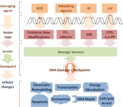
It was estimated that DNA damage occurs on the order of tens of thousands per genome on a daily basis (Lindahl and Nyberg 1972). Genotoxic insults can arise from a large variety of endogenous and exogenous sources (Figure II.1). Cellular metabolism can produce reactive oxygen species (ROS) and alkylating agents, while cells can be exposed to ultraviolet (UV) light, ionizing radiation (IR), and a variety of genotoxic chemicals (Loeb and Harris 2008). The type of lesion can vary widely and depends on the source of DNA damage. For example, ROS induces oxidative base modifications, IR typically leads to single- and double-strand breaks (SSB and DSB, respectively), DNA alkylation can lead to adduct and interstrand crosslink (ICL) formation, and UV light triggers the formation of thymidine dimers (Hurley 2002). The toxicity of DNA damage depends on the structural changes they inflict as well as the characteristics of the cell they occur in. Proliferating cells have a different repertoire of DNA repair pathways than quiescent cells and, therefore, the same lesion might have different effects in different tissues. In cycling cells, for instance, a single DSB is sufficient to impair chromosome segregation during mitosis and ICLs lead to replication fork collapse. For these reasons even a small number of DSBs and ICLs can be cytotoxic. In contrast, oxidative base modifications are generally less obstructive, while UV-induced cyclobutane pyrimidine dimers (CPDs) can be read through by specialized DNA polymerases and thus, can persist through replication, but pose an obstacle to transcription and lead to stalling of RNA polymerases (RNAPs).
Despite the toxic potential of DNA breaks, they are also needed for normal developmental and metabolic processes. Induction of DSBs is the basis for diversity of the immune system during V(D)J recombination in lymphocytes and allows generation of a enormous variety of antigen receptors (Bednarski and Sleckman 2012). Moreover, programmed DSBs and subsequent strand invasion into homologous sequences are the foundation for recombination to ensure biological diversity and are required for proper chromosome segregation during meiosis (Lemmens and Tijsterman 2011). Nevertheless, even deliberately introduced lesions are obstructive to cells if not sealed correctly.
Given the frequency and impact of DNA damage, highly sophisticated DNA repair systems have evolved. These systems recognize specific types of lesions and induce DNA damage signaling.
9 Failure of DNA repair has been associated with severe disorders in humans, often associated with occurrence of cancer and/or premature aging.
2.1 C. elegans as a model organism for studies on DNA repair
C. elegans is especially suitable for characterization of DNA repair pathways because it is susceptible to genetic manipulation, has a short life cycle and produces self-fertilized progeny.
Moreover, it is a multicellular organism that can also reproduce sexually, therefore being a good model for genetic experiments. Furthermore the whole genome is sequenced and the major DNA
Figure II.1. Diverse lesion types trigger DNA damage responses.
DNA damage can be caused by various genotoxic agents, such as reactive oxygen species (ROS) produced during cellular metabolism, alkylating agents that find application in cancer therapy, ionizing irradiation (IR), which is used for radio therapy, or ultraviolet (UV) irradiation presenting a daily threat as it is contained in sunlight. The inflicted lesions are just as diverse, since ROS usually lead to base modifications; alkylating agents form adducts, while bifunctional alkylating agents crosslink DNA to form interstrand crosslinks (ICLs). IR typically induces double-strand breaks (DSBs), and UV light triggers the formation of cyclobutane pyrimidine dimers (CPDs) and 6,4-photoproducts (6,4-PPs). Cells have a repertoire to sense the different lesions and subsequently activate DNA damage checkpoint proteins.
Ultimately, cells respond to the DNA damage by chromatin remodeling, modified transcription, fine- tuning of energy metabolism, cell cycle arrest, activation of DNA repair pathways and, in the case of irreparable damage load, induction of senescence or apoptosis.
(taken from Wolters and Schumacher, 2013)
10 repair machineries found in mammals are conserved (Boulton et al. 2002). A large number of mutant strains are available through well organized online databases (e. g. www.wormbase.org and Caenorhabditis Genetics Center at the University of Minnesota). In vivo observation of fluorescence-tagged proteins and phenotypical analysis of germ cells is facilitated by transparency of the nematode. The developmental program of C. elegans is well characterized and invariable in the course of events (Goenzyz and Rose 2005). There are two sexes of C.
elegans, hermaphrodites with XX genotype and males with X0 genotype. Although the general body plan is the same for both sexes they can be easily distinguished by behavior and appearance (Herman 2006). Usually C. elegans are self-fertilizing hermaphrodites but under unfavorable conditions or by chance they might lose an X chromosome by non-disjunction mutation and produce males (0.1% in wildtype worms at standard laboratory conditions). Therefore increased abundance of males (referred to as High Incidence of Males (him) phenotype) is indicative of genomic instability of a worm strain and often seen for DNA repair mutants (e.g. brc-1 mutant) (Lemmens and Tijsterman 2011).
In the fertilized embryo asynchronous cell divisions determine the cell fate. Already the first division gives rise to the germline precursor P1. At L1 stage, primordial germ cells Z2 and Z3, as well as somatic germline precursors Z1 and Z4 are formed. Before the end of L1 stage Z1 and Z4 give rise to two DTCs that provide the signal for proliferation of the germ cells (Hubbard and Greenstein 2005). When the egg is laid it contains already a majority of the cells that make up the adult animal (Clejan, Boerckel, and Ahmed 2006). An adult hermaphrodite has 959 somatic cells that are mainly post-mitotic (Herman 2006). In contrast, germ cells continue to divide throughout adulthood.
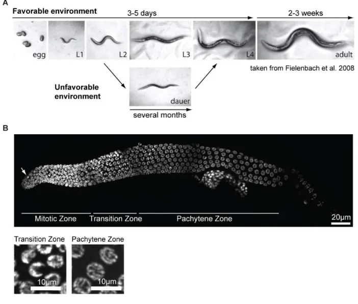
During lifecycle C. elegans passes through four larval stages (L1, L2, L3 and L4), each of them ending with molting to enter the next stage (Figure II.2 A) (Altun and Hall 2006). Within three days under optimal conditions it develops from egg to a reproducing adult. The total lifespan is about three weeks but could be much longer under unfavorable conditions when worms enter diapauses stage (Figure II.2 A). If worms hatch in the absence of food they arrest at L1 stage but resume development once food is available again. In the laboratory this feature of C. elegans is used to obtain synchronous populations. However, in response to stress, like high population density, starvation or high temperatures, worms enter dauer stage (P. J. Hu 2007). The decision for this alternative second molt occurs late in the L1 larval stage and is communicated via
11 pheromones. Dauer stage worms differ from normal L3 larvae in their morphology (Figure II.2 A), metabolism and transcriptome and are highly stress-resistant to various kinds of agents and
Figure II.2 The model organism Caenorhabditis elegans.
A C. elegans life cycle. In favorable environments worms develop from egg to reproducing adults within 3-5 days depending on the temperature. The adult lifespan is about 2-3 weeks. Under unfavorable conditions worms develop into an alternative larval stage called dauer. In this state they can outlast for several months and resume development once environmental conditions improve.
B DAPI staining of dissected hermaphrodite germline. The germline of C. elegans is a polar organ with mitotic cells at the distal end and nuclei progressing through meiosis towards the proximal end. Cells of each zone can be easily identified by their characteristic shape. While nuclei of the mitotic region have a uniform round shape with exception of few mitotic figures (indicated by an arrow), transition zone cells are crescent shaped. Once cells enter pachytene region, the DNA forms long strings followed by condensation into six bivalents at diplotene and diakinesis.
12 conditions. Under current investigation is a third diapauses state, the adult reproductive diapauses, that enables fully developed worms to survive stressful conditions, for instance starvation or hypoxia, and delay reproduction (Angelo and Van Gilst 2009; Leiser et al. 2013).
The reproductive system of C. elegans consists of two U-shaped tubes at which proximal end oocytes get fertilized by passing the spermatheca and finally eggs are laid through the vulva at the ventral midline of the worms’ body (Greenstein 2005; Altun and Hall 2006). Germ cells are organized in a syncytium where nuclei with incomplete borders are connected through an inner canal. The germline is a convenient organ to study DNA damage pathways because of its spatial separation of mitotic and meiotic cells (Figure II.2 B). Proliferation is mediated by the somatic distal tip cell (DTC) that determines a stem cell niche at the distal end of the gonad arm by secretion of pro-mitotic molecules (Kimble and Crittenden 2005). Through continuous proliferation at the distal end of the germline, nuclei move out of the area of influence of the DTC. This transition zone is marked by DNA condensation giving rise to crescent-shaped 4',6- diamidino-2-phenylindole (DAPI)-stained structures and entry into meiosis (Figure II.2 B) (leptotene and zygotene stage of prophase I). Nuclei at pachytene stage of meiosis have characteristic “bowl of spaghetti” morphology (Lints and Hall 2009). Cells in this region induce DSBs using topoisomerase VI-related spo-11 to activate homologous recombination (HR) repair.
One DSB per chromosome is repaired by crossover formation to ensure proper chromosome segregation in subsequent steps of meiosis and may lead to recombination of sequence information from the two homologous chromosomes. To maintain tissue homeostasis, cells at pachytene stage near the loop region undergo programmed cell death. In 1999 Gumienny et al.
estimated that about one half of all germ cells die, possibly to nurse the surviving cells by providing cytoplasmatic components (Gumienny, Lambie, Hartwieg, Horvitz, & Hengartner, 1999). Corpses can be identified using DIC microscopy by their typical round and detached shape. In addition to tissue homeostasis, apoptosis of germ cells is also utilized to eliminate damaged cells, for instance upon DNA damage. Unlike pachytene cells, cells of the mitotic region are not capable of apoptosis but arrest cell cycle when encountering damaging insults (Stergiou, Doukoumetzidis, Sendoel, & Hengartner, 2007). DAPI staining of mitotic nuclei easily identifies arrested nuclei because in comparison to cycling cells they are heavily enlarged (Gartner, MacQueen, and Villeneuve 2004). In contrast, worms defective for checkpoint response do not arrest germ cells. In the loop region, nuclei enter diplotene stage where
13 chromosomes condense and then continue to diakinesis (Figure II.2 B). Here, the chromosomes are most condensed and form six discrete bivalents. In parallel the cytoplasmatic volume increases and oocyte maturation is completed before oocytes enter into the spermatheca. During accumulation of cytoplasm, oocytes are equipped with maternal molecules, which is why homozygous progeny of a heterozygous mother may show a less pronounced phenotype than expected (Hubbard and Greenstein 2005). After fertilization the embryo enters the uterus and is laid.
2.2 Double Strand Break Repair
As the presence of a DSB poses a major obstacle for further cell division a sophisticated network of DNA damage response (DDR) signaling is ignited (Ciccia and Elledge 2010). Genetic experiments that were performed in yeast nearly 25 years ago established that DNA damage checkpoints transiently halt cell cycle progression in the presence of genotoxic stress to assure that the repair is completed before cell division (Weinert and Hartwell 1989; Rowley, Hudson, and Young 1992). The recognition that DDR defects are causal for cancer development has sparked major research efforts employing model systems from yeast to mammals. The DNA damage checkpoint mechanisms turned out to be highly conserved throughout evolution. For instance, C. elegans has been used intensively for characterization and identification of known and new DSB-responsive molecules (Lemmens and Tijsterman 2011). Cells, however, not only respond by transient cell cycle arrest but also by inducing cellular senescence, thus permanently withdrawing from cell division. Dependent on the circumstances cells induce apoptosis upon DNA damage and, thus, no longer pose a threat to the organism (Harper and Elledge 2007).
Intriguingly, the DDR not only impacts on regulators of cellular proliferation and cell death but impinges on a variety of cellular processes such as transcription, DNA repair, respiration, energy metabolism, chromatin remodeling, and others (Figure II.1) (Jackson and Bartek 2009).
In human cells the initial recognition of DSBs involves binding of the trimeric Mre11-Rad50-Nbs1 (MRN) complex to the broken DNA ends (Figure II.3) (Bartek and Lukas 2007). The MRN complex activates the PI3 kinase-like kinase ataxia telangiectasia mutated (ATM), which in turn phosphorylates a plethora of targets (Shiloh 2003). ATM targets include Mediator of DNA Damage Checkpoint 1 (MDC1) as well as the checkpoint kinase CHK2, which in turn activates p53. p53 then induces cell cycle arrest and, amid severe damage, apoptosis.
14
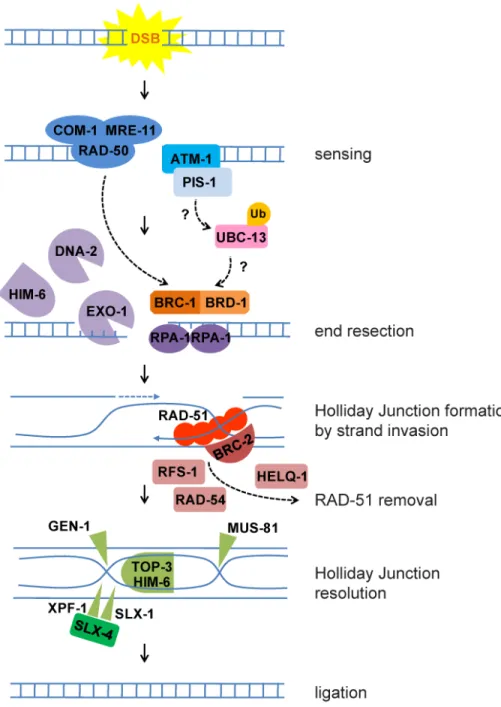
Figure II.3 Homologous Recombination in C. elegans.
DSBs are sensed by ATM kinase and MRN complex comprised of MRE-11, RAD-50 and COM-1, homolog of human NBS1. Subsequently, a number of repair factors are recruited and phosphorylated by ATM including MDC1 (the ortholog of which is PIS-1 in C. elegans). Studies in cell culture suggest that ubiquitination events mediated by UBC-13 lead to recruitment of BRC-1. BRC-1/BRD-1 mediates end resection together with the MRN complex, the nucleases EXO-1 and DNA-2, and the homolog of RecQ helicase, HIM-6. The single stranded DNA ends are protected by RPA-1. Next RAD-51, stabilized by BRC-2, induces strand invasion to a homologous DNA sequence. After induction of double Holliday Junction, RAD-51 is removed with the help of RFS-1, RAD-54 and HELQ-1. Holliday Junction resolution requires nucleases GEN-1, MUS-81 and SLX-4 that serves as a scaffold for SLX-1 and XPF-1.
Furthermore helicase/topoisomerase complex HIM-6/TOP-3 promote dissolution. Finally the nicks are sealed to restore the intact DNA strands.
15 These key factors are highly conserved in C. elegans, like for example cep-1 homologue of p53.
While DNA damage checkpoint signaling halts the cell cycle, at least two distinct DSB repair machineries are activated depending on the phase of the cell cycle (Chapman, Taylor, and Boulton 2012). In S/G2 phase HR uses the sister chromatid as template for accurate repair, while during G1 non-homologous end joining (NHEJ) ligates the broken ends after end resection.
Thereby NHEJ comprises a fast and efficient but error prone DNA repair method. However, for large parts of the genome the NHEJ-induced errors can be tolerated when genes are not affected.
Since NHEJ is utilized when no sister chromatid is available, it is thought to function mainly in non-proliferating cells, like neurons (Jeppesen, Bohr, and Stevnsner 2011).
2.2.1 Homologous Recombination Repair
After sensing of a DNA DSB by MRN complex and MDC1 phosphorylation by ATM, MDC1 recruits additional repair factors (Figure II.3). A well-known factor of HR repair is breast cancer type 1 susceptibility protein (BRCA1). Its recruitment requires ubiquitination events at the site of DSBs mediated by UBC13 E2, RNF8 and RNF168 E3 enyzmes. Subsequently RAP80 binds to the ubiquitin chains and recruits BRCA1 (Huen, Sy, and Chen 2010; Rosen 2013). In how far these events are conserved in C. elegans should be studied in future because this organism lacks obvious homologues of RNF8 and RAP80. However, BRCA1 forms a heterodimer with BRCA1-associated RING domain protein 1 (BARD1). The RING domain classifies BRCA1 as an E3 ubiquitin ligase which enzymatic activity is potentiated by binding to BARD1 (Xia et al.
2003). Ubiquitination is a highly dynamic posttranslational modification of proteins to control diverse cellular processes, such as protein degradation, signal transduction and DNA repair (Fournane et al. 2012; Pinder, Attwood, and Dellaire 2013). Protein modification by ubiquitin requires at least three enzymes called E1, E2 and E3. First, E1 activates ubiquitin moieties, which are then transferred to E2 and finally, ubiquitin is conjugated to a lysine residue of the target protein with the help of an E3 ligase. BRCA1 has been shown to bind different E2 ubiquitin ligases through its RING domain (Christensen, Brzovic, and Klevit 2007). H2AX, H2A, CtIP and topoisomerase IIA (Lou, Minter-Dykhouse, and Chen 2005) are targets of BRCA1 in human cells, however, the functions of most of these interactions remain elusive (Huen, Sy, and Chen 2010). At its C-terminus BRCA1 has conserved BRCT, phosphopeptide-
16 binding domains. Interaction of BRCA1 with a number of DNA damage response factors is mediated through its BRCT domains. By binding different subsets of cofactors BRCA1 forms at least four distinct protein complexes that fulfill different functions (Huen, Sy, and Chen 2010;
Rosen 2013). By now BRCA1 has been implicated in several processes during DNA repair.
Through interaction with CtIP and the MRN complex BRCA1 promotes end resection at the site of a DSB (Figure II.3). The formation of single stranded stretches is crucial to make the DNA ends accessible and find a homologous template for repair (Lemmens and Tijsterman 2011). The 5‘-3‘ DNA degradation is mediated redundantly by several genes in addition to BRCA1 complex. Experiments in yeast identified Mre11, Rad50, Exo1, Dna2 and Sgs1 (Bloom Syndrome helicase) (Huertas, 2010; Manfrini, Guerini, Citterio, Lucchini, & Longhese, 2010) as such resection factors. However, resection needs to be controlled. Therefore BRCA1 interaction with MRN is linked to the cell-cycle to ensure that end resection is only executed in S-phase in the presence of a homologous template. The DNA single stranded stretches are protected by replication protein A (RPA) until RAD51 protein forms nucleofilaments along the DNA to induce strand invasion. Nucleofilament formation and removal of RPA are mediated by BRCA2 (Martin et al. 2005; Petalcorin et al. 2007; Ward et al. 2007). Through strand exchange two Holliday Junctions are formed by invasion of the broken 3’ end into the donor DNA to prime DNA synthesis. In C. elegans RAD-51 is removed from double stranded DNA redundantly by helq-1 and rfs-1 (Ward et al. 2010) (Figure II.3). Furthermore it was suggested that RAD-54 is important for nucleofilament removal because RAD-51 persists on the DNA in C. elegans rad- 54 mutants (Mets and Meyer 2009). Resolution of the Holliday Junction relies on a number of nucleases that are conserved from C. elegans to human, namely gen-1 (Bailly et al. 2010), mus- 81 (Oh et al. 2008; Lemmens and Tijsterman 2011), slx-4 that serves as a scaffold protein to direct slx-1 and xpf-1 nucleases and a helicase-topoisomerase complex (Saito et al. 2009) (Figure II.3). To facilitate homology search during HR, cohesins keep sister chromatids in close proximity to each other. Alternatively, the homologous chromosome may be used as template for repair. During meiosis DNA DSBs are intended because they are prerequisite for crossover formation and correct chromosome segregation. Outside meiosis crossovers are usually suppressed during HR repair of DSBs.
In the last few years HR has also been proven to be important to overcome replication fork barriers. During replication DNA polymerase may stall at obstacles arising from DNA secondary
17 structures (e.g. quadruplex DNA), protein-DNA complexes, intersections with other replication forks or the transcription machinery. Exogenous sources of replication fork stalling are DNA damage as well as inhibition of replication by nucleotide depletion (e.g. by hydroxyurea (HU) treatment). To cope with this potentially toxic insult the nascent strand needs to be protected, the fork needs to be stabilized and finally restarted, all of which is mediated by HR (Lambert et al.
2005). It was shown that BRCA1 and BRCA2 form foci specifically in S-phase in response to HU and UV irradiation (J. Chen et al. 1998). These foci colocalize with RAD51 (Huen, Sy, and Chen 2010) and can be easily detected by immunofluorescence or fluorescence-tagging of repair proteins. This is because DNA damage response signal spreads up to 1Mb up- and downstream to the initial DSB and forms therefore huge complexes at the site of damage (Costes et al. 2010).
Nevertheless, excessive end resection and exaggerated spreading of the DNA damage response must be avoided. While BRCA1 complex has been shown to be important for end resection induction by RNF8 and RNF168 ubiqitination events, it also restricts resection via BRCC36 deubiquitination (Coleman and Greenberg 2011; Shao et al. 2009; J. Wu et al. 2012; Bartocci and Denchi 2013).
In parallel to DNA repair, cell cycle must be stopped transiently to give time for repair. It was suggested that BRCA1 triggers G2-M checkpoint through its promotion of DNA end resection.
Furthermore it delays the onset of mitosis upon DSB recognition by control of intra-S checkpoint (B Xu, Kim St, and Kastan 2001; Bo Xu et al. 2002).
Taken together, HR is an error-free repair process of DSBs that is utilized in S- or G2-cell cycle phase when a homologous sequence is available and BRCA1 plays a crucial role in orchestrating HR at different steps.
2.2.2 Non-Homologous End Joining
When no homologous chromosome is available, error-prone NHEJ is utilized to repair DSBs instead of HR. In C. elegans CKU-70/CKU-80 heterodimer senses DSBs, protects the broken ends and recruits LIG-4 ligase for sealing of the gap (Clejan, Boerckel, and Ahmed 2006;
Lemmens and Tijsterman 2011).
Surprisingly, although HR and NHEJ are specifically repairing DSBs of the DNA, it was shown that they are not acting redundantly and may not compensate for each other in C. elegans (Clejan, Boerckel, and Ahmed 2006). While HR is primarily utilized during the fast
18 proliferations in young embryos and in the germline, NHEJ is important only later during embryogenesis and in non-dividing somatic cells. Therefore, knockdown of HR by rad-54 or rad-51 RNAi elicits germline radiation sensitivity whereas mutations in NHEJ factors, like cku-80 and lig-4, do not. In contrast, NHEJ mutants display somatic defects and slow growth upon IR that are not found in HR-deficient worms. Moreover, genomic instability in the absence of RAD-54 in worms was shown to be partially rescued by lig-4 mutation (Ryu, Kang, and Koo 2013), which could be explained by the variability in end resection, nucleotide addition and ligation process producing different NHEJ products even when starting from identical substrates (Lieber 2010).
Hallmarks of NHEJ deficiency in human are not only hypersensitivity to IR but also immunodeficiency as it is crucial for V(D)J recombination and class switch recombination (Lieber 2010).
2.3 Interstrand Crosslink Repair
Like DSB repair, the employment of removal mechanisms of ICLs alters depending on cell cycle stage. During G1 the excision repair cross-complementation group 1–xeroderma pigmentosum group F (ERCC1–XPF) endonuclease initiates the ICL removal (Deans and West 2011). When ICLs are encountered by the replication fork a Fanconi anemia (FA) protein complex comprised of FANCA, -B, -C, -E, -F, -G, -L, and -M mono-ubiquitylates FANCD2 that interacts with DSB repair proteins including BRCA1, FANCD1/BRCA2, FANCJ, and the MRN complex (Kee and D’Andrea 2010). BRCA1/FANCD2 and RAD51/FANCD2 complexes accumulate at the site of damage and form foci during S-phase of cell cycle. Subsequent repair is thought to be achieved by HR pathway (Taniguchi et al. 2002).
2.4 Nucleotide Excision Repair
Different types of helix distorting lesions, like those inflicted by UV radiation of the sunlight, chemotherapeutics or cigarette smoke, can be removed by nucleotide excision repair (NER) (Lans and Vermeulen 2011). Mutations in NER underlie a variety of skin cancer predisposing and degenerative disorders (Cleaver, Lam, and Revet 2009). Mutations that affect the two distinct branches of NER are linked either to cancer susceptibility or to premature aging. Defects
19
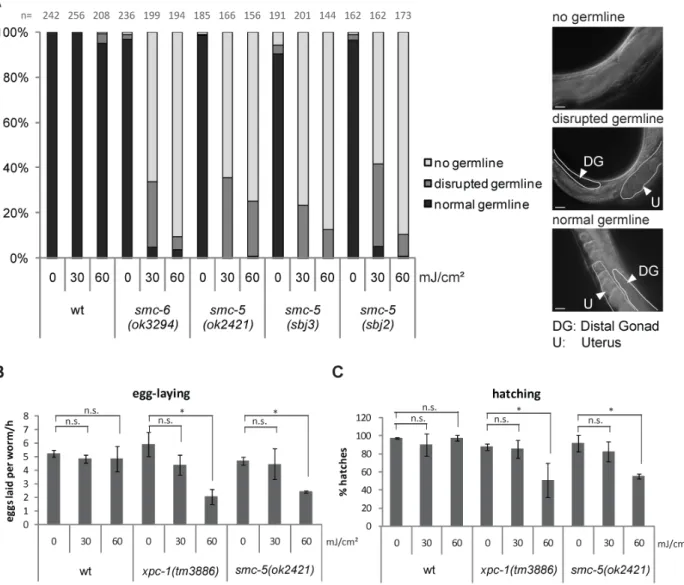
Figure II.4 Mutations in GG-NER and TC-NER lead to embryonic lethality and developmental arrest, respectively.
A NER pathway. After sensing transcription stalling by CSB-1 or bulky lesion sensing elswhere in the genome by RAD-23/XPC-1, the helix is locally opened by TFIIH and the lesion is validated by XPA-1. The endonucleases ERCC-1/XPC-1 and XPG-1 cut the damaged strand to release an oligonucleotide containing the lesion. The DNA is restored by gap filling and ligation with the help of the replication machinery.
B Average number of eggs laid per hour, 24h post irradiation of L4 larvae.
C Percentage of worms hatched from eggs quantified in B.
B and C Error bars indicate standard deviation between three independent replicates. Asterisks denote p value
≤0.01 calculated applying two-tailed students T-Test.
D and E Percentage of larval stages 48h and 72h after irradiation at L1 stage, respectively. Number of worms used (n) for analysis is indicated at the top of the columns.
20 in the global genome (GG-)NER branch cause the skin cancer susceptibility syndrome Xeroderma pigmentosum (XP), while mutations affecting the transcription-coupled (TC-)NER branch lead to progeroid syndromes such as Cockayne syndrome (CS) that is characterized by postnatal growth retardation and accelerated aging but not cancer (Alan R Lehmann 2003).
Hence, even in the absence of exogenous DNA damage NER is important for normal development, growth and tissue maintenance. The two branches of NER differ in their recognition of the lesion but funnel into the same pathway for processing and removal of the damage. The CSB protein is associated with RNAPII and, upon stalling at a lesion, recruits CSA and activates TC-NER. Outside of actively transcribed genes the UV-DDB ubiquitin complex and the trimeric XPC/hHR23/Centrin complex scan for lesions disrupting the helical structure known as GG-NER. Upon lesion sensing, the NER machinery is recruited including XPA and the 10-subunit transcription factor II H (TFIIH) that comprises XPB, XPD, and TTDA (p8).
TFIIH locally unwinds the DNA and recruits XPG to the 3′ side of the lesion, which in turn stabilizes binding of the XPF–ERCC1 heterodimer (also called XFE) 5′ to the lesion. Both, XFE and XPG, are endonucleases that incise the damaged strand 25–30 nucleotides apart. The single- stranded stretch is coated by RPA before the gap is filled by DNA polymerases δ and ε that are recruited through RFC and PCNA. Finally, the nick is sealed by DNA ligase.
In C. elegans NER has been extensively studied and revealed conservation of most of the NER genes, such as csb-1, rad-23/xpc-1 homologues of the mammalian TC-NER and GG-NER factors, respectively (Figure II.4A) (Lans et al. 2010; Lans and Vermeulen 2011). Although it remains unclear in how far C. elegans is exposed to UV light as a genotoxin in its natural habitat (Félix and Duveau 2012; Barriere and Felix 2006), worms might be exposed to normally occurring DNA damaging chemicals. Therefore, C. elegans relies on NER to repair these lesions.
For examination of helix-distorting lesions in the laboratory, however, it is convenient to use UV irradiation of worms. UV light is divided into three wavelength ranges: UVA (320–400 nm), UVB (290–320 nm) and UVC (<290 nm). Sunlight filtered through the ozon layer is mostly composed of UVA and UVB. While it has been shown that UVC and UVB can induce CPDs and 6,4-photoproducts (6,4-PPs) directly by photoreaction of the DNA, UVA has also been implicated in radiation-mediated oxidative DNA damage (Ravanat, Douki, and Cadet 2001).
Both types of DNA damage lead to bulky lesions in the DNA and a disruption of the helical structure. In the absence of helix-distorting lesion, C. elegans strains deficient in any of the NER
21 factors are phenotypically wildtype-like. However, upon UVB-irradiation, hallmarks of GG- NER and TC-NER deficiency are embryonic lethality (Figure II.4B and C) and developmental arrest (Figure II.4D and E), respectively. This suggests that GG-NER is acting mainly in the germline whereas TC-NER is employed in somatic cells. Worms mutant for factors of the common NER pathway or TC- and GG-NER double mutants arrest already at UV doses that do not harm mutants for TC-NER or GG-NER only. Hence, both pathways act synergistically, and soma and germline cells depend on the other NER branch if one is not functional (Lans et al.
2010; Lans and Vermeulen 2011).
2.5 Translesion synthesis
Alternatively to NER and HR, lesions in proliferating cells may be overcome by replicating over the damage to avoid replication fork stalling. Translesion polymerases are characterized by a more open active site compared to high fidelity DNA polymerases and therefore, are able to read over DNA lesions. Known translesion polymerases in vertebrates are Polη, Polκ, Polι and Rev1, of the Y-family of polymerases and Polζ, B-family member. The structure of the active site confers the lesion specificity of the different polymerases but is also the cause for the lack of proofreading activity. Because of this translesion synthesis (TLS) is considered as an error-prone DNA damage response. Polη is mutated in Xeroderma Pigmentosum Variant (XPV) disorder (Masutani et al. 1999) which is characterized by increased susceptibility to UV-induced carcinogenesis like NER-deficient patients (Alan R Lehmann et al. 2007). In line with the disease pattern, Polη is specific for UV-induced CPDs as well as some other lesions (Roerink et al. 2012). The C. elegans homologue of Polη, polh-,1 is crucial during the first rapid divisions in the embryo upon fertilization (Ohkumo et al. 2006; Roerink et al. 2012). While in the absence of DNA damaging insults, polh-1 mutant worms are wildtype-like, embryos are extremely UV sensitive as measured by survival of progeny form UV-treated L4 larvae (Roerink et al. 2012).
The early divisions of the C. elegans are fixed in their spatiotemporal progression and any deviation or delay from this pattern may be detrimental to the embryo. Therefore, it is very important to strictly control this process and prevent any delay. It was shown that embryos silence the checkpoint response with the help of POLH-1 by reading over lesions (Holway et al.
2006). In contrast to polh-1 embryonic sensitivity, worms defective for xpa-1, an essential component of NER pathway (Figure II.4 A), are not sensitive. However, development of L1
22 larvae and also germ cell maturation mainly relies on NER whereas polh-1 is dispensable (Roerink et al. 2012). Double mutants compromised for xpa-1 and polh-1 are even more sensitive to DNA damage than the respective single mutants, suggesting that they are acting in parallel pathways. Upon stalling of the replication fork at a lesion, TLS is mainly promoted by two posttranslational modification events. Mono-ubiquitination of PCNA homologue, PCN-1, (Alan R Lehmann et al. 2007) induces polymerase switching and POLH-1 is protected from degradation by SUMOylation (Kim and Michael 2008). In addition to UV- and cisplatin- sensitivity, it was reported that polh-1 deficient worms are as sensitive to IR as brc-1 (ortholog of human BRCA1) mutants. Yet, evidence is accumulating that sensitivity to IR is not caused by polh-1 acting in HR, as suggested earlier, but is rather a consequence of IR-induced lesions other than DSB (McIlwraith et al. 2005; Alan R Lehmann et al. 2007).
23
III BRC-1/BRD-1 inactivation suppresses genome instability during replication blockage in smc-5 mutants
(modified from Wolters et al. 2013, under review at Genetics)
3.1 Introduction
3.1.1 Discovery of a novel radiation-sensitive gene
In the 1970s several radiation-sensitive (rad) mutant Schizosaccharomyces pombe (S. pombe) were isolated and UV- as well as IR-sensitivity was characterized (Nasim and Smith 1975). The S. pombe rad18-X mutant was described to be a Structural Maintenance of Chromosomes (SMC) family member judged from its sequence motifs (A R Lehmann et al. 1995). Later, rad18 was renamed to smc6. Structurally smc6 compromises an ATP-binding domain at its N- and C-terminus and disruption of the N-terminus leads to dysfunction of the protein. Moreover, the protein harbors two extended coiled coil domains which are separated by a hinge in the middle (Figure III.1) (Fousteri and Lehmann 2000; T. Hirano 2006). The protein folds back in an antiparallel coiled coil that brings together the ATPase domains on the N- and C-terminus. The hinge domain is the interacting motif for heterodimerization of Smc6 with Smc5. Mutations impairing either Smc6 or Smc5 are thought to disrupt the functionality of Smc5/6 complex and, hence, elicit the same phenotypes.
3.1.2 The Smc family
Like Smc5/6 also the other SMC family members form heterodimers, which are Smc2 and Smc4, and Smc1 and Smc3, commonly known as condensin and cohesin, respectively (T. Hirano 2006;
Figure III.1 Basic achitecture of SMC complexes.
Each SMC binding partner of a heterodimer folds back in a coiled coil bringing together the N- and the C-terminus. Thereby an ATP-binding head region is formed. The hinge domain is the interacting motif with the other SMC subunit. Non-SMC subunits mainly bind to the head region to close the ring formation.
24 N. Wu and Yu 2012). Furthermore, all three complexes are associated with non-Smc elements (Nse). Cohesin has a well-established role in sister-chromatid cohesion by embracing the sister chromatids inside its ring (M. Hirano and Hirano 2006; Sun et al. 2009). It is loaded during telophase and G1 phase before the onset of replication. Chromatids are released by cleavage of cohesin employing seperase at meta-anaphase transition. Similar to Smc5/6, cohesin-defective yeast was reported to be sensitive to killing by UV irradiation and IR. Based on this observation, further studies suggested implications of cohesin in DSB repair. Here, cohesin is important to keep sister chromatids in close proximity as a prerequisite for strand invasion and intra-S as well as G2/M checkpoint activation upon DNA damage (N. Wu and Yu 2012).
The third SMC complex, condensin, functions in chromosome organization and condensation. In human two condensin complexes differing in their subunit composition have been reported.
While condensin I was shown to be linked to single strand break DNA repair, condensin II has a role in DSB repair by HR. C. elegans encodes an additional condensing-like SMC complex important for dosage compensation (N. Wu and Yu 2012).
3.1.3 The subunits of the Smc5/6 complex
In yeast the Smc5/6 complex is associated with at least six Nse proteins and so far only four were found in human (reviewed in De Piccoli, Torres-Rosell, and Aragón 2009).
Nse1 resembles structurally an ubiquitin ligase due to its RING finger motif. The enzymatic action of this protein, however, remains to be shown. Nevertheless, S. cerevisiae carrying a mutation in Nse1 display growth defects and human Nse1 was reported to be ubiquitinylated in vivo. Furthermore Nse1 interacts with Nse3, forming a subcomplex together with Nse4 (Palecek et al. 2006).
Nse2 is also known as Mms21 and confers DNA damage resistance in S. cerevisiae through its action as a SUMO E3 ligase (X. Zhao and Blobel 2005). Deficiency for this enzymatic function lead to MMS-, UV- and bleomycin- (inducing DNA damage similar to IR) sensitivity as well as defects in nucleolar organization and telomere integrity. Also in human MMS21 was shown to be important for DDR and prevention of inappropriate apoptosis induction (Potts and Yu 2005).
Targets of Mms21 SUMO ligase are Smc5 and Yku70, NHEJ factor, as indicated in yeast (X.
Zhao and Blobel 2005) as well as SMC6 in human (Potts and Yu 2005).
25 Nse3 is structurally related to the MAGE (melanoma antigen gene) family of proteins and leads to MMS sensitivity in human cells carrying MAGEG1, homologue of Nse3, mutation (Taylor et al. 2008). In Drosophila, mutation of MAGE sensitizes to IR, HU and MMS comparable to Smc5 and Smc6 mutations (X. Li et al. 2013). Furthermore, it was reported that dysfunctional MAGE leads to pupal lethality upon treatment with ATR and ATM kinase inhibitor, caffeine.
Nse4 belongs to the kleisin superfamily that are characterized by interaction with SMC proteins to form ring-like structures (Schleiffer et al. 2003). In particular, Nse4 was shown to bridge Smc5 and Smc6 to form a ring (Palecek et al. 2006).
Finally, Nse5 and Nse6, which are reported to be yeast-specific, were also found to interact with Smc5/6 and, like Nse4, might join the arms of the Smc5/6 complex (Palecek et al. 2006).
3.1.4 Characteristics of the Smc5/6 complex
The initial publication about rad18 (smc6) by Lehmann et al. reported that this novel protein acts in parallel to rad13, the homologue of NER factor XPG, and is epistatic with the RAD51 homologue, rhp51 (A R Lehmann et al. 1995). Furthermore the authors noted that rad18 is important for replication. Concerning the radiation sensitivity, it was suggested that smc6 could act by promoting Rhp51 strand invasion or hold together the ends of a DNA DSB (Fousteri and Lehmann 2000). Further evidence for smc6 acting in HR came from the finding that smc6 is also epistatic with mus81 endonuclease (Sheedy et al. 2005). Another allele of smc6 was identified later in a screen for DNA damage checkpoint-defective strains, namely rad18-74 (Verkade et al.
1999). The point-mutation found in this strain affected the C-terminal ATP-binding domain and conferred Methyl methanesulfonate (MMS)- and HU-sensitivity. In particular, G2 checkpoint was not triggered in response to IR, MMS or UV-mimetic 4-NQO agent treatment in the rad18- 74 mutant strain in contrast to wildtype S. pombe. While chk1 phosphorylation kinetics after IR and UV irradiation were normal at the beginning, rad18-74 mutants were unable to maintain chk1 activation in the presence of DNA damage. Genetic analysis of rad18 revealed that overexpression of brc1, containing a BRCT domain, suppressed rad18-74 and rad18-X UV-, MMS-, 4-NQO and HU-sensitivity (Verkade et al. 1999; Sheedy et al. 2005). In contrast, brc-1Δ was found to be synthetically lethal with rad18-74 (Sheedy et al. 2005). S. pombe BRC1 is the homologue of human PAXIP1 (also known as PTIP), which is implicated in NHEJ-mediated V(D)J recombination of T-killer cells (Callen et al. 2012). In line with its role in NHEJ, PAXIP1
26 has been shown to interact with 53BP1 (Cho et al. 2007). After testing a number of other DNA repair and HR factors it was hypothesized that smc6-74 mutants might accumulate aberrant DNA structures that rely on recombination for resolution. Furthermore Sheedy et al. stated that this recombination is mediated by Brc1, Slx1 (homologue of human endonuclease SLX1), Rhp18 (homologue of human RAD18) and Mus81 (homologue of human endonuclease MUS81).
Consistent with a role in HR, temperature sensitive smc6-9 mutant S. cerevisiae were found to arrest in G2/M induced by Rad53 (Chk2) activation (Torres-Rosell et al. 2005). The same was true for a mutant of the binding partner of smc6, smc5. 2D gel electrophoresis revealed that smc5 and smc6 mutants accumulate X-shaped DNA structures that are likely HR repair intermediates.
A fraction of these intermediates could be still resolved in smc6 mutants with the help of SGS1 RecQ helicase and TOP3 topoisomerase III (Torres-Rosell et al. 2005). Studies using HU to induce replication stress illustrated that spatiotemporal recruitment of repair factors, like Rad22 (homologue of human RAD52) and Rhp51 (homologue of human RAD51) does not differ from wildtype in smc6 defective S. cerevisiae. Taken together this indicates that Smc5/6 is dispensable for HR factor recruitment but is essential for Holliday Junction resolution (Ampatzidou et al. 2006). Yeast two-hybrid screen and co-immunoprecipitation experiments identified helicase Mph1, FANCM homologue, as direct binding partner of Smc5/6 complex.
Mph1 does not only dissociate DNA D-loops which are precursor structures of double Holliday Junctions (Y.-H. Chen et al. 2009) but also supports lagging-strand synthesis by Okazaki fragment processing (Kang et al. 2009).
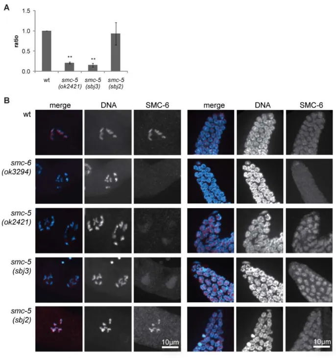
Although initially identified in yeast, Smc5/6 is conserved up to human. In 2010 the first study about SMC-5/6 in a multicellular organism, C. elegans, was published. In contrast to defects of Smc5/6 in yeast, C. elegans mutant for either of the proteins are viable (Bickel et al. 2010).
Nevertheless, worms displayed some degree of genomic instability as seen by chromatin bridges in intestinal cells as well as transgenerational sterility due to defects in the germ cells. Similar to the yeast mutants, also C. elegans smc-5 and smc-6 mutants were sensitive to IR and displayed two times more germ cell corpses (Bickel et al. 2010). Moreover it was found that SMC-5/6 defective C. elegans accumulate RAD-51 foci in meiotic germ cells at mid-pachytene that could be rescued by mutating spo-11 meiotic recombination initiator. Genetic analysis revealed that SMC-5/6 has some role in promoting inter-sister recombination repair in C. elegans.
27
3.2 Results
3.2.1 Screening for UV-sensitive alleles in L1 larvae.
We sought to identify new genes involved in the response to UV-induced DNA damage. We employed a forward ethyl methanesulfonate (EMS) mutagenesis screen in C. elegans to recover mutant worms that are hypersensitive to UVB-irradiation (Figure III.2 A). EMS as a mutagen has been used for a long time and is an established tool. First it was employed in 1959 (Loveless and Stock) for examination of the effect of alkylating agents. In 1974 it was established for the nematode model system as a tool for mutagenesis by Sydney Brenner (S. Brenner 1974).
Generally EMS has a tendency to make GC to AT transitions, which is an advantage because exons are GC rich compared to the rest of the C. elegans genome (Flibotte et al. 2010). By screening over 2,500 worms, five strains could be identified displaying UV-sensitivity. This was confirmed by repetition of the experiment using a synchronized L1 larvae population and treatment with UV light. While two strains arrested at L1 stage upon irradiation, three strains were sterile in adulthood. The novel strains and the respective alleles were named BJS1(sbj1), BJS2(sbj2), BJS3(sbj3), BJS4(sbj4) and BJS19(sbj19). In the next step we backcrossed the strains to N2 wildtype worms to outcross mutations not linked to the UV-specific phenotype.
Since those strains were indistinguishable from wildtype worms when not treated with UV- radiation, we had to screen the F2 generation of the crossed P0 worms for UV-sensitivity. This was done similarly to the initial screening method by irradiation of the F3 generation while single F2 worms were kept in liquid culture backup plates.
In parallel, the mutation was mapped to the chromosome by single nucleotide polymorphism (SNP) mapping technique (Davis et al. 2005). After several rounds of backcrossing, Solexa whole-genome sequencing was performed. The sequencing data were analyzed using the
‘Mapping and Assembly with Qualities’ (MAQ) Gene mapping software with the C. elegans genomic sequence version WS201 from www.wormbase.org as reference. The data were processed using standard settings for MAQ Gene software. Overall around 1,900 to 2,000 mutations were found in each of the mutants. However, all five strains sequenced had 1,013 mutations in common with each other. Most likely these aberrations display differences between the wildtype strain in our laboratory compared to the wormbase reference and are not induced by EMS. Therefore, these variations were not taken into account for further analysis.
28
Figure III.2 EMS-based forward genetic screening.
A Worms were mutagenized with EMS and F2 generation synchronized. After egg-laying for 2-3h, F2 adults were backed up in 96-well plate liquid culture. F3 larvae were irradiated with 60mJ/cm² UV light and screened for impaired development and reproduction 48h later. The phenotypes were confirmed using the worms from the 96-well backup plate.
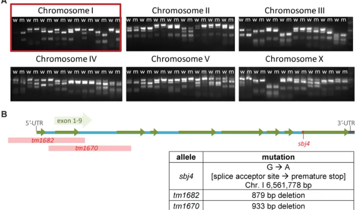
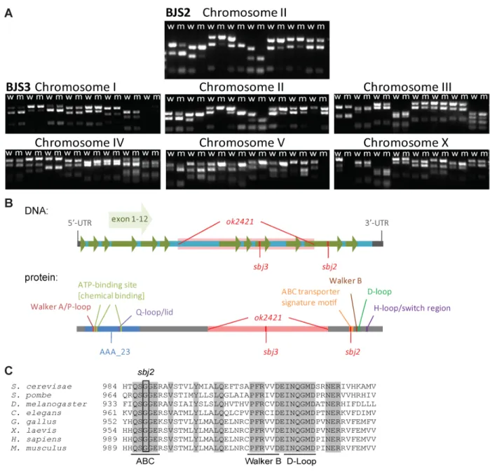
B Table showing the number of mutations found in five worm strains using whole-genome sequencing and MAQ gene analysis (Bigelow et al. 2009). C. elegans genomic sequence version WS201 from www.wormbase.org served as reference. The number of ‘unique mutations’ represents mutations not common to all five strains to account for differences of the wildtype used here and the wormbase reference. The chromosome linked to the mutation conferring UV-sensitivity is marked with an asterisks (*). BJS1, BJS2 and BJS19 were identified by Dr. Maria A. Ermolaeva.
C Graph displays the relationship between the number of mutations in a given worm strain and the number of crosses to N2 wildtype worms. The linear regression line (y = -13.591x + 1034.9) and R² are shown.
D Percentage of nucleotides mutated on the six chromosomes of C. elegans in the five sequenced candidate strains. The chromosome linked to the mutation conferring UV-sensitivity is marked with an asterisks (*).
E Distribution of mutations in BJS4 strain. Plus-signs denote mutations solely found in BJS4 and not in any other of the four strains. The mutation linked to the UV-sensitivity phenotype is marked with an asterisks (*).