Glutamate oxidase advances the selective
bioanalytical detection of the neurotoxic amino acid -ODAP in grass pea: A decade of
progress*
Ghirma Moges
1,‡, Negussie Wodajo
1,2,†, Lo Gorton
3,
Yirgalem Yigzaw
3, Kurt Kalcher
2, Abebaw Belay
1,**, Girma Akalu
4,***, Baboo M. Nair
4, and Theodros Solomon
11Department of Chemistry, Addis Ababa, Ethiopia;2Department of Analytical Chemistry, Institute of Chemistry, Karl-Franzens University, Graz, Austria;
3Department of Analytical Chemistry, University of Lund, Sweden;4Applied Nutrition and Food Chemistry, University of Lund, Sweden
Abstract: The search for an enzyme as a reagent for selective bioanalytical detection of the neurotoxic amino acid, β-N-oxalyl-L-α,β-diaminopropionic acid, β-ODAP (found in grass pea, Lathyrus sativus) led to its redox catalytic reaction by glutamate oxidase (GluOx).
Homogeneous kinetic studies and an immobilized GluOx reactor-based flow-injection assay were initially made for β-ODAP with small immobilized GluOx/catalase glutamate-destroy- ing prereactors. The method was applied to examine the toxin content in processed grass pea.
The kinetics and the equilibrium of the thermal isomerization of β-ODAP to the nontoxic iso- mer α-ODAP established that GluOx is specific to the neurotoxin. The first ever GluOx-based amperometric biosensor for liquid chromatography (LC) detection was re- ported in 1997. This biosensor coupled with a refractive index detector improved LC per- formance. The most recent work with GluOx resulted in MnO2-based screen-printed ampero- metric biosensor, with offline elimination of glutamate interference by glutamate decarboxylase. A single-shot chemiluminescent sensor developed for hydrogen peroxide is also proposed for β-ODAP with GluOx application. This decade of progress resulted from studies that included four Ph.D. (Ethiopia, Sweden, Austria), four M.Sc. (Ethiopia, Sweden) and Licentiate (Sweden) theses projects, plus one collaborative project in Sweden. The ad- vances in grass pea research may be regarded as a model north–south cooperation for re- search and education.
INTRODUCTION
Grass pea (Lathyrus sativus, also known as guaya and sebere in Ethiopia and khesari dhal in the Indian subcontinent) is a food, feed, and fodder crop that belongs to the family Fabaceae. It is produced in India, Pakistan, Bangladesh, Ethiopia, Syria, China, and Nepal, giving rise to different names in differ-
*Plenary lecture presented at the Southern and Eastern Africa Network of Analytical Chemists (SEANAC), Gaborone, Botswana, 7–10 July 2003. Other presentations are published in this issue, pp. 697–888.
‡Corresponding author: current address: OPCW, Johan de Wittlaan 32, R The Hague, The Netherlands; E-mail:
ghirma2001@yahoo.com
†N. W. Beyene: current address: University of Pretoria, Department of Chemistry, South Africa
**A. B. Jemere: current address: University of Alberta, Department of Chemistry, Edmonton, Canada
***G. Akalu: current address: Ethiopian Health and Nutrition Research Institute, Addis Ababa, Ethiopia
ent regions [1]. This hardy crop thrives under poor soil and climate conditions, resists drought and flooding, and is cultivated when other crops fail to grow [2].
Grass pea has a high nutritional value containing up to 31 % protein and 65 % carbohydrate [3]
and is a good source of minerals. However, consumption of grass pea by humans causes an irreversible paralysis of the lower limbs called neurolathyrism [4]. The disease is prevalent in times of drought and flood as a result of frequent consumption of grass pea meals. Consumption of meals solely made from the seed for more than three months may cause this crippling disease [2]. Epidemic cases of this dis- ease have been reported in Ethiopia, India, Pakistan, China, and Bangladesh when famine is triggered by drought. The earlier history of neurolathyrism, which dates back to the time of Hippocratus, has been reviewed recently [1].
The most remarkable account of neurolathyrism comes from the description of the epidemic among the prisoners of war at a camp in Vapniarca, Ukraine, occupied by Germany during World War II [5]. This is reported as the highest incidence of neurolathyrism on record, developed between December 1942 and February 1943. The diet of all inmates had included up to 400 g of boiled grass pea seed per day. Within months, 60 % of the 1200 or so inmates developed different symptoms and levels of neurolathyrism. Lathrysim was also a major outbreak during the Spanish Civil War, 1936–1939, due to heavy consumption of lathyrus gruels by peasants mainly in the Spanish provinces Catalonia and Castilla [6]. In the Castillian town of Consuegra, at least 1 % of 5000 were reported to have developed neurolathyrism. Citing a recent article on the adverse effect of neurolathyrism in Ethiopia on the productive population of a rural community may suffice to illustrate the effect of neu- rolathyrism on humans [7].
The causative agent for neurolathyrism, β-N-oxalyl-L-α,β-diaminopropionic acid (β-ODAP) was identified and characterized by Rao and coworkers [8]. It is a non-protein amino acid (HOOCCONHCH2CHNH2COOH) found in grass pea.
ANALYTICAL METHODS FOR THE DETERMINATION OF -ODAP
Research to eradicate neurolathyrism has been intensified since the identification of the neurotoxic amino acid, β-ODAP, in grass pea. This covers investigation of clinical symptoms, biosynthesis, toxicity mech- anism, traditional genetic breeding, and genetic engineering, as well as food processing. The latter in- cludes comparisons between boiling or cooking, roasting, autoclaving, and fermentation to produce meals with none or low content of β-ODAP. Grass pea research obviously requires a reliable analytical method for assaying the toxin in presence of the nontoxic isomer and other possible interferences.
Historically, the first known method to determine β-ODAP is the ninhydrin method, which in- volved electrophoresis of the grass pea extract on a paper strip followed by spraying with 0.5 % nin- hydrin in acetone and heating at 80 °C for 15 min before eluting the ninhydrin positive band with 75 % ethanol containing copper sulphate [9]. The absorbance reading is at 510 nm. The method, however, was not sensitive and was soon replaced by a newer colorimetric method developed by Rao [10], which is the most widely used method up to now. Rao reported in 1978 an indirect assay for the toxin involv- ing a colorimetric determination of L-α,β-diaminopropionic acid (DAP), the alkaline hydrolysis prod- uct of the toxin. DAP forms a yellow adduct (1-alkylthio-2-alkylisoindole), the absorbance of which is measured at 420 nm, with o-phthalaldehyde (OPT) in basic aqueous buffer and in the presence of a re- ducing agent (mercaptoethanol).
Various modifications of this method were made to optimize its reliability, including changes in the extraction time, extraction medium, constituents of the OPT reagent, and the buffer system. Hussain et al. compared six modifications [11] (as practiced in six different laboratories) with the original method by Rao. They reported results with considerable variations in the β-ODAP content of the same sample. Besides these discrepancies, the OPT method lacks selectivity since the nontoxic isomer, α-ODAP, also hydrolyzes to yield DAP. In addition to the long period of time required for extraction, assaying the toxin involves a 1-h hydrolysis and color reaction. An evident disadvantage of the OPT
method is that it assays total ODAP and cannot be used to monitor the decrease in toxin content in meals from grass pea processed under different temperature and pH. It is established that β-ODAP isomerizes to α-ODAP with heating (boiling, cooking) to an equilibrium value with a ratio of about 60:40 [12].
In the search for more selective and rapid assays, high-performance liquid chromatographic (HPLC) methods were developed initially with offline precolumn derivatization steps. Derivatizations with dansyl chloride [13] and 9-fluorenylmethyl chloroformate [14] do not differentiate between the two isomers, β-ODAP and α-ODAP. Separation of the isomers was achieved by phenyl isothiocyanate (PITC) derivatization, but the experimental inconvenience is aggravated since it is difficult to isolate the derivatizing reagent [15]. Reversed-phase high-performance chromatography with 6-aminoquinolyl-N- hydroxy-succinimyl carbamate (AQC) derivatization was also reported [16], but the derivatives easily decompose at room temperature. Capillary zone electrophoresis was also considered [17], which has the drawback of a higher working pH (9.2), a condition that may cause β-ODAP hydrolysis to DAP. An option that had not been much explored is the possibility of applications of enzymes for selective bio- analytical detection of the neurotoxin. Moges discussed the possibilities of direct and indirect enzyme- based detections for the toxin [18].
ENZYME-BASED DETECTIONS
The use of enzymes as biocatalysts for selective detection of substrates is well established, including for L-amino acids. Until a report in 1993 [19], there had not been any known enzyme that could cat- alyze the oxidation of the neurotoxin as an L-amino acid. In general, proposals that could lead to sus- tainable research on enzyme-based assays for the compound had not been put forward.
Earlier developments
Mehta and coworkers reported that tryptophanase degraded ODAP to pyruvate and other products [20].
The attempt to reproduce and detect the reported activity of tryptophanase on ODAP, by monitoring any pyruvate formation, was not successful [21]. Another earlier work was also reported on the use of DAP-ammonia lyase (from Pseudomonas sp.) for the determination of ODAP after its alkaline hydrol- ysis to DAP [22]. Evidently, the approach would be equally unselective as the OPT method. There has not been any report on its sustained application after the paper was published in 1971.
Ammonia is an enzymatic reaction product of several nitrogen-containing compounds, which typ- ically includes L- and D-amino acids catalyzed by amino acid redox enzymes. Ammonia is also liber- ated in α,β-elimination reactions of a number of amino acids catalyzed by tryptophanase. DAP is one of these amino acids involved in this type of reaction [23]. DAP is obviously the hydrolysis product of ODAP which, as intermediate, is used for the colorimetric determination of the toxin in grass pea [10].
Indirect detection of β-ODAP with this approach was given consideration since ammonia could easily be detected in a cuvette or by flow-injection method with glutamate dehydrogenase (GlDH) in the pres- ence of α-ketoglutarate and nicotinamide adenine dinucleotide (NADH). The disappearance of NADH can be conveniently monitored with different detectors [24]. The reaction equilibrium is quite favorable toward the formation of glutamate. Hence, an FI ammonia assay system with a GlDH reactor was ini- tially developed to test the action of tryptophanase on DAP [25].
The formation of ammonia from DAP was detected from a crude tryptophanase-catalyzed reac- tion at 37 °C by injecting samples into the FI system described. The activity of crude tryptophanase was, however, very low, only about 1.5 ×10⁻⁴µmol min–1per mg of the crude enzyme [25]. This result was noted as a prospect for possible indirect semienzymatic analytical method for β-ODAP if a purified and a more active enzyme could be made available.
Moges and coworkers reported the result of a screening procedure with seven enzymes for any possible catalytic action over β-ODAP, or its products [21]. The selection of the enzymes was based on the catalytic activities with respect to known substrates possessing similar functional groups or struc-
tural features shared with β-ODAP as well as on its hydrolysis product, DAP. One of the tests made was purified tryptophanase on DAP but had still low activity on the substrate, and it was concluded that the enzyme had no analytical potential. The tests continued for other enzymes, if possible, which could di- rectly act on β-ODAP.
Glutamate oxidase: A new enzyme for -ODAP
One of the enzymes considered for testing was L-glutamate oxidase (EC 1.4.3.11, from Streptomyces sp.), whose selectivity to L-glutamate arises from the presence of additional terminal carboxylic group other than the α-carboxylic group. The enzyme is also slightly active to L-aspartate. β-ODAP has also a terminal carboxylic group (Fig. 1).
One of the screening experiments, resulted in the catalytic oxidation of the β-ODAP by the redox enzyme, reflecting that the action of the enzyme exhibits structure–activity relationship. In the presence of this enzyme, β-ODAP generated hydrogen peroxide and ammonia, showing that the compound un- dergoes a typical L-amino acid oxidase catalyzed reaction [21]. Hence, the oxidation reaction product is attributed to the formation of the corresponding α-keto acid, β-N-oxalyl-α-keto-β-aminopropionic acid.
HOOCCONHCH2CHNH2COOH + O2+ H2O →
HOOCCONHCH2COCOOH + H2O2+ NH3 (1)
The kinetics of the redox enzyme reaction in solution was studied and compared with that of glu- tamate and aspartate. The rate of the reaction of the neurotoxin was found much slower, only about 0.8 % of the rate for glutamate and slightly higher than that of aspartate. Developing a soluble enzyme assay method for the substrate was, therefore, hindered by this factor, which otherwise would be an ex- pensive method, due to the very high cost of this redox enzyme.
FLOW-INJECTION ANALYSIS OF-ODAP
The option considered for a flow system application with an immobilized glutamate oxidase (GluOx) reactor led to a successful development for assaying β-ODAP, which included an immobilized horse- radish peroxidase (HRP) reactor for detecting the generated hydrogen peroxide [19]. Studies continued to eliminate the possible interference of glutamate in grass pea or preparations from the crop. Moges and Johansson described the first flow-injection assay for the neurotoxin β-ODAP in grass pea using an immobilized GluOx reactor with two prereactors to eliminate the interference from glutamate. [26]. The system consisted of four packed-bed enzyme reactors in series: GluOx (20 µl), catalase (20 µl), GluOx (250 µl), and horseradish peroxidase, HRP (50 µl). Residence time studies and practical tests for the enzyme loaded showed, at a flow rate of 0.3 ml/min, 100 % oxidation of up to 500 µM glutamate in the 20-ml glutamate oxidase (GluOx) reactor followed by a complete destruction of its product, hydro- gen peroxide, in the 20-µl catalase reactor. This was verified by the absence of a glutamate FI signal.
Tests up to the studied limit of hydrogen peroxide, 2000 µM, also showed no FI response because of the high activity of the small catalase reactor. Only 5 % of the β-ODAP was destroyed by the two small Fig. 1 Structures of the neurotoxic amino acid β-ODAP and L-glutamate.
prereactors. Good conversion efficiency for β-ODAP (80 %) was obtained with the third 250-µl GluOx reactor (with higher enzyme loading) at a flow rate of 0.3 ml min–1. Exploiting kinetic differences for interference elimination is not new in analytical chemistry, but the approach for destroying glutamate (and hydrogen peroxide) was reported as a new kinetic approach for enhancing specificity with FI re- actors. The complete FI set-up with the four reactors was employed to monitor the toxin in grass pea with 20 samples per hour at a flow rate of the carrier stream, 0.3 ml/min. The hydrogen peroxide from the oxidation of β-ODAP was detected via red quinoneimine formation, λmax 512 nm (Trinder method) [27], in the presence of 4-aminoantipyrene and 2,4-dichlorophenol-6-sulphonate, in the HRP reactor.
Injections of 20-µl ODAP standards into the four-reactor FI system delivered a response curve, which was linear within the range of 10–650 µM β-ODAP using a phosphate buffer at pH 7. The system was used to determine β-ODAP in grass pea extracts in phosphate buffer after offline separation of macro- molecules with ultrafiltration membranes (MW cut-off value 10 000). The proposed FI method was very fast compared with the usual colorimetric and the earlier-reported HPLC methods. Further, β-ODAP so- lutions after an offline heating at 80 °C for 1 h showed 62 % FI response compared to the unheated so- lutions due to the isomerization of β-ODAP to α-ODAP, which agreed with the well-known equilibrium ratio of the β- and α-isomers, about 60:40 [12]. This is the most important achievement of the gluta- mate oxidase method as the enzyme selectively oxidizes the β-isomer in presence of the α-isomer.
Effect of processing grass pea
The FI system with the four enzyme reactors was intensely used later in a work involving a Ph.D. proj- ect fully committed to the chemical composition of grass pea [3]. It aimed at developing a process for reducing the β-ODAP content in grass pea seeds. The publications generated additional data on the con- sequences of processing grass pea on the chemical composition (protein, starch, total dietary fiber, fat, and ash), in vitro and in vivo digestibility, and physicochemical as well as functional properties of starch [3,28–29].
The flow-injection assay revealed that most of the β-ODAP (93 %) in the seed is located in the cotyledons. It was also shown that there is a significant variation in the β-ODAP content (from 520 to 942 mg/100 g) among the different varieties investigated. It was shown that the β-ODAP content in the whole seeds could be reduced (up to 87 %) by roasting, cooking, autoclaving, and soaking.
Recommendations for effective reduction of the neurotoxin, monitored on the FI assays, consid- ered the desired nutritional characteristics. Soaking for 12 h of 1.5–2.0 mm grits from the cotyledons and roasting at a temperature of 200 °C for a period of 37 min was recommended to significantly re- duce β-ODAP in combination with improved in vitro digestibility. The seeds could also be processed by soaking the grits for 12 h from the cotyledons and cooking in water at pH 8 for 75 min in order to achieve similar effect of reducing the content of the neurotoxin and improving the in vitro digestibility of starch.
Kinetics of thermal isomerization of -ODAP
Although the phenomenon and β-αequilibrium ratio (60:40) after isomerization was described as long ago as 1966, no detailed quantitative study on the β-αisomerization of the toxin, that included ener- getics and kinetic order, had been earlier studied [12]. An interesting NMR study of thermal isomer- ization of β-ODAP and its higher homologs of β-ODAP [HOOHCONH(CH2)nCHNH2COOH where n = 3 or 4] had been reported, which compared their isomerization products and suggested a mecha- nism [30].
As indicated earlier, 62 % toxin was detected with the FI assay after offline heating experiments of solutions of β-ODAP [26]. Further work continued with the FI method to produce detailed quantita- tive data on the kinetics of β-αisomerization and propose a mechanism for the process (the GluOx and HRP reactors in the FI set-up were sufficient for this study). A fixed volume of phosphate buffer (0.1 M,
pH 7) was introduced into a screw-capped test-tube and heated in a water bath thermostatted at 60, 70, 80, and 90 °C. A 10-mM β-ODAP solution in the same buffer was added to the preheated buffer to maintain its final concentration within 500–2000 µM [31,32]. A 100-µl sample was withdrawn from the heated solution after 1 min and injected into the FI system following 2–10-fold dilution in the same buffer at 4 °C. More samplings were made every 5 or 10 min for injecting into the system.
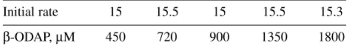
The study showed that at a given temperature, the initial rate constant of isomerization of the toxin to the nontoxic isomer , k0, was independent of its initial concentration (Table 1 below). The mean rate at 80 °C was 15.3 µM/min at pH 7. The plot of log(rate) against log[β-ODAP] revealed a horizontal line with a slope of zero and with an intercept equal to ln ko. This finding suggests that the isomerization process follows zero-order kinetics.
Table 1 Data on the β-αthermal isomerization of β-ODAP with concentration at 80 °C.
Initial rate 15 15.5 15 15.5 15.3
β-ODAP, µM 450 720 900 1350 1800
The results also showed that the equilibrium time to reach 62–63 % β-ODAP was linearly de- pendent on the initial concentration. Both the initial apparent rate constant of isomerization and linear relation between the equilibrium time and the initial concentration confirmed a zero-order kinetics, sug- gesting rapid formation of an intermediate, whose decomposition to both isomers should be the rate- limiting step [31]. The results support a previously suggested mechanism, the formation of a 5-ring cyclic intermediate via intramolecular rearrangement, since it was found that higher homologs of β-ODAP, [HOOHCONH(CH2)nCHNH2COOH where n = 3 or 4], did not favor oxalyl transfer to the α-amino group [30].
Sample pretreatment studies
In the original FI method, ultrafiltration membranes (MW cut-off value 10 000) were employed to sep- arate the macromolecules from grass pea extracts, prior to injection into the system. This technique is not affordable for routine applications in the laboratories of the developing countries severely affected by neurolathyrism. In an attempt to solve this problem, Wodajo and coworkers, Addis Ababa, utilized chemical precipitation of proteins with perchloric acid and trichloroacetic acid (TCA) for sample pre- treatment of grass pea extracts prior to injection [33]. The effects of removing the protein from the ex- tracts by the two precipitation methods (and ultrafiltration) were compared with that of the directly in- jected crude extracts after spiking with 300–400 µM glucose. The FI response to the spiked glucose in the grass peas samples was tested with a 50-µl immobilized glucose oxidase, GOD, reactor (a much cheaper enzyme than glutamate oxidase) and an HRP reactor in series. Protein removal by precipitation was estimated by the method of Lowry [34]. Responses to injections of grass pea extracts after separa- tion of the protein with precipitation exhibited enhanced reactor stability. Between 40–50 injections of the pretreated extracts spiked with glucose were made into the FI system. The recorded glucose signals showed no significant changes in the responses for all cases of samples treated with precipitation as well as with ultrafiltration. The FI responses in the case of the directly injected extract, spiked with 300 µM glucose, declined gradually, without any response after the 36thinjection. The result of the sample treat- ments by offline precipitation methods was proposed for stability tests with GluOx reactors for fur- thering its applications to β-ODAP assays.
The same group also studied different extraction procedures of the neurotoxin from grass pea to improve the sample pretreatment [35]. Extractions in water and phosphate buffer, with and without son- ication, were made. Extractions with and without EDTA in extraction were also made. The different ex-
traction approaches had given comparable results in all cases, as the calculated maximum for the neu- rotoxin content was found only 5 % higher than the minimum recorded value for the same sample.
-ODAP BIOSENSOR FOR LIQUID CHROMATOGRAPHIC DETECTION
Enzyme-based biosensors are integrated into online analysis systems for selective detection of analytes of interest in the biomedical industry, including in vivo studies and monitoring fermentation processes after chromatographic separation. Oxidases are the most studied enzymes for bioanalytical detections, including biosensors. The finding that GluOx catalyzes the selective oxidation of β-ODAP for bioana- lytical detection in flow systems spurred on the development of its first biosensor based on glutamate oxidase/peroxidase amperometric detection [36]. The biosensor developed in Lund, Sweden was suc- cessfully applied to measure the β-ODAP content in grass pea, after its chromatographic separation from glutamate.
Further improvements of the amperometric biosensor
Yigzaw and coworkers improved the performance of the biosensor [37] earlier reported by Belay and coworkers [36] for quantitative chromatographic determination of β-ODAP and free glutamate in raw grass pea seed samples using bioelectrochemical and online refractive index detection, thus enabling two important steps, i.e., (i) the chromatographic column separates glutamate and ODAP and (ii) the biosensor selectively monitors the β-ODAP content, whereas the RI detector gives the total concentra- tion of ODAP. The biosensors developed [36,37] are based on cross-linking HRP and an Os-containing mediating polymer [38] with poly(ethyleneglycol)(400) diglycidyl ether (PEGDGE), forming an inner hydrogel layer and then immobilizing GluOx as an outer layer on top of a graphite electrode [39].
Addition of polyethylenimine (PEI) to the hydrogel is believed to have sensitivity- and stability-en- hancing effect on the biosensor. The double-layer approach in the biosensor construction avoided direct electrical wiring of GluOx, resulting in a higher sensitivity of 4.6 mA/M cm2with respect to β-ODAP and a wider linear range (1–250 µM) for both L-glutamate and β-ODAP when compared with a single- layer approach where GluOx, HRP, and Os-polymer are cross-linked together. The limit of detection for the chromatographic–biosensor system was found to be 2 µm with respect to β-ODAP and 0.7 µM with respect to glutamate. The refractive index detection, online with the biosensor, enabled full control of the chromatographic system for assaying the amounts of β-ODAP, glutamate, and total ODAP (α- and β-forms). To test the applicability of the sensors to real sample analysis, the chromatographic system was employed for 10 grass pea samples collected from lathyrism-prone areas of Ethiopia. On a dry mass basis, toxin concentrations of the grass pea collections ranged from 0.52 to 0.76 %. Comparison of the results of an established spectrophotometric assay [10] and that of the present system showed an ex- cellent degree of agreement as revealed by parallel “t” test (90 % confidence limit). The analysis time per sample was 10 min after an extraction time of 90 min. The operational stability of the improved biosensor tested by continuous injections of L-glutamate and β-ODAP in the flow-injection mode was more than 50 h.
More improvement was reported later on the bienzyme electrodes for β-ODAP by the same group based in Lund University [39] by coating solid graphite rods with a premixed solution containing GluOx and HRP cross-linked with a redox polymer formed from poly(1-vinylimidazole) complexed with [osmium (4-4′-dimethylbpy)2 Cl]II/III. Poly(ethylene glycol) diglycidyl ether (PEGDGE) was used as the cross-linker, and the modified electrodes were inserted as the working electrode in a con- ventional three-electrode flow through amperometric cell operated at –0.05 V vs. Ag|AgCl (0.1 M KCl).
The bienzyme electrode was optimized with regard to wire composition, Os-loading of the wire, en- zyme ratio, coating procedure, flow rate, effect of poly(ethyleneimine) addition, etc. The optimized electrodes were characterized by a sensitivity of 88.36 +/– 0.14 µA mM–1cm–1, a detection limit of 0.3µM (calculated as 3 times the signal-to-noise ratio), a response time of less than 10 s and responded
linearly between 0.3 and 250 µM (linear regression coefficient = 0.999) with an operational stability of only 3 % loss of sensitivity as tested in an 8-h continuous flow-injection (FI) operation at a sample throughput of 30 injections h–1.
Fermentation studies
The improved biosensor reported in [39] was also used in fermentation studies of grass pea [40]. Solid- state fungal fermentation of pure grass pea was carried out with strains of Rhizopus oligosporous and Aspergillus oryzae in succession in that order at 35 °C with autoclaving at the start and in-between the inoculations at 100 °C for 10 min. The substrate was allowed to ferment for about 48 h with each in- oculation (106spores/5 g fermenting sample). Grass pea grits were subject to fermentation without any further sample pretreatment, except adjusting the pH to 4 with lactic acid (50 g l–1). This fermentation process reduced the toxin level in grass pea on the average by 82 % for the high toxin variety, and by up to 97 % for the low-toxin variety. The concentration levels of the neurotoxic amino acid were meas- ured by the chromatography-biosensor system. The respective β-ODAP levels in the original grass pea samples were 0.76 and 0.52 % on a dry weight basis.
SCREEN-PRINTED MnO2-BASED -ODAP BIOSENSORS
Kalcher’s group in Graz, Austria has been active in thoroughly investigating the role of manganese dioxide (MnO2) as a modifier for heterogeneous carbon transducers and developed amperometric sen- sors and biosensors using MnO2modified pastes and screen-printed carbon electrodes (SPCEs) [1,41].
In further increasing the applications of their MnO2-modified SPCEs, the group immobilized GluOx in a neutralized Nafion®film for applications to assaying glutamate and β-ODAP. The resulting GluOx electrode responded to injections of standard β-ODAP solutions when used in a flow-injection amper- ometric mode. Using the optimum parameters obtained for the primary substrate glutamate (applied po- tential of 440 mV vs. Ag|AgCl, flow rate of 0.1 ml min–1, and pH 7.75 of the carrier) the biosensor ex- hibited a linear range of 50–500 mg l–1, a detection limit of 29 mg l–1, and a relative standard deviation of 4.5 % (c = 50 mg l–1) for β-ODAP. This is the first ever report on a β-ODAP biosensor based on SPCEs [41]. To avoid interferences from glutamate, its enzyme glutamate decarboxylase (GlDC) was tested successfully by incubating grass pea extracts with the enzyme (see reaction below) [1,42].
Initially the action of GlDC on known glutamate solution was studied by incubation with the enzyme at 37 °C for 3 h. The GlDC-treated glutamate solution showed no response when it was injected into the system.
HOCO-CH2CH2CH(NH2)CO2H →HOCO-CH2CH2CH2NH2+ CO2 (2) The procedure involved extraction of β-ODAP from grass pea seed samples using dihydrogen phosphate solution (pH 4.5), treated with GlDC and the pH adjusted to 7.75 with disodium phosphate solution before injecting it into the FI with the β-ODAP biosensor. The system became specific to β-ODAP. The same sample without GlDC treatment had higher FI peaks, which accounted for re- sponses to glutamate. This is a new advance since GluOx is made specific to β-ODAP by destroying the main substrate with its other enzyme.
The result with GlDC pretreatment is quite significant in the context of relative simplicity, op- tions, and cost. The cost of instrumentation that could be applied for interference elimination with GlDC in FI modes compared with the earlier reviewed chromatographic β-ODAP biosensors would be considerably less. The first FI applied for assaying the neurotoxin employed a kinetic approach to ini- tially destroy glutamate in small GluOx catalase prereactors [26]. Unlike the GlDC method, a GluOx catalase combination in solution cannot obviously be applied for offline grass pea sample pretreatment to eliminate the main substrate.
An FI system with a GlDC glutamate-destroying prereactor is proposed for future studies. An al- ternative approach that employs a multienzyme-layer electrode, to contain GlDC at the upper most layer and GluOx at the inner layer, has also have been suggested.
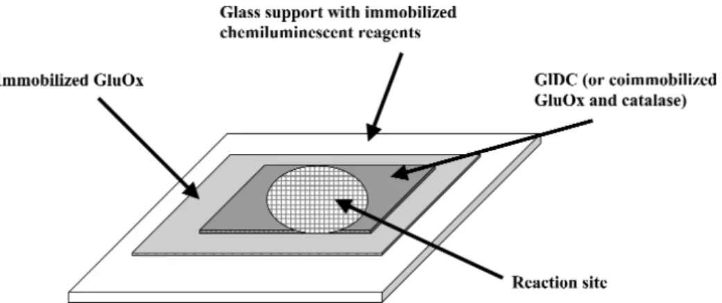
CHEMILUMINESCENT BIOSENSOR
The same group in Graz also envisages a solid-phase chemiluminescent biosensor for β-ODAP. They developed a simple, homemade, battery-driven field device based on light-dependent resistor and/or photodiode as a chemiluminescent light detector for hydrogen peroxide [1,42]. All chemiluminescent reagents (luminol, the catalyst cobalt chloride, the surfactant Triton® X100) were immobilized in a polymeric matrix (hydroxyethyl cellulose, polyvinylpyrrolidone). The device developed using photo- diode performed very well when used for detection of hydrogen peroxide [42]. Work is in progress to couple it with oxidase-catalyzed reactions that generate hydrogen peroxide. The group proposes the de- velopment of a single-shot chemiluminescent β-ODAP biosensor that incorporates multiple enzymatic layers (Fig. 2). The upper-most layer would contain either GlDC (or coimmobilized GluOx-catalase) to destroy interference from glutamate inherent in a grass pea sample and the inner layer to contain GluOx for the oxidation of β-ODAP.
CONCLUSIONS
The work with GluOx in the three institutions during the last decade has shown that β-ODAP can be selectively detected in both reactor and biosensor modes. The LC-biosensor systems coupled with RI detector could be used to assay β-ODAP and total ODAP in grass pea. Small GluOx-catalase reactors (or their mixture in one reactor) enhance β-ODAP selectivity over glutamate. The elimination of gluta- mate interference with glutamate decarboxylase catalysis will offer simplicity and economic advantage over the previously reported applications of the FI and LC-biosensor systems. An interlaboratory study may be proposed for a systematic validation of the GluOx-based bioanalytical detection methods for the neurotoxin amino acid in grass pea against the established Rao colorimentric method.
Fig. 2 Possible pattern for the proposed single-shot chemiluminescent β-ODAP biosensor.
REFERENCES
1. N. W. Beyene. Ph.D. dissertation, Institute of Chemistry, Analytical Chemistry, Karl-Franzens University of Graz, Austria (2003).
2. Grass Pea and Lathyrus/Lathyrism, The Second International Lathyrus/Lathyrism Conference in Ethiopia Addis Ababa, 16–17 Dec. 1992, B. M. Abegaz. R. Tekle-Haimanot, V. S. Palmer, P. S.
Spencer (Eds.), Third World Medical Research Foundation, Mountain Lakes, New Jersey (1994).
3. G. Akalu. Ph.D. dissertation, Department of Applied Nutrition and Food Chemistry, Lund University, Sweden (1999).
4. S. L. N. Rao, K. Malathy, S. Sarma. In World View of Nutrition and Dietetics, Vol. 10, G. H.
Bourne (Ed.), pp. 214–238, Karger AG, Basel (1969).
5. F. Lambein, D. Diasolua Ngudi, Yu-H Kuo. Lathyrus Lathyrism Newsletter 1, 1 (2001).
6. S. Gimenez-Roldan, A. C. Ludolph, J. Hugon, M. Hens, D. Mateo, G. E. Kisby, P. S. Spencer.
Grass Pea and Lathyrus/Lathyrism, The Second International Lathyrus/Lathyrism Conference in Ethiopia Addis Ababa, 16–17 Dec. 1992, B. M. Abegaz. R. Tekle-Haimanot, V. S. Palmer, P. S.
Spencer (Eds.), pp. 10–25, Third World Medical Research Foundation, Mountain Lakes, New Jersey (1994).
7. G. Haileyesius. Lathyrus Lathyrism Newsletter 2, 59 (2001).
8. S. L. N Rao, P. R Adiga, P. S. Sarma. Biochemistry 3, 432 (1964).
9. P. S. Cheema, G. Padmanaban, P. S. Sarma. J. Neurochem. 18, 2137 (1971).
10. S. L. N. Rao. Anal. Biochem. 86, 386 (1978).
11. M. Hussain, B. Chowdhurry, R. Haque, G. Wouters, C. G. Campbell. Phytochem. Anal. 5, 247 (1994).
12. E. A. Bell and P. O’Donovan. Phytochem. 5, 1211 (1966).
13. G. E Kisby, D. N. Roy, P. S. Spencer. J. Neurosci. Meth. 26, 45 (1988).
14. A. Geda, C. J. Briggs, S. Venkatarm. J. Chromatogr. 635, 338 (1993).
15. J. K. Khan, Y-H Kuo, N. Kebede, F. Lambein. J. Chromatogr. A 687, 113 (1994).
16. X. Chen, F. Wang, Q. Chen, X. C. Qin, Z. Li. J. Agric. Food Chem. 48, 3383 (2000).
17. L. Zhao, X. Chen, Z. Hu, Q. Li, Q. Chen, Z. Li. J. Chromatogr. A 857, 295 (1999).
18. G. Moges. Grass Pea and Lathyrus/Lathyrism, The Second International Lathyrus/Lathyrism Conference in Ethiopia Addis Ababa, 16–17 Dec. 1992, B. M. Abegaz. R. Tekle-Haimanot, V. S.
Palmer, P. S. Spencer (Eds.), pp. 97–105, Third World Medical Research Foundation, Mountain Lakes, New Jersey (1994).
19. G. Moges, T. Solomon, G. Johansson. In Proceedings of Lathyrus and Lathyrism: Progress and Prospects, Daka (1993), H. K. M. Yusuf and F. Lambein (Eds.), pp. 73–79, Dhaka University Press, Dhaka (1995).
20. T. Mehta, A.-F. Hsu, B. E. Haskell. Biochemistry 11, 4053 (1972).
21. G. Moges, T. Solomon, G. Johansson. Anal. Lett. 27, 2207 (1994).
22. D. R. Rao, K. Hariharan, K. R. Vijayalakshmi. J. Agr. Food Chem. 22, 1146 (1974).
23. Y. Morino and E. E. Snell. In Methods in Enzymology, Vol. XVIIA, S. P. Colowick and N. O.
Kaplan (Eds.), p. 439, Academic Press, New York (1970).
24. G. G. Guilbault. Handbook of Enzymatic Methods of Analysis, Marcel Dekker, New York (1976).
25. G. Moges, T. Solomon, G. Johansson. Bull. Chem. Soc. Ethiop. 7, 99 (1993).
26. G. Moges and G. Johansson. Anal. Chem. 66, 3834 (1994).
27. P. Trinder. Ann. Clin. Biochem. 6, 24 (1969).
28. G. Akalu, G. Johansson, B. M. Nair. Food Chem. 62, 233 (1998).
29. G. Akalu, F. C. Tufvesson, G. Johansson, B. M. Nair. Starch/Stärke 50, 374 (1998).
30. B. B. Abegaz, P. B. Nunn, A. De Brun, F. Lambein. Phytochem. 33, 1121 (1993).
31. G. Moges, A. Belay, T. Solomon, G. Johansson. In Lathyrus and Lathyrism: A Decade of Progress, Proceedings of an International Conference, Addis Ababa, November 1995, R. Tekle Haimanot and F. Lambein (Eds.), pp. 63–67, University of Ghent, Belgium (1997).
32. A. Belay, G. Moges, T. Solomon, G. Johansson. Phytochem. 45, 219 (1995).
33. N. Wodajo, G. Moges, T. Solomon. Bull. Chem. Soc. Ethiop. 10, 129 (1996).
34. R. K. Scopes. Protein Purification: Principles and Practice, 3rded., Springer Verlag, New York (1994).
35. N. Wodajo, G. Moges, T. Solomon. Bull. Chem. Soc. Ethiop. 11, 151 (1997).
36. A. Belay, T. Ruzgas, E. Csöregi, G. Moges, M. Tessema, T. Solomon, L. Gorton. Anal. Chem. 69, 3471 (1997).
37. Y. Yigzaw, N. Larsson, L. Gorton, T. Ruzgas, T. Solomon. J. Chromatogr. A 929, 13 (2001).
38. A. Heller. J. Phys. Chem. 96, 3579 (1992).
39. Y. Yigzaw, L. Gorton, T. Solomon. Curr. Separations 19, 119 (2001).
40. Y. Yigzaw, L. Gorton, T. Solomon, G. Akalu. J. Agric. Food Chem. 52, 1163 (2004).
41. N. W. Beyene, H. Moderegger, K. Kalcher. Electroanalysis 16, 268 (2004).
42. H. Moderegger. Ph.D. dissertation, Institute of Chemistry, Analytical Chemistry, Karl-Franzens University of Graz, Graz, Austria (2003).