AUS DER ABTEILUNG FÜR NEUROPATHOLOGIE Prof. Dr. med. Markus J. Riemenschneider
DER FAKULTÄT FÜR MEDIZIN DER UNIVERSITÄT REGENSBURG
REELIN/DISABLED-1-SIGNALING
IN GLIOMA CELL MIGRATION AND INVASION
Inaugural Dissertation
zur Erlangung des Doktorgrades der Medizin
der Fakultät für Medizin der Universität Regensburg
vorgelegt von Stefan Bernd Swoboda
2018
Dekan: Prof. Dr. Dr. Torsten E. Reichert
1. Berichterstatter: Prof. Dr. med. Markus J. Riemenschneider
2. Berichterstatter: Prof. Dr. rer. nat. Eugen Kerkhoff
Table of Contents
- 3 -
Table of Contents
Abbreviations ... - 7 -
1 Introduction ... - 9 -
1.1 Reelin/Disabled-1 signaling pathway ... - 9 -
1.1.1 Reelin ... - 9 -
1.1.2 Disabled-1 ... - 15 -
1.1.3 Overview of known downstream targets ... - 17 -
1.1.4 Reelin/Disabled-1 signaling in other cancers ... - 20 -
1.2 General information on glioblastoma ... - 21 -
1.2.1 Overview and epidemiology ... - 22 -
1.2.2 Etiology ... - 24 -
1.2.3 Symptoms, diagnosis and treatment ... - 25 -
1.2.4 Molecular diagnosis and pathology of gliomas ... - 26 -
1.3 Reelin/DAB1 signaling in glioblastoma ... - 28 -
1.3.1 RELN promoter hypermethylation ... - 29 -
1.3.2 DAB1 induced proliferation block ... - 29 -
1.4 Aims of this study ... - 30 -
2 Materials ... - 32 -
2.1 Chemicals ... - 32 -
2.2 Cell culture reagents ... - 33 -
2.3 Cell culture materials ... - 34 -
2.4 Primers ... - 34 -
2.5 Antibodies for western blotting ... - 34 -
2.5.1 Primary antibodies ... - 34 -
2.5.2 Secondary antibodies ... - 35 -
Table of Contents
- 4 -
2.6 Antibodies and dyes for immunocytochemistry ... - 35 -
2.7 Cell lines ... - 36 -
2.7.1 HEK 293T cells ... - 36 -
2.7.2 Human glioblastoma cells ... - 36 -
2.8 Devices ... - 39 -
2.9 Software ... - 40 -
2.10 Kits ... - 40 -
2.11 Other materials ... - 41 -
2.12 Solutions, buffers and media ... - 41 -
2.12.1 Cell culture ... - 41 -
2.12.2 Western blotting ... - 42 -
2.12.3 2D migration assay ... - 44 -
2.12.4 Transwell invasion assay ... - 44 -
2.12.5 Immunocytochemistry ... - 44 -
3 Methods ... - 46 -
3.1 Cell biological methods ... - 46 -
3.1.1 Cell lines and cell culture conditions ... - 46 -
3.1.2 Plate coating ... - 47 -
3.1.3 Preparation of Reelin/GFP conditioned medium ... - 47 -
3.2 Molecular biological methods ... - 48 -
3.2.1 RNA isolation ... - 48 -
3.2.2 cDNA synthesis ... - 48 -
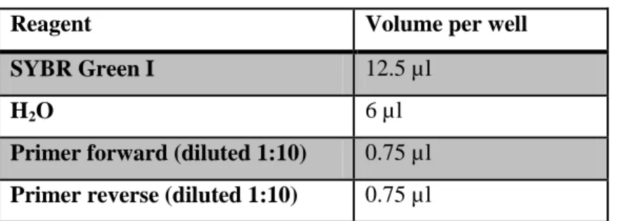
3.2.3 Real-time quantitative PCR ... - 49 -
3.3 Protein biochemistry ... - 51 -
3.3.1 Western blotting ... - 51 -
3.3.2 Immunocytochemistry and F-actin staining ... - 55 -
3.4 2D migration assay ... - 58 -
Table of Contents
- 5 -
3.4.1 Cell seeding ... - 58 -
3.4.2 Cell tracking ... - 58 -
3.5 Transwell invasion assay ... - 59 -
3.5.1 Cell seeding ... - 59 -
3.5.2 Cell staining and computer-based analysis ... - 60 -
3.6 Data analysis ... - 60 -
4 Results ... - 61 -
4.1 Reelin induced DAB1 phosphorylation in U87 and U251 glioblastoma cell lines . - 61 - 4.1.1 Reelin produced by HEK 293T ... - 61 -
4.1.2 DAB1 phosphorylation in U87/U251 cells ... - 62 -
4.2 Analysis of migration under the influence of Reelin/DAB1 signaling ... - 64 -
4.2.1 Fibronectin ... - 65 -
4.2.2 Laminin ... - 69 -
4.3 Influences of the Reelin/DAB1 pathway on cell invasion ... - 73 -
4.4 Analysis of the cytoskeleton under the influence of Reelin/DAB1 signaling ... - 76 -
4.4.1 Analysis of the actin cytoskeleton ... - 77 -
4.4.2 Analysis of the P-Cofilin to Cofilin ratio ... - 81 -
4.5 Analysis of downstream targets in Reelin/DAB1 signaling ... - 83 -
5 Discussion ... - 87 -
5.1 Reelin stimulation induces DAB1 phosphorylation in glioblastoma cells ... - 88 -
5.1.1 DAB1 levels in glioblastoma cell lines U87 and U251 ... - 88 -
5.1.2 Reelin induced DAB1 activation levels in U87 and U251 ... - 89 -
5.2 Reelin/DAB1 preferentially reduces cell migration in glioblastoma cells ... - 90 -
5.2.1 Cell migration ... - 90 -
5.2.2 Matrigel invasion assay ... - 94 -
5.2.3 Cofilin, actin cytoskeleton and other downstream targets ... - 95 -
Table of Contents
- 6 -
5.3 Conclusions ... - 97 -
6 Abstract ... - 98 -
7 Deutschsprachige Zusammenfassung ... - 99 -
8 References ... - 100 -
9 Acknowledgements ... - 111 -
10 Curriculum Vitae ... - 112 -
11 Eidesstattliche Erklärung ... - 113 -
12 Supplement ... - 114 -
Abbreviations
- 7 -
Abbreviations
Abbreviations
' minute(s)
'' second(s)
2D two dimensional
3D three dimensional
A ampere
ADF actin depolymerizing factor ADP adenosine diphosphate APS ammonium persulfate ARF1 ADP-ribosylation factor 1 ATP adenosine triphosphate
bp base pairs
BSA bovine serum albumin CaCl
2calcium chloride
cDMEM complete Dulbecco's modified Eagle's medium cDNA complementary DNA
CNS central nervous system
CO
2carbon dioxide
d distance
D distance from origin DAB1 Disabled-1
DAPI 4',6-diamidino-2-phenylindole dH
2O destillated H
2O
DMEM Dulbecco's modified Eagle's medium DMSO dimethyl sulfoxide
DNA deoxyribonucleic acid
dNTP deoxyribonucleotide triphosphate E. coli Escherichia coli
ECM extracellular matrix EGF epidermal growth factor
EGFR epidermal growth factor receptor
EGFRvIII epidermal growth factor receptor variant III
F phenylalanine
F-actin filamentous actin FCS fetal calf serum
g acceleration due to gravity (9.81 m/s2) G418 geneticindisulfat
G-actin globular actin
GBM glioblastoma multiforme
GFP green fluorescent protein
Abbreviations
- 8 -
h hour(s)
HEPES 4-(2-hydroxyethyl)-1-piperazineethanesulfonic acid HRP horseradish peroxidase
IDH isocitrate dehydrogenase KCl potassium chloride
kDa kilo Dalton
Kip1 Cyclin-dependent kinase inhibitor 1B
LIMK LIM (Lin-11/Isl-1/Mec-3)-domain-containing protein kinase MgCl
2magnesium chloride
MGMT O-6-methylguanine-DNA methyltransferase MnCl
2manganese (II) chloride
mRNA messenger RNA
MRT magnetic resonance tomography NaCl sodium chloride
NaOH sodium hydroxide NP-40 nonylphenolethoxylate
O/N overnight
p27 Cyclin-dependent kinase inhibitor 1B p53 tumor protein p53
PAGE polyacrylamide gel electrophoresis PBS phosphate buffered saline
P-cofilin phosphorylated cofilin PCR polymerase chain reaction
PDGFR platelet-derived growth factor receptor
PFA paraformaldehyde
PI3K phosphatidylinositol-4,5-bisphosphate 3-kinase RNA ribonucleic acid
RT room temperature
SDS sodium dodecyl sulfate
sec second(s)
STAT signal transducer and activator of transcription TBS Tris-buffered saline
TCGA The Cancer Genome Atlas TEMED tetramethylethylenediamine Triton X-100 t-octylphenoxypolyethoxyethanol
Tween 20 polyoxyethylene (20) sorbitan monolaurate
V volt
v volume
w weight
w/o without
WT wildtype
Y tyrosine
Introduction
- 9 -
1 Introduction
Primary tumors of the brain and spinal cord represent about 2 - 3% of all diagnosed tumors and are therefore rather rare cancer entities (Schlegel et al. 2003). Gliomas comprise about 80% of all malignant brain tumors and more than half of all gliomas are glioblastomas (Ostrom et al. 2016). Despite extensive research, treatment of glioblastomas is still lacking highly effective therapies leaving patients with an average survival time of less than two years (Tonn et al. 2006, Louis 2007). In terms of basic research, the Reelin/Disabled-1 signaling pathway has emerged as a new potential tumor suppressor over the last decade.
1.1 Reelin/Disabled-1 signaling pathway
The role and the biological mechanisms of the Reelin/Disabled-1 signaling pathway have already been an object of research for the last decades. Especially its function during embryonic brain development as an inhibitor of neuronal migration has been the topic of intensive research. Much less is known about the meaning of Reelin/Disabled-1 signaling in cancers, especially in brain tumors. However, previous work of our laboratory for Reelin and Disabled-1 suggested a potential role as tumor suppressor in high-grade gliomas.
1.1.1 Reelin
Several studies have unraveled the functions in regulation of neuronal migration of the
extracellular glycoprotein Reelin but, primarily, it has been discovered by D. S. Falconer at
the Institute of Animal Genetics, Edinburgh, in the 1950s. The name of this protein is derived
from the reeler mouse, an autosomal recessive mutant mouse showing pronounced
neurological symptoms, like dystonia, ataxia and tremors (Falconer 1951) and striking
abnormalities in the telencephalic and cerebellar cortices (Goffinet 1984). These mice were
later found to lack functioning Reelin due to a certain homozygous gene mutation
(D'Arcangelo et al. 1995). Over the last six decades, Reelin and the reeler mouse have been
Introduction
- 10 -
extensively studied as a model for brain development, proclaiming Reelin as a key regulator of neuronal migration.
The Reelin gene (RELN, GenBank-Accession-No.: AAC51105.1), is widely conserved through vertebrates and transcribed into a linear mRNA of 11’571 bp with an open reading frame (ORF) of 10,383 nucleotides. RELN is encoding for a large protein consisting of 3461 amino acids resolving in a molecular mass of about 388 kDa (DeSilva et al. 1997). Three different alleles, situated at the chromosomal region 7q22, were found responsible for the reeler mutant, with each of them displaying a partial deletion of the RELN coding sequence (D'Arcangelo et al. 1995, Takahara et al. 1996).
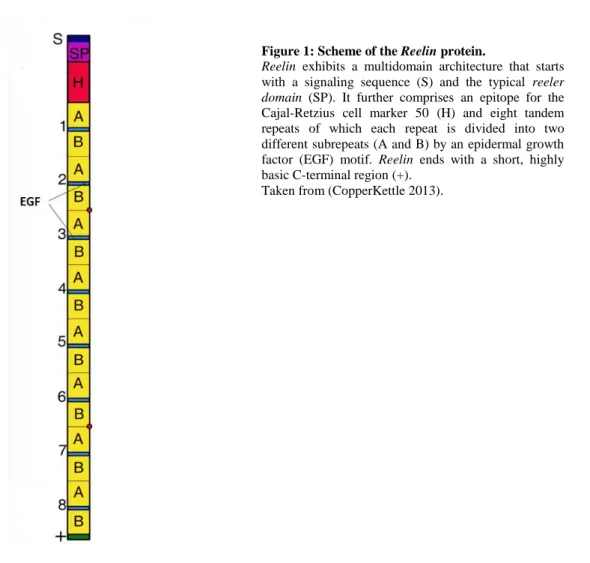
X-ray cristallography has revealed that Reelin comprises several domains (see Figure 1) consisting of a signaling sequence (S), followed by the reeler domain which can also be found in the extracellular proteins F-spondin and mindin (SP) (Feinstein et al. 1999). Next comes another unique region containing an epitope for the Cajal-Retzius cell marker 50 (CR-50) (H), an immunohistochemical marker for cells that are producing Reelin. The Reelin producing Cajal-Retzius cells are important during corticogenesis (see Figure 2). Then, the combination of a central epidermal growth factor (EGF) motif surrounded by two different sequences, A and B, is repeated eight times, each of these containing 350-390 amino acids (Yasui et al.
2007). The final reelin domain contains a highly basic and short C-terminal region (+) with a
length of 32 amino acids. This region is highly conserved, being 100% identical in all
mammals. In contrast to previous results, this region is not required for secretion but is
necessary to activate downstream signaling efficiently (Nakano et al. 2007).
Introduction
- 11 -
Canonical signaling downstream of Reelin is mediated by binding to the very-low-density lipoprotein receptor (VLDLR) and the ApoE receptor 2 (ApoER2) which results in the activation of the cytoplasmic adapter protein Disabled-1 (DAB1) by tyrosine phosphorylation (Hiesberger et al. 1999). Both receptors are able to stimulate DAB1 phosphorylation, however clustering of these receptors at the plasma membrane is necessary for recruitment of SRC family tyrosine kinases (SFKs) (see chapter 1.1.2), further downstream signaling and especially for its effect on neuronal migration (Strasser et al. 2004).
The role of Reelin during embryonic brain development has been most extensively studied in the neocortex of mice as a model system. Here, Reelin is required for the proper formation and positioning of the neuronal cell layers in the cortical plate (Honda et al. 2011). The cortical plate is a neuronal region that neuroblasts reach through migration guided by radial glia during corticogenesis to form the six layers of the neocortex (Herz & Chen 2006)
.A very profound study on the influences of deficient Reelin signaling during embryonic development is summed up in Figure 2:
Figure 1: Scheme of the Reelin protein.
Reelin exhibits a multidomain architecture that starts with a signaling sequence (S) and the typical reeler domain (SP). It further comprises an epitope for the Cajal-Retzius cell marker 50 (H) and eight tandem repeats of which each repeat is divided into two different subrepeats (A and B) by an epidermal growth factor (EGF) motif. Reelin ends with a short, highly basic C-terminal region (+).
Taken from (CopperKettle 2013).
Introduction
- 12 -
Generally speaking, lack of Reelin leads to an inversion of the six cell layers of the neocortex.
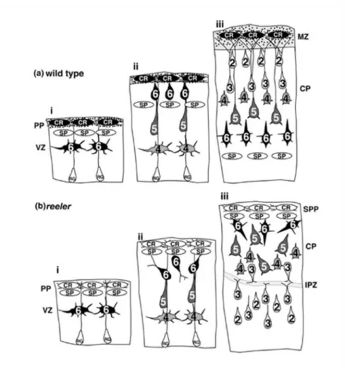
The schematic drawings show a part of the cerebral cortex of an embryonic mouse brain at three successive points in time (i, ii and iii). The presence of Reelin is shown by the blackened Cajal-Retzius neurons (CR) and black dots in wild type mice (a). These cells, together with subplate neurons (SP), represent the preplate (PP) which will later be the marginal zone (MZ) of the brain. The Arabic numbers of the indicated neurons mark the future layers of the neocortex (1-6). The newly born neurons that are formed by wild-type mice in the ventricular zone (VZ) migrate past the subplate and stop beneath the marginal zone. Later generated neurons overtake the subplate as well as the earlier neurons and reach the top of the developing cortex. As a consequence, the earlier-generated neurons are located beneath the later-born ones.
In reeler mice, the preplate appears to be normal. However, the Cajal-Retzius cells are unable to generate Reelin which is indicated by the uncolored CR neurons. Furthermore, the neurons of this cortex are not able to migrate past the subplate and therefore the preplate does not split into two layers but becomes the superplate (SPP). Thus, earlier-generated neurons stack at the top of the cortex whilst later-generated neurons reside in the deeper layers. Moreover, neurons in the upper layer near the internal plexiform zone (IPZ) show a formation of horizontally or even inversely oriented dendrites. Briefly summarized, the neocortex of reeler mice, opposed to that of wild-type mice, shows an outside-in sequence of neuronal positioning (Honda et al.
2011).
Introduction
- 13 -
However, proper lamination in the central nervous system is not only a result of direct interaction of Reelin with migrating neurons. Additionally, radial glial cells are activated by the Reelin signaling cascade. Radial glial cells of the neocortex seem in general only mildly affected, whereas the development of the dentate gyrus shows severely altered neuronal positioning and formation of radial processes in the absence of Reelin (Förster et al. 2002, Hartfuss et al. 2003). This considerable morphological phenotype was found independently of
Figure 2: Development of the cerebral cortex in wild type and reeler mice.
(a) depicts the influences that Reelin, secreted by Cajal-Retzius neurons (CR, black cells), has on corticogenesis at three successive points in time (i, ii, iii) whereas (b) is showing failed cortical development due to absence of Reelin (CR, white cells). Cajal-Retzius cells (CR) and subplate neurons (SP) represent the preplate (PP) becoming the marginal zone (MZ) later. The Arabic numbers of the indicated neurons mark the future layers of the neocortex (1-6).
(a) In wild-type mice newly born neurons are formed in the ventricular zone (VZ) and migrate past the subplate (SP). Finally, earlier-generated neurons are located beneath the later-born ones.
(b) In reeler mice neurons are not able to migrate past the subplate (SP) and therefore earlier-generated neurons stack at the top of the cortex, whilst later-generated neurons reside in the deeper layers. Further cortical anomalities found here, are formation of a superplate (SPP) and an internal plexiform zone (IPZ) where neurons show a formation of horizontally or inversely oriented dendrites. The neocortex of reeler mice shows an outside-in sequence of neuronal positioning.
Modified from (Honda et al. 2011, S. 1272).
Introduction
- 14 -
Reelin signaling in neurons. This suggests that Reelin is directly affecting glial cells (Brunne et al. 2013).
Although the functions of Reelin/DAB1 signaling during embryonic development are very well understood, only little is known about its importance for the adult brain. It has been shown that Reelin is expressed continuously even after disappearance of the Cajal-Retzius cells at the end of the neuronal migration phase. Instead, Reelin is produced by a group of GABA-ergic (γ-aminobutyric acid) interneurons (Herz & Chen 2006). Some studies indicate that Reelin/DAB1 signaling influences the growth of dendrites (Jossin & Goffinet 2007) and is a potential modulator of neurotransmission. It was shown that Reelin and its receptors ApoER2 and VLDLR influence cognition and plasticity of synapses in mice. Loss of one of these receptors leads to defects in long term potentiation, a major cellular mechanism that underlies learning and memory. These findings might contribute to understand the pathophysiology of alterations of cognitive functions and loss of synapses in patients with Alzheimer’s disease (Weeber et al. 2002).
Besides the importance for Alzheimer’s disease, further analyses showed that Reelin/DAB1 signaling seems to play an emerging role in other neurological and neuropsychiatric disorders of the adult brain. Lower levels of Reelin have been found in patients suffering from schizophrenia, bipolar disorder and autism (Fatemi et al. 2000, Fatemi et al. 2005). Again, the effects of Reelin on long term potentiation which lead to a reduction of NMDA receptors were proposed to contribute to these disorders (Coyle 2006). Other findings suggest that Reelin/DAB1 signaling is required to maintain neuroanatomical integrity in the adult brain as Reelin malfunction leads to dispersed positioning of dentate granule cells in patients with epilepsy (Heinrich et al. 2006). This malpositioning of neurons is correlated to the wake of seizures, especially to that of temporal lobe epilepsy (Haas et al. 2002, Heinrich et al. 2006).
Genetic defects of the Reelin gene were found to result in a form of lissencephaly with cerebellar hypoplasia. Lissencephaly is a human brain disorder that is characterized by the absence or reduction of cerebral convolutions (Hong et al. 2000).
For quite a long time it was believed that only traces of Reelin could be detected outside the
brain, however, about 15 years ago, it was shown that Reelin is present in plasma of rats, mice
and humans. Moreover, western blotting and immunocytochemistry revealed expression in
the liver, the pituitary gland and a subgroup of chromaffin cells in the adrenal medulla
(Smalheiser et al. 2000). Only little is known about the physiological role of Reelin/DAB1
signaling in these and other peripheral tissues but findings suggest rather little importance of
Introduction
- 15 -
Figure 3: Protein structure of DAB1.
Human DAB1 comprises three domains (PTB domain, DabH1 and DabH2) that show high sequence-homology to its related mouse gene. The percentage of identy is given below each region. Furthermore, DAB1 consists of four genetic regions (a, b, c and d) that contain five potential phosphorylation sites. Taken from (Howell et al. 2000).
this pathway for normal organic functions. Nonetheless, some studies on non-brain cancers proposed a function in tumor suppression for the Reelin protein (see chapter 1.1.4 for more information).
1.1.2 Disabled-1
As mentioned above, the key target for further transmission of Reelin signaling in embryonic development of the brain is the protein Disabled-1 (DAB1) (~ 63 kDa), whose gene represents a homologue of the drosophila protein disabled (Ware et al. 1997). This insight was gained by experiments with mice that were deficient of the DAB1 gene. These mouse mutants, called scrambler and yotari (both of them containing different mutations of the DAB1 gene), show a phenotype that is indistinguishable from the one in reeler mice (Howell et al. 1997b).
DAB1 interacts with the Reelin receptors ApoER2 and VLDLR through binding to a cytoplasmic domain that contains a typical sequence of amino acids. This amino-terminal phosphotyrosine binding (PTB) domain contains a sequence of asparagin (N), proline (P), an arbitrary amino acid (x) and tyrosine (Y) and is therefore abbreviated as NPxY motif (Howell et al. 2000). Binding of Reelin leads to clustering of its receptors which finally induces tyrosine phosphorylation of DAB1 (Hiesberger et al. 1999). Additionally, it could be shown that binding of Reelin to co-receptors like Cadherin-related neuronal receptors (CNRs) is necessary for the initiation of DAB1 phosphorylation through Src family kinases (SFKs) (Senzaki et al. 1999).
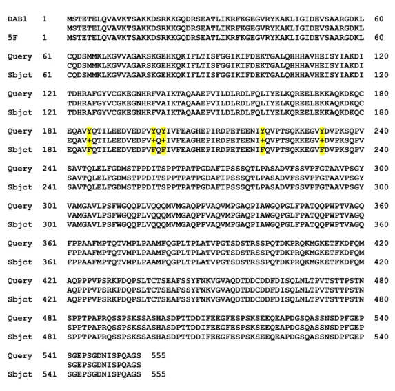
Figure 3 explains the structure of the human DAB1 protein. Human DAB1 contains three domains, the PTB, DabH1 and DabH2 domain, that represent regions of homology to the related mouse gene. The percentage of identity varies between 54 and 73%. Next to the
PTB domain, four genetic regions, containing five phosphorylation sites, Tyr185 (a),
Tyr198/Tyr200 (b), Tyr220 (c) and Tyr232 (d), are marked (Howell et al. 2000). Additionally,
the sequence around Tyr198 resembles a sequence of the platelet derived growth factor
receptor containing two tyrosines (equivalent to Tyr198 and Tyr200) which are both supposed
Introduction
- 16 -
to be phosphorylated (Mori et al. 1993). Two of these four sites have already been proved as targets for phosphorylation by analyzing several mutants replaced by phenylalanine instead of tyrosine. A DAB1 protein with mutation of all five tyrosines is called 5F mutant (Howell et al.
2000).
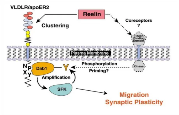
Tyrosine-phosphorylated DAB1 only extents its whole potential as a transmitter of Reelin signaling in the presence of Src family kinases (SFKs), especially of the members Src, Fyn and Abl. SFKs are non-receptor tyrosine kinases that interact with multiple cytosolic, nuclear and membrane proteins (Howell et al. 1997a). Studies show that DAB1 is a physiological substrate and also an activator of SFKs in neurons, meaning that DAB1 can potentiate its own phosphorylation level through these kinases. Furthermore, mutations of certain SFKs lead to similar changes of mouse cortices present in the reeler mutant, suggesting that the cellular reaction to Reelin normally includes activation of SFKs (Bock & Herz 2003). The above information on DAB1 so far is summed up in Figure 4.
Another interesting finding is that the intracellular protein DAB1 is generally stable in its inactivated form. However, in response to its activator Reelin, it is polyubiquitinated and
Figure 4: Hypothetical model for SFK activation by Reelin.
Reelin binds to its receptors, the very-low-density lipoprotein receptor (VLDLR) and the ApoE receptor 2 (apoER2), which leads to clustering of these receptors and binding of DAB1 to the intracellular NPxY amino- acid sequence. Additionally, Reelin might bind unknown coreceptors that induce primary tyrosine- phosphorylation (Y) of DAB1. Once activated, DAB1 is capable of amplifying its own phosphorylation through Src family kinases (SFK). This finally leads to reduced migration and enhanced synaptic plasticity of neurons. Taken from (Bock & Herz 2003).
Introduction
- 17 -
degraded by proteasome complexes, proposing that tyrosine phosphorylation is important for the degradation of DAB1 (Arnaud et al. 2003). This conclusion is also in accordance with findings in reeler mutants: The levels of DAB1 protein are much higher in embryonic brain tissue from reeler mutant mice than compared to controls (Howell et al. 2000).
DAB1 contains not only tyrosine phosphorylation sites but can also be phosphorylated at serine491 through the Cyclin-Dependent Kinase 5 (Cdk5). It has been shown that Cdk5 as well as its regulatory subunits p35 and p39 are regulating neuronal migration during corticogenesis independently of Reelin signaling. Mice that lack Cdk5, p35, or both p35 and p39 have defects of lamination in the neocortex similar yet not identical to reeler mice.
Further significance of DAB1 serine phosphorylation is unknown. However, these findings suggest that DAB1 represents a molecular interface of two distinct signaling pathways regulating cell positioning (Keshvara et al. 2002).
1.1.3 Overview of known downstream targets
Proper development of the mammalian brain requires extensive coordination between migration, differentiation, proliferation and survival of future neurons and glial cells. Some of the known downstream targets of Reelin/DAB1 signaling are contributing to these cellular functions during embryonic development.
As briefly mentioned above, it was shown that Cadherin-related neuronal receptors (CNRs)
are co-receptors of Reelin in the cell membrane of neurons. CNR family proteins are
supposed to be involved in formation and reorganization of synaptic connections in the
nervous system. Inhibition of Reelin binding through antibodies against proteins of the CNR
family (anti-RBD CNR antibody) leads to reduced tyrosine phosphorylation of DAB1
(Senzaki et al. 1999). It has also been proved that CNRs interact with Fyn, a member of the
SFKs (Hamada & Yagi 2001), suggesting that Reelin is also capable of modulating the
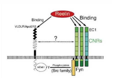
phospho-tyrosine level of DAB1 through other receptors than ApoER2 and VLDLR. Figure 5
gives an overview of this hypothetical pathway of Reelin signaling.
Introduction
- 18 -
A proved target further downstream of Reelin/DAB1 signaling is the phosphatidylinositol 3- kinase (PI3K). Stimulation of PI3K by Reelin leads to activation of the protein kinase B (PKB, also known as Akt). PI3K/PKB signaling is highly conserved in all cells of higher eukaryotes (Manning & Cantley 2007) and known to control a very diverse group of cellular functions like cell growth, proliferation, migration and intracellular trafficking. These components of Reelin signaling are enriched in axonal growth cones, contributing to proper neuronal positioning during brain development (Beffert et al. 2002). Further studies revealed that PI3K and PKB activity regulated through Reelin are both crucial for corticogenesis (Jossin & Goffinet 2007, Leemhuis & Bock 2011, Ohkubo et al. 2003).
Besides the downstream targets above, migration and cellular motility is also often corresponding to expression of and interaction with integrins. These proteins are necessary for establishment of cell-matrix junctions. Integrins are transmembrane receptors containing two different chains, an α and a β subunit, and can trigger intracellular signaling pathways.
Research on Reelin signaling has particularly turned the spotlight on two specific integrins:
α3β1 integrin (also known as laminin receptor) and α5β1 integrin (also called fibronectin receptor).
The receptor α3β1 integrin is mainly known for binding laminin but collagen I and IV or entactin/nidogen are described as ligands as well. However, α3β1 integrin is more than a
Figure 5: Hypothetical pathway of Reelin interacting with CNR family proteins.
Binding of Reelin to the N-terminal EC1 domain of Cadherin-related neuronal receptors (CNRs) is capable of modulating the tyrosine-phosphorylation (Y) of mouse DAB1 (mDab1) through Fyn, a Src family kinase. Probably, an interaction of CNRs and the main Reelin receptors, the very-low-density lipoprotein receptor (VLDLR) and the ApoE receptor 2 (ApoER2), has additional influences on the activation of DAB1.
Modified from (Senzaki et al. 1999).
Introduction
- 19 -
simple adhesion receptor, but has a rather complex role as signaling molecule that can modulate multiple cellular functions like proliferation, differentiation and migration (Kreidberg 2000). It was also proved that α3β1 integrin is able to bind the extracellular protein Reelin which leads to inhibition of neuronal migration during brain development.
Further data suggests that activation of the laminin receptor modulates targets of Reelin signaling, as a reduction of DAB1 levels could be found in α3β1-deficient mice (Dulabon et al. 2000).
α5β1 integrin is the primary receptor for fibronectin and able to support matrix assembly of fibronectin as well as promoting cell migration (Arnaout et al. 2007, Zhang et al. 1993).
Recent studies showed that binding of Reelin to its receptors can activate integrin α5β1 through a pathway that includes DAB1. Consequently, the activation of α5β1 integrin through this inside-out signaling promotes neuronal adhesion to fibronectin. Additional data indicates that this pathway influences neuronal migration and is crucial for proper layering of the neocortex during brain development (Sekine et al. 2012).
Migrating cells do not only show changes of integrin activity but most often also exhibit mechanisms to modulate their cytoskeletal structure. It is known that Reelin is capable of controlling at least two pathways that reorganize the actin cytoskeleton.
The neuronal Wiskott-Aldrich syndrome protein (N-WASP) is known to promote invasion by activating the Arp2/3 complex, a protein initiating the polymerization of actin and inducing formation of filopodia (Tang et al. 2013). Unphosphorylated/inactive DAB1 binds N-WASP directly and starts the signaling pathway by activating N-WASP. Phosphorylated/active DAB1 in turn is degraded through ubiquitinylation and thus can no longer activate these mechanisms leading to reduced filopodia formation and decreased cell motility (Suetsugu et al. 2004).
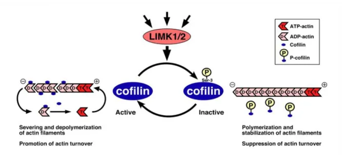
A second pathway influencing actin filament dynamics and reorganization includes
ADF/cofilin family proteins (hereafter referred to as cofilin for simplicity). Active,
unphosphorylated cofilin binds globular (G)-actin as well as filamentous (F)-actin and can
enhance the disassembly of F-actin by stimulating severance and depolymerization of actin
filaments. This results in higher actin turnover (Pollard & Borisy 2003). Reelin/DAB1
signaling leads to activation of the enzyme LIM kinase 1 and 2 (LIMK1/2) which
phosphorylates cofilin at serine3 and converts the protein into an inactive form. With this
modification it is no longer capable of binding actin and thus promotes actin polymerization
and cytoskeletal stability. As a consequence, actin turnover is suppressed and cell motility
Introduction
- 20 -
reduced (Chai et al. 2009, Kruger et al. 2010). Figure 6 illustrates the interactions of cofilin and actin.
1.1.4 Reelin/Disabled-1 signaling in other cancers
As mentioned above in chapter 1.1.1, expression of Reelin is not restricted to the brain, but can be found in many other tissues. Although existing data rather suggests little importance for physiological mechanisms of Reelin/DAB1 signaling (Ikeda & Terashima 1997), Reelin might act in reducing invasion of multiple non-brain cancers. This presumption was established after having compared the expression of Reelin protein in several cancers (e.g.
hepatocellular carcinoma, breast cancer, esophageal cancer or pancreatic cancer) to levels in normal tissues. Most of the samples showed a reduced level of expression of Reelin compared to the control cases (Okamura et al. 2011, Sato et al. 2006, Stein et al. 2010, Yuan et al.
2012).
Research on hepatocellular carcinoma revealed that up to 80% of the samples showed a clearly reduced level of Reelin expression. Again, about half of these tumors were found to be hypermethylated on the RELN gene promoter preventing synthesis of Reelin mRNA.
Figure 6: Control of actin filament dynamics by phospho-regulation.
Amongst other proteins, LIMK1/2 is activated by Reelin, which leads to phosphorylation of cofilin at serine3 converting it from an active into an inactive state. Unphosphorylated cofilin is binding ADP-actin and induces formation of ATP-actin, leading to enhanced severing and depolymerization of actin filaments and stimulation of actin turnover. Phosphorylated cofilin is unable of binding actin and is therefore suppressing actin turnover by promoting polymerization and stabilization of actin filaments.
Modified from (Mizuno 2013).
Introduction
- 21 -
Moreover, clinical data suggested Reelin as a regulator gene being inversely correlated to the reoccurrence of hepatocellular carcinoma (Okamura et al. 2011).
Similar results were found in a study on breast cancer that found that hypermethylation of RELN promoter leads to poorer prognosis and positive lymph node status. Besides that, it was proved that overexpression of Reelin and incubation with exogenous Reelin was capable of suppressing cell migration, invadopodia formation and invasiveness in vitro (Stein et al.
2010). Similar findings are provided in a study on pancreatic cancer. Knockdown of RELN or its downstream target DAB1 using small interfering RNA in cells that retained expression of Reelin exhibited enhanced cell migration and invasiveness (Sato et al. 2006).
However, a study by Perrone et al. showed a completely different result: whereas a down- regulation of Reelin was found in the carcinomas described above and several other entities, an increased expression of this protein was associated with high-grade disease in prostate cancer. Elevated levels were significantly correlated with aggressive phenotypic behavior of cancer cells. Interestingly, benign prostatic tissue did not show any expression of Reelin (Perrone et al. 2007).
Moreover, also the protein DAB1 has been found to function in a pathway that is promoting invasion and metastasis of colorectal cancers. The phosphorylation level of DAB1 as well as interaction with the tyrosine kinase ABL seem to play a crucial role in a signaling system that is induced by the transmembrane receptor protein NOTCH (Sonoshita et al. 2015).
1.2 General information on glioblastoma
The preceding chapter gave an overview of the pathological role of Reelin/DAB1 signaling in non-neoplastic tissue and multiple cancers. However, there is no existing study investigating the influence of this pathway on glioblastoma or other brain tumors, except for two immunohistochemical studies in gangliogliomas. This low-grade tumor which is often associated with focal epilepsy in young patients, showed significantly reduced levels of DAB1 protein in its tissue but no mutations of the DAB1 gene could be detected (Becker et al. 2002, Kam et al. 2004).
The chapter below will provide some general information on glioblastoma that is necessary to
understand the contents and experiments of this doctoral thesis and will also give a short
summary of the data on Reelin/DAB1 signaling in glioblastoma that was raised in our
workgroup so far.
Introduction
- 22 -
1.2.1 Overview and epidemiology
Tumors of the central nervous system (CNS) represent about 2 - 3% of all cancers diagnosed displaying a rather seldom but most often highly-malignant tumor entity in adults (Schlegel et al. 2003). In childhood, in reverse, brain tumors are the most common type of neoplasms after leukemias (Kaatsch 2010).
Gliomas are subdivided according to histological and biochemical similarities to astrocytic, oligodendroglial or ependymal cells. However, the cellular origin of gliomas is still unknown but studies of tumor suppressor mouse models suggest that gliomas arise from stem/progenitor cells (Alcantara Llaguno et al. 2009). Another study states that gliomas can also be induced by oncogenes via dedifferentiation of both neurons and astrocytes (Friedmann-Morvinski et al. 2012). Astrocytic gliomas are the most common primary tumors of the CNS (Tonn et al. 2006).
Gliomas are classified by means of histological and molecular criteria as stated in the World Health Organisation (WHO) classification of tumors of the central nervous system in its latest version from 2016 (first edition: 1979). The WHO classification is dividing brain tumors in four grades by assigning a certain grade of malignancy ranging from WHO grade I (benign) to WHO grade IV (highly malignant) to each histological subtype. Tumor classification is necessary for finding suitable therapeutic strategies and can give a prediction on the patient’s prognosis (Louis et al. 2016).
WHO grade I tumors display neoplasms that show only a low proliferation rate and little degeneration. As they are normally histologically well-circumscribed, they can be cured completely by radical and maximal surgical resection. A typical grade I glioma is the pilocytic astrocytoma which is also the most common primary brain tumor in pediatric patients (Tonn et al. 2006, Louis 2007).
Tumors classified as WHO grade II, e.g. the diffuse astrocytoma, also show a low proliferation rate but are infiltrating healthy surrounding brain tissue in contrast to WHO grade I tumors. Therefore, these tumors tend to reoccur after surgical excision and the median survival is reduced to 4 years (Medical Disability Guidelines 2012).
A WHO grade III neoplasia like the anaplastic astrocytoma is characterized by microscopic features of malignancy and is normally accompanied by restricted life expectancy.
Histologically, these tumors show nuclear and cellular atypia, as for example chromatin richness, disproportion of nuclear-cytoplasm ratio and an elevated mitotic activity (Tonn et al.
2006, Louis 2007).
Introduction
- 23 -
Glioblastomas, also known as glioblastoma multiforme (GBM), are highly malignant tumors due to diffuse infiltration of healthy brain tissue and high mitotic and proliferative activity.
They are therefore classified as WHO grade IV tumors. Besides high cellularity, nuclear and cytoplasmic pleomorphism, the presence of microvascular proliferation and central necrosis is essential for the diagnosis. Without effective treatment, these tumors have an average survival time of only a few months that can be extended to about 15 months with the standard of current therapy including surgery, chemotherapy and radiation. Due to the overall poor prognosis, patients have a 2-year survival rate of about 27% (Tonn et al. 2006, Louis 2007, Omuro 2013).
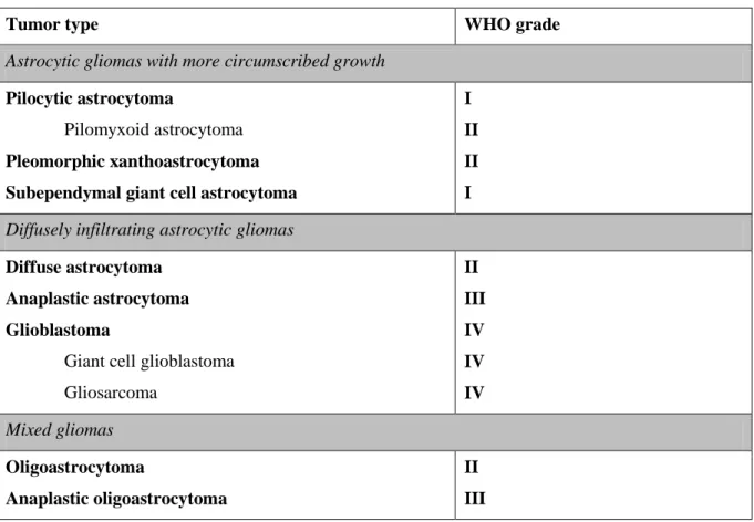
Table 1 gives an overview on classification and grading of the most common glioma entities.
Table 1: Classification and grading of the most common astrocytic gliomas according to the WHO classification of tumors of the central nervous system.
Modified from (Huse et al. 2013, Louis et al. 2007, Riemenschneider & Reifenberger 2009).
Tumor type WHO grade
Astrocytic gliomas with more circumscribed growth
Pilocytic astrocytoma
Pilomyxoid astrocytoma Pleomorphic xanthoastrocytoma Subependymal giant cell astrocytoma
I II II I Diffusely infiltrating astrocytic gliomas
Diffuse astrocytoma Anaplastic astrocytoma Glioblastoma
Giant cell glioblastoma Gliosarcoma
II III IV IV IV Mixed gliomas
Oligoastrocytoma
Anaplastic oligoastrocytoma
II III
Glioblastomas are deemed to be the most prevalent and also most aggressive primary brain tumors accounting for approximately half of all functional tissue CNS tumor cases and 20%
of all intracranial tumors (Ohgaki & Kleihues 2005). It is estimated that approximately 2 - 3
new cases per 100’000 residents occur annually in western countries affecting slightly more
Introduction
- 24 -
men than women. Glioblastomas may appear at any age, but older people with a peak of incidence between 50 and 70 years are more often afflicted (Tonn et al. 2006).
The majority of glioblastomas develops in the cerebral hemispheres and can spread into the basal ganglia or the contralateral hemisphere. The brain stem is a rare localization in adults but can be particularly found in children. Macroscopically, these tumors present most often as necrotic masses with frequent central hemorrhages that are surrounded by oedematous brain tissue (Tonn et al. 2006) as shown in Figure 7.
1.2.2 Etiology
Research efforts from many individual groups and more recent data collections from multiinstitutional research networks, like The Cancer Genome Atlas (TCGA) (Brennan et al.
2013), identified several genetic mutations (see chapter 1.2.4) and novel aspects on the etiology of glioblastomas. Besides that, genetic syndromes caused by inherited mutations, like Li-Fraumeni syndrome or Neurofibromatosis, are found in about 1% of all glioblastoma patients (Farrell & Plotkin 2007).
Most patients with glioblastomas present with a very short clinical history due to the very fast and spontaneous development of these tumors. Without any evidence of longer precedent malignant progression, these neoplasms are called primary glioblastomas. Opposed to these, cases of secondary glioblastomas are less common and evolve from pre-existing low-grade diffuse or anaplastic astrocytoma. Secondary glioblastomas are more common in younger adults (30-45 years), whereas primary glioblastomas occur mostly in older patients (mean age 55 years) (Kleihues et al. 2002). A morphological differentiation between both entities is not possible because of identical histological features; however, these tumors can be distinguished by their genetic and epigenetic aberrations (Huse et al. 2013). More information on the molecular pathology in glioblastoma can be found in chapter 1.2.4.
Figure 7: Glioblastoma of the temporal lobe.
A necrotic mass with central hemorrhages can be identified in the temporal lobe. Most likely, macroscopic appearance suggests glioblastoma multiforme.
Taken from (Tonn et al. 2006).
Introduction
- 25 -
1.2.3 Symptoms, diagnosis and treatment
Due to the rapid growth of glioblastomas, health complaints usually arise within only a few weeks or months. Most patients primarily suffer from non-specific neurological symptoms like nausea, vomiting and headache which can be accompanied by seizures, memory loss or hemiparesis. However, the array of symptoms produced by these tumors is very diverse and depends highly on their localization. A glioblastoma of the frontal cerebral lobe might lead to personality changes, whereas a tumor next to the optic nerve can cause visual loss. Other locations lead rather to cerebral oedema and increased cerebral pressure which explains the general symptoms listed above (Kleihues et al. 2002).
Malignant cells of glioblastomas carried in the cerebrospinal fluid can result in gliomatosis of the meninges or may spread to the spinal cord. Metastases beyond the central nervous system are extremely unusual but dissemination to regional lymph nodes, lung, pleura, liver and bone has been described in very rare cases (Frappaz et al. 1999, Pasquier et al. 1980).
Nowadays, patients with newly developed unclear neurological symptoms are usually quickly subjected to cranial tomography like magnetic resonance imaging (MRI). Here, glioblastomas normally appear as an intracerebral space-occupying lesion with ring-enhancement due to peripheral vascularization of the mass and a central hypointense area which is corresponding to tumor necrosis. These radiological findings underscore the suspicion of a glioblastoma, however, cerebral abscess or metastasis of another malignant tumor have to be taken into account as differential diagnoses (Smirniotopoulos et al. 2007). A definitive diagnosis of glioblastoma requires a stereotactic biopsy or a craniotomy with tumor resection and pathologic confirmation by an experienced neuropathologist. Lately, diagnostic methods have been extended by more advanced imaging, like measurement of blood perfusion or of tumor metabolite concentration. However, neuropathologic examination remains the gold standard for diagnosis of a glioblastoma (Nikiforova & Hamilton 2011).
The goal of glioblastoma therapy is to slow down the tumor growth and improve the quality of life. All therapy strategies are non-curative interventions but are at most life-prolonging.
The standard treatment of glioblastoma is based on three pillars: surgery, radiation and chemotherapy.
Surgical intervention is usually the first step intending to remove as much tumor as possible
without destroying any functional brain tissue and thus increasing survival (Lacroix et al.
Introduction
- 26 -
2001). Normally, the operation is followed by a combined radio- and chemotherapy in which radiation is the mainstay of treatment for glioblastoma. Studies could show that patients who received a radiotherapy had a median survival time of more than double of those who did not (Walker et al. 1978). The chemotherapeutic drug of choice at the moment is temozolomide which seems to sensitize the tumor cells to radiation but also has direct anti-tumorigenic effects itself (Chamberlain et al. 2007). This treatment showed a significant influence as patients receiving radio- and chemotherapy survived 2.5 months longer than a control group with radiation alone, extending the median survival from 12.1 to 14.6 months (Stupp et al.
2005). Besides this, older patients whose tumor tissue exerts MGMT-methylation (see chapter 1.2.4) are exclusively treated with temozolomide due to the high toxicity of combined chemoradiation. These patients have a higher benefit from temozolomide alone (Malmstrom et al. 2012).
1.2.4 Molecular diagnosis and pathology of gliomas
Molecular markers are gaining more and more importance in neurooncology as histology no longer serves as sole diagnostic tool for correct brain tumor classification (Louis et al. 2016).
Additionally, molecular testing attributes to survival prognosis of tumor patients and can give predictive information on response to certain treatments (Dietmaier et al. 2015).
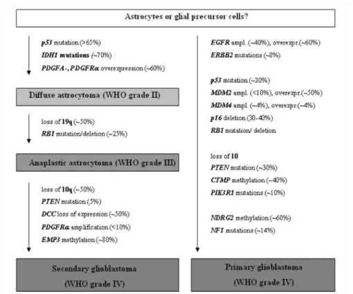
As the schematic representation of molecular markers during pathogenesis of gliomas in Figure 8 explains, a large number of biological alterations is known in glioblastomas and astrocytic tumors of which the following represent important markers for everyday clinical practice or have been reported to be involved in glioma cell migration and invasion.
The methylation level of the O6-Methylguanin-DNA-methyltransferase (MGMT)-gene promoter is among the most frequently analyzed molecular markers in human brain tumors (Dietmaier et al. 2015). This gene, located on the chromosomal region 10q26, encodes for a DNA repair gene that removes alkyl groups from the O
6of guanine (Watanabe et al. 2007) and is hypermethylated in 40% of all glioblastomas. Elevated methylation levels of the MGMT promoter predict a better response to treatment with the alkylating chemotherapeutic drug temozolomide which is translating into a prolonged patient survival (Hegi et al. 2005).
Mutations of the substrate binding domain of the isocitrate dehydrogenase (IDH) 1 gene and
its homologue IDH2 display another biological marker analyzed frequently that allows to
differentiate between several low-grade astrocytomas. For example, approximately 70% of
Introduction
- 27 -
diffuse astrocytomas show a mutation of amino acid 132 in the IDH1 gene whereas pilocytic astrocytomas express mainly wildtype IDH1. Furthermore, 90% of all secondary glioblastomas, opposed to primary glioblastomas, are also tested positive for this mutation as they most often develop from these precursor lesions (Dietmaier et al. 2015, Yan et al. 2009).
Besides MGMT promoter methylation and IDH1/2 methylation, complete loss of the long arm of chromosome 1 and the short arm of chromosome 19 (1p/19q deletion) is an entity-defining alteration in oligodendroglial tumors and may guide to consideration of oligodendroglial differentiation in diagnosis of oligoastrocytomas (Louis et al. 2016). Additionally, detection of 1p19q deletion predicts favorable prognosis for tumors with oligodendroglial features and responsiveness to polychemotherapy with procarbazine, lomustine and vincristine (Cahill et al. 2015, Cairncross et al. 2006, Dietmaier et al. 2015, Riemenschneider et al. 2013).
Apart from these biochemical markers analyzed with regard to diagnosis of brain tumors, gliomas show multiple genetic alterations like mutation of the epidermal growth factor receptor (EGFR), of the tumor suppressor protein p53 or of PI3 kinase pathway genes. These markers are less applicable for molecular diagnosis as usually no information on prognosis or response to treatment is gained due to their identification (Ceccarelli et al. 2016, Huse et al.
2013, Riemenschneider & Reifenberger 2009).
Numerous studies have shown that the EGFR is not only frequently amplified or overexpressed in glioblastomas but also in multiple other cancers. Very often, a truncated EGFR protein which forms a constitutively active mutant called EGFR variant III (EGFRvIII) is present in these tumor cells (Libermann et al. 1985, Schlegel et al. 1994, Sugawa et al.
1990). With regards to gliomas, a positive correlation between EGFR overexpression and enhanced invasiveness of tumor cells (Lund-Johansen et al. 1990) as well as promotion of glioblastoma cell migration and tumor growth by the epidermal growth factor (EGF), a ligand of the EGFR (Tysnes et al. 1997), was reported.
The p53 protein is among the best known of all tumor suppressors and – when mutated – is capable of promoting migration and invasion, as well as inhibiting cellular aging and apoptosis in various human cancers (Muller et al. 2011). It was recently shown that p53 has a crucial role in gliomagenesis by inducing accumulation of cooperative oncogenic alterations (Wang et al. 2009). It is therefore reasonable that mutations of p53 are frequently found and can be similarly observed in lower grade malignant gliomas like diffuse astrocytoma as well as in secondary glioblastomas (Ohgaki et al. 2004).
Besides EGFR and p53 mutations, oncogenes of the PI3 kinase pathway play a crucial role in
development of glioblastomas. PI3 kinases are involved in many cellular functions such as
Introduction
- 28 -
cell growth, differentiation, motility and survival and are hence capable of promoting tumorigenesis when misregulated. Additionally, the phosphatase and tensin homologue (PTEN), an antagonist of the PI3 kinase pathway, is absent in up to 30% of all primary glioblastomas (Bleeker et al. 2014, Huse et al. 2013, Riemenschneider & Reifenberger 2009).
An overview on known molecular alterations that are frequently found in astrocytic gliomas is given in Figure 8. Some of the genetic mutations listed below have been explained in greater detail in this chapter. Additionally, a distinction is made between alterations that are preferentially found in primary glioblastomas on the one hand and secondary glioblastomas on the other.
1.3 Reelin/DAB1 signaling in glioblastoma
As explained extensively in chapter 1.1.4, Reelin/DAB1 signaling has an important effect on non-brain tumors suggesting a tumor supressive function in several cancers like hepatocellular carcinoma, breast cancer, esophageal cancer or pancreatic cancer (Okamura et al. 2011, Sato et al. 2006, Stein et al. 2010, Yuan et al. 2012). Although Reelin acts particularly during development of the central nervous system, none of these studies have
Figure 8: Schematic representation of the molecular pathogenesis and progression of astrocytic gliomas.
Note that a distinction between genetic mutations that are preferentially found in primary or secondary glioblastomas can be made. Taken from (Riemenschneider & Reifenberger 2009).