Acute bilateral serous retinal detachments with spontaneous resolution in a 6-year-old boy
Abstract
A healthy 6-year-old boy presented with acute bilateral vision loss, multiple serous retinal detachments between the vascular arcades and
Sophie Van Camp
1Steffi Vande Walle
1a thickened choroid. Spontaneous resolution occurred over several
weeks.
Ingele Casteels
1Julie Jacob
1We hypothesize that the clinical constellation in our patient is suggestive of acute exudative polymorphous vitelliform maculopathy (AEPVM) or
Cathérine Cassiman
1might be an atypical presentation of Vogt-Koyanagi-Harada (VKH) dis-
Carine Wouters
2ease. We propose that it was caused by an autoimmune-mediated ac-
Pieter-Paul Schauwvlieghe
1tivation of inflammatory cells at the level of the choroid, induced by an unknown trigger.
Keywords:serous retinal detachment, young child, acute exudative
polymorphous vitelliform maculopathy 1 Department of
Ophthalmology, University Hospitals Leuven, Belgium 2 Department of Pediatrics,
University Hospitals Leuven, Belgium
Introduction
negative for ophthalmological problems. There was no history of travelling to exotic regions. The patient had Acute exudative polymorphous vitelliform maculopathy suffered from a cold a few weeks before the onset of (AEPVM) is a rare disorder, first described by Gass et al. symptoms.in 1988 [1]. Patients usually present with bilateral blurred We measured a best corrected visual acuity of Snellen 0.6 vision, often associated with headache. The typical fun- in the right eye and Snellen 0.16 in the left eye. Slitlamp doscopic findings consist of serous retinal detachments biomicroscopy of the anterior segment showed no abnor- in both eyes, accompanied by scattered yellowish subret- malities and a normal intraocular pressure. Multiple large inal lesions in a honeycomb-like pattern. These lesions serofibrinous detachments with hyperreflective dots below contain lipofuscin, which can be demonstrated on auto- the neurosensory retina were seen bilaterally on fundo- fluorescence imaging. The clinical features can resolve scopic examination and optical coherence tomography spontaneously over the course of several months. Thus (OCT) of the retina. These lesions were found to be hyper- far no treatment has been found effective, nor has a autofluorescent. A thickened choroid – estimated at single etiological hypothesis been proven. 1000 µm – was detected on enhanced depth imaging optical coherence tomography (EDI-OCT) (Figure 1). In-
Case description
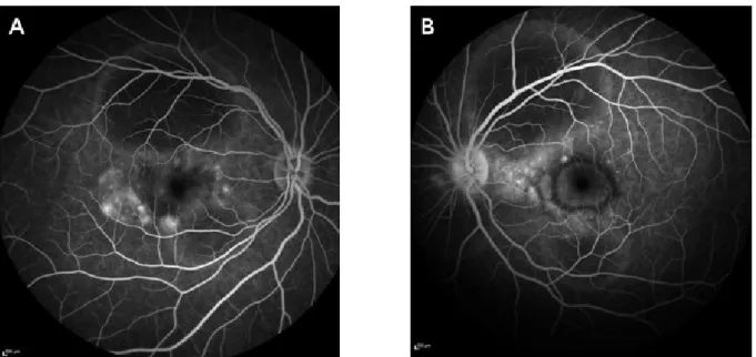
frared imaging showed serous detachments with a center of slightly increased intensity surrounded by decreased intensity (Figure 1). Fluorescein angiography (FA) revealed An otherwise healthy 6-year-old boy with no relevant small leakage points at the level of the serous detach- medical history was referred to our department because ments, limited staining of the optic disc in the left eye of acute onset of bilateral blurry vision, metamorphopsia (without staining in the right eye) and masking effect due and scattered scotomas. He has a mother of Turkish to neurosensory detachments (Figure 2).descent and a Belgian father. The family history was
Case Report
OPEN ACCESS
Figure 1: OCT and infrared images show multiple serofibrinous retinal detachments in the right (A, C) and left (B, D) eye and a thickened choroid measured with EDI-OCT (A).
Figure 2: Late-phase fluorescein angiography shows tiny leakage points with limited leakage in the subretinal space (A, B) and discrete staining of the left optic disc (B).
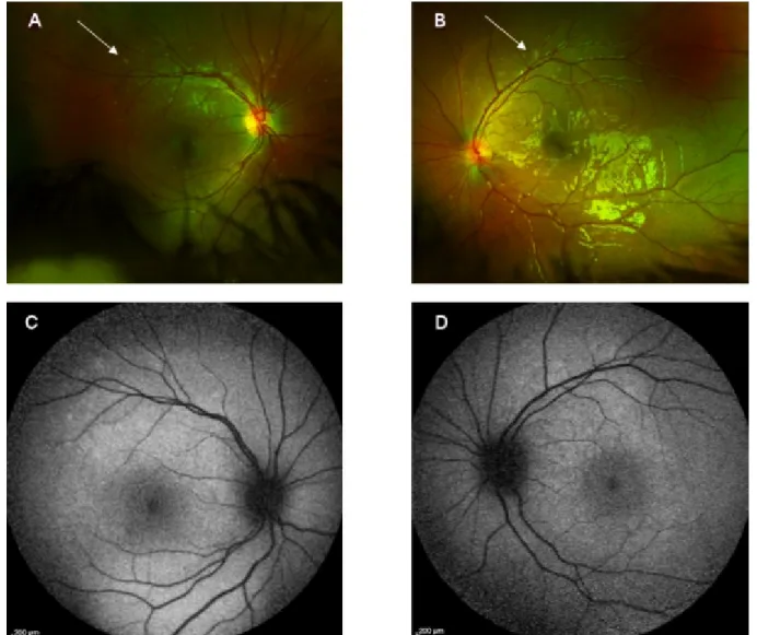
Figure 3: Fundoscopic images three weeks after initial presentation show multiple yellow deposits scattered around the vascular arcades (A and B, see arrows), with corresponding mild hyperautofluorescence (C, D).
The patient was referred to our department of pediat- Over the next two weeks, the serous retinal detachments rics, where a total clinical work-up did not show any resolved completely in the left eye and decreased evidence of systemic inflammation, infection or malignant markedly in the right eye, with distinct deposition of some
diseases. yellowish, slightly hyperautofluorescent vitelliform material
Serology for borreliosis, syphilis, toxoplasmosis, human in the subretinal space (Figure 3). Further improvement immunodeficiency virus, herpes simplex, rubella, cyto- of visual acuity to Snellen 0.6 in both eyes (OU) occurred.
megalovirus and Epstein-Barr virus was negative, in the Since intraocular inflammation was no longer present, absence of leukocytosis. A chest X-ray and magnetic topical steroids were tapered.
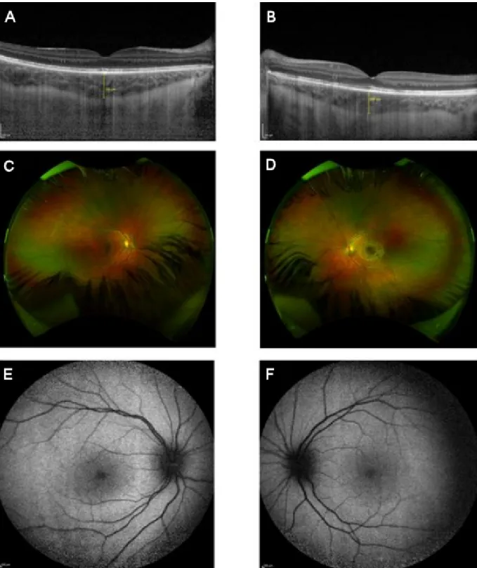
resonance imaging (MRI) of the brain showed no abnor- Three months after initial presentation, complete resolu- malities except for a thickened choroid in both eyes. tion of retinal lesions was noted with recovery of vision One week after initial presentation, spontaneous improve- to Snellen 0.9 OU (Figure 4).
ment of vision occurred with regression of the lesions on OCT. Minimal inflammation in the anterior chamber (without keratitic precipitates) and vitreous was seen and topical steroids were administered six times per day.
Van Camp et al.: Acute bilateral serous retinal detachments with spontaneous ...
Figure 4: Three months after initial presentation, there was total resolution of the exudative retinal detachments in the right (A) and left (B) eye with normalization of choroidal thickness and an almost complete resorption of the vitelliform lesions (C,
D). Autofluorescence was normal (E, F).
Discussion: differential diagnosis
Acute exudative polymorphous vitelliform maculopathy (AEPVM)
AEPVM is a rare disorder, with symptoms of (sub)acute bilateral vision loss, frequently – but not always – pre- ceded by headache [1], [2], [3]. Usually there are no or very few signs of intraocular inflammation. The fundoscop-
ic findings are remarkable with bilateral multifocal central serous retinal detachments and round to oval white- yellowish lesions in a peculiar honeycomb-like pattern at the level of the retinal pigment epithelium, between and around the vascular arcades. These lesions do not occur in every patient, and they usually present after several months [2]. Fluorescein angiography shows early hyper- fluorescence of the multifocal yellow lesions and staining in the late phase. Leakage of dye is minimal to absent.
On autofluorescence imaging, lesions are hyperautofluo- rescent, suggesting the presence of lipofuscin [4]. Indo- cyanine green (ICG) angiography shows an affinity of the dye for these yellow lesions, exhibiting involvement of the choriocapillaris. One case reported choroidal thickening measured by EDI-OCT, with striking elevation in the acute phase [2], as seen in our patient. The serous retinal de- tachments typically resolve spontaneously over the course of weeks to months. This tends to occur faster if few or no yellow lesions are visible. These vitelliform lesions can gravitate and form a pseudo-hypopyon of yellow material at the level of the inferior macula, blocking fluorescence on FA. They can persist for years despite complete reso- lution of symptoms. This is the most typical presentation, although AEPVM can present with a more variable clinical course than initially described. We believe the lesions in our patient are very similar to the subtype ‘bleb-like le- sions along vascular arcades’ of AEPVM [2].
The etiology of AEPVM remains unclear. Dysfunction of the retinal pigment epithelium (RPE) with decreased phagocytosis of secreted photoreceptor outer segments has been suggested, leading to secondary serous retinal detachment. This can cause precipitation and formation of yellow lesions, consisting of lipofuscin. OCT images have shown thickened outer segments, compatible with this hypothesis. The favorable response to corticosteroid therapy in some patients suggests an inflammatory base for this dysfunction [1].
A few cases have been reported following a viral [5] or bacterial infection [6], [7], [8]. These cases were thought to be based on an autoimmune-mediated activation of inflammatory cells (presumably T lymphocytes as in Vogt-Koyanagi-Harada disease) at the level of the cho- roid, leading to profound choroidal swelling. This could cause saturation of the choroid with subretinal accumu- lation of fluid rich in lipofuscin and exudative retinal de- tachment. A subsequent decrease in choroidal swell- ing – either spontaneously or by treatment with cortico- steroids – can restore fluid absorption in the choroid with resolution of the retinal detachments. An incomplete clearance of lipofuscin causes yellow subretinal deposits.
In our patient, a cold preceded the onset of symptoms, but we could not identify a specific causative micro-organ- ism.
AEPVM has also been reported as a paraneoplastic ret- inopathy (e.g. metastatic melanoma, small cell lung car- cinoma), where antibodies against an extraocular tumor supposedly cross-react with retinal or RPE antigens.
Therefore, referral for systemic evaluation is always indi- cated [9], [10].
Treatment of AEPVM is controversial. In the first described cases of Gass et al., there was improvement after system- ic prednisolone [1]. However, literature shows little evi- dence for the efficacy of corticosteroids; resolution of the lesions has been documented in many patients without any form of treatment.
One patient with AEPVM was treated in one eye with an intravitreal implant for sustained corticosteroid release [11]. There was no difference in the clinical course
between both eyes, suggesting corticosteroids affect neither resolution of the lesions nor visual outcome. In another patient, intravenous corticosteroids led to a quick recovery of visual acuity, but the added value of cortico- steroids on final visual acuity remains uncertain [12].
Vogt-Koyanagi-Harada (VKH) disease
An atypical presentation of VKH disease was considered in our case, due to the bilateral presentation of serous retinal detachments in a child of Turkish descent. How- ever, the typical vitelliform lesions in AEPVM do not occur in VKH disease and FA usually shows bilateral hot discs, which in our case was only present in the left eye (due to subretinal fluid – originating from the retinal detach- ments – in the peripapillary region, which led to staining of the optic disc). Since systemic features (auditory dis- turbances, skin changes or neurological symptoms) and panuveitis were absent, the diagnostic criteria were not met. Additionally, spontaneous recovery without aggres- sive treatment (i.e. systemic corticosteroids or other im- munosuppressive agents) is not described in VKH disease [13], [14]. However, it must be mentioned that – although rare – VKH disease can be seen in children. There are only few case series describing VKH disease in children, in which a more aggressive course of disease and more frequent sight-threatening complications (e.g. cataract, glaucoma, retinal pigment epithelium atrophy) are noted [15], [16]. Thus, it is incorrect to assume that the disease has the same characteristics as in adult patients. Human leucocyte antigen (HLA) typing could help determine whether there is a genetic predisposition for VKH disease, but it cannot confirm the diagnosis. Therefore it was not tested in our case.
Conclusion
A 6-year-old boy showed characteristics suggestive of the subtype ‘bleb-like lesions along the vascular arcades’ of AEPVM, but other features – such as a thickened choroid in the acute phase – resembled VKH disease, though as an atypical manifestation. To our knowledge, he is the youngest patient reported with the tentative diagnosis of AEPVM.
We hypothesize that the clinical picture was caused by a transient activation of inflammatory cells at the level of the choroid, resulting in choroidal swelling and sub- sequent accumulation of lipofuscin-rich fluid in the sub- retinal space. Spontaneous recovery of choroidal and RPE function occurred. Though asymptomatically, depo- sition of lipofuscin in the subretinal space, clinically ap- pearing as yellow deposits, remained detectable for a limited period of time.
The underlying pathophysiological mechanism remains unclear. Infectious and paraneoplastic hypotheses have been suggested; we could not confirm any causative micro-organism in this case, nor withhold any arguments for RPE dysfunction or involvement of the choriocapillaris.
Van Camp et al.: Acute bilateral serous retinal detachments with spontaneous ...
Notes
Informed consent
The parents of the patient gave consent to publish this case report.
Competing interests
The authors declare that they have no competing in- terests.
References
1. Gass JDM, Chuang EL, Granek H. Acute exudative polymorphous vitelliform maculopathy. Trans Am Ophthalmol Soc. 1988;86:354- 66.
2. Barbazetto I, Dansingani KK, Dolz-Marco R, Giovannini A, Piccolino FC, Agarwal A, Lima LH, Vianna RN, Yannuzzi LA.
Idiopathic Acute Exudative Polymorphous Vitelliform Maculopathy:
Clinical Spectrum and Multimodal Imaging Characteristics.
Ophthalmology. 2018 Jan;125(1):75-88. DOI:
10.1016/j.ophtha.2017.07.020
3. Murtagh P, Treacy M, Stephenson K, Dooley I. Acute Exudative Polymorphous Vitelliform Maculopathy Syndrome; natural history and evolution of fundal and OCT images over time. BMJ Case Rep. 2018 Mar;2018. pii: bcr2018224241. DOI: 10.1136/bcr- 2018-224241
4. Vaclavik V, Ooi KG, Bird AC, Robson AG, Holder GE, Webster AR.
Autofluorescence findings in acute exudative polymorphous vitelliform maculopathy. Arch Ophthalmol. 2007 Feb;125(2):274- 7. DOI: 10.1001/archopht.125.2.274
5. Zhang W, Sharma S, Reichstein D. Possible Herpetic Viral Association in Cases of Acute Exudative Polymorphous Vitelliform Maculopathy. J Vitreoretin Dis. 2018 Nov 28;3(1):31-5. DOI:
10.1177/2474126418806830
6. Singh RS, Tran LH, Kim JE. Acute exudative polymorphous vitelliform maculopathy in a patient with Lyme disease.
Ophthalmic Surg Lasers Imaging Retina. 2013 Sep- Oct;44(5):493-6. DOI: 10.3928/23258160-20130909-14 7. Wolff B, Mrejen S, Freund KB. A case of neurosyphilis revealed
by acute exudative polymorphous vitelliform maculopathy.
Ophthalmic Surg Lasers Imaging Retina. 2014 Apr 4;45:e29- e31. DOI: 10.3928/23258160-20140331-06
8. Massaro D, Pece A, Pichi F, Yannuzzi LA, Gilardoni F, Carrai P, Bonsignore F, Nucci P. A case of acute exudative polymorphous vitelliform maculopathy: follow-up and wide-field spectral-domain optical coherence tomography. Eur J Ophthalmol. 2015 Jul 30;25(5):e91-e94. DOI: 10.5301/ejo.5000616
9. Li DQ, Golding J, Glittenberg C, Choudhry N. Multimodal Imaging Features in Acute Exudative Paraneoplastic Polymorphous Vitelliform Maculopathy. Ophthalmic Surg Lasers Imaging Retina.
2016 Dec 1;47(12):1143-6. DOI: 10.3928/23258160- 20161130-09
10. Koreen L, He SX, Johnson MW, Hackel RE, Khan NW, Heckenlively JR. Anti-retinal pigment epithelium antibodies in acute exudative polymorphous vitelliform maculopathy: a new hypothesis about disease pathogenesis. Arch Ophthalmol. 2011 Jan;129(1):23-9.
DOI: 10.1001/archophthalmol.2010.316
11. Scupola A, Abed E, Sammarco MG, Traina S, Villano A, Grimaldi G, Blasi MA. Intravitreal dexamethasone implant for acute exudative polymorphous vitelliform maculopathy. Eur J Ophthalmol. 2014 Sep-Oct;24(5):803-7. DOI:
10.5301/ejo.5000477
12. Grenga PL, Fragiotta S, Cutini A, Vingolo EM. Acute exudative polymorphous vitelliform maculopathy: To bolus or not to bolus?
Oman J Ophthalmol. 2018 Sep-Dec;11(3):280-3. DOI:
10.4103/ojo.OJO_12_2017
13. Chan CK, Gass JD, Lin SG. Acute exudative polymorphous vitelliform maculopathy syndrome. Retina. 2003 Aug;23(4):453- 62. DOI: 10.1097/00006982-200308000-00002
14. O’Keefe GA, Rao NA. Vogt-Koyanagi-Harada disease. Surv Ophthalmol. 2017 Jan-Feb;62(1):1-25. DOI:
10.1016/j.survophthal.2016.05.002
15. Tabbara KF, Chavis PS, Freeman WR. Vogt-Koyanagi-Harada syndrome in children compared to adults. Acta Ophthalmol Scand. 1998 Dec;76(6):723-6. DOI: 10.1034/j.1600- 0420.1998.760619.x
16. Rathinam SR, Vijayalakshmi P, Namperumalsamy P, Nozik RA, Cunningham ET Jr. Vogt-Koyanagi-Harada syndrome in children.
Ocul Immunol Inflamm. 1998 Sep;6(3):155-61. DOI:
10.1076/ocii.6.3.155.4041
Corresponding author:
Sophie Van Camp
Department of Ophthalmology, University Hospitals Leuven, Herestraat 49, 3000 Leuven, Belgium, Phone:
+32 216332372, Fax: +32 16332367 sophie.vancamp@uzleuven.be
Please cite as
Van Camp S, Vande Walle S, Casteels I, Jacob J, Cassiman C, Wouters C, Schauwvlieghe PP. Acute bilateral serous retinal detachments with spontaneous resolution in a 6-year-old boy. GMS Ophthalmol Cases.
2020;10:Doc37.
DOI: 10.3205/oc000164, URN: urn:nbn:de:0183-oc0001644
This article is freely available from
https://www.egms.de/en/journals/oc/2020-10/oc000164.shtml Published:2020-08-25
Copyright
©2020 Van Camp et al. This is an Open Access article distributed under the terms of the Creative Commons Attribution 4.0 License. See license information at http://creativecommons.org/licenses/by/4.0/.