Function of the trigger loop
in distinct steps of the transcription cycle
Dissertation zur Erlangung des Doktorgrades der Naturwissenschaften (Dr. rer. nat.) der Naturwissenschaftlichen Fakultät III – Biologie und Vorklinische Medizin
der Universität Regensburg
vorgelegt von Thomas Fouqueau aus Paris, Frankreich
im Jahr 2013
Promotionsgesuch eingereicht am: 06.08.2013
Diese Arbeit wurde angeleitet von: Prof. Dr. Michael Thomm
Unterschrift:
Table of contents
Tables of contents I-‐III
I ) Introduction
A. DNA-‐dependent RNA polymerase 2
B. Structure of multisubunit RNAPs 5
1. Stalk (E/F subcomplex) 6
2. Clamp domain 6
3. Switch region 8
4. Active site 9
a. The trigger loop 9
b. The bridge helix 11
c. Further active site elements 12
C. Transcription cycle 12
1. Initiation of transcription 14
2. Elongation 18
a. Nucleotide addition cycle 19
b. Nucleotide selection 20
c. Proofreading 21
α. Intrinsic RNA cleavage 22
β. Factor-‐stimulated RNA cleavage 23
d. Processivity 26
3. Termination 27
D. Aims of this thesis 28
II) Materials
A. Suppliers 29
1. Chemicals 29
2. Enzymes and other proteins 30
3. Column chromatography 30
B. Genetic materials 31
1. Strains 31
2. Plasmids 31
3. Primers for mutagenesis 31
4. Primers for promoter mutagenesis and oligonucleotides 32
III) Methods
A. Cloning 33
1. Gel purification of primers 33
2. Sequence-‐specific mutagenesis of plasmid 33
3. Ligation of linear plasmid 34
4. Transformation of E. coli 34
B. Protein overexpression and purification 34
1. Protein overexpression 34
2. Purification of recombinant P. furiosus RNAP subunit 35 a. Purification of the subunit from inclusion bodies 35 α. Purification of A’ and K subunits 35
β. Purification of A’’ subunit 35
b. Purification of soluble subunit 36
3. Purification of recombinant TFS 36
4. Reconstitution of RNAP from P. fusiosus 36
C. DNA templates preparation 37
1. Standard promoter-‐dependent transcription templates 37
2. Pre-‐opened templates 37
3. KMnO4-‐footprint template 37
4. Radioactively 5´end labeled EMSA template 38
D. Assays 38
1. In vitro promoter dependent transcription assays 38
2. Band shift assays (EMSA) 39
3. KMnO4-‐footprint assays 39
4. Bead-‐based RNA extension and TFS induced cleavage assays 39
5. Bead-‐based RNA intrinsic cleavage assays 40
6. Data analysis 40
IV) Results
A. Recombinant TL mutant RNAPs 41 B. Reconstitution of TL mutants RNAPs and binding on the promoter 42 C. Function of the TL in transcription initiation 43 D. TL function in catalysis 46
1. Complement UTP addition 46 2. Complement ATP addition 47 E. TL function in NTP selection and transcription fidelity 48 F. TL function in NTP over 2'dNTP discrimination 50 G. TL is not required for intrinsic RNA cleavage 51 H. TL is not required for TFS-‐stimulated RNA cleavage 54 I. The TL functions in suppressing abnormal transcription termination 56
V) Discussion
A. The essential role of the TL during transcription initiation 58 B. The function of A'' L83 in transcription fidelity 58 C. Substrate binding and catalysis 59 D. Discrimination against the wrong nucleotide 59 E. TL-‐dependent and TL-‐independent RNA proofreading 60 F. Implications for the mechanism of transcription termination 61 G. TL dynamics in the transcription cycle 62
VI) Bibliography
64VII) Appendix
85A. Abbreviations 85
B. Supplemental figures 87
Summary
93Acknowledgements
94Erklärung
95
I) Introduction
This work aims to have a better understanding of transcription machinery, using Pyrococcus furiosus (Pfu) as a model organism. This organism was isolated in 1986 from geothermally heated marine sediments collected at the beach of Porto Levante in Vulcano Island, Italy (Fiala and Stetter, 1986). Pyrococcus (literally "ball of fire") is a genus of Archaea, which represents one of the three domains of life (with Eukarya and Bacteria (Woese et al., 1990)).
Archaea were originally seen as extremophiles that lived in harsh environments in terms of temperature, pH, salanity and pressure, such as hot springs and salt lakes, but they have been found in a broad range of mesophilic habitats including oceans (Adams, 1998; Delong, 1998), soils (Bintrimet al., 1997; Leininger et al., 2006) and human intestinal mucosa (Miller et al., 1982;
Matarazzo et al., 2012).
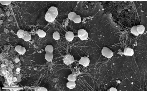
Figure 1. Growth of P. furiosus on the surface of sand grains from its natural habitat, visualized by scanning electron microscopy. Flagella attach the cells of the microcolony to the sand grain and to each other. Bar = 2 μm (Närther et al., 2006).
Archaea are very diverse organisms from morphological and metabolic point of view. They are single-‐celled organisms lacking nuclei and are therefore prokaryotes. Individual archaea range from 0.1 μm to over 15 μm in diameter, and some form aggregates or filaments up to 200 μm in length (Figure 1). Archaea are characterized by their unique ether-‐linked membrane lipids (Koga and Morii, 2007), and also by their unique enzymes such as specific DNA topoisomerases (Forterre et al., 2007) and DNA polymerases (Ishino et al., 1998). However, Archaea share some characteristics with the other two kingdoms. Thus, like bacteria, archaea usually has a single circular genome, their genes are grouped in operons and are regulated by bacteria-‐like transcription regulators (Bell et al., 1999). But it is with Eukarya that Archaea share most of its
information-‐processing systems (replication, transcription and translation). Indeed the majority of translation (Bell and Jackson, 1998), DNA replication (Kelman and White, 2005) and DNA repair factors (Kelman and White, 2005) are specifically shared between Archaea and Eukarya but are not present in Bacteria. Moreover, Archaeal RNA polymerases are closely related to their eukaryotic counterparts, in terms of both subunit composition and structure (Werner, 2008), and by the basal transcription factors required for initiation (Bell and Jackson, 2001). Thus, because of these similarities and because in archaea the number of factors involved are generally lower, archaeal systems act as simplified model systems for complex eukaryal processes. The investigations of transcription using Archaea provide therefore not only insights into the biology of archaeal cell, but allow better understanding of the experimentally limited eukaryotic transcription machinery too.
Study of archaeal transcription machinery using the hyperthermophilic organism P.
furiosus has several advantages. The organism is capable heterotrophic growth on a wide range of substrates (starch, peptone, complex organic substrates, casein, and maltose). Under optimal conditions (100°C, pH 7), P. furiosus has a rapid doubling time of 37 minutes and can grow to high cell density (>1010 cells/ml). These characteristics are helpful for the isolation of endogenous RNAP (Fiala and Stetter, 1998). Although the organism is strictly anaerobic, the purification and the transcription activity of RNAP can be done under aerobic experimental conditions (Hethke et al., 1996). In addition, all basal transcription factors, transcription regulators and the eleven RNAP subunits can be individually purified from E.coli (Hausner et al., 1996; Goede et al., 2006). P.
furiosus RNAP can be reconstituted from the individual RNAP subunits. This allows the design of RNAP substitution/deletion mutations that are potentially lethal in vivo and subsequent specific in vitro analysis (Naji et al., 2007; Naji et al., 2008). Since the sequence of the complete genome of P.furiosus is known (Robb et al., 2001), the identification and the characterization of transcription factors and regulators could be significantly improved. Moreover, by using a cryo-‐electron microscopy approach a relatively accurate prediction of P.furiosus RNAP architecture was obtained (Kusser et al., 2008). Thus, to over 15 years, the group of Prof. Dr. Michael Thomm contributes to the improvement of the knowledge of the transcription machinery by using the transcription system of P. furiosus. Recently, in addition to biochemical approaches and in vitro characterization, a genetic system was developed in this organism (Waege et al., 2010), allowing an enhancement of characterization of the transcription machinery of P. furiosus by in vivo data.
A. DNA-‐dependent RNA polymerase
All cells accomplish the transcription by one or more DNA dependent multisubunit RNAPs, which consist of 5-‐15 subunits and a molecular weight of up to 0.7 MDa (Cramer et al., 2008).
Bacteria, archaea and chloroplast (PEP, plastid encoded polymerase) contain a single type of RNAP, while the eukaryotes contains three to five distinct types (RNAP I, II, III, IV and V) (Darst,
2001; Kanamaru and Tanaka, 2004; Cramer and Arnold, 2009; Grohmann et al., 2009a; Pikaard and Tucker, 2009; Ream et al., 2009). In addition to those enzymes, single-‐subunit RNAPs were also described in certain cells, like in mitochondria and chloroplast (NEP, nuclear encoded polymerase) (Gaspari et al., 2004; Kanamaru and Tanaka, 2004). Those enzymes are related to the single-‐
subunit RNAPs from bacteriophages, such as T7, T3 or SP6, from which T7 RNAP is, structurally and functionally, best characterized (Steitz, 2009).
In eukaryotes, RNAP I synthesizes ribosomal RNAs (pre-‐rRNA 45S in yeast) which will form the major RNA sections of the ribosome. RNAP II synthesizes pre-‐messenger RNA (pre-‐mRNAs), small nuclear RNAs (snRNAs, ~125 nt) and small non-‐coding RNAs (microRNAs, ~22 nt), and RNAP III synthesizes transfer RNAs (tRNAs) and other small RNAs. Finally, RNAP IV and RNAP V, which are specific to the plants, are essential for the synthesis of small interfering RNAs (siRNAs) and other RNAs required for heterochromatin formation and gene silencing (Pikaard et al., 2008.; Wierzbicki et al., 2008;. Ream et al., 2009).
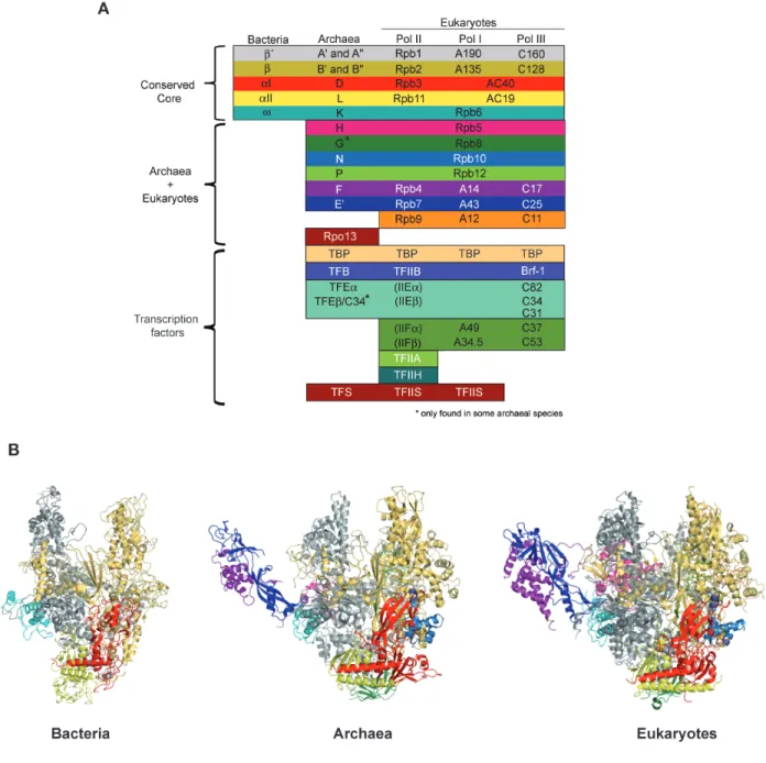
Archaea and bacteria contain only a single RNAP that catalyses the synthesis of all cellular RNAs (Darst, 2001; Grohmann et al., 2009a). Archaeal RNAP is, structurally and mechanistically, closely related to eukaryotic nuclear RNAP II (Langer et al., 1995). Figure 2A shows a comparison of the topology of the essential subunit and transcription factors in bacterial, archaeal and eukaryotic RNAPs (Werner and Grohmann, 2011). Sequence comparisons of the RNAP subunits lay that all multisubunit RNAPs derive from a common precursor enzyme (Huet et al., 1983). The bacterial RNAP has five subunits and any of the bacterial subunits has an archaeal/eukaryotic homologue (Sweetser et al., 1987; Ebright, 2000). The two largest RNAP subunits, β and β´ in bacteria, Rpb1 and Rpb2 in eukaryotes, and RpoA and RpoB (also known as Rpo1 and Rpo2) in archaea form about two-‐thirds of RNAP to form the catalytic centre and are derived from a common ancestor (Figure 2B) (Zhang et al., 1999; Cramer et al., 2001; Hirata et al., 2008b). In P. furiosius, and other archaea, the Rpb1 homologue is split into two subunits denoted RpoA´ and RpoA´´, respectively (Pühler et al., 1989). In Methanogenes and extreme Halophiles, the Rpb2 homologue is also split into two subunits (RpoB 'and RpoB''). The Rpb1/Rpb2 complex is anchored at one end into the Rpb3/Rpb11 heterodimer. Eukaryotic Rpb3/Rpb11 heterodimer (RpoD/L in archaea) together with Rpb10/Rpb12 (RpoN/P in archaea), as well as α-‐subunit homodimer in bacteria, form the assembly platform required for the efficient assembly and stability of RNAP (Werner et al., 2000; Werner and Weinzierl, 2002; Grohmann et al., 2009a). The smallest bacterial RNAP subunit ω, corresponding to the Rpb6 and RpoK in eukaryotes and archaea, respectively, also promots the RNAP assembly by latching the assembly platform (Minakhin et al., 2001).
The archaeal RpoH subunit lacks the N-‐terminal domain forming the lower jaw domain in eukaryotic homologue Rpb5. The C-‐terminal domain makes intricate contacts with the C terminus of the largest subunit (Rpb1 in eukaryotes, RpoA in archaea). Rpb8 and RpoG are located at the bottom of the RNAP between the assembly platform and the pore. Yeast Rpb8 is essential but its precise function remains unclear (Briand et al., 2001). In archaea, RpoG is present only in the
Crenarchaeota (Koonin et al., 2008; Kwapsiz et al., 2008; Korkhin et al., 2009). Recently, good indications of subunits and transcription factor homologies between the three nuclear RNAPs were also obtained by (Kuhn et al., 2007;. Carter and Drouin, 2009).
Figure 2: Composition and structure of multisubunit RNAPs. (A) Homology pattern of the subunits in the RNAPs of bacteria, archaea and eukaryotes (Werner, 2012). The specificity of the RNAP subunits is indicated in left. (B) Overall architecture of RNAPs from bacteria (Thermus aquaticus (1HQM) Minakhin et al., 2001), archaea (Sulfolobus shibatae (2Y0S) Wojtas et al., 2011) and eukaryotes (Saccharomyces cerevisiae (1Y1V) Kettenberger et al., 2004). The color code of the RNAP subunits is same as panel A.
Some subunits are specific for a single kingdom of life. Rpb9 is the only subunit found exclusively in eukaryotic RNAPs (Figure 2A). Rpb9 is related to the transcription factor TF(II)S, but with a loss of efficient RNA cleavage activity (Walmacq et al., 2009; Ruan et al., 2011), suggesting that Rpb9 was obtained through gene replication and alteration of catalytic C-‐ribbon. Rpo13 is the only archaea-‐specific RNAP subunit, and it is only present in a subset of archaeal genomes (Korkhin et al., 2009). Its function is unclear, but recent biochemical studies suggest that Rpo13 stabilize RNAP-‐DNA interaction by binding non-‐specifically to double strand DNA (Wojtas et al., 2012).
The previously mentioned subunits in archaea (RpoB, A ', A'', D, L, N, P, K, H and additional G, Rpo13 in Crenarchaeota) and their eukaryotic homologues in RNAP II (Rpb1, 2, 3, 5, 6, 8, 9, 10, 11 and 12) form the core part of the enzyme RNAP that resembles a crab claw (Cramer et al., 2001).
The most pronounced difference between archaeal and eukaryotic enzymes and the bacterial one, is the presence of a stalk-‐like protrusion (RpoE/F and Rpb4/7 subcomplexes) (Cheetham and Steitz, 2000; Cramer et al., 2001; Hirata et al., 2008a; Grohmann and Werner, 2011). Indeed, the crystal structures of RNAP II and the archaeal RNAP, and also the ones of RNAP I and RNAP III, show the presence of the heterodimer forming the stalk above Rpb6/RpoK subunit (Armache et al., 2003;
Bushnell and Kornberg, 2003; Jasiak et al., 2006; Kuhn et al., 2007; Korkhin et al., 2009)
B. Domains and structural elements of RNAPs
Figure 3. Structural elements of multisubunit RNAPs. Important domains and structural elements of multisubunit RNAPs are shown in RNAP II of S. cerevisiae (Kettenberger et al., 2004). The top view shows the active site (Metal ion A) at the centre of the enzyme. The Helix Bridge connects the two halves of the “crab claw", which each consist of mainly Rpb 1 (Clamp) and Rpb2 (Lobe and Protrusion) subunits domains. The front view shows the “Wall” and the position of the “Funnel”, which forms the outer edge of the pore or the secondary channel.
1. Stalk (E/F subcomplex)
Archaeal and eukaryotic RNAPs (including RNAP IV and V) contain homologous subunits, which are not present in bacteria (Werner, 2008). E and F subunits (homologous to the eukaryotic Rpb4 and 7, respectively), form a stalk-‐like protrusion (Figure 3) which plays an important role during transcription initiation (Edwards et al., 1991; Armache et al., 2005; Grohmann et al., 2009). In archaea, those two subunits were shown to facilitate DNA melting and are required for the function of TFE (Werner and Weinzierl, 2005; Naji et a., 2007). During elongation, the E/F subunits interact with the nascent RNA emerging from the RNA exit channel of RNAP, and thus increase the processivity (Ujvári and Luse, 2006; Andrecka et al., 2009; Hirtreiter et al., 2010a). In addition, E/F may stabilize the elongation complex by inducing a conformational change in RNAP, such as the closure of the RNAP clamp (Armache et al., 2005). Recent studies on archaeal transcription termination showed that E/F significantly increases termination efficiency at weak termination signal (five dT stretch) (Hirtreiter et al., 2010a). In vivo, archaeal rpo4 and eukaryotic rpb4 genes are essential for survival, while archaeal rpo7 and eukaryotic rpb4 can be deleted with viability retained at moderate temperatures (Sheffer et al., 1999; Hirata et al., 2008a). Purified fractions of RNAP II of S. cerevisiae had substoichiometric amounts of Rpb4/7 that made its structural elucidation difficult for a long time (Cramer, 2004a). Reconstitution of the complete RNAP II from endogenous yeast core and recombinant Rpb4/7 allowed this obstacle to be overcomed structurally (Armache et al., 2003; Bushnell and Kornberg, 2003) and functionally (Edwards et al., 1991; Naji et al., 2007). The idea emerged that, in the yeast system, the stalk can assemble and disassemble during transcription cycle (Edwards et al., 1991). The relative ratio of RNAP II and Rpb4/7 in S. cerevisiae is dependent on the growth phase (Choder and Young, 1993). However, recent studies showed that E'/F on the archaeal RNAP from Methanocaldococcus jannaschii is stably incorporated into RNAP and that dynamic equilibrium with E'/F does not occur (Grohmann et al., 2009b).
2. Clamp domain
The high stability of RNAP elongation complexes prevents dissociation of RNAP from DNA and allows efficient transcription. This stability is mainly caused by the tight binding of the RNA/DNA hybrid to RNAP (Kireeva et al., 2000; Sidorenkov et al., 1998). In the elongation complex, the hybrid is nested in a highly complementary binding site, created by the closure of the mobile module called the “clamp” (Figure 3). The clamp is open in free RNAP and early transcription initiation complexes but a dramatic 30° rotation of the clamp occurs with the binding of the DNA template strand to three out of five “switch” regions (Gnatt et al., 2001). In the open state, the clamp allows promoter DNA to be loaded into and unwound in the active centre cleft. The binding of RNA/DNA hybrid to the folded switches stabilizes the closed state which accounts for the high
stability of initiation complexes and the high stability and processivity of elongation complexes (Cramer et al., 2001; Gnatt et al., 2001; Chakraborty et al., 2012).
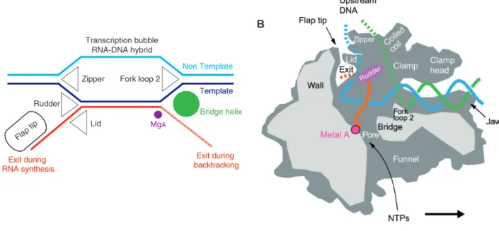
Three loops that protrude from the clamp maintain the arrangement of the nucleic acids during the elongation (Figure 4A). The “rudder” is required for promoter opening in bacteria (Kuznedelov et al., 2002) and for transcription in archaea (Naji et al., 2008). The “lid” is important to stabilize the open promoter complex (Toulokhonov and Landick, 2006), in abortive transcription and serves as a wedge to facilitate RNA displacement by sterically blocking the formation of the overextended hybrid (Gnatt et al., 2001; Naji et al., 2008; Naryshkina et al., 2006). Finally, the double strand DNA is reformed at the back end of the transcript bubble by the “zipper” (Gnatt et al., 2001; Cramer et al., 2001). In bacteria, the zipper also contributes in promoter element (called
“Z-‐element”) recognition (Yuzenkova et al., 2011). In addition to these loops, the mobile part of the “flap loop” (flap tip) on top of the “wall” contributes in bubble maintenance and binds to nascent RNA hairpins that pause or terminate bacterial transcription (Figure 4B) (Toulokhonov and Landick, 2003; King et al., 2004). In Archaea and Eukaryotes, RNA hairpins do not affect transcription, probably because the flap tip is shorter in archaeal and eukaryotic RNAPs (Cramer, 2002). Moreover, unlike bacterial RNAP, eukaryotic RNAP II flap loop is not essential for transcription initiation (Palangat et al., 2011).
Figure 4. The RNAP elongation complex. (A) Schematic presentation of the arrangement of nucleic acids during RNA chain elongation. The DNA template and nontemplate strands are in blue and cyan, respectively, and the RNA is in red.
The active site metal ion A is indicated by a pink sphere. Protein elements that are proposed to be involved in the maintenance of the arrangement of nucleic acids are indicated. (B) Cutaway view of the RNAP elongation complex. Cut surfaces are lightly shaded. During transcription, DNA enters the enzyme from the right (the polymerase moves to the right). Structural features that appear to be important for function are labeled. The DNA template and nontemplate strands are in blue and green, respectively. (Modified from Cramer, 2002).
The RNAP clamp coiled-‐coil motif was recently shown to be a binding site for several transcription factors pointing to its importance in transcription initiation and elongation (Figure 4B). The TF(II)B B-‐linker domain and bacterial σ2 domain, which are involved in promoter opening, were shown to bind to the clamp coiled-‐coil and the rudder (Kostrewa et al., 2010). Moreover, transcription initiation factor TF(II)E and universally conserved NusG/Spt5 elongation factor compete to bind on clamp coiled-‐coil motif (Grohmann et al., 2011; Grünberg et al., 2012;
Martinez-‐Rucobo et al., 2011; Werner, 2012). The binding affinities of these factors are context dependent: TFE prevails over Spt4/5 in the initiation complex, whereas Spt4/5 prevails over TFE in the elongation complex. Thus, TFE prevents the inhibitory affect of Spt4/5 on transcription initiation and, during early elongation, Spt4/5 displaces TFE resulting in a high-‐processivity elongation complex.
3. Switch region
The “switch region” is located at the base of the clamp and serves as the hinge on which the clamp swings during clamp opening and clamp closure (Cramer et al., 2001; Cramer, 2002).
Five segments of the switch region, termed “switch 1” through “switch 5”, undergo different conformations in open and closed clamp conformational states. It has been proposed that direct contacts between the switch region and DNA phosphates might coordinate clamp closure and DNA loading into the RNAP active centre (Gnatt et al., 2001; Vassylyev et al., 2007). In bacteria, this region is a target for several antibiotics that inhibit distinct steps of transcription initiation (Belogurov et al., 2009; Mukhopadhyay et al., 2008; Srivastava et al., 2011).
Switch 3 is a polypeptide loop which binds to each RNA base in a nascent transcript as it dissociates from the RNA/DNA hybrid (Kent et al., 2009). In archaea, it was shown to be crucial in transcript elongation, unlike bacteria, in which it is required to form stable complexes with nucleic acid scaffolds by controlling clamp closure (Santangelo and Reeve, 2010; Wiesler et al., 2012). This divergence is likely caused by the differences in charge and flexibility of archaeal and eukaryotic switch 3 loops (Santangelo and Reeve, 2010).
Recent studies on the bacterial switch region suggest that switch 1, 2, 4 and 5 contribute in thestart site melting mechanism (Wiesler et al., 2012). Indeed, a number of substitutions in the switch region affected transcription initiation. Analysis of switch 2 substitutions suggested that this region may be involved in start site selection, abortive initiation, promoter escape and transcript elongation (Majovski et al., 2005; Naji et al., 2007; Pupov et al., 2010). Furthermore, the invariant arginine (Pfu A´-‐R313; Sce Rpb1-‐R337; Eco ß´R339) of switch 2 was recently proposed to, in cooperation with switch 1, 4 and 5, undergo conformational changes that stabilize the DNA melting around the start site (Naji et al., 2007; Wiesler et al., 2012).
4. Active site
The catalytic cycle of RNAP (called nucleotide addition cycle) is driven by complex conformation changes that accompany NTP binding, catalysis, and RNAP translocation. When the NTP enters in the RNAP active site, via the secondary channel, a network of interactions between the incoming NTP and active site elements allow the proper positioning of the NTP and its incorporation into the nascent RNA. Recent studies identified two elements in the active centre of RNAP, the “Trigger loop” and the “Bridge helix”, which appear to play key roles during the nucleotide addition cycle (Brueckner et al., 2009).
a. The trigger loop
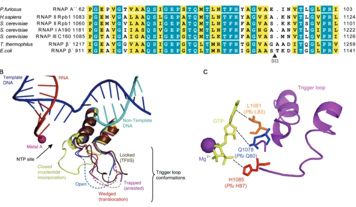
The trigger loop (TL) is a polymorphous element of RNAP active site that is highly conserved among the three domains of life (Figure 5A). The TL is present in the largest subunit of eukaryotic RNAP II Rpb1 and the analogous β′ subunit of bacterial RNAP, and A subunit of archaeal RNAP (A′′
in Pfu RNAP). In E.coli RNAP the TL contains a sequence insertion of 188 aa, called SI3. Structural and biochemical studies in yeast RNAP II and bacterial RNAPs, revealed the importance of the TL in substrates selection and catalysis. The conformational changes of the TL were proposed to link TL-‐
NTP interaction with the substrate positioning and selection but also to be critical in translocation and proofreading (Kaplan et al., 2008; Brueckner et al., 2009; Huang et al., 2010; Yuzenkova et al., 2010; Yuzenkova and Zenkin, 2010; Zhang et al., 2010). Five distinct TL conformations have been observed: “open”, “closed”, “wedged”, “trapped”, and “locked” (Figure 5B) (Martinez-‐Rucobo and Cramer, 2013).
During nucleotide addition, in the absence of substrate, the TL adopts an “open” conformation in which its central part is unstructured (Kettenberger et al., 2004). Binding of an incoming NTP in the +1 site induces folding of the TL, resulting in extension of two helixes at the base of the TL and creating a closed, catalytically competent conformation of the active centre in which the NTP is properly aligned with the 3´-‐OH of the nascent RNA to facilitate catalysis (Vassylyev et al., 2007b;
Wang et al., 2006). The “closed” TL forms a three-‐helix bundle with the Bridge helix (BH) that interacts with the substrate NTP and the template DNA base, resulting in the closure of the active site. Recent structural analysis on bacterial RNAP and yeast RNAP II proposed that TL residues Rpb1 Q1078, L1081 and L1085 (Pfu A´´ Q80, L83, and H87, respectively) contact the 2´-‐OH group, the base and the triphosphate moieties of the incoming NTP, respectively (Figure 5C), whereas the central part of BH contacts the template base (Vassylyev et al., 2007a; Wang et al., 2006;
Yuzenkova et al., 2010; Zhang et al., 2010). However, many additional active centre residues make also essential interaction with the NTP substrate (Nudler, 2009; Cheung et al., 2011). The direct contact between TL residues with the substrate was proposed to link substrate positioning and
recognition, and to be critical for catalysis (Kaplan et al., 2008; Yuzenkova et al., 2010; Cheung et al., 2011).
Moreover, the TL was proposed to participate in translocation during the nucleotide addition cycle, and to be critical in intrinsic cleavage activity in bacteria (see below).
Figure 5. Conserved active site element: the trigger loop (TL). (A) The sequences alignment of the TL from archaeal RNAP (P. furiosus), eukaryotic RNAP II (H. sapiens and S. cerevisiae), RNAP I and RNAP III (S. cerevisiae) and bacterial RNAPs (T. thermophilus and E. coli). The black triangle indicates the position of insertion site of SI3 (188 aa) in the E.
coli RNAP. (B) Comparison of TL conformations (Martinez-‐Rucobo and Cramer, 2013). Superposition of the five RNAP II TL conformations known structurally. “Open” TL in the post-‐translocation state (PDB 1Y1W (Kettenberger et al., 2004), blue), “closed” TL in the nucleotide incorporation state (PDB 2E2H (Wang et al., 2006), yellow), “wedged” TL in the translocation intermediate (PDB 2VUM (Brueckner and Cramer, 2008), red), “trapped” TL in the arrested complex (PDB 3PO2 (Cheung and Cramer, 2011), violet), and “locked” TL in the reactivation intermediate (PDB 3PO3 (Cheung and Cramer, 2011), brown). DNA template (blue), DNA non-‐template (cyan), RNA (red) and metal A (pink) are from the open state. (C) Closed TL forms a network of interactions with a nucleoside triphosphate (NTP) in the active centre (Wang et al., 2006). When correct NTP enter to the insertion site, TL invariant glutamine residue ((Sce Rpb1 Q1078;
Pfu A´´ Q80), leucine reissue (Sce Rpb1 L1081; Pfu A´´ L83) and invariant histidine residue (Sce Rpb1 H1085; Pfu A´´
H87) were suggested to form a network of interaction with the base, the sugar 2´OH-‐group and β-‐phosphate of the NTP, respectively.
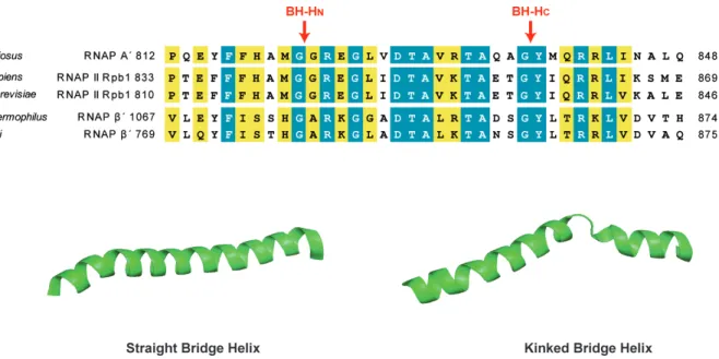
b. The bridge helix
The BH, an α-‐helix spanning the active site, is a highly conserved element which acts as pawl in a ratchet-‐like translocation mechanism to move DNA through the RNAPs (Bar-‐Nahum et al., 2005). The BH forms a stable three-‐helix bundle that is structurally flexible and isomerizes between a straight and a kinked conformation. Particularly, two flexible sites of BH, called N-‐
terminal Hinge (BH-‐HN) and C-‐terminal Hinge (BH-‐HC), were shown to induce the kinked conformation (Weinzierl, 2011). Because BH kinking was observed on NTP-‐bound elongation complexes, and an increased BH kinking at the two hinges correlates directly with an increased rate of nucleotide addition, the BH dynamics were suggested to play a role in catalysis (Tan et al., 2008; Vassylyev et al., 2007a; Wang et al., 2006; Weinzierl, 2010). The BH N-‐terminus is tightly surrounded by other elements, such as experimentally uncharacterized “link domain” and the “F-‐
loop”. During nucleotide addition, the BH and F-‐loop form a gateway that contacts the link domain and the tip of the TL (Miropolkaya et al., 2009). Moreover, C-‐terminal BH influences the TL conformation and the BH-‐HN may alter the position and conformation of the Link domain that is in direct physical contact with the nucleotide (Wienzierl, 2010). Taken together, because of its crucial role in translocation and in catalysis, and because of its contacts with other cleft loops, the BH is proposed to act as a central switchboard for catalysis and substrate movement coordination.
Figure 6. Conserved active site element: the bridge helix (BH). (A) The sequences alignment of the BH from archaeal RNAP (P. furiosus), eukaryotic RNAP II (H. sapiens and S. cerevisiae) and bacterial RNAPs (T. thermophilus and E. coli).
The BH N-‐termus and C-‐termus Hinge sites are indicated with red arrows. (B) Comparison of two BH conformations from T. thermophilus RNAP. “Straight” state (PDB: 2O5I (Vassylyev et al., 2007a)) and kinked (PDB: 1IW7 (Vassylyev et al., 2002)) BH.
c. Further active site elements
Other conserved structural features of the RNAP active centre include “fork loops 1 and 2”
(FL1 and FL2) and “F loop” (FL).
The FL1 is a small conserved segment of the larger fork domain, in the proximity of the active centre. The FL1, with the lid and the rudder, plays a key role in DNA/RNA strand separation.
FL1 contacts the base pairs -‐6 and -‐7 in hybrid region, limiting strand separation (Westover et al., 2004a). Structural analysis suggested that FL1 conformation may fluctuate, engaging the single-‐
strand DNA or RNA/DNA hybrid during transcription initiation or elongation respectively. Thus, after the formation of the nascent RNA (>8 nt), FL1 interacts with the rudder to lock the hybrid into a more stable interaction (Meyer et al., 2009).
The flexible FL2 directly interacts with an unpaired DNA residue in the non-‐template DNA strand, one nucleotide ahead from the active centre (the +2 site) and thus sterically preventing reannealing of the DNA strands (Andrecka et al., 2009; Cramer et al., 2001). This interaction also facilitates NTP sequestration through interaction with the adjacent segment of the fork subdomain I involved in the active centre of RNAP (Kireeva et al., 2011). Thus, FL2 may facilitate the non-‐
catalytic (TL-‐independent) NTP incorporation in the active centre of RNAP and increase the rate of phosphodiester bond formation (Kennedy and Erie, 2011; Kireeva et al., 2011).
FL is located near the N-‐terminus of BH and directly contacts the closed TL in the NTP bound transcription elongation complex. Together with the BH, the FL forms a gateway that accommodates the folded TL during nucleotide addition. The FL may be required for the proper folding of the TL and may stabilize the closed conformation of the active centre during catalysis (Miropolskaya et al., 2009; Miropolskaya et al., 2010).
C. Transcription cycle
The synthesis of RNA from a DNA template is conserved among all RNAPs. The transcription cycle is divided into three distinct phases, initiation, elongation and termination, each of which is regulated by various factors (Figure 7). The structure and function of some factors are conserved across the three domains of life (NusG and Spt5), whereas other non-‐
homologous factors show structural and/or functional similarities, suggesting that convergent evolution occurred to allow the same process (For example: Gre and TF(II)S, Sigma and TF(II)B).
Figure 7. The archaeal transcription cycle. During initiation TBP and TFB assemble on the promoter and recruit RNAP. TFE stimulates DNA melting and the template strand loading into the active site during the next step of initiation. Spt4/5 and TFS associate with the elongation complex and stimulate processivity and proofreading, respectively. The DNA template (T) and non-‐template (NT) strands are in blue and cyan, respectively.
RNA is in red and the active site is in purple.
1. Initiation of transcription
Promoter-‐directed transcription requires sequence-‐specific recruitment of RNAP to the promoter, initiation of RNA polymerization in a primer-‐independent manner and efficient escape from the promoter. This transcription phase is stimulated by evolutionarily unrelated basal initiation factors in all domains of life. However, as the molecular mechanisms of initiation are the same in all three domains and these non-‐homologues factors utilize the same RNAP-‐binding sites, they stimulate closely related mechanisms (Grohmann and Werner, 2011).
In bacteria, gene specific Sigma(σ)-‐factors interact with the core RNAP (ββ´ααω) to form holo-‐RNAP and enables specific binding of the enzyme to promoters (-‐10 and -‐35 elements). In addition to increasing RNAP sequence-‐specificity for promoters, it also facilitates DNA melting and template strand loading during the closed to open complex transition (Campbell et al., 2008, Murakami and Darst, 2003).
In eukaryotes, distinct general transcription factors (GTFs) are required to form, with the RNAP, the transcription initiation complex. The archaeal RNAP have identical but simplified set of minimal transcription initiation factors to eukaryotic RNAP II (Langer et al., 1995; Bartlett, 2005).
Transcription initiation by RNAP II begins with assembly of polymerase and all five general initiation factors into a pre-‐initiation complex at the promoter and culminates in formation of an open complex and synthesis of the RNA transcript. In the first step, TBP (TATA-‐binding protein) subunit of TFIID complex (12 TAFs in yeast, TBP associated factors) binds specifically to the TATA box and induces bending of DNA by approximately 90°. TFIIA, by interacting with TBP, can stabilize this complex. In the second step, TFIIB functions as an adaptor by binding specifically to TATA-‐box-‐
TBP complex and RNAP. TFIIF (Tetramer of two TFIIFα/RAP74 and two TFIIFβ/RAP30) strongly stabilizes this complex and recruits TFIIE (Dimer of TFIIEα and TFIIEβ in Metazoa, trimer of Tgf1, Tgf2 and Tgf3 in yeast) and TFIIH (10 subunits) into the complex. TFIIE binds on the clamp coiled-‐
coil element and is required for open complex formation, that occurs by DNA melting generated by ATP-‐dependent DNA helicase activity (SSL2/XPB and RAD3/XPD) of TFIIH (Grünberg et al., 2012). The transcription initiation required phosphorylation of CTD provided by Kin28/CDK7 subunit of TFIIH, followed by promoter escape. TFIIF, E and H also suppress promoter-‐proximal pausing of RNAP (Dvir et al., 2001; Woychik and Hampsey, 2002).
In archaea, in contrast, there are only 3 GTFs named TBP, TFB and TFE, of which only TBP and TFB are essential for promoter-‐specific in vitro transcription initiation (Qureshi et al., 1995;
Hausner et al., 1996). While the GTFs of RNAP II machinery are around 30 polypeptides with about 1560 kDa, the three archaeal proteins are only about 80 kDa. Moreover, the melting of the promoter DNA occurs without ATP hydrolysis, and there is so far no evidence of transcription cycle-‐dependent phosphorylation of archaeal RNAP (Hausner and Thomm, 2001).
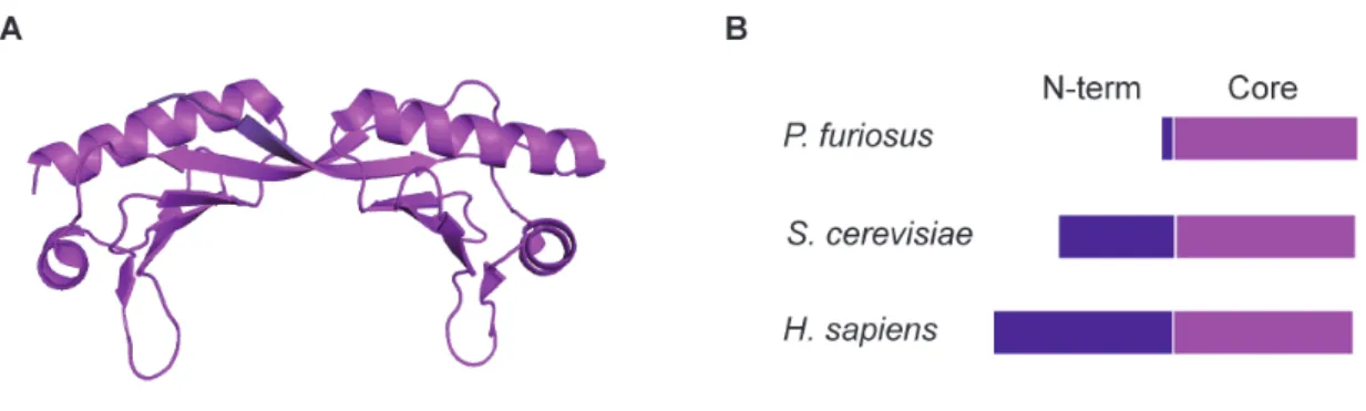
Archaeal TBP, as eukaryotic TBP, has a symmetric saddle-‐shaped structure that is formed by two homologous domains (Figure 8A) (Nikolov et al., 1992). Eukaryotic TBP contains an amino-‐
terminal domain that is absent in archaeal TBP (Figure 8B). Archaea, however possess 6-‐10 acidic amino acids at the C-‐terminus which are not observed in eukaryotic TBPs (Bell and Jackson, 1998).
The highly conserved core domain (saddle) is responsible for DNA binding, both upstream and downstream of the TATA box (Cox et al., 1997, Kosa et al., 1997).
Figure 8. Structure and domain organization of TBP. (A) Structure of archaeal TBP from M. jannaschii (PDB: 2Z8U (Adachi et al., 2008). (B) TBP consists of an N-‐terminal domain (purple) which is absent in archaea and a highly conserved Core domain (magenta).
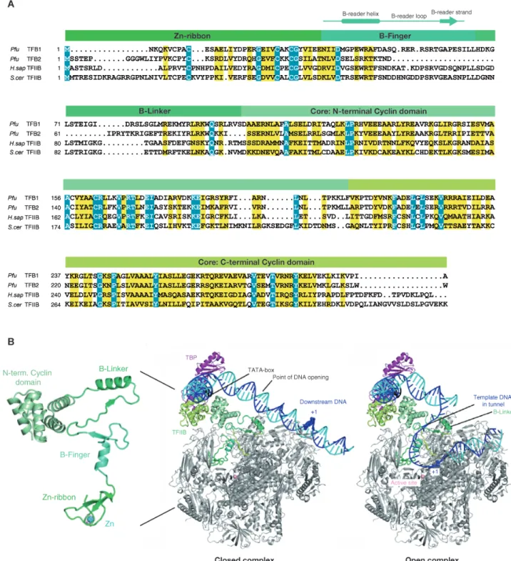
Archaeal TFB is a single polypeptide that is highly related to eukaryotic TFIIB (Figure 9A) (Ouzounis and Sander, 1992; Creti et al., 1993). It consists of N-‐terminal zinc-‐ribbon domain (Zn-‐
ribbon) (Zhu et al., 1996), which interacts with the dock domain of RNAP, and the C-‐terminal core domain recognizes the BRE element of the promoter and ensures the correct orientation of the initiation complex (Bell and Jackson, 2000; Qureshi and Jackson, 1998; Lagrange et al., 1998). The highly flexible linker region that connects the TF(II)B domains (consisting of B-‐reader helix and B-‐
linker) penetrates deep into the active centre of RNAP (Figure 9B). The B-‐reader is displaced by the growing RNA transcript (> 6 nt), whereas the B-‐linker is displaced by the rewinding of upstream DNA during TF(II)B release and promoter escape (Bushnell et al., 2004; Kostrewa et al., 2009).
While eukaryotes have only one TFIIB, archaea encode mostly for two with Halophilic archaea even up to 6 TBPs and 7 TFBs (Werner, 2007). The additional copies of TFB often exhibit N-‐ or C-‐
terminal truncations or deviations in the functional areas (Werner, 2007). The assumption is that different TBP-‐TFB-‐sets, similar to the various σ factors in bacteria, recognize different subsets of promoters efficiently (Facciotti et al., 2007). In Pfu, the second TFB (TFB2) functions poorly in promoter-‐dependent transcription initiation, probably because of a truncation in B-‐finger/B-‐linker region (Figure 9A) (Micorescu et al., 2007).
Figure 9. Structure and domain organization of TF(II)B. (A) The sequences alignment from archaeal TFB1 and TFB2 (P.
furiosus) and eukaryotic TFIIB (H. sapiens and S. cerevisiae). The B-‐finger organization is indicated. (B) Structure of yeast TFIIB as observed in its complex with RNAP II and model of closed and open initiation complexes (Kostrewa et al., 2009). The DNA template and nontemplate strands are in blue and cyan, respectively. The TATA element is in black.
Archaeal TFE corresponds to the N-‐terminal part of the large subunit TFIIE-‐α (Figure 10A) (Kyrpides and Ouzounis, 1999). This consists of a winged-‐helix (WH) motif, which is a special form of the HTH motif (Brennan, 1993), typically found in transcription factors and DNA-‐binding proteins (Gajiwala and Burley, 2000). The structure of the S. solfataricus WH motif has been solved, due to its good preservation within the Archaea, this structure is likely to be applicable to the other archaeal TFEs (Meinhart et al., 2003). The preservation in TFIIE is lower, but sufficient to create homology models. A specific feature of the WH motif of TF(II)E is the extension of the canonical winged helix fold at the N and C termini, and the canonical three helices of the hydrophobic core. Located in the central part of TFE, there is also a conserved Zinc-‐binding motif and a predicted HTH motif at the non-‐crystallized C-‐terminus (Figure 10B) (Meinhart et al., 2003).
TFE has a slight stimulatory effect on the transcription at limiting TBP concentrations or at weakly expressed promoters by stabilizing the open pre-‐initiation complex (Bell et al., 2001; Hanzelka et al., 2001). TFE binds to single stranded DNA (Grünberg et al., 2007), but the effect of TFE depends on the presence of E'/ F subcomplex. Indeed, TFE has, in the presence of E´ subunit, a stimulatory effect on promoter opening and on abortive transcription (Grünberg et al., 2007; Naji et al., 2007).
Moreover, the RNAP clamp coiled coil domain and E´/F subcomplex were shown to be crucial for TFE binding and its effect on transcription activity (Ouhammouch et al., 2004; Naji et al., 2007;
Grohmann et al., 2011). This suggests that, during transcription initiation, TFE is able to prevent binding of the elongation factor Spt4/5 on RNAP clamp coiled coil domain. Thus, by remaining associated with RNAP during early elongation, TFE can efficiently inhibit the inhibitory effect of Spt4/5 on transcription initiation (Grünberg et al., 2007; Grohmann et al., 2011; Werner, 2012).
Recently, archaeal homologue of RPC34 (homologue of TFIIEβ subunit) was identified via computational search, but its function in transcription initiation has not yet been validated experimentally (Blombach et al., 2009).
Figure 10. Structure and domain organization of TF(II)E. (A) TF(II)E consists of a highly conserved WH domain (cyan) and a Zn-‐ribbon domain (magenta). (B) Structure of archaeal TFE WH domain from S. solfataricus (PDB: 1Q1H (Meinhart et al., 2003) and eukaryotic TFIIE-‐α Zn-‐ribbon domain from H. sapiens (Okuda et al., 2004).