• Keine Ergebnisse gefunden

Cell death and transcriptional signalling mediated by the coiled-coil domain of the barley resistance protein MLA

N/A
N/A
Protected

Academic year: 2021

Aktie "Cell death and transcriptional signalling mediated by the coiled-coil domain of the barley resistance protein MLA"

Copied!
252
0
0

Wird geladen.... (Jetzt Volltext ansehen)

Volltext

(1)

NNT : 2016SACLE007

THESE DE DOCTORAT L’UNIVERSITE DE COLOGNE de

et de

L’UNIVERSITE PARIS-SACLAY,

préparée à l’Université d’Evry Val d’Essonne ÉCOLE DOCTORALE N° 577

Structure et dynamique des systèmes vivants Spécialité de doctorat Sciences de la Vie et de la Santé MATHEMATISCH-NATURWISSENSCHATLICHE FAKULTAET

Par

M elle Florence Jacob

Cell death and transcriptional signalling mediated by the coiled-coil domain of the barley resistance protein MLA

Thèse présentée et soutenue à Cologne, le 25/04/2016.

Composition du Jury :

Pr. Döhlemann, Gunther Professor, Institut für Botanik, Köln Président

Pr. Hofmann, Kay Professor, Institut für Genetik, Köln Rapporteur

Pr. Panstruga, Ralph Professor, RTWH Aachen Rapporteur

Pr. Dron, Michel Professor, Université Paris Sud Examinateur

Dr. Somssich, Imre Research Group Leader, MPIPZ Examinateur

Pr. Hirt, Heribert Research Group Leader, IPS2, Paris Saclay Directeur de thèse

Pr. Schulze-Lefert, Paul Professor, Institut für Genetik, Köln Co-directeur de thèse

(2)

Cell death and transcriptional signalling mediated by the coiled-coil domain of the barley resistance protein MLA

Inaugural-Dissertation zur

Erlangung des Doktorgrades

der Mathematisch-Naturwissenschaftlichen Fakultät der Universität zu Köln

vorgelegt von

Florence Jacob

(3)
(4)

Die vorliegende Arbeit wurde am Max-Planck-Institut für

Pflanzenzüchtungsforschung in Köln in der Abteilung für Pflanze-Mikroben

Interaktionen (Direktor: Prof. Dr. P. Schulze-Lefert) und am Institute of Plant Sciences Paris-Saclay in Orsay in der Abteilung für Physiology and Signalling (previously URGV, Direktor: Prof. Dr. M. Crespi) angefertigt.

Berichterstatter: Prof. Dr. Kay Hofmann

Prof. Dr. Ralph Panstruga

Prof. Dr. Jürgen Zeier

(5)
(6)

Savoir s'étonner à propos

est le premier pas fait

(7)
(8)

Cell death and transcriptional signalling mediated by the coiled- coil domain of the barley resistance protein MLA

Abstract

Plants rely entirely on innate immunity to prevent infection by pathogens. Extracellular

perception of evolutionarily conserved pathogen/microbe-associated molecular patterns

(P/MAMP) by membrane-resident pattern recognition receptors (PRRs) leads to pattern-

triggered immunity (PTI). Host-adapted pathogens intercept PRR-mediated immunity by

delivering effectors into host cells. These polymorphic effectors can be recognized by

intracellular immune receptors of the nucleotide-binding domain leucine-rich repeat (NLR)

family. Upon effector recognition, NLRs trigger a rapid immune response, termed effector-

triggered immunity (ETI), which is typically associated with a host cell death response. In

barley, the NLR MLA confers ETI against the pathogenic powdery mildew fungus, Blumeria

graminis f. sp. hordei. Although MLA orthologues are present only in the Triticeae family of

monocotyledonous plants, barley MLA is functional in transgenic dicotyledonous A. thaliana,

indicating that the underlying disease resistance mechanism has been evolutionarily

conserved for at least 150 Mya in monocot and dicot plants. In dicotyledonous Nicotiana

benthamiana, transient gene expression of the coiled-coil (MLA CC ) domain, consisting of the

N-terminal 160 amino acids of the MLA receptor, was sufficient to activate a cell death

response, raising the possibility that MLA initiates a conserved signalling mechanism through

the MLA CC domain. This thesis aimed at identifying signalling mechanism(s) acting

downstream of the MLA CC module in transgenic A. thaliana. Conditional MLA CC expression

triggered immune-related responses, characterized by a rapid onset of massive changes in

(9)

implying that the barley MLA CC domain is sufficient to stimulate an ETI-like response in A.

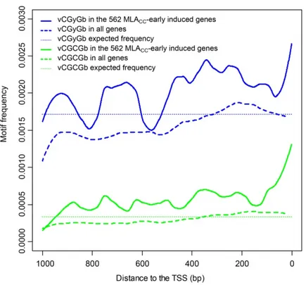
thaliana. This also suggests that activation of these immune response genes can occur independently of PTI. Most (> 74.7%) of the 562 genes that were significantly upregulated at 2 hpi in MLA CC -expressing plants are immediate early response genes since their induction does not depend on de novo protein synthesis, suggesting that these genes are activated by the removal of short-lived repressors. In the 5’ regulatory regions of the early induced genes, I found a striking enrichment of cis-acting motifs that serve as binding sites for Ca 2+ -responsive transcription factors, including the calmodulin-binding transcription activator 3 (CAMTA3) which is known to be rapidly degraded during ETI. This might explain complete inhibition of MLA CC –mediated responses by the Ca 2+ channel inhibitor LaCl 3 , which was previously also reported for several NLR-mediated and P/MAMP-triggered responses. Using chemical mutagenesis, I identified three candidate suppressor mutants of MLA CC -mediated responses.

Affinity purification of MLA CC complexes and a yeast two-hybrid screen identified several MLA CC candidate interacting proteins. Together this has revealed novel candidate components engaged in MLA CC signalling.

Keywords: plant biology, phytopathology, immune system, resistance protein, immune

signalling, hypersensitive response

(10)

Régulation transcriptionnelle et induction de la mort cellulaire par le domaine coiled-coil de MLA, protéine de résistance de l'orge

Résumé

La réponse immunitaire des plantes contre les pathogènes végétaux dépend entièrement du

système immunitaire inné. La perception extracellulaire de motifs moléculaires conservés

associés aux pathogènes/microbes (P/MAMP) par des récepteurs de reconnaissance de motifs

moléculaires (PRR) induit un type de réponse immunitaire dénommé PTI (pattern-triggered

immunity). Certains pathogènes interceptent cette réponse en injectant, dans les cellules hôtes,

des protéines appelées effecteurs. Ces effecteurs, souvent polymorphiques, peuvent être

détectés par des récepteurs immunitaires intracellulaires de la famille des NLR (nucleotide-

binding domain leucine-rich repeat containing). En cas de détection, les NLRs induisent une

réponse immunitaire rapide, appelée ETI (effector-triggered immunity), souvent associée à la

mort des cellules hôtes. Un des NLRs présents chez l’orge, MLA, confère une ETI protectrice

contre Blumeria graminis f. sp. hordei, l’agent pathogène du mildiou. Bien que MLA n’ait

d’homologues qu’au sein de la famille monocotylédone des Triticeae, MLA est fonctionnel

dans une lignée transgénique chez l’espèce dicotylédone A. thaliana. Ceci indique que le

mécanisme de résistance sous-jacent a été conservé depuis plus de 150 Ma chez les plantes

dicotylédones et monocotylédones. Chez la plante dicotylédone Nicotiana benthamiana,

l’expression transitoire du domaine coiled-coil (MLA CC ) qui correspond aux 160 premiers

acides aminés en N-terminus de MLA, suffit à induire la mort cellulaire. Cette observation

suggère que MLA pourrait initier un mécanisme de signalisation conservé, via son domaine

MLA CC . Le but de cette thèse a été de décrire le(s) mécanisme(s) de signalisation en aval de

(11)

cours de l’ETI et de la PTI chez A. thaliana, a révélé des patterns largement similaires lors de la réponse précoce. Ceci suggère que la signalisation initiée par MLA CC , l’ETI et la PTI pourrait converger rapidement vers une machinerie de transcription commune qui active les gènes de la réponse immunitaire. De plus, ces données indiquent que le domaine MLA CC de l’orge suffit à induire la réponse immunitaire normalement associée à l’ETI chez A. thaliana, et que l’activation des gènes de la réponse immunitaire peut se produire indépendamment de la PTI. La plupart (> 74.7%) des 562 gènes significativement induits à 2 hpi chez des plantes exprimant MLA CC sont caractéristiques des gènes de type « immediate early response » puisqu’ils sont induits rapidement sans nécessiter la synthèse de novo de protéine. Ces gènes pourraient donc être activés par élimination d’un répresseur à courte durée de vie. Dans les régions régulatrices en 5’de ces gènes, des motifs sont fortement enrichis qui correspondent à des sites de liaison pour des facteurs de transcription sensibles au Ca 2+ , tels que le calmodulin-binding transcription activator 3 (CAMTA3) qui est rapidement dégradé au cours de l’ETI. Ceci pourrait expliquer l’inhibition totale de la réponse médiée par MLA CC en présence de LaCl 3 , un inhibiteur des canaux calciques, inhibition qui a aussi été décrite par le passé concernant les réponses induites par plusieurs NLRs et P/MAMPs. J’ai effectué un crible génétique de mutants obtenus par mutagénèse chimique et identifié par ce biais trois candidats suppresseurs de la réponse médiée par MLA CC . La purification des complexes protéiques associés à MLA CC , ainsi qu’un crible en double-hybride chez la levure ont permis d’identifier plusieurs candidats interragissant avec MLA CC . Ces approches ont révélé de nouveaux candidats impliqués dans la signalisation médiée par MLA CC .

Mots-clés : biologie végétale, phytopathologie, système immunitaire, protéine de

résistance, signalisation immunitaire, réponse hypersensible

(12)

Zelltod- und transkriptionelle- Signalisierung aktiviert durch die Coiled-Coil Domäne von der Gersten-R-Protein MLA

Zusammenfassung

Pflanzen sind voll und ganz auf ihr angeborenes Immunsystem angewiesen um Infektionen durch Krankheitserreger zu verhindern. Extrazelluläre Erkennung von evolutionär konservierten Pathogenen/Mikroben-assoziierten molekularen Mustern (microbe/pathogen- associated molecular patterns; P/MAMP) durch membranassoziierte P/MAMP-Rezeptoren (pattern recognition receptors, PRRs), was zur sogenannten Muster-ausgelösten Immunität (pattern-triggered immunity, PTI) führt. Wirtsadaptierte Krankheitserreger hemmen diese Immunität durch die Sekretion von Effektoren in die Wirtszelle. Als Folge der Effektor- Erkennung, lösen NLRs eine rasche Immunantwort aus, welche als Effektor-ausgelöste Immunität (effector-triggered immunity, ETI) bekannt ist, und oft einen programmierten Zelltode beinhaltet. In Gerste vermitteln die MLA NLRs Resistenz gegenüber dem Mehltau Pilz Blumeria graminis f. sp. hordei. Obwohl MLA-Orthologe nur in anderen Mitgliedern der Triticeae Familie von Monokotyledonen Pflanzen bekannt sind, ist MLA in der Dikotyledonen Pflanze Arabidopsis thaliana voll funktionsfähig; was heißt, dass die MLA- zugrundeliegenden Widerstandsmechanismen über mindestens die letzten 150 Millionen Jahre evolutionär konserviert wurden. Transiente Genexpression der MLA Coiled-Coil‘

(MLA CC ) Domäne, bestehend aus den 160 N-terminalen Aminosäuren von MLA, in der der Dikotyledonen Pflanze Nicotiana benthamiana ist ausreichend um den Zelltod auszulösen.

Daher besteht die Möglichkeit, dass die MLA CC Domäne verantwortlich für die Auslösung

der konservierten Signalmechanismen ist. Diese Doktorarbeit bezweckt die Signalwege zu

(13)

exprimierenden, transgenen Pflanzen, zeigte eine erhebliche Überschneidung der Transkriptionsprofile in den frühen Phasen nach Exprimierung und Aktivierung der jeweiligen Rezeptoren. Dies weist darauf hin, dass die frühen, durch MLA CC , ETI und PTI induzierten Signalwege zu einem gemeinsamen Transkriptionsmechanismus konvergieren, welcher zur Aktivierung von Immunitätsgenen führt. Diese Resultate deuten auch darauf hin, dass die MLA Coiled-Coil Domäne von Gerste tatsächlich ausreichend ist um in A. thaliana ETI-ähnliche Reaktionen herbeizuführen, was auch bedeutet, dass diese Immunreaktionen PTI-unabhängig sind. Die meisten (>74,7%) der 562 Gene, welche ca. zwei Stunden nach Induktion der MLA CC Expression signifikant höher exprimiert sind, gehören zu den sogenannten „immediate early“ Genen, da deren Expression unabhängig von der de novo Proteinbiosynthese ist. Das wiederum bedeutet, dass die Expression dieser Gene durch den Verlust eines Transkriptionsrepressors ist. In der 5‘ Regulationsregion der früh induzierten Reaktionsgenen, identifizierte ich eine auffällige Anreicherung von cis-Regulationselementen, welche als Motive für Ca 2+ -regulierte Signal-Transkriptionsfaktoren dienen. Einer dieser Faktoren ist der calmodulin-binding transcription-activator 3 (CAMTA3), welcher während ETI abgebaut wird. Das wiederum könnte erklären warum Reaktionen von MLA CC und anderen NLRs, durch den Kalciumkanalblocker LaCl 3 komplett gehemmt werden. Zusätzliche habe ich durch chemische Mutation drei Mutanten identifiziert welche auch die von MLA CC

herbeigeführten Reaktionen inhibieren. Eine Affinitätsreinigung von MLA CC

Proteinkomplexen und eine Hefe-Zwei-Hybrid-Suche (yeast-2-hybrid screen) identifizierten zusätzliche, mit MLA-assoziierende Proteine. Diese bisher nicht charakterisierten Kandidaten fungieren möglicherweise als Komponenten für die MLA CC Signalwege.

Stichwörter: Pflanzenbiologie, Phytopathologie, Immunsystem, Resistenz-Protein,

Immun-Signalweg, Hypersensitive Response

(14)

Preface and Acknowledgements

This thesis concludes my PhD work conducted jointly at the Max Planck Institute for Plant Breeding research (Cologne) and at the URGV-IPS2 (Evry/Orsay), in the frame of a

“cotutelle”. This work has benefited from several contributions. Takaki Maekawa provided me with some of the starting material such as pENTR clones and later A. thaliana transgenic lines expressing full-length MLA variants. Several students performed under my supervision some work related to the project: Betel Endeshaw, Sebastian Böckmann and Parmida Jamali.

Barbara Kracher provided a major contribution in the bioinformatics analysis of the raw RNA-seq data, the whole-genome resequencing data, and in the SHOREmap analysis.

Xiangchao Gan supported the mapping of the MLA CC suppressor mutation by analyzing the sequence information with a pipeline which he developed. The Y2H screen for MLA CC

interaction partners was performed in the group of Nobutaka Mitsuda (AIST, Japan). The group of Kenichi Tsuda and the group of Jane Parker shared some unpublished RNA-seq data which I integrated into a comparative transcriptomic analysis. Iris Finkemeier, Katharina Kramer and Anne Harzen from the MPIPZ proteomics facility performed the LC-MS-MS analysis of the affinity-purified protein complexes and provided scientific advising and support for the data analysis. All sequencing reactions were performed by the MPIPZ Genome Center. The French-German University financially supported the extra travel and accommodation costs related to the “cotutelle”.

Parts of the introduction have been taken from my review „Evolution and conservation of

plant NLR functions“, published in Frontiers, in accordance with Frontiers terms and

(15)

both a scientific and a personal point of view, but also had a lot of pleasure working together with him.

Many thanks go to Pr. Ralph Panstruga and Pr. Kay Hofmann for reviewing my thesis as well as to the other members of my defence jury: Pr. Gunther Döhlemann, Dr. Imre Somssich, and Pr. Michel Dron.

I would like to thank Pr. Thomas Kufer for being part of my PhD committee and Jean Colcombet for both his involvement in my PhD committee and as a PhD co-director.

Many thanks to Takaki Maekawa, Isabel Saur, Eva Willing and Johan Zicola for their conscientious correcting of my thesis.

To the group members at the MPIPZ: Takaki, Xunli, Makoto, Stéphane, Hyeran, Saskia V.

and B., Isabel, Sabine, Petra, lots of thanks for the great work atmosphere, the nice discussions, and the help provided. Without you, it would not have been the same great adventure.

To the group members at the URGV (now IPS2 Saclay): Jean B., Jean C., Marie, Marie-Lu, Cécile, Baptiste, Eleonora, Sebastian, Santiago, Tiffany, Ana, Yunhe, and Axel; a big merci for everything and for accommodating my presence over all the short periods of intensive work. I still owe you quite many cakes for this. Special thanks go to Jean Bigeard for sharing with me his knowledge and experience on tandem affinity purification.

I would like to thank my beloved partner in life, Vipul, for his patience and for standing by

me during the ups and downs alike, and my parents for their useful advice and support despite

the geographical distance.

(16)

Table of content

ABSTRACT ... I RESUME ... III ZUSAMMENFASSUNG ... V PREFACE AND ACKNOWLEDGEMENTS ... VII TABLE OF CONTENT ... IX INDEX OF TABLES ... XV INDEX OF FIGURES ... XVI INDEX OF SUPPLEMENTARY DATA ... XVIII

INDEX OF SUPPLEMENTARY FIGURES ... XVIII INDEX OF SUPPLEMENTARY TABLES ... XIX

ABBREVIATIONS ... XX

INTRODUCTION ... 1

1. THE PLANT IMMUNE SYSTEM ... 1

1.3.1 The early events downstream of pathogen recognition ... 3

1.3.2 Transcriptional reprogramming in immunity ... 5

2. EFFECTOR-TRIGGERED IMMUNITY AND PLANT NLRS ... 9

1.1. A N OVERVIEW OF THE MULTI - LAYERED PLANT INNATE IMMUNE SYSTEM ... 1

1.2. D IVERSITY OF PATHOGENS AND DIVERSITY OF PATHOGEN DETECTION MECHANISMS ... 1

1.3. I NNATE IMMUNE SIGNALLING ... 3

1.4. H YPERSENSITIVE RESPONSE AND OTHER DEFENCE OUTPUTS ... 7

(17)

RESULTS ... 19

CHAPTER 1. FUNCTIONAL CHARACTERIZATION OF THE MLA COILED-COIL MODULE IN A. THALIANA ... 21

1.2.1. Growth phenotype of stable transgenic lines expressing MLA CC ... 23

1.2.2. Selection and characterization of a DEXp:MLA CC -mYFP line conferring stable transgene expression over multiple generations ... 25

1.2.3. Conditional expression of MLA CC induces a rapid cell death response and MAPK activation ... 25

1.2.4. MLA CC -mediated cell death requires extracellular calcium influx but not NADPH-dependent oxidase activity... 31

1.2.5. MLA CC -mediated cell death is homodimer-dependent and similar to the HR induced by the autoactive full length MLA in A. thaliana ... 33

1.2.6. The MLA CC domain induces cell death but not MAPK activation in partially immunocompromised pen2 pad4 sag101 dde2 ein2 sid2 sextuple mutant plants ... 34

1.3.1. MLA CC rapidly induces a transcriptional reprogramming which does not require the major defence phytohormones and MAPK activation ... 36

1.3.2. The MLA CC –mediated transcriptional response is similar to several other NLR mediated- and P/MAMP- triggered early responses ... 41

1.3.3. Investigation of the 562 genes rapidly induced by MLA CC and identification of immediate early immune response genes... 48

1.4.1. RAR1 is dispensable for MLA CC -mediated responses ... 55

1.4.2. Robustness of MLA CC -triggered responses in diverse environmental conditions ... 57

CHAPTER 2. A GENETIC SUPPRESSOR SCREEN TO UNRAVEL EVOLUTIONARILY CONSERVED SIGNALLING COMPONENTS REQUIRED FOR THE MLA CC -TRIGGERED IMMUNE RESPONSES IN A. THALIANA ... 61

2.2.1. Mutagenesis and generation of the M 2 seed batches ... 64

2.2.2. Primary suppressor screening of the M 2 population ... 66

2.2.2.1. Primary screening on MS plates ... 66

2.2.2.2. Primary screening on soil ... 66

1.1. I NTRODUCTION ... 22

1.2. E XPRESSION OF THE MLA CC MODULE INDUCES HR- LIKE RESPONSES IN A. THALIANA ... 23

1.3. B ARLEY MLA CC JUMP - STARTS AN IMMUNE TRANSCRIPTIONAL PROGRAM INDEPENDENT OF MAPK ACTIVATION AND DEFENCE PHYTOHORMONES IN A. THALIANA ... 36

1.4. R OBUSTNESS OF THE MLA CC - TRIGGERED SIGNALLING MECHANISM ... 55

2.1. I NTRODUCTION ... 62

2.2. T HE SCREENING PROCEDURE : FROM PRIMARY SCREENING TO CONFIRMED CANDIDATE SUPPRESSORS .... 64

(18)

2.2.4. Parallel screening of the ~1,900 M 2 suppressor candidates for hyper-responsivity to Pst DC3000 AvrRpm1

... 67

2.2.5. Integration of the different screening data and selection of 58 candidates for further characterization ... 69

2.2.6. Validation of the 58 mutant candidates by analysis of their M 3 progeny phenotype ... 70

2.3.1. Introduction ... 72

2.3.2. Determination of the SNP profile of 21 M 2 candidates using whole genome resequencing ... 73

2.3.3. Targeted analysis of prospective suppressor loci ... 76

2.3.4. Genome-wide analysis of the mutant allele frequencies ... 77

2.4.1. Generation and analysis of the segregating F 2 populations ... 78

2.4.2. Mapping by sequencing applied to candidates 1A, 1E, 2B and 2H ... 80

CHAPTER 3. IDENTIFICATION OF THE MLA CC -ASSOCIATED SIGNALLING PARTNERS IN A. THALIANA ... 93

3.3.1. Design of the experimental setup and material preparation ... 98

3.3.1.1. Aims of the approach and experimental setup ... 98

3.3.1.2. Generation and characterization of the transgenic plant material ... 101

3.3.2.Optimization of the tandem affinity purification protocol ... 102

3.3.2.1. Optimization of the affinity purification procedure ... 102

3.3.2.2. Processing of the TAP samples ... 105

3.3.2.3. Analysis of the pull down input fractions for quality control and investigation of the proteome-wide MLA CC - dependent responses ... 107

3.3.3. Quantitative MS data analysis of the pull downs leads to the identification of a MLA candidate 2.3. P RELIMINARY MUTATION MAPPING BASED ON THE WHOLE GENOME SEQUENCES OF THE M 2 MUTANT POPULATION ... 72

2.4. M APPING OF THE CAUSAL MUTATIONS BY CLASSICAL “ MAPPING BY SEQUENCING ” ... 78

2.5. C HARACTERIZATION OF AT3G02840, A CANDIDATE GENE ENCODING AN ARM- CONTAINING PROTEIN SHARING HIGH SIMILARITY WITH A T CMPG1/PUB20 AND A T PUB21 ... 85

3.1. I NTRODUCTION ... 94

3.2. S CREENING FOR PHYSICAL INTERACTION BETWEEN MLA CC AND A. THALIANA TRANSCRIPTIONAL REGULATORS BY YEAST TWO HYBRID ... 94

3.3. I SOLATION AND CHARACTERIZATION OF MLA CC - ASSOCIATED PROTEIN COMPLEXES IN A. THALIANA ... 98

(19)

DISCUSSION ... 119

CONCLUSION AND PERSPECTIVES ... 137

MATERIALS AND METHODS ... 139

1. MATERIAL ... 139

1.1.1. Arabidopsis thaliana ... 139

1.1.2. Nicotiana benthamiana ... 140

1.5.1. Restriction endonucleases ... 148

1.5.2. Nucleic acid modifying enzymes ... 148

2. METHODS ... 152

2.4.1. Standard seed surface sterilization ... 153

2.4.2. Vapor phase seed surface sterilization ... 153

2.5.1. Pst spray-infection ... 153

2.5.2. Pst detached leaf dipping ... 153

2.5.3. Pst infiltration assay for ion leakage measurement ... 154

2.5.4. Pst infiltration assay for RNA-seq ... 154

1. MLA CC INDUCES EFFECTOR - TRIGGERED IMMUNITY - LIKE RESPONSES IN A. THALIANA ... 120

2. T HE EARLY TRANSCRIPTIONAL RESPONSES DURING PTI, ETI AND THOSE MEDIATED BY MLA CC AND TEMPERATURE - DEPENDENT RPS4 ARE LARGELY OVERLAPPING ... 124

3. E ARLY TRANSCRIPTIONAL REPROGRAMMING JUMP - STARTED BY NLR S IN THE ABSENCE OF ET, JA, SA AND EDS1. 129 4. F ORWARD SUPPRESSOR SCREENING OF MLA CC - MEDIATED RESPONSES : OUTCOME AND CONCLUSIONS .... 130

5. I DENTIFICATION OF CANDIDATE MLA CC INTERACTION PARTNERS ... 133

1.1. P LANT MATERIALS ... 139

1.2. B ACTERIAL STRAINS ... 140

1.3. V ECTORS ... 141

1.4. O LIGONUCLEOTIDES ... 143

1.5. E NZYMES ... 148

1.6. C HEMICALS ... 149

1.7. A NTIBIOTICS ... 149

1.8. M EDIA 149 1.9. A NTIBODIES ... 150

1.10. B UFFERS AND SOLUTIONS ... 150

2.1. M AINTENANCE AND CULTIVATION OF A. THALIANA PLANTS ... 152

2.2. G ENERATION OF A. THALIANA F 1 , F 2 , AND F 3 PROGENY ... 152

2.3. A GROBACTERIUM - MEDIATED STABLE TRANSFORMATION OF A. THALIANA ( FLORAL DIP ) ... 152

2.4. A. THALIANA SEED SURFACE STERILIZATION ... 153

2.5. P SEUDOMONAS SYRINGAE PV . TOMATO INOCULATION ... 153

(20)

2.6.2. Temperature shift for RNA-seq ... 155

2.8.1. Y2H screening for AT3G02840 interaction partners ... 155

2.8.2. Y2H screening for MLA CC interaction partners (AIST system)... 156

2.9.1. Leaf disc assays ... 156

2.9.2. Leaf infiltration ... 157

2.10.1. Seedling assays... 157

2.10.2. Large scale induction of older plants ... 158

2.11.1. Primary screening on plates ... 158

2.11.2. Primary screening on soil ... 158

2.11.3. Secondary screening ... 158

2.11.4 Analysis of the M 3 and F 2 .BC T progeny ... 159

2.11.4.1. Scoring and selection of mutant plants ... 159

2.11.4.2. Selection of wild-type plants in the F 2 .BC T progeny (negative segregant bulk) ... 159

2.13.1. A. thaliana total protein extraction for immunoblot analysis ... 159

2.13.2. Denaturing SDS-polyacrylamide gel electrophoresis (SDS-PAGE) ... 159

2.13.3. Immunoblot analysis (Western Blot) ... 160

2.13.4. Detection of phosphorylated MAPK by Western Blot ... 160

2.13.4.1. MAPK extraction ... 160

2.13.4.2. Immunodetection of phosphorylated MAPKs ... 161

2.13.5. Semi-Denaturing Detergent-Agarose Gel Electrophoresis (SDD-AGE) ... 161

2.13.6. Tandem affinity purification (TAP) of protein complexes ... 162

2.13.7. Mass spectrometry (MS) analysis of A. thaliana protein extracts ... 162

2.13.7.1. Proteolytic digestion and desalting ... 162

2.13.7.2. LC-MS/MS data acquisition ... 163

2.13.7.3. Data analysis... 163

2.7. EMS- INDUCED MUTAGENESIS ... 155

2.8. Y EAST TWO HYBRID (Y2H) SCREENING ... 155

2.9. D EXAMETHASONE (DEX)- INDUCIBLE EXPRESSION IN STABLE A. THALIANA TRANSGENIC LINES ... 156

2.10. E STRADIOL (ER)- INDUCIBLE EXPRESSION IN STABLE A. THALIANA TRANSGENIC LINES ... 157

2.11. S CREENING FOR SUPPRESSORS OF MLA CC - MEDIATED SIGNALLING ... 158

2.12. T RYPAN BLUE STAINING ... 159

2.13. B IOCHEMICAL METHODS ... 159

(21)

2.14.9. Agarose gel electrophoresis of DNA ... 165

2.14.10. Isolation of DNA fragments from agarose gel ... 166

2.14.11. Transformation of chemically competent E. coli cells ... 166

2.14.12. Transformation of electro-competent A. tumefaciens cells ... 166

2.14.13. Genome Walking ... 166

2.25.1. Whole genome resequencing ... 169

2.25.2. SNP mapping and analysis in the M 2 candidates ... 169

2.25.3. SNP mapping and analysis in the F 2 .BC T bulks ... 170

REFERENCES ... 171

SUPPLEMENTARY DATA ... 193

ERKLÄRUNG ... 219

CURRICULUM VITAE ... 221

2.15. C ONFOCAL LASER SCANNING MICROSCOPY (CLSM) ... 166

2.16. M ICROARRAY DATA ANALYSIS ... 167

2.17. RNA- SEQ ASSAY ... 167

2.18. RNA- SEQ DATA ANALYSIS ... 167

2.19. P ROMOTER ELEMENT ENRICHMENT ANALYSIS ... 168

2.20. G ENE ONTOLOGY (GO) TERM ENRICHMENT ANALYSIS ... 168

2.21. H EATMAPS ... 168

2.22. V ISUALIZATION AND ANALYSIS OF COEXPRESSION NETWORKS ... 169

2.23. C OMPARATIVE TRANSCRIPTOMIC ANALYSIS ... 169

2.24. V ENN D IAGRAMS ... 169

2.25. A NALYSIS OF SINGLE NUCLEOTIDE POLYMORPHISMS (SNP) IN THE CANDIDATE MUTANTS ... 169

1. S UPPLEMENTARY FIGURES ... 193

2. S UPPLEMENTARY TABLES ... 212

(22)

Index of tables

Table 1-1: Overrepresentation of cis-regulatory sequences in the promoter of the 562 MLA CC

early-induced genes ... 52 Table 2-1: Calculation method for the candidate mutant score ... 70 Table 2-2: Description of the 21 M 2 mutant candidates selected for characterization ... 71 Table 2-3: Analysis of the SNPs number and their effects in the 21 M 2 candidate mutants .. 76 Table 2-4: Candidate SNPs with significant impact on genes identified on chromosome 1 of

candidate mutants 2B and 2H. A, SNP description. B, gene description ... 84 Table 2-5: AT3G02840 candidate interactors identified by Y2H. ... 92

Table 3-1: Description of the clone libraries used for yeast two hybrid screening. ... 95 Table 3-2: Number of candidate interactions identified by Y2H. ... 95 Table 3-3: Description of the MLA CC candidate interactors identified by Y2H. ... 96 Table 3-4: TF family representation in the 31 MLA CC candidate interactors ... 97 Table 3-5: Summary of the samples processed by TAP and analysed by MS. ... 105 Table 3-6: Protein groups identified by MS analysis exclusively in the MLA CC -PC2 pull

down fractions but not in the pull down controls. ... 110

Table 3-7: MLA CC candidate interactors identified by MS analysis of MLA CC pull down. . 111

(23)

Index of figures

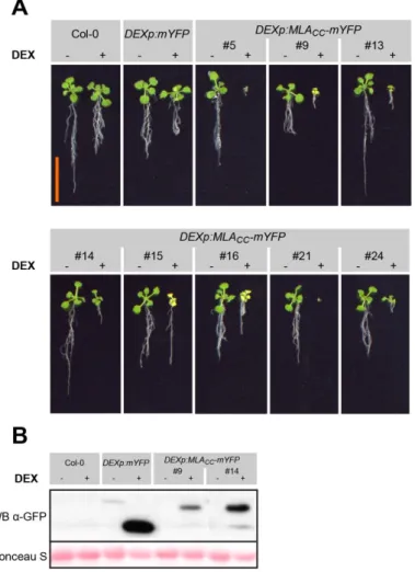
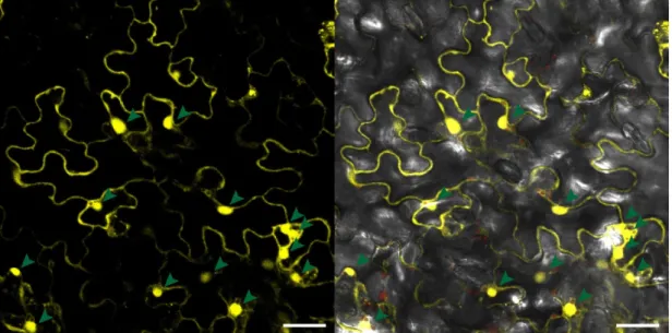
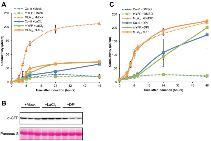
Figure 1-1: Plant growth phenotype and leaf chlorosis of DEXp:MLA CC -mYFP expressing transgenic lines. ... 24 Figure 1-2: Timing of MLA CC -dependent activation of cell death and MAPKs in leaves. .... 27 Figure 1-3: Subcellular localization of MLA CC -mYFP fusion protein in A. thaliana. ... 28 Figure 1-4: Time series of confocal images of epidermal cells expressing MLA CC -mYFP. .. 30 Figure 1-5: Effect of the calcium channel blocker LaCl 3 and the NADPH oxidase inhibitor

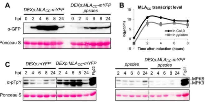
DPI on MLA CC -mediated cell death. ... 32 Figure 1-6: MLA CC -mediated MAPK activation is abolished in the ppsdes mutant

background. ... 35 Figure 1-7: MLA CC rapidly induces a similar transcriptional reprogramming in wild type A.

thaliana (Col-0) and ppsdes mutant background. ... 39 Figure 1-8: GO term enrichment analysis of MLA CC -upregulated genes at 2 hours after

induction. ... 42 Figure 1-9: Transcriptome-wide correlation analysis among MLA CC -, ETI- and PTI- associated gene expression patterns. ... 45 Figure 1-10: Expression profile of the genes rapidly induced upon MLA CC expression in the

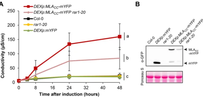
early response to diverse biotic, abiotic, hormone and chemical treatments. ... 50 Figure 1-11: Analysis of CAMTA binding motif frequency and distribution in 5’ cis- regulatory sequences. ... 54 Figure 1-12: RAR1 is dispensable for MLA CC function. ... 56 Figure 1-13: MLA CC -mediated growth phenotype is only partially temperature sensitive. ... 58 Figure 2-1: DEX-inducible expression of MLA CC -mYFP leads to growth arrest. ... 62 Figure 2-2: Flowchart of the genetic suppressor screen for MLA CC -dependent growth arrest.

... 63 Figure 2-3: Nomenclature used in this chapter to identify each analysed generation and

lineage. ... 65 Figure 2-4: Examples of M 2 candidates displaying enhanced symptoms after inoculation with

Pst DC3000 AvrRpm1. ... 67 Figure 2-5: Position of the confirmed and putative loss of function mutations identified in

RPM1. ... 68

Figure 2-6: Number of homozygous SNPs identified in each of the 22 M 2 plants. ... 74

Figure 2-7: Overall SNP analysis before and after curation for background SNPs. ... 75

(24)

Figure 2-9: Allele frequencies identified by sequencing of segregant bulks for the candidate mutants 1A, 2B and 2H. ... 83 Figure 2-10: Candidate SNPs identified on chromosome 1 by analysis of positive and

negative segregant bulks of the mutant candidates 2B and 2H. ... 83 Figure 2-11: Comparison of AT3G02840 and AtCMPG1 protein structure and domain

composition. ... 85 Figure 2-12: AT3G02840 sequence analysis. ... 86 Figure 2-13: Analysis of AT3G02840 expression in A. thaliana upon MLA CC expression or

MLA1 activation. ... 88 Figure 2-14: Effect of AT3G02840 deficiency on HR cell death triggered by RPM1. ... 90

Figure 3-1: Overview of the TAP strategy and the construct used. ... 99 Figure 3-2: Bait constructs expressed in the A. thaliana transgenic lines for the TAP analysis.

... 100 Figure 3-3: Characterization of the bait-PC2 constructs used for TAP. ... 102 Figure 3-4: Abundance-based ranking of the proteins identified in the different pull downs.

... 104 Figure 3-5: Immunoblot analyses of tandem affinity-purified plant extracts expressing

MLA CC -PC2, mYFP-PC2 and MLA CC L36E-PC2. ... 106 Figure 3-6: Volcano plot of the protein groups identified in the TAP input samples of the

MLA CC line and the mYFP line. ... 108 Figure 3-7: Overview of the protein groups identified by MS analysis in the TAP samples.

... 110 Figure 3-8: Volcano plot of the protein groups identified by MS analysis in MLA CC -PC2 pull

down versus the control pull downs. ... 112 Figure 3-9: Abundance-based ranking of the protein groups confirms the enrichment of

several candidate interactors in the MLA CC pull down compared to the control pull downs. ... 114 Figure 3-10: Expression profile of the MLA CC candidate interactors in A. thaliana upon

MLA CC expression. ... 116

Figure 3-11: Effects of PLA2A inhibitors on MLA CC -mediated cell death. ... 117

(25)

Index of supplementary data

Index of supplementary figures

Figure S1-1: Genomic location of the DEXp:MLA CC -mYFP transgene in line #5.1 as defined by sequencing of the flanking regions. ... 193 Figure S1-2: Ion leakage measure in the DEXp:MLA CC -mYFP line upon DEX treatment... 193 Figure S1-3: Cell death induction by MLA CC and MLA MHD in A. thaliana. ... 195 Figure S1-4: Analysis of the top 3,153 differentially expressed genes upon MLA CC

expression in wild-type or ppsdes mutant background. ... 197 Figure S1-5: Expression profile of EDS1, SA and, ET/JA marker genes upon MLA CC

expression in wild-type and ppsdes mutant. ... 198 Figure S1-6: GO term enrichment analysis in genes differentially expressed upon MLA CC

expression in wild type A. thaliana (Col-0) and ppsdes mutant background. ... 199 Figure S1-7: Expression profile of various gene clusters after inoculation with different

pathogens and MLA CC inducible expression. ... 200 Figure S1-8: Extended expression profile of the genes rapidly induced upon MLA CC

expression in the early response to diverse biotic, abiotic, hormone and chemical treatments. ... 201 Figure S1-9: Effect of sucrose concentration and day length on the MLA CC -mediated growth

phenotype. ... 202 Figure S2-1: Pictures of four representative screening plates. ... 203 Figure S2-2: Pictures of the pilot screen conducted on soil. ... 204 Figure S2-3: Distribution of the suppressor candidate scores. ... 204 Figure S2-4: Effect of DZNep and Zeb on transgene silencing. ... 205 Figure S2-5: Segregation analysis of the F 2 .BC T progeny. ... 206 Figure S3-1: Plasmid maps of the estradiol inducible vectors used in this study. ... 207 Figure S3-2: Optimization of the TAP extraction buffer composition. ... 208 Figure S3-3: Clustering analysis of the TAP input and eluate samples. ... 210 Figure S3-4: Multi scatterplot showing the correlation in protein group abundance (LFQ

intensities) between all samples of the TAP input fractions. ... 211

(26)

Index of supplementary tables

Table S1-1: Description of the transcriptomic datasets used for comparative transcriptomic analysis together with the MLA CC -related transcriptomic profiling. ... 213 Table S2-1: List of loci selected for the targeted analysis of the 21 M 2 candidate mutants

sequenced ... 214 Table S2-2: SNPs isolated by the targeted (A) and the untargeted (B) analysis in the 21 M 2

suppressor mutants sequenced ... 215 Table S2-3: Mutations identified in the candidate mapping intervals for the candidate mutants

1A, 2B and 2H ... 216 Table S3-1: Top 16 differentially expressed protein groups (DEPGs) in lines expressing

MLA CC -PC2 compared to lines expressing mYFP-PC2. ... 217 Table S3-2: Comparison of proteomic and transcriptomic data. ... 217 Table S3-3: Protein abundance of the 9 MLA CC candidate interactors. ... 218 Table S3-4: Co-expression degree between the candidates. ... 218 Table S3-5: Ortholog search for the nine MLA CC candidate interactors in Phytozome v10.3.

... 218

(27)

Abbreviations

- translational fusion (in the context of protein fusion constructs)

∅ diameter

°C degree Celsius

A.A amino acid

ABA abscissic acid

AIST National Institute of Advanced Industrial Science and Technology

ARA aristolochic acid

ARM armadillo

Avr avirulence

BEL bromoenol lactone

BLAST basic local alignment search tool

bp base pair

C-terminal carboxy-terminal

CaMV cauliflower mosaic virus

CC coiled-coil

CHAPS 3-[(3-Cholamidopropyl)dimethylammonio]-1-propanesulfonate

CMPG protein family Cys, Met, Pro and Gly

CNL CC-NBS-LRR

cpm count per million

DAMP damage-associated molecular pattern

DDM β-D-Maltopyranoside

DEG differentially expressed gene(s)

DEPG differentially expressed protein group(s)

DEX dexamethasone

DEXp dexamethasone-inducible promoter

DMSO dimethylsulfoxid

DNA deoxyribonucleic acid

dpg day(s) post germination

DPI diphenylene iodonium

DZNep 3-Deazaneplanocin A

EDS1 Enhanced disease resistance 1

EMS ethyl methanesulfonate

ER estradiol

ET ethylene

FC fold change

FDR false discovery rate

Fig. figure

FL full length

FW fresh weight

g gram(s)

gDNA genomic DNA

(28)

Het heterozygous

Hom homozygous

hpi hour(s) post inoculation/induction/infiltration

HR hypersensitive response

HRP horseradish peroxidase

iBAQ intensity-based absolute quantification

JA jasmonic acid

KO knock out

L litre

LaCl3 lanthanum(III) chloride

LC liquid chromatography

LFQ label free quantification

log logarithm

log 2 logarithm base two

LRR leucine-rich repeats

m milli

M molar (mol/L)

MAMP microbe-associated molecular pattern (=PAMP)

MAPK mitogen-activated protein kinase(s)

max. maximum

min minute(s)

min. minimum

MLA CC MLA coiled-coil domain

MPIPZ Max Planck Institute for Plant Breeding Research

mRNA messenger ribonucleic acid

MS Murashige and Skoog medium, or mass spectrometry

MW molecular weight

mYFP monomeric yellow fluorescent protein

NB-ARC nucleotide-binding adaptor shared by APAF-1, R proteins, and CED-4

NBS nucleotide binding-site

NDR1 Non race-specific disease resistance 1

NLR nucleotide-binding domain and leucine rich repeats-containg protein(s)

N-terminal amino-terminal

ng nanogram(s)

NP-40 nonidet P-40

(29)

R-gene resistance gene

RNA ribonucleic acid

RNAseq RNA sequencing

rpm round per minute

RPM resistance to Pseudomonas syringae pv. maculicola

RPS resistant to Pseudomonas syringae

RT room temperature

s second(s)

SA salicylic acid

SDS sodium dodecyl sulphate

SNP single nucleotide polymorphism

Stdev standard deviation

T-DNA transfer DNA

TAP tandem affinity purification

TBS tris buffered saline

TF transcription factor

TIR Toll/interleukine-1 receptor homology domain

TNL TIR-NBS-LRR

TR transcription regulator

Tris Tris-(hydroxymethyl)-aminomethane

TUM Technische Universität München

U unit(s)

UCSD University California, San Diego

URGV/IPS2 Unité de Recherche en Génomique Végétale/Institute of Plant Science - Paris Saclay

UTR untranslated region

V Volt(s)

v/v volume per volume

w/v weight per volume

WB western blot (immunoblot

WT wild-type

Y2H yeast two hybrid

Zeb zebularine

(30)

1. The plant immune system

Introduction

1. The plant immune system

1.1. An overview of the multi-layered plant innate immune system

Contrary to vertebrates, plants possess neither an adaptive immune system nor specialized immune cells. Therefore each plant cell relies entirely on its own innate immunity to defend itself against pathogens (Maekawa et al., 2011a). To achieve a specific and localized immune response, plants have evolved several lines of defence against pathogens. Plasma membrane- localized pattern-recognition receptors detect in the extracellular space the presence of conserved pathogen-derived epitopes, such as bacterial flagellin and fungal chitin, and stimulate an immune response which limits pathogen proliferation (Boller and Felix, 2009).

However, host-adapted pathogens can suppress this immune response by delivering highly polymorphic effector arsenals inside host cells (Jones and Dangl, 2006; Rafiqi et al., 2012).

As a counter mechanism, plants deploy resistance (R) proteins to detect the presence of the effectors, which in turn trigger a potent immune response that terminates pathogen growth (Chisholm et al., 2006; Jones and Dangl, 2006). The former type of immune response is called

“pattern-triggered immunity” (PTI), whereas the latter is called “effector-triggered immunity”

(ETI). ETI is often associated with a local and rapid host cell death at sites of attempted pathogen ingress, designated the hypersensitive response (HR) (Maekawa et al., 2011a).

1.2. Diversity of pathogens and diversity of pathogen detection mechanisms

(31)

pathogens, denoted as biotrophs, can engage in a feeding process only with living plant cells.

This strategy implies the capacity to inhibit host defence and host death. In the more specific case of obligate biotrophy, the pathogen can grow and complete its reproductive cycle only on its living host. Thus biotrophs, and to an even higher degree obligate biotrophs, are tightly co- evolving with their hosts and usually display a narrow host range. In contrast, necrotrophic pathogens kill host cells in order to feed on dead plant tissues (e.g. the fungus Fusarium oxysporum, causal agent of vascular wilt disease in many species such as banana tree, cotton, melon, and tomato). Other pathogens have adopted an intermediate strategy referred to as hemibiotrophy which starts with a biotrophic phase followed by a shift to a necrotrophic phase. From the plant side, such pathogen diversity implies the activation of specific defence responses.

Upon pathogen challenge, plants recognize pathogens via pattern recognition receptors

(PRRs). Plant PRRs are surfaced-localized receptor kinases or receptor-like proteins which

recognize a wide range of microbe- or plant-derived molecules (Zipfel, 2014). Since the

molecules recognized by PRRs are not specific to phytopathogens, PRR-triggered immunity

(PTI) is activated by, and efficiently limits the growth of various pathogenic and non-

pathogenic organisms in contact with the plant. PRRs can be either broadly conserved across

flowering plants, such as FLS2 (FLAGELLIN-SENSITIVE-2), or present only in restricted

taxa, such as EFR (EF-TU RECEPTOR) in Brassicacea. FLS2 recognizes an epitope

consisting of 22 amino acids of the bacterial flagellin (flg22). Among the other recognized

bacterial-derived molecules are the elongation factor Tu (EF-Tu, recognized by EFR), HrpZ,

peptidoglycans and lipopolysaccharides (LPS). On the fungus and oomycete side, chitin and

NPP1 (NECROSIS-INDUCING PHYTOPHTORA PROTEIN-1) induce PTI. Mechanisms

for recognition of viruses and insects have been so far less documented. In addition to non-

self-sensing, PRRs also detect self-molecules, so-called DAMPs (damage-/danger- associated

molecular patterns) which are released upon cell damage or pathogen detection. Three

DAMPs have been documented so far: the Pep peptide family, oligogalacturonides, and

extracellular ATP. The green leaf volatile E-2-hexenal is produced upon wounding, herbivory

and pathogen infection, and act as a potent inducer of general defence response (Mirabella et

al., 2015). Thus, hexenal is also a potential DAMP. DAMP perception can activate defence

responses similar to PTI and contributes to amplification of the PTI in a positive feedback

loop (Zipfel, 2014).

(32)

1. The plant immune system

A key for successful pathogen invasion is the release of effector proteins which can sabotage

the PTI and manipulate the host cell to promote efficient colonization. Therefore, plants have

evolved a highly specific pathogen recognition mechanism mediated by so-called resistance

(R) proteins which can sense the presence of pathogen effectors. The recognition results in a

potent immune response designated effector triggered immunity (ETI). The corollary of this

system is that pathogen effectors are under selection pressure to escape recognition whereas

selection pressure promotes allelic diversification of R proteins to expand the recognition

repertoire and keep up with effector evolution. This layer of the plant immune system has

been at the beginning genetically described as a “gene for gene” interaction, since ETI is

controlled by a pair of matching genes: the plant R gene on one side, and its cognate pathogen

avirulence (Avr) gene on the other side (Flor, 1955). This model has set the basis for the

definition of virulence/avirulence and compatible/incompatible interaction: a virulent

pathogen is a pathogen whose effectors are not recognized by any of the R proteins of the host

plant and thus does not trigger the ETI (compatible interaction). By opposition, an avirulent

pathogen has at least one effector recognized by at least one R protein of the host plant and

does trigger the ETI (incompatible interaction). The highly pathogen-specific property of ETI

makes it resemble to some extent the acquired immunity in mammals. The mechanisms of

effector recognition by plant R proteins has been well established. Plant R proteins utilize two

major modes of effector recognition: a direct and an indirect recognition mode (Chisholm et

al., 2006; DeYoung and Innes, 2006; van der Hoorn and Kamoun, 2008; Jones and Dangl,

2006). In case of a direct recognition, an effector is detected by direct physical interaction

with its cognate R protein, whereas during the indirect recognition, an R protein senses

modifications of host proteins caused by the cognate effector action. Experimental evidence

supports that the indirect recognition enables a single R protein to recognize multiple effectors

irrespective of effector structures when effectors target the same host protein (Chisholm et al.,

2006; Jones and Dangl, 2006). However, detection of multiple effectors by a single R protein

(33)

1.3. Innate immune signalling

1.3.1 The early events downstream of pathogen recognition

Although the knowledge in this field remains largely incomplete, qualitatively similar signalling mechanisms seem to occur upon ETI and PTI activation. It has been proposed that quantitative and temporal differences mainly account for the distinct outputs of ETI and PTI, and that different types of R proteins also trigger similar responses (Jones and Dangl, 2006;

Maleck et al., 2000; Navarro et al., 2004; Tao et al., 2003; Tsuda and Katagiri, 2010).

Pathogen perception induces a rapid and transient increase of calcium (Ca 2+ ) concentration in the cytosol or other subcellular compartments. This elevation of Ca 2+ concentration represents one of the earliest common signalling events downstream of all modes of pathogen detection.

Ca 2+ signals are decoded by calcium sensors (calmodulin [CaM], calmodulin-like proteins, calcium dependent protein kinases [CDPK], and calcineurin B-like proteins [CBL]), and regulate several pathways involved in immune signalling (Poovaiah et al., 2013). CDPKs are important players in immunity (Boudsocq and Sheen, 2013). For example, CDPKs can directly regulate the NADPH oxidase RBOHB (RESPIRATORY BURST OXIDASE HOMOLOG B) leading to the production of reactive oxygen species which act as both antimicrobial compounds and signalling molecules further contributing in the immune signalling (Baxter et al., 2013). Pathogen recognition also activates a signalling cascade mediated by mitogen-activated protein kinases (MAPKs). In A. thaliana, functionally redundant AtMPK3 and AtMPK6 are activated upon several stresses including pathogen detection by a partially unknown mechanism. The immune signalling also involves a complex interplay between several plant hormones. Salicylic acid (SA), jasmonic acid (JA), and ethylene (ET) are considered as the three major defence phytohormones but abscisic acid (ABA), gibberellins (GA), auxin, cytokinins, brassinosteroids and nitric oxide can also modulate the immune signalling network (Pieterse et al., 2012). In a simple model, SA and ET/JA act antagonistically to positively regulate plant defence against biotrophic pathogens and against necrotrophic pathogens and herbivorous insects, respectively (Bari and Jones, 2009; Glazebrook, 2005). However, accumulating evidence suggests a more complex hormone cross-talk whose molecular bases are being gradually resolved (Pieterse et al., 2012;

Tsuda and Somssich, 2015).

Two interesting facts have emerged from the study of the immune signalling network. First,

(34)

1. The plant immune system

signalling pathways converge on transcription factor regulation. It has been proposed that transcription factors (TFs) act as convergence points which integrate the signals from a complex signalling network, and that integration of the multiple inputs defines the response specificity at the transcriptional level. Transcriptional reprogramming, which is crucial for plant immunity, thus acts as a major effector arm in immunity. The mechanisms involved in the immune transcriptional reprogramming are detailed in the next section.

1.3.2 Transcriptional reprogramming in immunity

A rapid and massive transcriptional reprogramming occurs upon pathogen detection (Boudsocq et al., 2010; Eulgem et al., 2004; Gao et al., 2011; Maekawa et al., 2012; Maleck et al., 2000; Navarro et al., 2004; Ramonell et al., 2005; Tao et al., 2003). Transcriptional reprogramming is an essential process in plant immunity. This is supported by the facts that several TFs are required for resistance (Ando et al., 2014; Padmanabhan et al., 2013; Shimono et al., 2007; Xu et al., 2014; Zheng et al., 2006), a large part of the induced genes are involved in critical defence processes, and transcription factors are frequently targeted by pathogen effectors (Kazan and Lyons, 2014). For example, in A. thaliana, synthesis of camalexin, an antimicrobial compound critical for resistance to the hemibiotroph Botrytis cinerea, is induced via WRKY33-dependent activation of the camalexin biosynthesis genes, and the TF WRKY33 is a target of pathogen effectors (Mao et al., 2011; Sarris et al., 2015). The main TF families involved in immunity are AP2/ERF, bHLH, bZIP, MYC, NAC, WRKY and CAMTA (Tsuda and Somssich, 2015).

How are then the different immune signalling pathways integrated into a specific immune response at the transcriptional level?

MAPK cascades regulate directly or indirectly several TFs by phosphorylation. Direct

(35)

directly regulated by CaM proteins (Poovaiah et al., 2013; Tsuda and Somssich, 2015). For example the CAMTA (CALMODULIN-BINDING TRANSCRIPTION ACTIVATOR) family of TF is regulated by Ca 2+ via interaction to CaM, and CAMTA3 is involved in the transcriptional regulation of EDS1 (ENHANCED DISEASE SUSCEPTIBILITY-1) and NDR1 (NON DISEASE RACE SPECIFIC DISEASE RESISTANCE 1), two major regulators of plant immunity (Du et al., 2009; Nie et al., 2012). Similarly, CDPKs can regulate defence- related TFs as exemplified by the phosphorylation of AtWRKY8, 28 and 48 by AtCDPK4,5,6 and 11 (Gao et al., 2013).

Plant hormones also mediate defence-related transcriptional reprogramming. It has been proposed that AtNPR1 (NON-EXPRESSOR OF PATHOGENESIS-RELATED GENES1), which can indirectly sense SA accumulation, interacts with TFs of the TGA family to drive expression of most of the SA-responsive genes (Tsuda and Somssich, 2015). Similarly, the JA-dependent regulation of the TF AtMYC2 and its role in defence have been well described (Chini et al., 2007; Thines et al., 2007) as well as the ET-mediated regulation of AtEIN3 (ETHYLENE INSENSITIVE3) and AtEIL1 (EIN3-LIKE1) which control expression of some defence genes such PDF1.2 (Tsuda and Somssich, 2015).

Interestingly, TFs can also be the site of convergence for signals coming from different pathways. For instance, the activity of AtEIN3 and AtEIL1 is synergistically regulated by both JA and ET whereas AtMYC2 and AtEIN3 antagonize each other. Several other convergence points important for immunity have been described (Tsuda and Somssich, 2015).

Recent reports indicate direct interactions between R proteins and transcription factors. The

barley NLR MLA10 interacts with three transcription factors upon activation (HvWRKY1,

HvWRKY2, HvMYB6), and the interaction releases HvMYB6 from the HvWRKY1-mediated

repression (Chang et al., 2013; Shen et al., 2007). Pb1, a rice R protein, has been shown to

interact with the transcription factor OsWRKY45. However, in contrast to the MLA-

HvMYB6 interaction, the transcriptional activity is regulated via OsWRKY45 abundance,

since Pb1 protects OsWRKY45 from degradation upon pathogen attack (Inoue et al., 2013). A

third example aiding in our understanding of nuclear activity of R proteins is the interaction of

N with the transcription factor SPL6 (SQUAMOSA PROMOTER BINDING PROTEIN-

LIKE 6) in Nicotiana benthamiana (Padmanabhan et al., 2013). The association of N and

SPL6 at subnuclear bodies occurs only in the presence of the cognate effector. A genetic

(36)

1. The plant immune system

responsive genes are differentially regulated upon AtSPL6 silencing (Padmanabhan et al., 2013). The bHLH TF AtbHLH84 and its homologs interact with two R proteins, RPS4 and SNC1, and all three homologues act redundantly homologues in the ETI mediated by the R protein RPS4 and in the constitutive ETI-like response mediated by snc1 (Xu et al., 2014).

The R protein RRS1 possesses a WRKY domain which can bind DNA in vitro (Noutoshi et al., 2005) and RRS1 might directly contribute to transcriptional regulation (Heidrich et al., 2013). Close re-examination of yeast-two-hybrid data generated by Mukhtar et al., 2011, indicate at least 14 other potential R protein-TF in A. thaliana, further supporting that R protein-TF interactions constitute a more common mechanism of R protein actions.

Interaction between transcriptional regulators and NLRs has also been demonstrated, such as the interaction between the transcriptional co-repressor AtTPR1 (Topless-related 1) and the A.

thaliana R protein SNC1 (Zhu et al., 2010). Taken together, these studies draw an emerging picture in which nuclear localized R proteins mediate transcriptional reprograming via interaction with transcription factors in various plants species. Interaction with transcriptional regulators appears not to be limited to just a few specialized R proteins. Instead, this type of interactions might be a more common phenomenon, implying a possible general mechanism of direct regulation of transcriptional reprogramming via plant R proteins. Nonetheless, among the TFs found to interact with R proteins, none of them seems to be commonly targeted by multiple R proteins. Therefore the existence of TFs which act as signalling targets for multiple R proteins is still debated.

Overall, TFs appear to integrate several immune signals and the network of recruited TFs likely defines and fine tunes the response output. However the current knowledge still needs to be integrated into a finer spatio-temporal model to accurately determine the specific pattern of immune transcriptional regulation at different time and in local or distant cells.

Furthermore, the predictable common immune mechanisms downstream of ETI and PTI

(37)

1.4. Hypersensitive response and other defence outputs

Interestingly, both ETI and PTI converge on qualitatively similar defence outputs.

Quantitative differences seem to account for most differences between PTI and ETI. ETI is indeed considered as an accelerated and amplified PTI (Jones and Dangl, 2006). ETI and PTI result in a wide range of defence outputs: cell wall thickening, lignification, callose deposition, alkalinisation of the extracellular space, production of antimicrobial or repellent compounds, production of pathogen-degrading enzymes and reactive oxygen intermediates, activation of systemic acquired resistance, and stomatal closure. Defence against viruses is mainly mediated by activation of RNA silencing which is targeted against the viral RNAs, and suppression of the virus replication. These responses can occur not only locally but also in distant cells since some immune signals are spread via hormones or other mobile signalling mechanisms, and induce a fitness cost for the plant (Walters and Heil, 2007). Typically, ETI also results in the induction of a hypersensitive response (HR), which is a specific type of cell death occurring at the site of attempted infection. The HR is usually used as readout for the ETI activation. However the HR does not occur in all incompatible interactions and can be uncoupled from disease resistance, indicating that the HR is not always required to confer disease resistance to certain pathogens and that other defence outputs can be more efficient in conferring disease resistance (Bai et al., 2012; Bendahmane et al., 1999; Chang et al., 2013;

Coll et al., 2010; Yu et al., 1998, 2000). Whilst the HR can in some cases contribute to restricting the growth of biotrophs, it can on the contrary promote infection by necrotrophs.

Necrotrophic pathogens secrete toxins, which function as effectors to promote host cell death

response. These toxins are often host-plant species-specific, thus called host-selective toxins

and mediate effector-triggered susceptibility (ETS), which mirrors ETI to some extent (Laluk

and Mengiste, 2010). It has been implicated that susceptibility to necrotrophic pathogens or

sensitivity to their host-selective toxins is associated with R loci in diverse plant species such

as A. thaliana (Lorang et al., 2007), sorghum (Nagy and Bennetzen, 2008), and wheat (Faris

et al., 2010). This indicates that some necrotrophic pathogens highjack the ETI to trigger an

HR, thereby promoting virulence. Therefore, resistance to host specific necrotrophs is mainly

mediated by PTI, detoxification of toxins, loss of toxin recognitions, or restricting toxin-

mediated cell death response (Mengiste, 2012). Plant R proteins seem to play minor roles in

disease resistance to necrotrophic pathogens. However the A. thaliana RLM3 R locus confers

resistance to a broad range of necrotrophs by unknown mechanisms (Staal et al., 2008).

(38)

2. Effector-triggered immunity and plant NLRs

The HR-like response triggered by the pathogen-derived protein harpin requires an active

metabolism and de novo protein synthesis indicating that the HR is likely transcriptionally

controlled (He et al., 1994). It is still debated whether HR cell death results from the

activation of a cell death-specific program or simply from exaggerated defence responses,

especially since the HR can be uncoupled from disease resistance (see above). The current

understanding of the mechanisms involved in the HR is not complete. Divergent results

suggest that several distinct cell death mechanisms are involved and that different R proteins

trigger an HR via distinct processes. Cell death processes similar to the well-studied

mammalian apoptosis have not been observed in plant cells. According to a morphological

classification, there are at least two types of plant cell death: vacuolar cell death involving

autophagic-like processes, and necrosis (van Doorn et al., 2011). Vacuolar and autophagic

processes as well as regulation by the defence phytohormone salicylic acid have been

associated to the HR cell death (Coll et al., 2011; Hatsugai et al., 2004, 2015; Hofius et al.,

2009; Munch et al., 2015; Yoshimoto et al., 2009). However the HR fails to be assigned to

any type of plant cell death since it displays mixed features of both vacuolar and necrotic cell

death (van Doorn et al., 2011). A conductivity increase in the extracellular fluids can be

measured rapidly after HR induction. Such phenomenon can be explained by several

processes including a massive cation efflux and the fusion of the plasma membrane with the

vacuolar membrane leading to the release of the vacuolar content into the extracellular space

(Atkinson et al., 1985; Hatsugai et al., 2009). The release of intracellular molecules and the

conductivity increase occur before the apparition of obvious cell death symptoms such as cell

shrinkage, vacuolization and cytoplasmic aggregation (Hatsugai et al., 2009). Therefore

conductivity measure of the extracellular fluid is a marker routinely used to follow ETI and

HR development.

(39)

NLRs are modular proteins that typically consist of three building blocks: an N-terminal domain, the central NB-ARC domain (named after nucleotide-binding adaptor shared with APAF-1, plant resistance proteins, and CED-4), and a C-terminal LRR (leucine-rich repeats) domain (Van der Biezen and Jones, 1998). The central domain of animal NLRs is also known as the NACHT domain (named after NAIP, CIITA, HET-E, and TP1) (Koonin and Aravind, 2000) which is structurally similar to the plant NB-ARC domain but distinctive of animal NLRs (van Ooijen et al., 2007; Takken et al., 2006). Despite their structural and functional similarities, plant and animal NLRs are likely the result of convergent evolution (Ausubel, 2005; Rairdan and Moffett, 2007; Staal and Dixelius, 2007). The presence of either a TOLL/interleukin 1 receptor (TIR) domain or a coiled-coil (CC) domain at the N-terminus is a plant-NLR-specific feature and defines two major types of plant NLRs termed the TIR-type NLRs (TNLs) and the CC-type NLRs (CNLs), respectively. Although the origins of TNLs and CNLs seem to date back to very early land plant lineages, TNLs are absent from monocots (Liu and Ekramoddoullah, 2007; Yue et al., 2012). The NLR family has massively expanded in several plant species. The massive expansions have rendered the NLR family one of the largest and most variable plant protein families (Clark et al., 2007; Ossowski et al., 2008). Expansion of the recognition repertoire and rapid evolution of the pathogens are likely the main forces which drive the NLR expansion and diversification in plants. This contrasts with the vertebrate NLR repertoires, typically comprising ca. 20 members (Lange et al., 2011;

Meyers et al., 2003; Zhang et al., 2010; Zhou et al., 2004). Rapid evolution of the NLR repertoire by a rapid “birth and death” mechanism accounts for highly species-specific NLR repertoires in plants expect for a few NLR families which follow different evolutionary dynamics and are conserved across several plant taxa (Jacob et al., 2013).

2.2. Function of the different NLR modules

NLRs are modular proteins consisting mainly of three modules: the N-terminal domain, the

central NB-ARC domain and the C-terminal LRR. The “Rosetta Stone Hypothesis” proposes

that when two proteins that are separate in some species are fused in another species, their

fusion likely reflects a previously hidden interaction between the two seemingly non-related

proteins (Marcotte et al., 1999). The Rosetta Stone Hypothesis might explain the plant NLRs

evolutionary history. Indeed, co-expression of individual NLR domains (i.e., N-terminal, NB-

(40)

2. Effector-triggered immunity and plant NLRs

originally separated and have been assembled into a single multi-domain receptor during evolution. Thus, each domain represents a functional unit with assembly-dependent and assembly independent functions. The experimental data largely support such model.

The LRR domain is the most polymorphic domain and the main determinant for effector recognition or detection of effector-induced host modifications. However the other NLR domains also function in effector recognition in some cases (Qi and Innes, 2013). Recognition of the effector or modified self induces a conformational change which represents the first step in the switch process from the “off” to the signalling competent “on” state of the NLR receptor.

The central NB-ARC domain has an intramolecular regulatory function. Following detection of the effector or modified self by the LRR, an ADP to ATP exchange takes place which trigger further conformational changes leading to the signalling competent conformation. ATP binding depends on a functional P-loop which is a conserved motif within the NB-ARC domain. Therefore mutations in the P-loop motif usually lead to NLR inactivation. On the contrary, specific mutations in the MHD motif of the NB-ARC domain mimic ATP binding and lead to effector-independent activation of the NLR (Qi and Innes, 2013). Although no crystal structure of a full-length plant NLR is available yet, the first crystal structure of an animal NLR monomer (mouse NLRC4) in its inactive state was resolved (Hu et al., 2013) and contributed to support the auto-inhibition mechanism mediated by both LRR and NB-ARC domain. Experimental data from plant NLRs further support the model which was inferred from the mouse NLRC4 structure (Ade et al., 2007; Bendahmane et al., 2002; Hwang et al., 2000; Qi et al., 2012; Slootweg et al., 2013). In the case of RPS5 and Rx, the NB-ARC domain has an additional function in downstream signalling (Ade et al., 2007; Rairdan et al., 2008).

The N-terminal domain, which typically consists of either a CC domain or a TIR domain, has

Abbildung

Figure 1-1: Plant growth phenotype and leaf chlorosis of DEXp:MLA CC -mYFP expressing transgenic lines
Figure  1-3:  Subcellular  localization  of  MLA CC -mYFP  fusion  protein  in  A.  thaliana
Figure  1-5:  Effect  of  the  calcium  channel  blocker  LaCl 3   and  the  NADPH  oxidase  inhibitor  DPI  on  MLA CC -mediated cell death
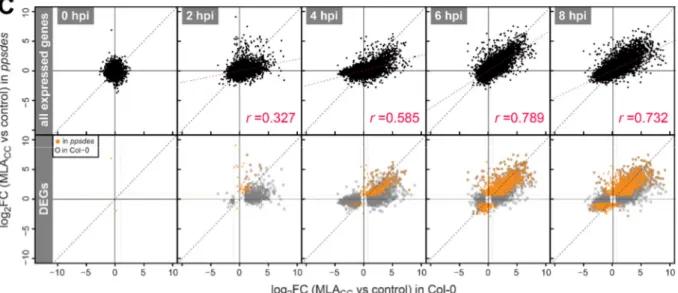
Figure 1-6: MLA CC -mediated MAPK activation is abolished in the ppsdes mutant background
+7

Referenzen

ÄHNLICHE DOKUMENTE

PD treated patients have shown decreased levels of naïve T cells, Tregs, dendritic cells, Th1, Th2, and Th17 cells, as well as increased counts of effector T, memory T, Th1,

In contrast to inflamma- tory cells, accelerated cell death is often observed in epithelial cells, a mechanism, which amplifies or at least maintains allergic inflammation..

Our observations strongly suggest that HAMLET stimu- lates a regression of epithelial structures and an apoptotic cell death of secretory epithelial cells in the lactating mammary

An increasing number of examples shows that bacterial pathogens utilize their secreted proteins to dampen host innate immune responses by interfering with two

Overexpression of Gli1 and Gli2 as the primary mediators of hedgehog signaling has no effect on the expression of the surface death receptor proteins Fas, TNFR

We first analyzed whether the expression of total H2AX was altered, and did indeed find that this molecule was reduced in CD8 + CD28 ) T cells when compared to CD8 + CD28 + T cells.

As mitochondria play a crucial role in the regulation and induction of apoptosis because of their ability to release several pro-apoptotic proteins, the Bcl-2 family of proteins,

Our findings strongly suggest that increased leakiness of the nuclear barrier affects nucleocytoplasmic transport, alters the localization of proteins across the