Cite this:Chem. Soc. Rev.,2014, 43, 3666
Optical methods for sensing and imaging oxygen:
materials, spectroscopies and applications†
Xu-dong Wang and Otto S. Wolfbeis*
We review the current state of optical methods for sensing oxygen. These have become powerful alternatives to electrochemical detection and in the process of replacing the Clark electrode in many fields. The article (with 694 references) is divided into main sections on direct spectroscopic sensing of oxygen, on absorptiometric and luminescent probes, on polymeric matrices and supports, on additives and related materials, on spectroscopic schemes for read-out and imaging, and on sensing formats (such as waveguide sensing, sensor arrays, multiple sensors and nanosensors). We finally discuss future trends and applications and summarize the properties of the most often used indicator probes and polymers. The ESI†(with 385 references) gives a selection of specific applications of such sensors in medicine, biology, marine and geosciences, intracellular sensing, aerodynamics, industry and biotechnology, among others.
1. Introduction
Almost all living organisms utilize oxygen‡for energy genera- tion and respiration. Discovered by Joseph Priestley in the 1770s, this event was elected as one of the top 100 greatest science achievements because of the significance of oxygen in
science and technology.1 Oxygen is involved in practically all forms of living organisms and played a major role in evolution.2 Adult humans metabolizeB200 g of oxygen per day. Human beings can live up to 1 month without food, up to 2 weeks without water, but not longer than maximally 10 min without supply of oxygen. It therefore does not come as a surprise that the (analytical) chemistry of oxygen plays a major role in biology and medicine. Sensing (i.e., continuous monitoring) of oxygen has attracted particular attention as can be seen from the numbers of respective references. A search for an ‘‘oxygen sensor’’ in SciFinder yields B8670 references, and B27 500 references are found that contain the concept ‘‘oxygen sensor’’ (search performed on August 1st, 2013).
Institute of Analytical Chemistry, Chemo- and Biosensors, University of Regensburg, D-93040 Regensburg, Germany. E-mail: otto.wolfbeis@ur.de;
Fax:+49 (0)941 943 4064; Tel:+49 (0)941 943 4065
†Electronic supplementary information (ESI) available. See DOI: 10.1039/
c4cs00039k
Xu-dong Wang (left) and Otto S. Wolfbeis (right)
Xu-dong Wang, born in 1985, received his PhD degree in Chemistry from Xiamen University (Xiamen, China) in 2011. He was an Alexander von Humboldt fellow in the group of Prof. Wolfbeis and is currently working at the Karlsruhe Institute of Technology. His research interests include the design of optical chemical and biosensors, nanosensors and molecular probes, developing novel (bio)sensing schemes, novel (nano)-materials and methods, and their applications to biological and chemical analysis. He has authored or coauthored more than 20 articles in the past five years, and his current h-index is 13 (as of Jan 2014).
Otto S. Wolfbeis was a Full Professor of Analytical and Interface Chemistry at the University of Regensburg from 1995 to 2012. He has authored numerous papers on optical (fiber) chemical sensors, fluorescent probes, labels and assays, on nanomaterials for use in sensing schemes and in spectroscopic methods including fluorescence (lifetime) imaging. He has acted as the (co)organizer of several conferences related to fluorescence spectroscopy (MAF) and to chemical sensors and biosensors (Europtrode). His current h-index is 75. He is one of the 10 curators of Angewandte Chemie and the editor of two other journals. Also see: www.wolfbeis.de.
Received 21st January 2014 DOI: 10.1039/c4cs00039k
www.rsc.org/csr
‡We use the common term ‘‘oxygen’’ for the more correct but less usual term
‘‘dioxygen’’.
REVIEW ARTICLE
Open Access Article. Published on 18 March 2014. Downloaded on 21/04/2015 12:40:30. This article is licensed under a Creative Commons Attribution 3.0 Unported Licence.
View Article Online
View Journal | View Issue
There are four major methods known for determination of oxygen. These are (a) the classical Winkler titration,3 (b) electroanalytical,4(c) pressure-based, and (d) optical methods.
The latter can be subdivided into numerous single methods that range from direct spectroscopy to indicator based methods.
Each method has its specific merits and applications.5–7 The Winkler method is precise but does not enable continuous sensing. The Clark electrode8 is the state of the art for the determination of oxygen at room temperature and in small volume. It works both in gaseous and fluid samples (whole blood included). Clark electrodes can be applied at temperatures up to B200 1C. They perform excellently but consume the analyte and are interfered by gases such as chlorine, ozone or nitrogen oxides. The quantitation of oxygen in (car) exhaust gases (which is challenging in view of the high temperatures of such samples) is performed using solid-state electrically conducting sensor materials to provide a feedback signal in catalytic converters. This so-called lambda probe (for a review, see ref. 9) was developed by the Bosch company during the late 1960s and is based on a zirconia ceramic coated on both the exhaust and reference sides with a thin layer of platinum to form a solid-state electrochemical fuel cell, where CO (if present) is oxidized by oxygen to form CO2. Both heated (43001C) and (less often) nonheated forms are known. The sensor does not actually measure oxygen concentration, but rather the difference between the amount of oxygen in the exhaust gas and the amount of oxygen in the supplied air.
Optical oxygen sensors have become attractive in the past four decades because of features such as (a) the lack of oxygen consumption during measurements; (b) full reversibility;
(c) good precision and accuracy; (d) the possibility of remote sensing using optical fibers; (e) the ease of miniaturization (down to the size of nanosensors); (f) the option of performing non-invasive measurements; and (g) the highly attractive feature of enabling imaging of oxygen both over large areas and on a micrometer scale. The success of optical sensors for oxygen is corroborated by the number of companies that are manufacturing respective instrumentation, examples being Presens
(probably the largest; www.presens.de); Centec (www.centec.de);
Ocean Optics, Inc. (www.oceanoptics.com); Oxysens, Inc.
(www.oxysense.com); Finesse, Inc. (www.finesse.com); PyroScience (www.pyro-science.com); and Hach-Lange (www.hach-lange.de), to mention the larger ones. In the medical field, OptiMedical Systems, Inc. (www.optimedical.com) and Terumo (www.terumo-cvs.com/
products/) probably are the largest.
There is some confusion in terms of definitions and terminology.
We refer to a sensing element as a material composed of both an oxygen-sensitive probe (OSP) and an appropriate (polymer) matrix that acts as a host or support (see Table 1). A complete sensor device will also incorporate a readout unit to give an electrical (or digital) signal. Ideally, the following (‘‘Cambridge’’) sensor definition applies: a sensor is a (small) instrumental system capable of (optically) detecting and quantifying a physical or chemical parameter over time and with high specificity (under the given circumstances). The vast majority of sensors for oxygen consists of an oxygen sensitive probe, a polymer or matrix for hosting the oxygen sensitive probe, a read-out (electronic) system, and a device to process data. A discussion on the definitions of sensors, probes and labels was presented.10
In addition to direct spectroscopic methods (see Section 3), oxygen can be sensedviaabsorptiometric probes,i.e.those that undergo a color change upon exposure to oxygen (see Section 4), but the vast majority of optical sensors for oxygen are based on luminescent probes (Section 6) that are contained in an oxygen-permeable polymer (Section 7.1). Fig. 1 shows a cross- section of a typical planar luminescent sensor for oxygen.
Table 1 Terms used in this review, and respective definitions. Several definitions are ambiguous
Term Definition
(Optical) probe, indicator, molecular probe
A molecule that displays an optical effect with a specific analyte such as oxygen (or pH, or a metal ion).
Whatever their name: they are not sensors. OSP is used here as an acronym for oxygen-sensitive probe.
Polymer, host, binder, matrix, paint
These terms relate to a polymer that hosts a probe (an indicator), for example an OSP. The choice of the polymer (and any additives) is a very critical step in sensor development.
Sensor chemistry,
‘‘stimulus-responsive polymer’’
A combination of materials (typically a polymer containing a probe and, possibly, additives) that enables continuous chemical sensing of oxygen; can be manufactured in various formats such as in the form of a sensor film, of sensor nanoparticles, or as a coating on (or at the distal end of) an optical waveguide.
Sensor cocktail, ink, paint Asolutionof a sensor ‘‘chemistry’’ in an appropriate solvent.
Sensor The definition of a sensor as a ‘‘miniaturized device that can deliver real-time and on-line information on specific parameters’’ was used for decades until organic chemists spoiled it by referring to molecules, probes, indicators and the like as ‘‘sensors’’. Sensors are devices according to this definition, and they are produced by the millions.
One may differentiate between physical sensors (such as those for temperature, pressure, acceleration) and chemical sensors (such as for pH, oxygen, methane, NOx, glucose). Sensors incorporate a readout system and are expected to provide a digital signal.
Fig. 1 Schematic cross-section of a typical planar luminescent sensor layer (not to scale).
Open Access Article. Published on 18 March 2014. Downloaded on 21/04/2015 12:40:30. This article is licensed under a Creative Commons Attribution 3.0 Unported Licence.
It consists of a solid but optically transparent support and a polymer matrix (referred to as the ‘‘sensing layer’’) permeable to oxygen that contains a quenchable probe. Optionally, a black cover (the so-called ‘‘optical isolation’’, see Section 8.4) is placed on top to prevent sample luminescence originating from blood or (plant) tissue, for example, from interfering. The sensor layer is illuminated on one side and luminescence is collected on the same side. The sample to be analyzed is in contact with the sensing element on the other side, in Fig. 1 placed on top.
Most oxygen-sensitive probes (OSPs) are used either in surface- absorbed or in polymer-dissolved form. In their pioneering work back in 1931, Kautsky and Hirsch11used probes such as trypaflavin adsorbed on a silica gel solid support. The use of solid (polymeric) supports not only separates the OSP from the sample, but also can increase the QY of an OSP (due to rigidization) and may also result in the formation of long- lived phosphorescent emissions resulting from long-lived triplet states.12–15This is quite beneficial because long decay times increase quenching efficiency.16,17The immobilization of OSPs in matrices offers additional advantages such as improved diffusion and permeability17 so that sensitivity and detection range can be adjusted. In addition, the sensing element can be used for a long time, and the polymer matrix can shield the OSPs from interferences by other quenchers. In fact, some polymers have excellent permeation selectivity. On the other hand, organic polymers and (metal) organic probes tend to decompose at temperatures o2001C. There is a vast variety of luminescent and other methods to readout the effects caused by oxygen (see Sections 4, 5 and 9).
Once an appropriate material has been identified for use in a sensing element, it can be applied in various formats as outlined in Section 11. The materials are often applied in the form of a (viscous) solution in a (usually organic) solvent, the so-called ‘‘sensor cocktail’’, which then may be deposited on a mechanical support such as a thin film of an inert and transparent polymer to form a sensor film (or sensor layer, or
‘‘paint’’). Following solvent evaporation, the resulting (solid) sensor element can then be investigated by various kinds of optical spectroscopies. The cocktail may also be deposited at the tip of an optical fiber, on another kind of waveguide, or even be incorporated into its core or cladding. This will result in so-called fiber optics or waveguide sensors. If placed on the clad of an optical fiber at intervals over a certain distance, so-called distributed sensors are obtained that can be investi- gated by time-resolved methods of spectroscopy. They allow for continuous and spatially resolved sensing of oxygen along an optical fiber. Sensor chemistries may also be shaped in the form of (nano)particles as will be outlined in Section 11.6. If the sensor material is deposited on the whole object or area of interest, 2-dimensional imaging of oxygen becomes feasible, for example in so-called pressure-sensitive paints that are used to measure the air pressure on cars and aircrafts, to sense oxygen in (cancerous) skin, to monitor photolytic or photosynthetic processes, or the consumption of oxygen in fuel cells.
In this review, we summarize the state of the art in optical sensing and imaging of oxygen. More specifically, we review
sensors based on the measurement of absorbance, reflectance and luminescence (including fluorescence, phosphorescence, bioluminescence and chemiluminescence). The spectral range spans the ultraviolet, visible, and near infrared regions. On the other hand, X-ray fluorescence, X-ray photoelectron spectroscopy, and electron paramagnetic resonance are not covered. Several reviews5–7,18–25and book chapters26–28related to aspects of optical oxygen sensing have appeared, but they usually cover a narrow field or a limited time frame. Some earlier but useful work has resided hidden for a long time but is cited here.
Papkovskyet al.29have summarized applications of optical oxygen sensors in biosciences, with a focus on enzymatic assays, respiration, food and microbial safety, bioreactors and fluidic chips. We summarize the subject as a whole and are presenting a comprehensive and hopefully clear blueprint for optical sensing of oxygen. It covers the state of the art from the first reported optical sensing scheme for oxygen11to the present.
Quarantaet al.30 have summarized the wealth of metal ligand complexes that is available for use in optical sensing of oxygen.
2. A look back
Quenching of luminescence by oxygen and other quenchers was long known but not understood until Stern and Volmer derived their famous equation.31Optical continuous sensing of oxygen probably started back in the 1930’s when Kautsky and Hirsch reported11,32 that the room-temperature phospho- rescence (RTP) of certain dyes adsorbed on silica particles is quenched by even trace quantities of oxygen. Sensing beads were prepared by soaking silica gel particles with a solution of dyes such as trypaflavine or fluorescein. They were then dried and placed in a flow-through cell schematically shown in Fig. 2.
RTP was monitored using a fluorometer and it was found that even ppm quantities of oxygen gas (equivalent to apO2 of as little as 0.5103Torr) were detectable. The effect was found to be fully reversible within 1–2 s. The device led to the discovery of the Kautsky effect,i.e.the delay in the production of oxygen following the illumination of a leaf. This is a good example on how chemical sensor technology can impact other kinds of science.
Fig. 2 Schematic of the first continuous sensor for oxygen. A sample gas containing oxygen (S) is passedviavalve H1over silica beads dyed with a quenchable probe for oxygen and placed in a small flow-through cell. The room temperature phosphorescence of the beads on the bottom is quenched by even traces of oxygen and was monitored with a simple fluorometer.
Open Access Article. Published on 18 March 2014. Downloaded on 21/04/2015 12:40:30. This article is licensed under a Creative Commons Attribution 3.0 Unported Licence.
This RTP method was later applied by others33,34to monitor oxygen in seawater. The first fluorescent sensor system was described by Bergman35 in 1968 and comprised a UV light source, an oxygen-sensitive fluorescent layer composed of porous glass (or a thin film of polyethylene) soaked with the oxygen-sensitive probe fluoranthene, and a photodetector.
Thus, it contained all the elements of a modern optical sensor.
Oxygen quenches the fluorescence of fluoranthene, and intensity served as the analytical information. The system responds to oxygen at levels above 1 Torr.
The milestone paper by Lakowicz and Weber36 in 1973 on the potential of probing structural fluctuations in proteins with oxygen-quenchable probes also revealed, for many, the potential of such probes for sensing oxygen in unknown (gaseous or fluid) samples. A device similar to the one of Bergman was described, a few years later, in a patent.37It also mentions the possibility of using a radioactive source (rather than a UV light source) to excite fluorescence. In 1974, Hesse38 described a device that appears to have been the first fiber optic chemical sensor. An oxygen-sensitive chemistry was placed in front of a fiber optic light guide through which exciting light was guided. The fluorescence emitted was guided back through either the same fiber, or through other bundle of fibers. The system is based on the measurement of either fluorescence intensity or fluorescence decay time, both of which are affected by oxygen. In 1975 and 1976, Lu¨bbers and Opitz39,40described instruments capable of monitoring oxygen or carbon dioxide. They first referred to it as an ‘‘optrode’’ (by lingual analogy to the electrode), and later as an ‘‘optode’’ (optiwosodos; Greek for ‘‘optical way’’). They were also the first to apply an oxygen sensor as a transducer in an enzyme-based biosensor.41
The fiber optic oxygen sensor described by Petersonet al.42 in 1984 was a milestone in optical fiber sensor technology for use in medicine. The sensor was used to monitor oxygen in the blood of an ewein vivo. A planar sensor for oxygen based on the covalent immobilization of the pyrene probe on a porous glass support and with milliseconds response time was reported in the same year.43 A chemiluminescence (CL) based sensor for oxygen was reported by the Seitz group44 that exploits the CL produced in the reaction of oxygen with an electron-rich ethylene. The AVL company has filed a patent45that describes oxygen sensitive materials based on polycyclic aromatic hydro- carbons dissolved in a film of poly(vinyl chloride) containing a plasticizer. Deposited on an acrylate glass and covered with microparticles of ferric oxide acting as optical isolations to prevent interferences caused by the intrinsic fluorescence of biological matter, they can be used to monitor oxygen in bioreactors, in blood samples and in breath gas.
The first dual sensor (for simultaneous determination of oxygen and the inhalation narcotic halothane) was reported46 in 1985. It is based on dynamic quenching of the fluorescence of the probe decacyclene in silicone rubber by both oxygen and halothane. Interferences by oxygen are taken into account by a second sensor layer covered with polytetrafluoroethylene (which is impermeable to halothane) and responds to oxygen only. Halothane concentrations can be calculated with the help
of an extended Stern–Volmer relation (Section 9.8.1). The probe is practically specific for the two analytes, since other gases present in inhalation gases or blood do not interfere.
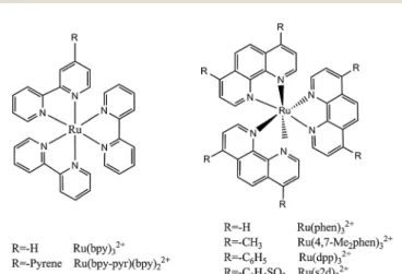
Quenchable ruthenium ligand complexes for use in optical oxygen sensing were reported47in 1986. Ruthenium tris(bipyridyl) was adsorbed onto silica gel and placed in a silicone membrane.
Ruthenium probes with better QY and known from the work of Alfordet al.48were applied as OSPs by Demas and coworkers49 in 1987. Such complexes have decay times of the order of a few microseconds. The first article50on luminescence decay time- based sensing of oxygen appeared in 1988. The Wilson group51 introduced a quenchometric scheme for oxygen based on the phosphorescence of metalloporphyrins in aqueous solution and bound to albumin, later to dendritic molecules. Both intensity and decay time were measured as a function of oxygen partial pressure. (Metallo)porphyrin-based sensor membranes were developed in the 1980s by the Gouterman group52that are suitable for phosphorescent sensing of oxygen, and a strong patent was published53 that covers pressure-sensitive paints based on the same effect. A 2-volume book that appeared in 1991 gives an account of the work on fiber optic chemical sensors and biosensors.27The first reliable fiber optic oxygen microsensors were introduced54 in 1995. A look back on the history of optical chemical sensor technology up to the year 2000 has been published.55
3. Methods for direct spectroscopic sensing of oxygen
Molecular oxygen has two main absorption bands in its UV-vis spectrum, one deep in the UV, the other at 760 nm which is very weak. It also has an intrinsic emission peaking at 1270 nm.
Geddeset al.56in 1992 monitored breath gas oxygenviaits UV absorption at 145 nm. Not unexpectedly, water vapor and carbon dioxide (and probably many other species) interfere.
The weak absorption lines at B760 nm can be exploited to sense oxygen in high concentration or if large penetration lengths can be accomplished such as in astronomy.57,58This direct spectroscopic method also enables monitoring of16O18O and16O17O isotopes, which provides valuable data with respect to the composition of the protoatmosphereviathe trapped gas in the polar ice. The oxygen line absorption was also used to monitor oxygen in harsh environments at high temperature, such as exhaust gas in combustion engines or burners.59 Respective instrumentation requires high-power lasers, long optical pathways (B100 m), and highly sensitive detectors.
Oxygen was also sensed directly by employing gas correla- tion absorption spectroscopy using multimode diode lasers.60 A diode laser was applied that has an emission spectrum that overlaps the oxygen absorption lines of the absorption band at 760 nm. A detection limit of 700 ppm was achieved with good accuracy (2%) and linearity (R2= 0.999). For comparison, measure- ments of ambient oxygen were also performed by tunable diode laser absorption spectroscopy (TDLAS) employing a vertical cavity surface emitting laser. The sensor is based on correlation Open Access Article. Published on 18 March 2014. Downloaded on 21/04/2015 12:40:30. This article is licensed under a Creative Commons Attribution 3.0 Unported Licence.
spectroscopy and displays good stability, is easy to use, and instru- mentation is more simple than the TDLAS-based instrumentation.
On the other hand, it can only be applied to gaseous samples.
In rather related work, wavelength modulation absorption spectroscopy of oxygen at 760.241 nm was applied61to determine its concentration in the 0–100% range at ambient pressure. TDLAS was applied, and the oxygen absorption was scanned with a tunable laser, while wavelength modulation spectroscopy was used to obtain the harmonics (1f, 2f, 3f and 4f) of the oxygen absorption signal. The modulation parameters such as the modulation voltage, modulation frequency, reference phase, time constant of the lock-in amplifier, the tuning voltage, and the tuning frequency were optimized to obtain the harmonics of high amplitude and narrow half width.
Oxygen concentrations were measured by the following three methods: (i) using only the 2nd harmonic; (ii) using the 2nd and 4th harmonics; and, (iii), using the 1st and 2nd harmonics.
Direct sensing of oxygenviathe intrinsic luminescence of singlet oxygen (1D) with its peak at 1270 nm has also been reported,62,63but emission is weak (QY in water is 9.3107).64 Sensing ofliquidoxygen at high pressure and at high flow rates is challenging. Dynamic quenching of the luminescence of OSPs is very strong at high oxygen concentrations as they occur in fluids, and therefore is not the method of choice to sense liquidoxygen. Here, Raman spectroscopy is superior, as shown by Tiwariet al.65who designed an integrated fiber optic Raman sensor that employs a frequency-doubled 532 nm cw Nd:YAG laser as the light source. A standard spectrometer can be used to collect the Raman spectrum ofliquidoxygen or its mixtures with liquid nitrogen (such as cryogenic fluids used in the supercritical environment of rocket engines). The method is safe, but background luminescence can interfere.
While direct spectroscopic sensing of oxygen appears to be much easier in terms of handling and effort, one has to face the fact that bulky and expensive instrumentation is needed to obtain a practical sensor because the UV absorbance and the emission of singlet oxygen are weak. This makes such approaches less economical and much less convenient. In addition, omnipresent substances such as water and carbon dioxide may interfere. Thus, these approaches cannot be recommended for practical uses except for very special situations.
4. Sensing oxygen using absorptiometric probes
Such sensors are based on the use of optical probes that undergo a chromogenic reaction with oxygen (mostly certain leuco dyes) or where oxygen causes a color shift (such as in the hemoglobin–
oxyhemoglobin system). Many of these sensors respond irreversibly.
Readout usually is performed by reflectometry, not by absorptio- metry. The law of Kubelka and Munk relates the intensity of reflectance at a specific wavelength to the concentration of the absorber dye formed with oxygen in a chromogenic reaction:
c¼Sð1RdiffÞ2
2eRRdiff (1)
Here,cis the concentration of the dye,Rdiffis the intensity of reflected light (maximally 1),eRis the molar decadic coefficient of reflectivity (almost identical with the molar absorbanceein the Lambert–Beer law), andSis a parameter that is specific for the surface of the sensor film and depends on its microstructure, material, and scattering properties.
Chromogenic (irreversible) test stripes are widely used in semiquantitative analysis, for example to determine pH values, blood glucose, or nitrate in water samples. The reflectivity of a sensing area typically is read outviasmall (mostly hand-held) reflectometers comprising (i) a first LED light source operated at the analytical wavelength, (ii) a second LED operated at a wavelength where reflectivity does not change with analyte concentration; (iii) a photodiode detector that alternatively (in ms intervals) reads the reflectivity of the colored and non-colored area; (iv) a power source (usually a battery); (v) an electronic circuit that amplifies the signal of the photodiode; and (vi) a microprocessor that converts the amplified signal into compre- hensive information such as a concentration unit.
Absorbance/reflectance-based oxygen sensors can be divided into three sub-groups,viz.(a) those using biological OSPs (such as hemoglobin), (b) those using synthetic oxygen binders (such as certain cobalt complexes), and (c) those using redox chemistry (using the oxidative power of oxygen). The probes can undergo a change in absorbance (intensity) or peak wavelength, or both. All suffer from the fact that they can readily detect the presence of oxygen, sometimes even quantify it, but that they are hardly capable of detecting anincreasein the concentration of oxygen over time, and – even less easily – a decrease. The various absorptiometric (reflectometric) methods will be discussed in the following.
4.1. Hemoglobin and myoglobin as optical probes for oxygen Hemoglobin and myoglobin, when binding oxygen, undergo a large spectral change. Seitz et al.66 constructed an optical oxygen sensor by immobilizing deoxyhemoglobin on a cation exchange resin. TheSoretabsorption band of deoxyhemoglobin shifts to longer wavelengths when exposed to oxygen to form oxyhemoglobin. The ratio of reflectances at 405 and 435 nm serves as the analytical information. This ratiometric method is suitable to measure oxygen partial pressure from 20 to 100 Torr.
However, the response time is long (around 3 min). This kind of oxygen sensor has several limitations: (1) the useful lifetime is short because of irreversible degradation of hemoglobin; (2) response to oxygen partial pressure is nonlinear and not readily described mathematically; (3) the sensor is interfered by gases that can also bind to hemoglobin (such as carbon monoxide), and by pH, because the affinity of hemoglobin for oxygen is a function of pH and ionic strength.
Myoglobin was used in another absorbance based oxygen sensor.
It reversibly binds dissolved oxygen to form oxy-myoglobin whose absorption spectrum is quite different. Valentineet al.67 used the effect by encapsulating myoglobin in a sol–gel glass matrix to prepare a reversible sensor for dissolved oxygen (DO).
The absorbances at 418, 432 and 436 nm change linearly on exposure to dissolved oxygen, but the signal change is small.
McCurley et al.68 also encapsulated myoglobin in a sol–gel, Open Access Article. Published on 18 March 2014. Downloaded on 21/04/2015 12:40:30. This article is licensed under a Creative Commons Attribution 3.0 Unported Licence.
reduced it to deoxy-myoglobin by bathing the gel in a dithionite solution, and exposed the sensor gel to DO upon which deoxy- myoglobin is oxidized to oxy-myoglobin. A fluorescent dye, brilliant sulfaflavine, was added to the system. It absorbs light at 430 nm and emits radiation at 520 nm. The excitation light for the dye is passed through the myoglobin-containing gel. The emission of the fluorescent dye changes as the absorbance of the myoglobin at 430 nm changes in response to DO. Carbon monoxide, HCN and SO2are likely to interfere in both methods.
4.2. Molecular absorptiometric probes for oxygen
Several cobalt–organic compounds are capable of reversibly binding molecular oxygen. The respective absorption spectra change with oxygenation. Baldiniet al.69screened a large number of oxygen carriers and found some of them to be viable optical probes, while others do not give large signal changes or have slow response. The bis(histidinato)cobalt(II) complex [Co(His)2] is well suited. Its absorbance at 408 nm increases strongly with oxygen concentration. An optical fiber sensor was designed that consisted of a polymeric hollow-fiber membrane filled with a solution of Co(His)2. However, its response time is quite long (up to 30 min) and the color of Co(His)2depends on pH. In order to improve performance, the probe was absorbed on thin layer chromato- graphy plates and then coated with silicone rubber.70If exposed to an oxygen free environment, the color of the plate changes markedly and can be detected with bare eyes. The effect is reversible and the response time is shorter.
The oxygen carrier cobalt(II)-tetrakis(o-pivalamidophenyl)- porphinato (CoP) has a unique structure in that it possesses a cavity for reversibly binding oxygen and a coordination site with a nitrogenous ligand to further increase the affinity for oxygen.
It was used71 along with two methacrylate-co-vinylimidazole copolymers (one partially fluorinated) to reversibly bind oxygen. The oxygenated complex exhibits a maximum absorbance at 547 nm and an isosbestic point at 536 nm. The sensor was used to determine oxygen in the 1–1000 hPa range, has a short response time (5–15 s) and good long-term stability, but is interfered by humidity. Tsuchida et al.72synthesized polymers with nitrogenous ligands capable of coordinating CoP. They found that the binding of oxygen by CoP is strongly affected by the kind of polymer depending on their nitrogen ligands. The CoP coordinated to fluorinated polymers is well suited to sense oxygen in water.
The iridium complex Ir(CO)Cl(PPh3)2undergoes a reversible(!) reaction with oxygen to form Ir(CO)Cl(O2)(PPh3)2(which however is photosensitive), and this is accompanied by substantial changes in the spectral properties of the complex.73 Various other oxygen- carriers (mainly complexes of cobalt, iron, manganese, platinum and iridium for potential use in oxygen transport) have been described.74,75TheVaskairidium complex has also been studied76 but was found to react too slowly and not to be sensitive enough.
An optical fiber sensor77,78utilizes the change in the contact charge-transfer absorption (CCTA) of N,N-dimethyl-p-toluidine in the presence of oxygen. The probe has a broad CCTA band in the UV/Vis region whose absorbance increases with increasing oxygen concentration. The band disappears if oxygen is removed.
There is a linear relationship between absorbance and oxygen
concentration in accordance with the Beer–Lambert law. Response is reversible, and the sensitivity is higher at shorter wavelength.
However, the slope decreases at high oxygen levels, and elemental chlorine and SO2interfere like in many other sensors for oxygen.
Absorption is not limited to transitions from the ground state to an excited state. The group of Amao79–81 used the transient triplet–triplet absorption of the excited states of the fullerenes C60and C70 to sense oxygen. The efficiency of this absorption depends onpO2. The method of triplet–triplet (excited state) absorption was also applied to platinum(II) complexes such as PtOEP,82PtTFPP,82and certain zinc porphyrins.83The photo- acoustic response84 may also be utilized to measure transient absorptions and thus to optically sense oxygen. This technique is based on photoacoustic probing of the excited state lifetime of Methylene Blue (MB). MB has an absorption peak at 660 nm.
A double pulse laser system is used to excite the dye and probe its transient absorption by detecting photoacoustic emission.
The relaxation rate of MB depends linearly on oxygen concen- tration. The measurements show high photoacoustic signal contrast at a wavelength of 810 nm, where the excited state absorption is more than four times higher than the ground state absorption.85
4.3. Methods based on the oxidative power of oxygen Oxygen is a powerful oxidant that can convert certain colorless species into colored products, or can cause a color change of a dye due to oxidation.86 The reduced (‘‘leuco’’) form of Methylene Blue (MB) is colorless and quickly converted back to blue MB by oxygen, a reaction that occurs at room tempera- ture.20In some cases it has been reported that the chromogenic reaction can be reversed by applying reducing agents or by electroreduction. Early work87involved the use of leuco MB to determine low levels (o100mg L1) of oxygen in power station water. Iron(II) and copper(II) ions interfere and must be removed before analysis by passing the water sample through a cation-exchange column. Others88have determined dissolved oxygen using photoreduced leuco phenothiazine dyes. Perlman and Linschitz89 have tested numerous oxygen indicators (by placing them in or on irreversibly responding test stripes) for use in packaging, including the leuco forms of MB. The leuco forms were generated by reaction with (strongly smelling) mercaptoethanol and deposited on filter paper, silica particles, nitrocellulose and other solid supports. Other leuco forms include those of thionine and methyl violet. Blue MB can be chemically reduced back to the leuco form with reducing agents such as dithionite, ferrous compounds, sulfite, ascorbic acid, or glucose in alkaline medium.
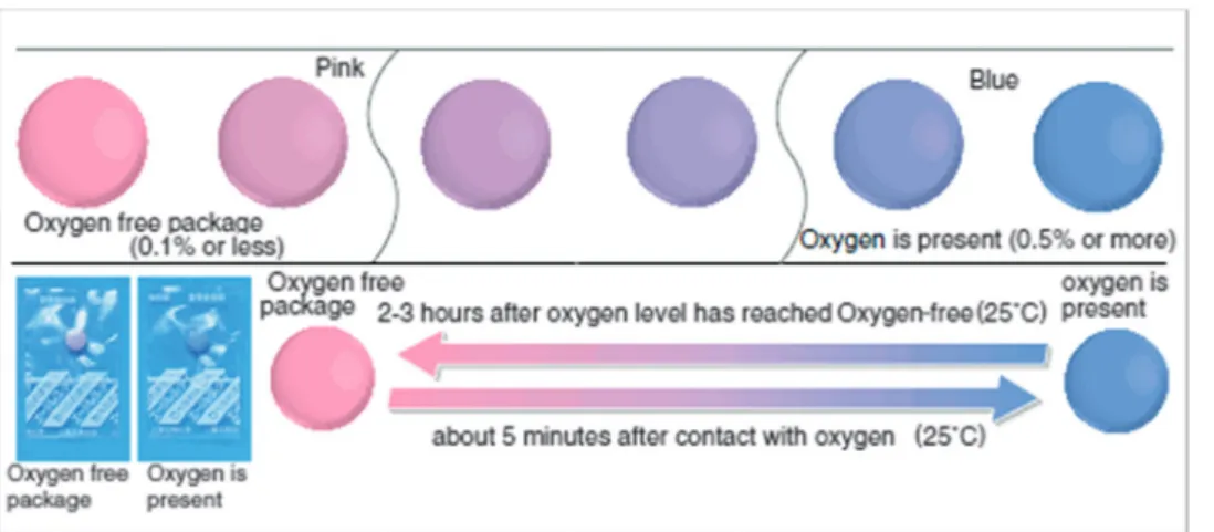
The Mitsubishi Gas Company has patented90,91and commer- cialized respective chromogenic oxygen indicators for use in food packaging (called the ‘‘Ageless Eye’’, Fig. 3). It is also based on leuco MB which was chemically reduced using glucose in alkaline medium. The exposure of the Ageless Eye to oxygen can oxidize the leuco form into the blue form.
MB can also be reduced by irradiation with UV light.92,93 Re-usable oxygen-sensitive inks were obtained by immobilizing TiO2, the sacrificial electron donor triethanolamine (TEOA), Open Access Article. Published on 18 March 2014. Downloaded on 21/04/2015 12:40:30. This article is licensed under a Creative Commons Attribution 3.0 Unported Licence.
and MB in a hydroxyethyl cellulose (HEC) matrix. The resulting blue TiO2-TEOA-MB-HEC film bleaches under UV irradiation, but not under ambient room light or visible light. If exposed to oxygen, the colorless film is irreversibly oxidized to give a blue film which again can be converted into its colorless form by UV irradiation for about 2.5 min. This cycle can be repeated at least 5 times. The working principle of this scheme is based on the formation of electron–hole pairs in the TiO2 semiconductor particles by UV light. The holes oxidize the TEOA, and the photogenerated electrons reduce the MB dye into its leuco form. Its irreversibility and reusability make this intelligent ink useful for applications in packed food technology. The feature of generating the leuco form by UV irradiation after packing simplifies the process, because handling of leuco dyes is complicated as it requires the complete absence of oxygen.
Semiconductors such as ZnO or SnO2 can also be applied for preparing the UV activated oxygen indicator film, but TiO2 works best. The response time can be adjusted by varying the thickness of the sensor film or by using polymers with different oxygen permeability.94
The same group95later modified the hydrophilic, water-soluble and cationic indicator MB into a hydrophobic MB by exchanging the anion. The hydrophobic indicator is soluble in organic solvents and the blue ink can be printed directly on food package.
A film made of TiO2, ion-paired MB, glycerol and a polymer named zein (a prolamine-type of protein found in maize) loses its color rapidly (o30 s) upon exposure to UVA light and remains colourless in an oxygen-free atmosphere, returning to its original blue color upon exposure to air. In the latter step the rate of color recovery is proportional to the level of ambient oxygen and the same film can be UV-activated repeatedly (see Fig. 4). This makes the ink useful for direct printing on food package.
A photoinitiator was employed96,97to photoreduce MBvia UV irradiation in an acrylate matrix. Irradiation generates radicals that reduce blue MB to its leuco form. Simultaneously, the acrylate monomer is polymerized to form a solid film that contains the indicator. This kind of photoreduction is reversible due to the cyclic processes of (1) oxidation on air, and (2) reduction in an oxygen-free environment under UV light. However, the cycles
come to an end once the photoinitiator is consumed. The absorp- tion of MB decreases with the number of cycles on air because of partial photo-decomposition of the indicator.
The polyviologens form a class of less sensitive irreversible oxygen indicators.98 They can also be reduced to colorless forms by exposure to UV light via mediated reduction in the presence of EDTA and TiO2 as described above for MB.
However, the rate for reduction is much faster than in the case of MB. The reduced (leuco) forms of thionine and 2,20-dicyano- 1,10-dimethylviologen were seen to persist until the oxygen concentration exceeded 2.3% and 4.0%, respectively. It was also reported99that beige-colored anthraquinoneb-sulfonate can be reduced to its red phenolate dianion by sodium thiosulfate in alkaline solution. When contacted with oxygen, the red color turns back to beige. This was suggested to serve as a time label to monitor the freshness of food. The rate of the color change can be adjusted by varying the chemical composition of the polyacrylate matrix.
In a fiber optic oxygen sensor for medical use, a viologen indicator is employed that becomes a strong absorber after brief stimulation with UV light. Its color thereafter disappears over time, and the rate of indicator return to transparency is Fig. 3 The color of Ageless Eye oxygen indicator changes from pink to blue in the presence of oxygen.
Fig. 4 Sensor film printed with an ink made of titanium dioxide, Methylene Blue, glycerol and a polymer. The film loses its color upon exposure to UV light but becomes blue again on exposure to air. (Reprinted with permission from ref. 95 Copyright Royal Society of Chemistry, 2008).
Open Access Article. Published on 18 March 2014. Downloaded on 21/04/2015 12:40:30. This article is licensed under a Creative Commons Attribution 3.0 Unported Licence.
proportional to the local concentration of oxygen.79Absorbance is monitored with a red LED and a photodiode, and data are processed by a dedicated processor. The solid-state sensor system has a performance that compares to existing oxygen techniques and may be applied to bothin vitroandin vivooxygen assays.
Colorimetric oxygen-sensitive films can also be based on the redox chemistry of 2,6-dichlorophenolindophenol (2,6-DCPIP) in the presence of fructose and an organic base in a thin film of ethyl cellulose.100The respective sensor film is colorless in the absence of oxygen, but turns to blue in its presence at levels of 30 Torr and above. This represents a simple means for colori- metric detection of oxygen. The oxidized form of 2,6-DCPIP can be reduced by fructose in the presence of a base contained in the polymer film. This ‘‘sensor’’ reversibly responds to oxygen over 10 nitrogen–oxygen switching cycles within a period of B5 h. At oxygen pressures between 0 and 50 Torr, there is a linear relationship between the absorbance of the film and oxygen partial pressure, and response occurs within 20 s.
The leuco forms of indigo and thioindigo were immobilized101 in poly(ethylene glycol), polyurethane hydrogel and poly(styrene-co- acrylonitrile) respectively, in order to adjust the permeability for oxygen and, thus, the response time. The reduced (leuco) forms are better soluble and almost colorless. On interaction with oxygen, a vivid blue or red color develops as can be seen in Fig. 5. The color change can be used to irreversibly detect oxygen. The sensor can be reversed (converted into the faintly colored form) by treatment with dithionite. In one further approach,102 an indicator system was described that gives color changes that result from the fact that the interaction of oxygen with the indicator layer causes a change in the local pH value. Numerous indicators were presented.
The same sensor material was also filled in thin capillaries that can serve as opto-chemical timers. The ‘‘clock’’ is started by opening one end of the capillary filled with leuco (thio)- indigo.103The length of the colored section increases over time as oxygen diffuses in. By using different molecular weight poly(ethylene glycols), one can control the permeability for oxygen and, thus, the time frame of the clock. Significant properties of several absorption-based irreversible probes for oxygen are summarized in Table 2. The above technologies are also covered by various patents.
4.4. Inorganic chromogenic materials
Butler and Ricco105coated optical fibers with nm-thin films of metals (such as nickel) and found that their reflectivity at
860 nm changes on exposure to oxygen as a result of the formation of a thin layer of an oxide. The film thickness also changes and so affects reflectivity. Instrumentation and techniques are simple and applicable to monitor the corrosion rates of metals. An oxygen sensor for car exhaust gases106 comprises a supported film of a heat-responsive inorganic sensing material having a light reflective surface which reversibly reacts with oxygen to form an oxide which causes a change in the reflectivity of the surface. The sensor also includes a temperature sensing means and a 3-way optical system.
5. Oxygen as a quencher of luminescent probes
Most sensors for oxygen are based on the effect of the quench- ing of the luminescence (fluorescence or phosphorescence) of oxygen-sensitive probes (OSPs). The process involves dynamic collision between molecular (triplet) oxygen and the excited electronic state of the OSP and leads to a reduction of its intensity and decay time. Quenching often is accompanied by the formation of singlet oxygen. It is noted in this context that not a single sensor is known for singlet oxygen. However, several irreversibly responding molecular probes have been reported that can be used to quantify singlet oxygen.107
The kinds of methods used in luminescence spectrometry vary to a large extent as will be briefly discussed in this section.
The analytically responsive (‘‘dynamic’’) range of a sensor material can be easily adjusted by (a) proper choice of the OSP (discussed in Section 6), (b) the matrix materials (discussed in Section 7), and (c) by various additives (Section 8). A discussion of the various read-out schemes and geometries of sensors will then be presented in Section 9.
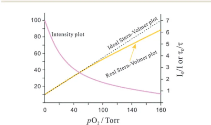
Dynamic (collisional) quenching by oxygen is a photophysical (rather than a photochemical) process. It is fully reversible, does not alter the optical probe, and thus has no effect on its absorption spectrum. Rather, it leads to a drop in luminescence intensity and decay time.108,109The relationship between inten- sity (or decay time) and the concentration of oxygen ([O2]) is reflected by the Stern–Volmer equation which, in its most simple form, reads as
F0/F=t/t0= 1 +KSV[O2] (2) whereF0andF, respectively, are the fluorescence (luminescence) intensities of a probe in the absence and presence of oxygen, Fig. 5 Left: real color images of an irreversibly responding test stripe (consisting of a solution of leuco indigo in a polyurethane hydrogel; LI–D4) for oxygen over a time interval of 20 min. Right: real color images of an irreversibly responding test stripe (consisting of a solution of leuco thioindigo in poly(styrene-co-acrylonitrile; LTI-PSAN)) for oxygen over a time interval of 36 h. (Reprinted from ref. 101 with permission from Elsevier).
Open Access Article. Published on 18 March 2014. Downloaded on 21/04/2015 12:40:30. This article is licensed under a Creative Commons Attribution 3.0 Unported Licence.
KSV is the Stern–Volmer constant which is a function of the lifetime of the probe and the (polymeric) solvent, and [O2] is the concentration of oxygen in the sample. The term [O2] (a concentration) may be replaced bypO2, the partial pressure of oxygen, and the numerical value ofKSVand its unit obviously has to be different then.
In an ideal quencher system, there is a linear relationship between F0/F (or t0/t) and oxygen concentration as shown in Fig. 6. Unfortunately, data forKSVare given by authors in various units including inverse (partial) pressure, (ppm)1, %1, inverse molarity, and others. If data are given in %1or (ppm)1unit, the barometric pressure most be indicated.
Stern–Volmer plots (SVPs) can be established by measure- ment of either luminescence intensity or decay time. However, luminescence intensity data can be adversely affected by poor stability of the light source, variations in the efficiency of the transmission optics, drifts in detector sensitivity, leaching and photodecomposition of probes, inhomogeneous probe distribution, background luminescence and stray light. In order to correct for these effects, an inert reference fluorophore emitting at a different wavelength is often used. See Section 9.9 on referenced sensing.
The Stern–Volmer constant determines the limits of detec- tion (LODs) of a sensor. The LOD is governed by both the initial slope (KSV) of the quenching plot and by the resolution of the instrument. Assuming a 0.1% uncertainty in light intensity measurement (which is the lower limit and requires a well- thermostatted device), the detection limit is 0.003/KSV (KSV
expressed in Torr1 units and at a signal-to-noise ratio of 3).
Phosphorescence based sensors, in contrast to fluorescent sensors, often have much largerKSVvalues and therefore have much lower LODs.
Many SVPs of sensor films exhibit downward curvature as shown in Fig. 6. This usually is indicating contributions by less efficient mechanisms of quenching. A general discussion of situations where both static and dynamic quenching occur can be found in respective textbooks.108,109Specifically for the situa- tion of sensing oxygen, Demaset al.110,111have investigated the photophysics and photochemistry of the quenching by oxygen of several Ru(II) polypyridyl complexes in various polymers.
Fig. 6 Intensity plot of the quenching of luminescence intensity by oxygen, and respective Stern–Volmer plot.
Table 2 Selected irreversibly acting chromogenic probes for oxygen along with respective polymeric supports and color transitions
Dye/matrix Color transition Comments Ref.
Phenothiazine Colorless-blue Photoreduced at pH above 9.5; Methylene blue is best because of its stability; the Methylene blue-EDTA solution undergoes more than 150 cycles of photoreduction-air oxidation without apparent degradation of the dye.
88
Malachite green (leuco form) on silica gel
Colorless-greenish blue Oxidized by reactive oxygen species only. 32
2,6-Dichloroindophenol (leuco form) in ethyl cellulose
Colorless-blue Indicator is co-immobilized with fructose and base; reversible response with short response time (B20 s); applicable to oxygen partial pressure between 0–50 Torr, linear relationship between absorbance and oxygen partial pressure.
100
Methylene blue (leuco form), TiO2and triethanolamine in hydroxyethyl cellulose
Colorless-blue UV-activated reusable ink; irreversible oxygen response but regenerableviaUV irradiation; regeneration time 2.5 min; stable in an oxygen-free environment;
colorimetric determination; response time adjustedviathe thickness of the matrix or using polymers with different permeabilities.
92, 94, 104
Anthraquinone b-sulfonate in pHEMA
Red-beige Reduced form (dianion) is red; color fades into beige with oxygen; useful for fadable printed signs for food packaging
99
Indigo (leuco form) and PEG
Colorless-blue Reduced in basic solution with Na2S2O4; time frame adjusted by using different molecular weight PEG; fairly stable but undergoes some
photobleaching upon exposure to visible light; also used as a timer material;
periods can be adjusted to up to 40 years.
101, 103
Thioindigo (leuco form) and PEG
Colorless-bed Reduced in basic solution with Na2S2O4; the timer covered time frame could be adjusted by using different molecular weight PEG; fairly stable but undergoes some photobleaching on exposure to visible light.
101, 103
Polyviologens (leuco form), TiO2, EDTA
Pale yellow-purple More quickly reduced than Methylene Blue upon UV exposure; high anodic redox potential; suitable for oxygen at above 0.5%, even above 4% (the upper LOD for MB is 0.1% only).
98
Open Access Article. Published on 18 March 2014. Downloaded on 21/04/2015 12:40:30. This article is licensed under a Creative Commons Attribution 3.0 Unported Licence.
Their results showed that downward curved SVPs originate from the heterogeneity of the microenvironment of the OSPs. They assumed that the Ru(II) polypyridyl complex exists in (at least) two distinctly different environments, one being quenchable, the other either not being quenched at all, or being quenched at a very different rate. A two-site model111,112was introduced to fit the curved SVPs, which has been widely used ever since. In the conventional form, it reads as follows (eqn (3)),
F
F0¼ f1
1þKSV1 ½O2þ f2
1þKSV2 ½O2 (3) whereFandF0, respectively, are the fluorescence (or phospho- rescence) intensities of the probe in the presence and absence of oxygen, respectively, f1 and f2 the fractions of the total emission for each component, respectively (withf1+f2being 1), and K1SV and K2SV are the Stern–Volmer constants for each component. The equation may also be written in the lifetime form by replacing (F/F0) for (t/t0).
It shall be reminded here that this model is sensitive to signals that contain contributions by stray light, ambient light and background luminescence. There are several reports on the literature where rather low values forK2SVhave been calculated, and where contributions from these two sources of error to the overall second quenching constant cannot be excluded. Stray light and background luminescence, but not ambient light, can be reduced – if not eliminated – if gating (time-resolved detection) is applied. Gating must not be confused with lifetime detection and is possible only if long-lived (41ms) OSPs are used.
Another model was presented113that relies on the analysis of the multi-exponential luminescence decay of a luminophore, specifically of Ru(dpp), in various polymers. A fit with a sum of exponentials gives physically unreasonable dependences of the pre-exponential factors of the decay components on the oxygen pressure. This is clear evidence that the decay profile is the total of many single but different relaxation rates. A spatial disorder model can relate the distribution of relaxation rates to a distribu- tion of different distances between the OSP and the sites of interaction in the polymer. The two models for multiple-site quenching have been applied to different situations. Generally, the two-site model is well-suited to describe quenching in hetero- geneous systems, while the spatial disorder model better fits homogeneous systems. On the other side, the two-site model can also describe nonlinear quenching in homogeneous systems.114,115 Phosphorescence lifetime analysis was also accomplished using a quadratic programming algorithm that can provide information on the distributions of quenchers in heteroge- neous systems.116The method is based on decomposition of the data vector to a linearly independent set of exponentials and uses quadratic programming principles. Solution of the resulting algorithm requires a finite number of calculations (it is not iterative) and is computationally fast and robust. The algorithm has been tested on various simulated decays and for analysis of phosphorescence data of palladium(II) porphyrins with discrete distributions of lifetimes. The technique is recommended for resolution of the distributions of quencher concentration in heterogeneous samples, of which oxygen distributions in tissue
is an important example. Improved calibration of phase- fluorometric oxygen sensors has been demonstrated on the basis of physical models,117 and the response of phase- fluorometric oxygen sensors can be modeled with respect to effects of temperature and operational requirements.118
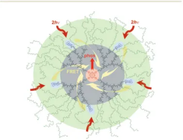
6. Luminescent probes for oxygen
These form by far the largest group of oxygen-sensitive probes (OSPs). Kautsky and coworkers11,16,32 in the 1930s observed that both the phosphorescence and fluorescence of surface- adsorbed OSPs such as acriflavin, trypaflavin, benzoflavin, safranin, chlorophyll, porphyrins and others are quenched by molecular oxygen. This effect was used to detect the formation of oxygen as a result of photosynthesis, and in turn led to the discovery of the Kautsky effect. After more than 80 years of development, numerous OSPs are available now,30with excitation and emission maxima ranging from the UV to the NIR, and with excited-state lifetimes ranging from a few nanoseconds to milli- seconds if not seconds in the case of phosphorescent probes.
Luminescent OSPs can be classified into four subtypes: (1) organic OSPs (mainly polycyclic aromatic hydrocarbons and fullerenes); (2) metal–ligand complexes (mainly transition metal–ligand complexes and metalloporphyrins); (3) luminescent nanomaterials; and (4) multiple emitters and related species.
Absorption-based (non-luminescent) OSPs are not included in this part but were discussed in Section 4. Luminescence-based oxygen sensor technologies are covered by numerous patents which cannot be included here.
6.1. Organic probes
6.1.1. Polycyclic aromatic hydrocarbons (PAHs).Lumines- cent polycyclic aromatic hydrocarbons can be regarded as the first generation OSPs. As summarized in Table 3, almost all PAHs (except those carrying nitro groups or carbonyl groups and some fullerenes) are strongly luminescent and have natural lifetimes of up to 200 ns. This makes them very amenable to quenching by oxygen. Some display fairly good photostability.
In 1939, Bowen and Norton119reported on the quenching of the fluorescence of anthracene by oxygen in various solvents.
However, the effect is small. Weil-Malherbe and Weiss120 in 1942 studied the response of several PAHs to oxygen and found that the fluorescence of 3,4-benzopyrene is completely quenched by oxygen and this effect is fully reversible. Bergman35 in 1968 described an apparatus for the determination of atmospheric oxygen by measuring the intensity of the fluorescence of fluor- anthene absorbed on porous glass or polyethylene. Oxygen acts as a quencher and can be detected in the 0–40 kPa range.
Decacyclene and benzo(g,h,i)perylene121 have much better photostability. Decacyclene was converted into a silicone-soluble derivative by alkylation with t-butyl groups.121 The resulting sensor film was used in a steam-sterilizable fiber waveguide sensor to monitor oxygen concentration in a bioreactor. The sensor layer was covered with a black layer that acts as an optical isolation to avoid interferences by the fluorescence of cellular matter.
Open Access Article. Published on 18 March 2014. Downloaded on 21/04/2015 12:40:30. This article is licensed under a Creative Commons Attribution 3.0 Unported Licence.
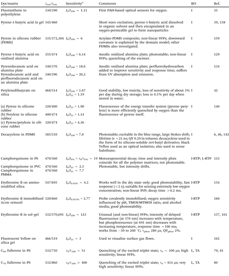
Table 3 Organic fluorescent probes for oxygen, their excitation/emission wavelengths (in nm), quenchability (‘‘sensitivity’’), along with the polymer solvent or support used, and the method for read-out (RO). Quenchability is expressed as eitherI0/I100the ratio of fluorescence intensity at zero% oxygen in the carrier gas and the intensity at 100% oxygen (at atmospheric pressure), or in a fluid equilibrated with such gases. Correspondingly,I21is the intensity under air at atmospheric pressure.Codes: I: intensity-based readout; I-RTP: intensity of room temperature phosphorescence; L-RTP: lifetime of RTP-based readout; TA: triplet absorption-based readout; LIM: lifetime imaging; IR: ratiometric intensity-based readout. QY: quantum yield; SVP:
Stern–Volmer plot. For other acronyms see the list in Section 14
Dye/matrix lexc/lem Sensitivitya Comments RO Ref.
Fluoranthene in polyethylene
330/390 I0/I100B1.11 First PAH-based optical sensors for oxygen. I 35
Pyrene-1-butyric acid in gel 345/460 Short wave excitation; pyrene-1-butyric acid dissolved in organic solvent and then encapsulated in an oxygen-permeable gel to form nanoparticles
I 39, 158
Pyrene in silicone rubber (PDMS)
335/372,384 I0/I100B6 Acrylate-PDMS composite; non-linear SVPs; downward curvature is explained by the domain model; other PDMSs also investigated.
I 159
Pyrene-1-butyric acid on oxidized alumina
355/474 I0/I100= 6.14 Anodic oxidized alumina plate; photostable; non-linear SVPs; quenching of the excimer.
I 129
Pyrenedecanoic acid on alumina plate
340/376 I0/I100= 18.6 Anodic oxidized alumina plate; perfluorohydrocarbon added to improve sensitivity and response time; suffers from UV absorption and emission.
I 134
Pyrendecanoic acid and perfluorodecanoic acid on an alumina plate
340/396 I0/I100= 20.2
Perylenedibutyrate on silica
468/514 I0/I100= 1.67 I0/I21= 1.19
Good stability, low toxicity, loss of sensitivity of about 3%
per day during dry storage; loss is 0.1% per day when stored in water.
I 42
(a) Pyrene in silicone rubber
320/400 I0/I21= 1.90 Fluorescence of the energy transfer system (pyrene–pery- lene) is more efficiently quenched by oxygen than the fluorescence of pyrene itself.
I 140
(b) Perylene in silicone rubber
400/474 I0/I21= 1.14 (c) Pyrene/perylene in sili-
cone rubber
320/474 I0/I21= 4.36
Decacyclene in PDMS 385/510 I0/I100= 7.8 Photostable; excitable in the blue range, large Stokes shift;
lifetime isB21 ns; QY 0.29 in toluene; decacyclene used in the form of its silicone-solubletert-butyl derivative; black Teflon used as an optical isolation; also used to sense halothane.
I 6, 46, 142
Camphorquinone in PS 470/560 I0/I6%=t0/t6%B19 Monoexponential decay; time and intensity plots coincide for all the polymer matrices; not photostable.
I-RTP; L-RTP 153 Camphorquinone in PVC 470/560 I0/I21B2.5 Photostable, but intensity drifts.
Camphorquinone in PMMA
470/560 I0/I21B7.7
Erythrosine B on amino- modified silica
547/695 I0/I0.05%B4.2 Works well in the dry state only; good photostability, fast response (o2 s); suitable for sensing extremely low oxygen concentration; non-linear SVP; decay time40.2 ms.
I-RTP 154
Erythrosine B immobilized in/on ormosil
520/660 I0/I0.015%= 2.77 Probe covalently immobilized; oxygen sensitivity influenced by pH, TMOS/MTMOS ratio, and alcohol media; good photostability.
I-RTP 160
Erythrosine B in sol–gel 532/570,691 I0/I100B123 Unusual (and non-linear) SVPs; intensity of delayed fluorescence (at 570 nm) increases with temperature, but phosphorescence (at 691 nm) decreases with increasing temperature, response timeB100 ms;
works from50 to 2001C;tphos280ms, QYphos2%.
I-RTP 157, 161
Fluorescent Yellow on silica gel
466/519 I0/I21B3 Used to visualize surface gas flows. I 162
C60fullerene in PS 532/750 t0/t100B51 Quenching of the excited triplet state;t0B100ms; high sensitivity; linear SVPs.
L, TA 79, 81
C70fullerene in PS 532/860 t0/t1004400 Quenching of the excited triplet state;t0= 414ms; very high sensitivity; linear SVPs.
L, TA 80
Open Access Article. Published on 18 March 2014. Downloaded on 21/04/2015 12:40:30. This article is licensed under a Creative Commons Attribution 3.0 Unported Licence.