Materials & Methods
5. MATERIALS AND METHODS 5.1 MATERIALS
All commonly used chemicals were from Merck, Sigma, J.B. Baker, Roth, Riedel de-Haen, Fluka, GERBU, Serva and AppliChem and were distributed by the university chemical centre Heidelberg, unless indicated otherwise. All water used to prepare solutions was filtered with a Mili-Q water purification system.
Solutions were either autoclaved or sterilized with filters unless indicated otherwise
Chemicals
Triton X-100 was purchased from Merck, Triton X-114 and Tween-20 from Sigma and NP-40 was obtained from Calbiochem. Both β-cycloheximide and Brefeldin A were purchased from Sigma and kept as a stock solution at 250 mg ml-1 in methanol (β-cycloheximide) or 7.5 mM (Brefeldin A) in PBS. The working concentration of β-cycloheximide was 50 µg ml-1, and 5 µM for Brefeldin A.
Protease inhibitor tablets were obtained from Roche and solved in appropriate buffers at 50 ml per tablet.
Enzymes
Restriction and modification enzymes were purchased from either New England Biolab, or Roche, or MPI, or Promega where specially indicated otherwise.
Buffers
Common used buffers were prepared as described below:
10 PBS: 1.36 M NaCl, 357 mM Na2HPO4, 143 mM KH2PO4 and 30 mM KCl.
PBST was generated from PBS by supplementing with TWEEN-20 to a final concentration of 0.05% or 0.1% (only for α-FLAG). PBS used for IF was prepared in 5 as 500 mM Na2PO4, 750 mM NaCl and 12.5 mM KCl. All PBS buffers were adjusted to pH 7.4.
Materials & Methods
10 TEN was prepared with a final concentration of 100 mM Tris/Cl (pH 7.4), 1.5 mM NaCl and 50 mM EDTA (pH 8.0), respectively.
PEN buffer was prepared as a 5 stock solution containing 125 mM PIPES, 10 mM EDTA and 1.5 M NaCl with pH 6.5.
50 TAE contains 242g of Tris base, 57.1ml of glacial acetic acid and 18.61g of EDTA in 1L solution.
20 SSC: 175.3g of NaCl and 88.2g of tri-sodium citrate were dissolved in 1L of water with pH 7.0 and autoclaved.
50 Denhardt’s solution: 1% (w/v) each of Ficoll-400 (Pharmacia), bovine serum albumin V (Sigma) and polyvinylpyrrolidone (Sigma). The solution was filtered and stored at -20°C.
10 MOPS was prepared by dissolving 20.6g of MOPS in 400ml of a 50mM sodium acetate solution pre-treated with DEPC, pH was adjusted to 7.0, 10ml of pretreated 0.5 M EDTA was added to the solution and the volume was adjusted to 500ml with DEPC-treated water. The solution was stored in dark after sterilization by filtration.
10 SDS-PAGE running buffer: 30.2g of Tris base, 188g of glycine and 10g of SDS in a 1L solution.
10 Blotting buffer for wet-blotting was prepared as 250mM of Tris base and 1.92M of glycine. To 1 blotting buffer, methanol was added to a final concentration of 20%.
Materials & Methods
Blotting buffer for semi-dry transfer:
Anode-I buffer contains 300mM Tris and 20% (v/v) methanol, whereas Anode- II buffer contains 25mM Tris and 20% methanol. Cathode buffer consists of 25mM Tris, 40mM aminocaproic acid and 20% methanol.
Sample buffer I was made as a 6 stock solution containing 0.25% bromophenol blue, 40% sucrose, 60mM Tris/Cl (7.4) and 6mM EDTA (8.0). This buffer was generally used for DNA samples.
Sample buffer II contains 50% glycerol, 1mM EDTA (8.0) and 0.25%
bromophenol blue. This buffer was treated with DEPC, autoclaved and used for RNA samples.
Sample buffer III was made as a 3 stock solution containing 187.5mM of Tris/Cl (6.8), 15% β-mercatoethanol, 6% SDS, 30% glycerol and 0.0675%
bromophenol blue and stored at -20°C as aliquots.
Coomassie bright blue staining and de-staining buffer were prepared according to G. Hervieu (MRC Molecular Neuroscience Group, Department of Neurobiology, Babraham Institute, Cambridge, UK CB2 4AT). Staining buffer was prepared by dissolving 2.5g of Coomassie blue R-250 (0.25%) in 500 ml of water and adding 400 ml of ethanol (40%), 100 ml of glacial acetic acid (10%), while De-staining buffer consists of 20% ethanol and 5% acetic acid.
Ponseau S solution: 0.8g of the dye was dissolved in 4% (w/v) TCA.
Media
α-MEM and DMEM powders were purchased from Gibco BRL (Invitrogen), solubilized into water, sterilized by passing through 0.2µM filters and
Materials & Methods
supplemented with 10% of fetal calf serum, 10 mg ml -1 of each of penicillin and streptomycin, L-Glutamine and non-essential amino acids (only for DMEM).
Ready-to-use Folate-free HAM was obtained from PAN biotech (Aidenbach, Germany).
Normal LB medium was prepared according to Molecular Cloning (3rd Edition):
10g of sodium chloride, 10g of Bacto-Trypton and 5g of yeast extract (DIFCO) were solved in 1L of water and the pH was adjusted to 7.4. For plates, 15g/L agar was supplemented.
Low salt LB media were prepared as Normal LB media except that 5g of NaCl was used.
BMGY/BMMY consists of 1% yeast extract, 2% peptone, 0.1M potassium phosphate (pH6.0), 1.34% YNB (yeast nitrogen bases), 4 10-5% Biotin and 1%
glycerol (BMGY) or 0.5% methanol (BMMY). To prepare this medium, 700ml autoclaved medium containing 10g yeast extract and 20g peptone was mixed with 100ml autoclaved 1M potassium phosphate (132ml 1M of K2HPO4 + 868ml of KH2PO4), 100ml filtered 13.4% YNB, 2ml filtered 0.02% Biotin and 100ml autoclaved 10% glycerol (BMGY) or filtered 5% (v/v) methanol (BMMY).
YPD was prepared by dissolving 10 g of BactoYeast extract, 20 g of each of BactoPeptone and Dextrose in 1 L of water and subsequent sterilization (autoclave).
Antibodies
Primary antibodies
Monoclonal anti-GM-130 was obtained from BD Biosciences. Anti-GRASP-65 antibodies were kindly provided by Dr. Barr (Max-Planck institute for
Materials & Methods
Biochemistry, Germany) and Dr. Lowe (University of Manchester, UK).
Monoclonal antibody against folate receptor (Mov19) was a kind gift of Dr.
Canevari (Istituto Nazionale Tumori, Italy). Polyclonal anti-luminal part of p23 was generated against the overexpressed and purified luminal domain of this protein (K Sohn, unpublished data). Polyclonal anti-Man II antibody was purchased from CHEMICON. Polyclonal antibodies against Gαi3 (C-10), Gβ (T- 20), Caveolin-1 (N-20) and Caspase-2L (C-20) were purchased from Santa Cruz Biotechnology. Polyclonal antibodies against the C-terminal peptide of Flotillin-1 and GAPR-1 were described previously (Gkantiragas et al. 2001, Eberle et al.
2002). M2 monoclonal anti-FLAG antibody and 9E10 monoclonal anti-myc antibody were obtained from Sigma. Monoclonal anti-α-tubulin antibody was generously donated by Ms. Wei Yao (EMBL, Germany)
Secondary antibodies
HRP-conjugated goat anti-rabbit IgG(H+L) and HRP-conjugated goat anti-mouse IgG(H+L) were obtained from Bio-Rad; TRITC-conjugated donkey anti-mouse Ig(H+L) and FITC-conjugated donkey anti-rabbit Ig(H+L) were purchased from Jackson ImmunoResearch Laboratories; Alexa Fluor® 488 goat anti-rabbit IgG(H+L) and Alexa Fluor® 568 goat anti-mouse IgG(H+L) were purchased from Molecular Probes.
Peptides
All peptides were synthesized at ZMBH, Heidelberg University, and lyophilized.
The sequences of peptides are: ALIQEQEAQIK (ALI), SEACRDGLRAQEEC (SEA), FYHYHPLPMDQKEPG (#2390) and VLAVPLFLLF (#2391).
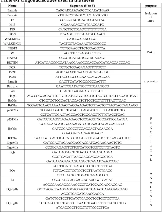
Primers
Oligoes were synthesized at either NAPS or Interactiva and HPLC purified. All oligoes solubilized in sterilized water to a final concentration of 100 µM.
Sequences are shown in Table 4-1.
Materials & Methods
Table 4-1 Oligonucleotides used in the thesis
Name Sequence (5’ to 3’) purpose
Band4s CARGARCARGARGCNCARATHAAR Band4a YTTDATYTGNGCYTCYTGYTCYTG
T7 CGCCCTAGTGAGTCGTATTAC M13 GGAAACAGCTATGAGCATG
Isolation of GREG P45 CAGCTTCTTCAGCTTCTGTTCGA P45N TCAAGCTTCTGGATGCGAACT WALKING CATGGGCAACGGGT WALKINGN TAGTGGTAGAAAGTGGGCGCC
NEST2 CCTGGAACCTTCTCGAGGTCA W AGCTTCCGAGGGCCCTG NNEST CGGGTGATAGTGGTAGAAAGT
BIOTIN ATGATGAGCGGGATAACCAAGGCCACCAGGATCAGGACCGAG PCR TCTGCTCGAGAGAGTTCTGCTT
RACE
P22F AGTCGAATTCAAACCACATGGCGC P22R ATTAGCGGCCGCAAAGAGCAGGAA Band4f GACTTCATATGGCGCCCACTTT B4trunc GAATTCGATATGGCCGTCAAGGCG B4ta CTACTCGAGAGAGTTCTGCTT
expression
FlagC AGCCGGCAGAGTTCTTGTCATCGTCGTCCTTGTAGTCCTGCTTAGATGTGAT BstXIs CTGGTGCTCGCAGTACCACTCTTCCTGCTCTTTTAGTTGAC BstXIa TCGAGTCAACTAAAAGAGCAGGAAGAGTGGTACTGCGAGCACCAGAAGG p23TMs TCGAGCGGGTCCTGTACTTCAGCATCTTTTCCATGTTCTG
CCTCATTGGACTAGCCACCTGGCAGGTCTTCTACCTGAG
p23TMa GATCCTCAGGTAGAAGACCTGCCAGGTGGCGATTCCAATGA GGCAGAACATGGAAAAGATGCTGAAGTACAGGACCCGC
BstYIs GATCGGAGGCCTCGAGGACTACAAGGA CGACGATGACAAGTGAGC
BstYIa GGCCGCTCACTTGTCATCGTCGTCCTTGTAGTCCTCGAGGCCTCC NgoMIs GATCGACTACAAGGACGACGATGACAAGAACTCTG NgoMIa CCGGCAGAGTTCTTGTCATCGTCGTCCTTGTAGTC EQs GATCAGGGCTCTGATCCAGGAGCAGGA
GGCTCAGATTAAGGAGCAGGAGGCTCA GATCAAGGAGCAGGAGGCTCAGATCAAGCCGC EQa
GGCTTGATCTGAGCCTCCTGCTCCTTGA TCTGAGCCTCCTGCTCCTTAATCTGAGC
CTCCTGCTCCTGGATCAGAGCCCT
Ls CGGGATCCAGGAGCAGAAGGCTCACAT EQ-BglIs
AGGCGAACAGCGAAGCCCTGATCCAGGAGCAGGAG GCTCAGATTAAGGAGCAGGAGGCTCAGATCAAGGAGCAGG
AGGCTCAGATCAAGGAGCA EQ-BglIa
GATCTGCTCCTTGATCTGAGCCTCCTGCTCCTTGA TCTGAGCCTCCTGCTCCTTAATCTGAGCCTCCTGCTCCTCG
ATCAGGGCTTCGCTGTTCGCCTTGA
F U N C T I O N A L
A N A L Y S I S
NB: R = A/G; Y = C/T; D = A/G/T; H = A/C/T; N = A/C/T/G.
Materials & Methods
Equipment
Thermal cycler was from ThermoHybaid, ultra-centrifuges were from either Beckman or Kontron Instruments (Watford, UK). Other centrifuges were from either Eppendorf or Heraeus. All rotors, including SW28, SW41, SW50, SW60, JA12.500, TLA45 and TLA55, were available from Beckman. Electrophoresis system, including plates, spacers and combs, and blotting systems, including semi-dry trans-blot and wet blot, were purchased from Bio-Rad (Munich, Germany). Cell incubators were from Heraeus. Other equipments not outlined here were as indicated in “Methods”.
5.2 METHODS
Standard methods were carried out according to Molecular Cloning (3rd Edition) with, in many cases, only minor modifications. All procedures were carried out at room temperature except where indicated otherwise.
5.2.1 CELL BIOLOGICAL AND IMMUNOLOGICAL METHODS 5.2.1.1 Cell culture
Cells in monolayer were maintained in α-MEM (CHO) or DMEM (NRK) media supplemented with 10% fetus calf sera, L-glutamine and 10 mg ml-1 of penicillin and streptomycin and cultured at 37°C in a 5% CO2 incubator. Stable cell lines constitutively expressing GREG-Flag were cultured in the same medium as their original cells except that 0.2 mg ml-1 of G418 was added. CHO cells stably transfected with FolR were cultured in Ready-to-use Folate-free HAM supplemented with 10 mg ml-1 of penicillin and streptomycin and 0.2 mg ml-1 of G418 at 37°C in a 5% CO2 incubator.
Just before reaching confluency, cells were either frozen or split. In both cases, cells were washed at least once with PBS after aspirating all cultured media. Trypsinized cells from plates were harvested by centrifugation at 4°C 1, 000xg for 5 min. Cell pellets were washed once in PBS and resuspended in appropriate volumes of normal growth media. For freezing cells, 1.8 ml of media
Materials & Methods
was used to resuspend cells either from one 20 cm plate or two 10 cm plates (around 1 107 cells). Cells (1.8 ml) were transferred into a frozen vial containing 0.2 ml of DMSO and mixed well by upside down movement for several times.
Cells were incubated at 4°C for 2 hrs and transfer to -80°C. For storage more than half a year, cells were immersed in liquid nitrogen.
For splitting cells, 10 ml of media were used to resuspend cells from one 10 cm plate (around 5 107 cells). 1 ml of cells was transferred to a fresh 10 cm plate and 9 ml of fresh growth media was added. Cells were incubated in a 37°C incubator.
5.2.1.2 Transfection of cells
Transfection procedures were conducted according to manufacturer’s guide (Invitrogen) with minor modifications. In each transfection, 1.5 µl of Lipofectamine™ 2000 and 1 µg of plasmid DNA were used for one well of a 6- well plate (5 µl and 5 µg for a 5cm dish). One day before transfection, cells were plated in normal growth media so that they were 60% confluent at transfection.
For transfection, appropriate amounts of plasmid DNA and Lipofectamine™ 2000 (in 100 µl of Opti-MEM® I reduced medium each) were diluted in different tubes.
After incubation at room temperature for 5 min, the two solutions (200 µl in total) were mixed gently by pipetting and incubated for 30 min at room temperature.
To the mixture 0.8 ml of Opti-MEM® I reduced medium was added and mixed gently. All mixtures were applied to cells pre-washed once with each of PBS and Opti-MEM® I reduced medium, respectively, and mixed gently by rocking the plate or dish back and forth. The cells were incubated in a 37°C CO2 incubator for 3 – 5 hrs. 2 volumes of normal growth media were added and cells were cultured in the incubator for appropriate times. Before processing for analysis by IF, cells transiently expressing constructs for mapping the Golgi targeting signal were treated with 50 µg ml-1 of β-cycloheximide at 37°C for 1 hr.
For transient expression of mutant proteins, cells were cultured for further 8 – 24 hrs after addition of normal media and immediately processed for analysis. For selecting stable cell lines, cells were cultured for 36 hrs after adding
Materials & Methods
normal media. Stable cell clones (in plates with cells diluted 1:5, 1:10 and 1:20) were selected in the presence of 0.6 mg ml-1 of G418. Around 2 weeks later, stable clones started to appear and were picked for further selection in 24-well plate.
When transferred into 5cm dishes, cells were also seeded on coverslips for determination of transfection by IF. Later, when enough cells are available, stable cell clones were further analyzed for expression of the desired construct by western blot or by IF with appropriate treatments, like 5 µM of BFA. Aliquots of stable cells were frozen for later usage as described above except supplementing 0.2 mg ml-1 of G418 to the medium.
5.2.1.3 Metabolic labeling of cells
CHO cells constitutively expressing GREG-Flag were maintained in α-MEM media supplemented with 10% fetus calf serum, L-glutamine, 0.2 mg ml-1 of G418 and 10 mg ml-1 of penicillin and streptomycin. When cells were around 80% confluent, 10 µCi of [14C]-ethanolamine was added to the media. Cells were metabolically labeled for 24 hrs. All media was carefully aspirated and cells were washed twice with PBS. Cells were scraped into 10 ml of TEN buffers and collected at 4°C 1, 000xg for 10 min. Cell pellets were resuspended in 0.1 ml of TEN containing protease inhibitors. Cells were lyzed by adding 0.1 ml of TEN containing 2% TX-100 and 2% NP-40. The lysates were cleared by centrifugation at 4°C 14, 000 rpm for 15 min and subsequently used for IP.
5.2.1.4 Interference mediated by dsRNA
The pellets of in vitro transcribed RNA (See 4.2.2.7) were resuspended in 10mM Tris/Cl (pH8.0) and the single-strand RNAs generated by T7 RNA polymerase was combined with the RNA obtained by the Sp6 RNA polymerase. To generate dsRNAs, the mixtures were first denatured at 95°C for 10 min and subsequently annealed slowly and incubated at 42°C for 2 hrs. The resulting dsRNAs were precipitated with 3 volumes of cold ethanol after adding 0.1 volume of 3M sodium acetate (pH 4.8) and 0.15 µg of circle plasmid DNA (pcDNA3) and
Materials & Methods
resuspended in 10 µl of sterilized water. CHO cells constitutively expressing GREG-Flag were seeded on coverslips one day before transfection. All transfection procedures were the same as the transfection procedures with plasmids. After transfection of dsRNAs for 24 hrs, cells were processed for analysis by IF.
5.2.1.5 Immunofluorescence microscope
Indirect immunofluorescence was carried out according to standard procedures.
Cells on coverslips were washed twice in cold PBS and immediately fixed in either 3.5% formaldehyde/PBS for 30 min at room temperature or methanol at - 20°C. After quenching formaldehyde with 50 mM NH4Cl in PBS, cells were permeabilized with 0.5 % TX-100 in PBS for 10 min. These two steps were not necessary for methanol fixation, which also permeabilizes cells. Cells were subsequently blocked in PBS containing 2% BSA for 1 hr at room temperature or overnight at 4°C. For double labeling, cells were incubated with antibodies against FLAG tag (1:2,000), α-tubulin (1:500), Mannosidase II (1:500), GRASP65 (1:400) or GM130 (1:500). Primary antibody labeling was then visualized by incubation with Alexa Fluor® 488 anti-rabbit IgG (1:1,000) and/or Alexa Fluor® 568 anti-mouse IgG antibodies (1:1,000). Cells were stained with DAPI at a final concentration of 1 µg ml-1 for 10 min immediately before mounting in Fluoromount G (Biozol). A Zeiss LSM510 with appropriate filters was used and images were collected and processed with LSM510 software. For cells stained with DAPI, images were taken with the use of a Zeiss inverted fluorescence microscope equipped with a cooled CCD camera.
3.5% formaldehyde was prepared by dissolving 7g of paraformaldehyde in 100ml of water at 80°C with stirring, adding 0.1M of each of CaCl2 and MgCl2
to a final concentration of 0.1mM while stirring, then this solution was mixed with 100ml of 2 PBS (IF) and aliquots were stored at -20°C
Materials & Methods
5.2.1.6 Peptide antibodies 5.2.1.6.1 Animal immunization
A short peptide is generally not enough to evoke an effective immune response, but can efficiently react with antibodies. Therefore, the short peptide should be coupled to a large protein carrier, in most cases, KLH (H-7017, Sigma-Aldrich) before immunizing animals. The peptide used here is the SEA peptide (SEACRDGLRAQEEC) corresponding to AA 51 – 64 of GREG. The coupling reaction was carried out according to standard procedures. 5 mg of the SEA peptide was solubilized in 0.5 ml of PBS and added to the tube containing 1ml of the reconstituted KLH (10 mg ml-1). The solution was mixed well and air bubbles were avoided. In 5 min intervals, 6 µl of freshly made 5% glutaraldehyde (EM level of 25% stock, Sigma-Aldrich) was added repetitively for 5 times (30 µl in total). All above procedures were performed at room temperature. The mixture was incubated on ice for 30 min and the excess glutaraldehyde was blocked by the addition of 40 µl of 1M glycine (pH8.5). Animal immunization procedures were as follows: Day 1: immunization of the animal; Day 21, 35, 49 and 63 boost;
Sera were collected at Day 42, 56 and 63, respectively; Final sera were collected at Day 90. Before immunization, sera were collected as a pre-immune control.
5.2.1.6.2 Affinity purification
1.0 g of CNBr-activated Sepharose 4B (Amersham/Pharmacia) was suspended in 10 ml of 1mM HCl and incubated for 20 min at room temperature. Beads were washed 5 times with 50 ml of ice-cold 1mM HCl and subsequently washed twice in 50 ml of coupling buffer (0.1M NaHCO3 and 0.5M NaCl, adjust pH to 8.3.). In each wash step, beads were collected by centrifugation at 4°C 500xg for 5 min. In between, 5 mg of the SEA peptide was solubilized in 1 ml of coupling buffer.
After all washes were completed, resins were mixed with the peptide solution and incubated at 4°C for 48 hrs with gentle rotation. Before and after incubation, 1 µl of the mixtures were separately diluted into 1 ml of coupling buffer for measurement of OD280 to determine the coupling efficiency (here more than
Materials & Methods
90%). After coupling, resins were collected by spinning down as above. To the resins 2 ml of 1M glycine was added and incubated at 4°C overnight with gentle rotation. The supernatants were discarded after spinning down the resins as described above. The resins were washed once with 50 ml of each of 0.1M Tris/Cl (pH8.0) and coupling buffer. Then, the resins were washed with 3 cycles of Wash buffer I (0.5M NaCl and 0.1N acetate buffer pH4.0) and Wash buffer II (0.5M NaCl and 0.1M Tris/Cl buffer pH8.0). The resins were washed twice with PBS and divided into two aliquots with one stored at 4°C after addition of NaN3
to a final concentration of 0.02%.
The other aliquot of the resin was rotated overnight at 4°C with 5 ml of sera pre-cleared at 4°C 10, 000xg for 10 min. The resins were collected by centrifugation at 4°C 500xg for 10 min. The resins were resuspended in PBS and transferred to a column. The column was washed with PBS until OD280 remained stable (generally around 20 column volumes). The column was eluted with 5 column volumes of 0.1M glycine (pH2.5) and each 1 ml fraction were collected into a 1.5 ml tube containing 50 µl of 1M Tris/Cl (pH8.8). According to OD280, fractions containing specific antibodies were pooled and immediately dialyzed against 4L of pre-cooled PBS overnight at 4°C. The column was washed with 10mM Tris/Cl (pH8.8) until pH remained 8.0. The column was eluted with 5 column volumes of 6M Guanidium HCl and each 1 ml fractions were collected and measured for OD280. According to OD280, the protein concentration was relatively low. All fractions with higher OD280 values were combined and dialyzed as above.
The column was washed with PBS and the resin was transferred into a 50 ml falcon tube. The resin was washed 3 cycles with Wash buffer I and II as above and once in PBS. The resin was stored at 4°C after addition of NaN3 to a final concentration of 0.02%.
5.2.1.7 Immunoprecipitation
IP was performed according to standard procedures. An appropriate amount of the primary antibodies, here 7 ng of anti-FLAG antibody, was first incubated
Materials & Methods
with 50 µl of protein A-beads pre-equilibrated in IP buffer at room temperature for 1 hr. Beads were washed twice with IP buffer prior to incubation with samples. The input materials for IP were either total cell lysates or Golgi membranes in IP buffer, both precleared for 15 min at 4°C 14, 000 rpm in an Eppendorf table-top centrifuge. IP was performed in TEN buffer containing protease inhibitors and 1% of each of TX-100 and NP-40 for 1 hr at room temperature (metabolic labeling) or 2 hrs at 4°C with gentle rotation. All centrifugations in every wash step were carried out for 2 min at 1, 000 rpm and at room temperature on a table-top centrifuge. After completion of incubation, the beads were spun down and washed 4 times with 0.5 ml of IP buffer and once in 1 ml of PBS. The precipitates were eluted into sample buffer III by incubation at 95°C for 5 min. An aliquot of eluates was loaded on SDS-PAGE and analyzed by western blot. For autoradiography, the gel was soaked in enhancing solution (Pharmacia/Amersham) and incubated for 20 min at room temperature. After drying the gel in a dryer at 80°C for 2 hrs, the gel was exposed to X-ray films at - 80°C.
5.2.2 MOLECULAR BIOLOGICAL METHODS
5.2.2.1 Extraction of total cellular RNA from monolayer cultured cells
Generally, all procedures were based on supplier’s instructions (QIAgen Mini RNeasy Kit) except some details, like duration of incubation time, centrifugation time etc. Care was taken to avoid RNase contamination during all steps. Tips and tubes used for RNA preparation were fresh and autoclaved. Water used for generating any solutions was sterilized by passing through 0.2 µm filters (Milipore). The detailed protocol was as follows:
CHO cells cultured in monolayer were harvested with the use of a disposal cell scraper, and washed twice with cold PBS. In these steps, cells were collected by centrifugation at 1,000 rpm 4°C for 5 min and a trivial amount of PBS was left to resuspend cell pellets in the last wash step. Cells were disrupted by adding 0.6 ml of RLT buffer supplemented with 6 µl of β–mercaptoethanol (β-
Materials & Methods
ME). The lysate was incubated for 5 to 10 min after mixing (upside down) several times. An equal volume of 70% ethanol was added to the lysate and mixed completely by upside down movement, repeated several times. Samples were loaded onto a column and spun down at full speed for 30 sec (from this step on, all centrifugations were performed at full speed for 30 sec except the last step). In many cases, the total volume of samples exceeded that of supplied columns;
therefore, aliquots were successively loaded. The column was washed twice, each with 0.5 ml of ethanol-containing RPE buffer and the flow-through discarded. The column was completely dried by further centrifugation for 2 min.
RNAs were eluted with 50 µl of RNase-free water. To remove the contaminated DNAs, RNA samples were incubated with RNase-free DNase (Invitrogen/Gibco) at 37°C for 30 min and subsequently precipitated by adding 0.1 volume of 3M sodium acetate (pH4.8) and three volumes of cold ethanol after extraction with an equal volume of phenol/chloroform. RNAs were quantified by determining OD260.
5.2.2.2 Reverse transcription
2 µg of total RNAs were used for each reaction, which was performed in 25 µl system. First, RNA samples were mixed well with generally 3 pmol of oligonucleotides, incubated together at 94°C for 10 min and then cooled down immediately on ice. Then, the required buffer, including DTT, was added. The reaction was carried out with 200 units of Superscript® II RNase- (Invitrogen/Gibco) in the presence of dNTPs each with a final concentration of 0.4 mM at 42°C for 1.5 hrs. Finally, enzyme was inactivated by heating samples at 75°C for 10 min and RNAs were removed by incubation with 10 units of RNase A.
The single-strand cDNA was purified with GlassMax (Invitrogen/Gibco).
Briefly, 3 volumes of binding solution were mixed together with reverse transcription mixtures. All samples were loaded onto the supplied cartridge, incubated at room temperature for 10 min and spun down at full speed for 30 sec. The columns were washed four times each with 0.4 ml of wash buffer and
Materials & Methods
once with 0.4 ml of cold 70% ethanol. In the last wash step, centrifugation was performed for 2 min to remove trace amount of ethanol. Single-strand DNA was eluted into 30 µl of filtered water.
5.2.2.3 Polymerase chain reaction
All reactions were carried out in either 25 or 50 µl system with either AmpliTaq (Applied Biosystems) or Turbo pfu (Invitrogen). The system contains: 1 reaction buffer, 0.4 µM of each primer, 1.5 mM MgCl2, 0.2 mM dNTPs and 0.5 or 1.0 U of AmpliTaq or 2.5 U of Turbo pfu (only for 50 µl system reactions). The PCR reaction was generally initiated from denaturation at 95°C for 3 min followed by denaturation at 94°C for 30 – 60 sec, annealing at the appropriate temperature (based upon different primer pairs, in principal 10 - 15°C lower than the Tm of the used primers) for 30 – 60 sec and elongation at 72°C for 1 min (if the fragment is longer than 5kb and Turbo pfu was used, the temperature is 68°C instead of 72°C and the time duration is calculated as 2 min/kb.) and stopped at 72°C for 5 min. In some cases, two annealing temperatures were adopted. In general, 5 – 10 cycles of reactions were first performed at lower annealing temperature and then 20 – 25 cycles at higher annealing temperature.
In general, 5 µl of PCR products were firstly analyzed by agarose gel electrophoresis to make sure that amplification was efficient. Then, the PCR amplified fragments were purified from agarose gel slices with the use of Geneclean II kit (Bio 101). Doubly digested plasmid vectors were also purified with this kit. Briefly, 3 volumes of 6M NaI were used to melt the gel slice at 55°C.
5 µl of completely mixed GLASSMILK® was added to the samples and incubated at room temperature for 10 min. The matrix was spun down and washed twice with 0.4 ml of reconstituted NEW™ Wash buffer each. The matrix was completely dried (it will take roughly 15 min in air.) and DNA was eluted with appropriate volumes of pre-heated to 65°C sterilized water. Purified PCR products were either directly used for cloning into pGEM-T (Promega) or digested with restriction enzymes (generally double enzymes). If the two used enzymes could
Materials & Methods
not work in the same buffer, the digestion mixtures were either purified from the gel or precipitated with 3 volumes of ethanol.
5.2.2.4 Subcloning
To map the signal for the Golgi localization of GREG, several constructs were prepared as described below and cloned into the EcoR I and Xho I sites of plasmid pcDNA3. To generate GPI-anchored N-termand GPI-anchored C-term constructs, GREG-Flag was digested with BstY I, which cuts at base 326 (109Q) and generates the N-terminal and C-terminal fragment. The C-terminal fragment (AA 100-211) was ligated with the signal peptide of GREG (AA 1-55). The N- terminal fragment (AA 1-110) was ligated with a FLAG-tagged GPI-Sp (GPI-Sp corresponds to AA 182-203). For the construction of ∆EQ, a fragment from amino acid 139 to 211 of GREG-Flag was amplified and ligated with the N-term fragment (AA 1-109). All constructs were verified by DNA sequencing. These constructs were used for transiently transfect CHO cells.
All ligation reactions were carried out in 10 µl at either 4°C overnight or room temperature for 1 hr. The cocktail contained 1 ligation buffer, 50 ng of DNA fragment, in some cases, 10 pmol of oligonucleotide pairs, 25 ng of vector pGEM-T or pcDNA3 (Invitrogen) or pPICZ (Invitrogen) and 2 U of T4 DNA ligase (MBI). 5 µl of ligation mixtures were used to transform competent cells, either DH5α (Invitrogen) or XL-1 Blue (Stratagene), by heat-shock. Briefly, 50 µl of competent cells were gently mixed with 5 µl of ligation mixtures and incubated on ice for 30 min. Cells were heat-shocked at 37°C (DH5α) or 42°C (XL-1 Blue) for 25 sec and incubated on ice again for about 2 min. 1 ml of pre- warmed LB medium was added to the transformation mixtures and incubated at 37°C for 1 hr with shaking. All cells were plated on LB agar plate containing appropriate antibiotics and incubated at 37°C overnight.
Several separate colonies were randomly picked and inoculated into 3 ml of LB medium supplemented with appropriate antibiotics. The cultures were incubated at 37°C with strong shaking (roughly approximately 180 – 230 rpm).
Materials & Methods
Plasmid DNA was prepared with QIAgen or Bio-Rad Mini-Prep kit. The protocol of these two kits is basically the same (except a modified alkaline lysis). Briefly, cells were pelleted by centrifugation at 4000 rpm 4°C for 10 min and resuspended in 0.2 ml of solution I containing RNase. Later on, all centrifugations were performed at full speed, room temperature. Cells were completely lyzed by adding 250 µl of solution II and gently mixing (upside down) several times. The lysates were neutralized with addition of 300 µl solution III and gentle mixing, and cleared by centrifugation for 15 min. The supernatants were transferred to a column. The flow-through was discarded.
Columns were washed each with PB and PE buffer once. In the last wash, centrifugation was performed for 2 min to remove trace ethanol. Plasmid DNA was eluted with 80 µl of 10 mM Tris/Cl (pH8.0).
All recombinant plasmids were identified by digestion with appropriate enzymes and confirmed by commercial DNA sequencing (TopLab or SeqLab).
5.2.2.5 Phage display
5.2.2.5.1 Preparation of host cells
Host cells (XL-1 Blue) were activated by plating on LB plates and overnight incubation at 37°C. One well separated colony was inoculated into 50 ml of LB broth, supplemented with 0.2% maltose (20% stock solution was sterilized by filtration with 0.22 µm filters) and 10 mM MgSO4, and cultured at 37°C for 11 hrs with shaking (200 rpm). Cells were collected at 2, 000 rpm 10 min (room temperature) and resuspended into 45 ml of 10 mM MgSO4 (OD600 = 1.4). Cells could be stored at 4°C for 3 weeks.
5.2.2.5.2 Titration of the cDNA library
The primary cDNA library was first diluted to 1:1, 000, 1:10, 000 and 1:100, 000. 4 µl of each of 1:10, 000 and 1:100, 000 dilutes were mixed well with 100 µl of competent host cells. The mixtures were incubated at 37°C with shaking at 190 rpm for 20 min. 3 ml of molten (around 50°C) top agarose (LB broth containing
Materials & Methods
0.7% agarose) was added, mixed well and poured onto a LB plate (Ø90mm). The plates were incubated at 37°C for 11 hrs and the colonies counted. The titer of the cDNA library was calculated as (pfu/ml):
(Number of colony dilution)/volume used for plating (µl) 1000 On the plate with 1:100, 000 dilutions, there were 250 colonies. Therefore, the titer of the cDNA library is around 6 109 ((250 105/4) 1000).
5.2.2.5.3 Screening of the cDNA library
In principal, 1 106 colonies should be screened in order to obtain one copy of cDNA in the cell. Two rounds were screened with each 5 105 colonies. All plating procedures were the same as for the titration. After incubation at 37°C for 7 hrs, the plates were left at 4°C for 4 hrs to harden the top agar. All further procedures were carried out at room temperatures except where indicated.
Nylon membranes (circular) were placed neatly onto the surface of the top agarose (air bulb should be avoided) and incubated 5 min. In between, membranes were marked in asymmetric loci with a needle. Membranes were carefully peeled off and placed with the DNA side up onto filter papers pre- soaked in respectively Denaturation solution (0.5N NaOH and 1.5M NaCl) (5 min), Neutralization solution (1.5M NaCl and 0.5M Tris/Cl pH7.4) (2x5 min), and 2 SSC (5min). Membranes were dried for more than 40 min and DNA was fixed on membranes by incubating membranes at 80°C for 1.5 hrs. After washing in solutions containing 5 SSC, 0.5% SDS and 1mM EDTA (pH8.0) at 42°C for 1.5 hrs, the membranes were blocked overnight in formamide hybridization system at 42°C. This system contains 6 SSC, 0.5% SDS, 5 Denhardts’ solution, 50% formamide and 100 µg ml-1 freshly denatured salmon sperm DNA.
In between, radio-labeled probes were prepared. For 15 filters (corresponding to around 1 106 plaques), 3 labeling reactions were performed with each in 30 µl. 25ng of DNA was used for each reaction. First, DNA was mixed with 1 labeling buffer containing 6 pmol of random primer and incubated at 95°C for 10 min and cool down on ice. Then at room temperature to
Materials & Methods
the DNA mixtures was added: 3 µl of 0.1 mM dA/G/TTP mixture, 5 µl of [α-
32P]dCTP (Amersham) and 5U of DNA polymerase I Klenow fragment (MBI).
The reaction was carried out at 37°C for 1 hr. The labeling efficiency was determined by paper chromatograph in 0.5M phosphate buffer (pH3.5). When the incorporation efficiency reached to about 60%, the labeling reaction was ceased by adding 6 µl of sample buffer I. The probes were purified by gel filtration using Sephadex G-25, denatured at 95°C for 10 min, then cooled down on ice for 3 min and added into hybridization solutions to hybridize membranes at 42°C for 24 hrs.
When hybridization was completed, the hybridization solutions were transferred into a fresh 50 ml of falcon tube and stored at 4°C. In all following wash steps, membranes were prevented from drying. Once hybridization mixtures were removed, membranes were immediately immersed in large volumes of 1 SSC, 0.5% SDS with gentle agitation at room temperature for 20 min. This buffer was change once and membranes were washed for a further 20 min. Then membranes were washed twice in 300 ml of each of 0.2 SSC, 0.1%
SDS buffer and 0.1 SSC, 0.05% SDS buffer at 60°C. Each wash with these buffers generally took 20 min. If the background was still high, membranes were washed in 300 ml of 0.1 SSC, 0.1% SDS at 60°C for further 30 min. In between, the background was monitored until acceptable. All wash solutions were removed by placing membranes on paper towels (membranes should not be completely dried.) and membranes were sealed with two sheets of cellophane. Membranes were exposed to X-ray films at -80°C overnight.
The films were developed and aligned with the corresponding membranes. Positive signals on films were marked and aligned with plaques on agar plates. Positive colonies were picked together with agars and placed in 1 ml of SM solutions (0.1M NaCl, 8mM MgSO4, 50mM Tris/Cl pH7.5, 0.01% gelatin) containing a drop of chloroform. The positive plaques were further identified by PCR with a specific primer of GREG and vector arm M13 primer. 4 clones were shown to contain longer inserts, which were commercially sequenced, and re- screened as described above.
Materials & Methods
5.2.2.6 Northern blotting
First, RNA samples were prepared containing 20 µg of total cellular RNAs, 2.2M formaldehyde, 50% formamide. RNA samples were incubated at 70°C for 10 min, chilled on ice and supplemented with sample buffer II. Before loading RNA samples, the gel was pre-run for 10 min at 5 V cm-1 and after loading RNA samples, gel electrophoresis was continued until the bromophenol blue migrated to 2/3 length of the gel. One lane cut from the gel was stained in 1 MOPS buffer containing 0.5 µg ml-1 of ethidium bromide for 20 min to visualize the position and the quality of 28S and 18S rRNAs. Before blotting, the gel was processed by: soaking subsequently in 0.05N NaOH for 20 min and twice in 20 SSC for 30 min each. After finishing the transfer procedure, all procedures to process the membranes were the same as those for processing membranes for phage display.
Agarose gel containing formaldehyde was prepared by melting appropriate amounts of agarose in an appropriate volume of DEPC-treated water by boiling three times and chilling down to about 60°C. 10 MOPS and formaldehyde were added to give a final concentration of 1 and 2.2M respectively, and the gel was cast in a chemical hood.
5.2.2.7 In vitro transcription
Procedures in these experiments were the same as those for preparation of RNAs. For in vitro transcription, the ∆ mutant cloned into the EcoR I and Xho I sites of pcDNA3 was used, which contains promoters of T7 and Sp6 RNA polymerase. According to instructions (Ambion), plasmid DNA was first linearized with the use of either EcoR I or Xho I and purified from gel slices as above. The eluted linearized DNA was precipitated with 3 volumes of ethanol and quantified by determining OD260. The total volume of each reaction was 20 µl. The reaction was performed in the presence of 1 buffer, 0.1mM of each NTP, 5 µg of linearized DNA and 10 U of T7 or Sp6 RNA polymerases at 37°C for 1 hr. To the reaction cocktail 10 U of RNase-free DNase I was added to remove
Materials & Methods
DNA template by incubation at 37°C for a further 15 min. After all reactions were completed, the in vitro transcribed RNA was extracted once with an equal volume of phenol/chloroform and precipitated with 3 volumes of cold ethanol after adding 150 ng of empty circlular pcDNA3 plasmids and stored at -80°C for later use.
5.2.2.8 In vitro translation
All procedures were performed under conditions to avoid RNase contaminations. For in vitro translation, some suppliers required linearized DNA as templates and translation reactions were separated from transcription ones. In this thesis, the coupled transcription and translation package (Promega) was used, which is easier and simpler to handle. In addition, linearization of the plasmid DNA is not mandatory. The template DNA is the complete ORF of GREG was used, with a single myc tag at the C-terminal end, cloned into pcDNA3. The reaction was carried out in the presence (50 µl) or absence (25 µl) of canine microsomes (Promega) at 30°C for 1.5 hrs. The system (25 µl) contains: 0.5 µg (1.0 µg for 50 µl system) of template DNA, 0.5 µl (1.0 µl for 50 µl) of amino acid mixture without methionine, 2.0 µl of [35S]-Met, 0.5 µl (2.0 µl for 50 µl) of TNT® buffer, 0.5 µl (1.0 µl for 50 µl) of RNasin (Promega), 1.0 µl (only for 50 µl system) of microsomes and 0.5 µl (1.0 µl for 50 µl) of T7 RNA polymerase. The final volume was adjusted with RNase-free water. In principal, 2 µl of reaction mixtures were analyzed on SDS-PAGE. After drying, the gel was autoradiographed at -80°C.
5.2.2.9 Over-expression of GREG in Pichia cells 5.2.2.9.1 Preparation of competent cells
The strain used here is GS115 (Invitrogen), which was activated on an YPD plate overnight. A single, well separated colony was inoculated into 10 ml of YPD media (generally supplemented with 100 µg ml-1 of amphicilin) and cultured overnight at 30°C with strong agitations (200 rpm). 0.6 ml of overnight cultures
Materials & Methods
were transferred into 1 L of YPD broth and cells were grown overnight again until OD600 reached to 1.3 – 1.5. Cells were collected by centrifugation at 4°C 1, 500xg for 5 min the pellets were resuspended in 500 ml of ice-cold sterilized water. The cells were spun down as above and washed again with 250 ml of water. The cells were pelleted again as above and the cells were washed once with 10 ml of 1M sorbitol. Finally, cells were resuspended in 1 ml of 1M sorbitol.
5.2.2.9.2 Transformation of cells
Before transformation, 15 µg of recombinant pPICZ plasmid was linearized with an appropriate enzyme (here Sac I). The enzyme digestion mixture was extracted with phenol/chloroform and precipitated with ethanol. 10 µg of linearized plasmid DNA (in 20 µl of water) was used to transform competent GS115 cells (80 µl) by electroporation at 3000 V, 25 µF and 329 Ω. When the pulse was completed, 1 ml of 1M cold sorbitol was added to cell mixture. After incubation at 30°C for 2 hrs without agitation, cells were plated on YPDS plates supplemented with 1 mg ml-1 of Zeocin® (Invitrogen) and incubated at 30°C until clone(s) appeared.
5.2.2.9.3 Purification of over-expressed GREG proteins
Single, well-separated clones were inoculated in 5 ml of BMGY media containing 100 µg ml-1 of ampicillin and 30 µg ml-1 of Zeocin® and incubated at 30°C overnight with agitation at 200 rpm. All overnight cultures were transferred into 1 L of BMGY media containing antibiotics and incubated as above for 36 hrs (OD600 = 4.5). Cells were collected by centrifugation at 500xg for 10 min (room temperature) and washed once with BMY. Cells were resuspended in 150 ml of BMMY broth containing antibiotics and cells were induced in the presence of methanol (inducer) to express GREG proteins at 30°C for 36 hrs with shaking at 180 rpm. Every 12 hrs, methanol was supplemented to a final concentration of 0.5%.
Materials & Methods
After induction, cells were collected at 500xg for 15 min and washed once with 100 ml of breaking buffer (50mM NaH2PO4, 300mM NaCl, 5% glycerol and 15mM imidazole). Cell pellets were resuspended in 25 ml of breaking buffer supplemented with protease inhibitors (Roche) and cells were broken mechanically with the use of a homogenizer (Avestin) at 20, 000 – 25, 000 psi for 5 cycles. Cell debris and membranes were removed by centrifugation at 4°C 20, 000xg for 20 min and 4°C 100, 000xg for 30 min. Supernatants were incubated with pre-equilibrated Ni-NTA beads with breaking buffer containing protease inhibitors at 4°C for 2 hrs or loaded directly onto a Ni-NTA column pre- equilibrated with breaking buffer containing protease inhibitors (the first flow- through should be loaded onto the same column once.). In the former case, a column was set up after incubation is completed. The column was washed with breaking buffer until OD280 remained stable (around 15 column volumes), washed with 10 column volumes of wash buffer I (50mM NaH2PO4, 300mM NaCl, 5% glycerol and 25mM imidazole) and with 4 bed volumes of wash buffer II (50mM NaH2PO4, 300mM NaCl, 5% glycerol and 50mM imidazole). An aliquot of each fraction was stored at -20°C for later analysis. The column was eluted four times with 800 µl of elution buffer (50mM NaH2PO4, 300mM NaCl, 5%
glycerol and 250mM imidazole) and an aliquot of each fraction was stored at - 20°C for later analysis by SDS-PAGE and western blot. The rest of each fraction from wash II and the elution were stored at -80°C.
5.2.3 BIOCHEMICAL METHODS 5.2.3.1 Golgi preparation
Golgi membranes were prepared as previously described (Balch et al. 1984b, Bruegger et al. 2000). All sucrose solutions were prepared in 10mM Tris/Cl (pH7.4) buffer. CHO cells from spinner cultures were harvested at 4°C 500xg for 10 min and washed twice in cold PBS. Cell pellets were further washed twice with cold breaking buffer (0.25M sucrose in 10mM Tris/Cl pH7.4). All wash steps were performed by centrifugation at 4°C 1, 000xg for 10 min (PBS washes)
Materials & Methods
and 5, 000xg for 15 min in washes with breaking buffer. Cell pellets were resuspended with 4 volumes of cold breaking buffer and homogenized with 20- 40 strokes using a Balch homogenizer (until more than 90% of cells were disrupted) and all homogenates were combined. To achieve a sucrose concentration of 37% ± 0.5% (w/w) in the homogenate, 62% (w/w) sucrose was added in the following proportion: 12 ml of homogenates, 11 ml of 62% (w/w) sucrose and 250 µl of 0.1M EDTA (pH7.1). The sucrose concentration was determined by use of a refractometer (Carl Zeiss). If necessary, the concentration was adjusted by adding sucrose or 10mM Tris/Cl (pH7.4) buffer. In an SW28 tube, the gradient was made as: 12 – 14 ml of 37% (w/w) sucrose-homogenates at the bottom, carefully overlayed with 15 ml of 35% (w/w) sucrose and finally with 9 ml of 29% (w/w) sucrose on the top. The gradient was centrifuged at 4°C 25, 000rpm for 2.5 hrs. Golgi membranes floated at the interface between 29%
and 35% while ER fractions were enriched at the interface between 35% and 37%.
2-3 ml Golgi fractions were collected from each gradient, combined, and divided into aliquots. An aliquot was stored at 4°C for analysis, including quantification (protein determination) and quality determination (enzyme activity determination). All other aliquots were frozen in nitrogen and stored at -80°C.
5.2.3.2 Preparation of Golgi-derived lipid rafts
Golgi-derived microdomains were prepared as described previously (Gkantiragas et al. 2001). All sucrose solutions were prepared in PEN buffer.
Briefly, 5 mg of Golgi membranes (protein concentration) were collected by centrifugation at 4°C 100, 000xg for 1 hr after dilution of thawed Golgi membranes (in 1M sucrose) with 4 volumes of PEN buffer. Golgi membranes were resuspended in 1 ml of ice-cold PEN buffer and then solubilized by addition of 1 ml of ice-cold PEN buffer containing 2% TX-100. After incubation on ice for 30 min, 2 ml of 80% (w/v) sucrose was added to the mixture and mixed well. This 4 ml of 40% sucrose mixture was transferred into a SW41 tube and sequentially overlayed with 1.3 ml of each of 30%, 25%, 20%, 15%, 10% and 5% sucrose (w/v). The gradient was centrifuged for 22 hrs at 4°C 39, 000 rpm.
Materials & Methods
The opalescent band at the interface between 10% and 15% was collected. An aliquot was used for quantification and the rest was stored at -80°C.
5.2.3.3 Quantification of proteins
The simplest method to quantify proteins in solution is to measure the absorbance at 280 nm and calculate the concentration based upon the extinction co-efficient. However, the extinction coefficient varies widely among proteins and is unknown for others. Therefore, measurement of A280 is only used as a rough estimate for protein concentrations. Several assays have been developed with each based on a chemical reaction with certain functional groups in proteins. During this thesis, the BCA protein assay (Pierce) was used. All samples, including a set of standards (BSA: 2.5 µg, 5.0 µg, 10 µg, 15 µg, 20 µg, 25 µg, 30 µg and 35 µg), were prepared in 10 µl aqueous solution. To each sample 200 µl of working reagent was added which was prepared by mixing well 50 parts of Reagent A with 1 part of Reagent B. All samples, including standards and blanks were incubated at 37°C for 30 min. The absorbance was measured at 562nm on a plate reader.
5.2.3.4 Trypsin digestion
Isolated Golgi membranes were first centrifuged at 4°C 100, 000xg for 1 hr after dilution with 4 volumes of PEN buffer. Membranes were resuspended in PEN buffer containing 0.25M sucrose and divide into aliquots. An aliquot of Golgi membranes (25 µg) was treated with or without trypsin in the presence or absence of 1% TX-100 at 30°C for 45 min. To each aliquot 1M sucrose in PEN buffer was added to adjust the final sucrose concentration at 0.25M. The intactness of Golgi membranes was determined by the difference in molecular mass of the integral Golgi protein p23 in the presence or absence of trypsin. After incubation, all samples were precipitated with 3 volumes of methanol/chloroform (2:1, v/v). Dried pellets were solubilized in sample buffer III by incubation at 95°C for 5 min. Samples were analyzed by western blot
Materials & Methods
5.2.3.5 Glycosidase digestion
Procedures were performed according to enzyme supplier’s instructions with minor modifications. Isolated Golgi membranes or total membranes were solubilized in 20 µl of aqueous solution containing 0.5% SDS and 1% β-ME at final concentrations for 10 min at 95°C. All liquids were spun down to the bottom and 5 µl of 10 buffer, 5 µl of 10% NP-40 and 0.75 mU of PNGase F (or Endo-H) was added. The final volume of the reaction mixture was 50 µl.
Deglycosylation was carried out at 37°C for 2 – 3 hrs or overnight. All samples were precipitated with 3 volumes of methanol/chloroform (2:1, v/v) and dried either at 37°C for 2 hrs or overnight at room temperature. The pellet was solubilized in sample buffer III for immunoblot analysis.
5.2.3.6 Detergent-phase separation using TX-114
These experiments were performed to confirm the GPI anchorage of GREG. The procedures were conducted as previously described (Bouder 1981). The pellets of Golgi membranes or total membranes were resuspended in 100 µl of ice-cold TEN buffer and mixed well. 100 µl of ice-cold TEN containing 2% TX-114 was added and the samples were incubated on ice for 30 min. In between, samples were gently mixed (air bubbles were avoided). Samples were put on 300 µl of a 6% (w/v) sucrose cushion in TEN containing 0.06% TX-114 and incubated on ice for 5 min. All samples were transferred to 37°C and incubate for 5 min. Samples became cloudy after incubation at 37°C. Centrifuge all samples for 3 min at 1, 000 rpm at room temperature in an Eppendorf centrifuge 5415C. The upper aqueous phase, which contains soluble proteins, was transferred into a fresh tube and the sucrose cushion was discarded. The detergent pellet, which contains integral membrane, including GPI-anchored, proteins, was washed once with 0.5 ml of cold TEN buffer. After spinning down, all TEN buffer was carefully removed.
The pellet was carefully resuspended in 100 µl of 10mM Tris/Cl (pH7.4) and 144mM NaCl. An aliquot (20 µl) was diluted by adding 180 µl of 10mM Tris/Cl
Materials & Methods
(pH7.4) and 144mM NaCl and incubated in the presence or absence of 10 U PI- PLC at 37°C 3 hrs or longer.
After the reaction was completed, all samples were cooled down on ice and extracted with 1% TX-114 by adding 25 µl of ice-cold 10% stock solution.
Samples were incubated on ice for 10 min and overlayed on a sucrose cushion as above. After incubation at 37°C, all samples were centrifuged as above. The resulting aqueous phase was transferred into a fresh tube and extracted each with 1% and 2% TX-114. All cushions and detergent pellets during extraction of the resulting aqueous phase were discarded. The final aqueous phase was precipitated with 3 volumes of methanol/chloroform (2:1, v/v). The resulting detergent pellet was washed twice with 0.5 ml of TEN. The final detergent pellet was resuspended in TEN and precipitated with 3 volumes of methanol/chloroform (2:1, v/v). The pellets were solubilized in sample buffer III and boiled for 5 min. Samples were analyzed by western blot.
5.2.3.7 Subcellular fractionation
The method adopted here was designed to isolate the ER (Fuellekrug et al. 1999) and which was modified according to (Jenne et al. 2003). The modifications are based on the number of fractions collected. This method was further modified here and used to determine a possible plasma membrane localization of GREG.
Cells in monolayer (around 100% confluency) were cultured in the presence or absence of 50 µg ml-1 of β-cycloheximide for 5 hrs. After drug treatment, cells were washed twice with cold PBS and all cells were scraped from dishes into PBS with the use of a disposal cell scraper. Cells were spun down at 4°C 500xg for 5 min and washed once with 50 ml of ice-cold homogenization buffer (25mM Tris/Cl pH7.5 and 130mM KCl). Cells were resuspended in 1.5 ml of homogenization buffer containing protease inhibitors and homogenized by passing through a 25-gauge needle 20 times at 4°C. The lysates were cleared for 5 min at 4°C 1, 000xg. Samples were overlayed on a Nycodenz gradient pre-made in a SW41 tube as: 0.66 ml of 40%, 5 ml of each of 25% and 5% Nycodenz in homogenization buffer. Samples were centrifuged for 1 hr at 4°C 30, 000 rpm. 12
Materials & Methods
fractions were collected from top to bottom. Each fraction was precipitated with TCA by adjusting sample volume to 1 ml and addition of 16.7 µl of 2% sodium desoxycholate. Solutions were mixed well by vortexing and incubated at room temperature for 10 min; 100 µl of 72% TCA was added and the samples were vortexed and incubated for 10 min; samples were spun down for 15 min at 4°C 14, 000 rpm in an Eppendorf centrifuge 5417R and supernatants were aspirated;
pellets were washed once with 1 ml of -20°C acetone and dried at room temperature. The pellets were solubilized in sample buffer III and incubated at 95°C for 5 min for subsequent analysis by western blot.
5.2.3.8 Isolation of DRMs
Cells harvested from monolayers, either trypsinized or scraped mechanistically using a disposal cell scraper, were centrifuged at 4°C 500xg for 5 min and washed twice with cold PBS. Cell pellets were resuspended in either PEN or TEN buffer containing protease inhibitors (at least 10 times of pellet volume, generally in 300 – 500 µl) and 0.25M sucrose. Cells were lyzed by passing through 25-gauge needle 20 times at 4°C. The lysates were cleared by centrifugation at 4°C 14, 000 rpm for 10 min using an Eppendorf centrifuge 5417R. The post-nuclear supernatants were further centrifuged at 4°C 100, 000xg for 1 hr. The pellet represents a total membrane protein fraction whereas the supernatant represents a soluble protein fraction (or cytosol).
Total membranes were resuspended in 50 µl of ice-cold PEN or TEN buffer and mixed well by pipetting up and down several times. Air bubbles were avoided. To total membranes (50 µl) , 50 µl of ice-cold PEN buffer containing 2%
TX-100 was added, and samples were mixed well and incubated on ice for 30 min. Samples were centrifuged at 4°C 100, 000xg for 1 hr. The pellet represents a detergent resistant fraction (total DRMs) whereas the supernatant represents a detergent soluble fraction. The pellet was directly resuspended in sample buffer III, whereas the supernatant was first precipitated with 3 volumes of methanol/chloroform (2:1, v/v) and resuspended in sample buffer III. All samples were boiled for 5 min and subsequently analyzed by western blot.
Materials & Methods
5.2.3.9 Western blot
Lamaeli SDS-PAGE gels were prepared according to Molecular Cloning (3rd ed.).
In general, 10%, 12% and 15% of the separation gels were used in this thesis.
After electrophoresis, the gel was stained in Coomassie blue staining buffer and subsequently destained, dried for autoradiography, or processed for blotting.
5.2.3.9.1 Transfer of proteins from an SDS-PAGE gel to a PVDF filter
Proteins were electro-transferred from a gel to PVDF membranes (Milipore) by either wet blotting or a semi-dry trans-blot system. In both cases, PVDF membranes were first soaked in methanol for a while and transferred to blotting buffer (submerge blotting) or Anode buffer II (semi-dry blotting). The gel was equilibrated in blotting buffer or Anode buffer II.
For wet blotting, a sandwich was made in the following order: pad, 2 sheets of Whatman 3MM filter paper pre-immersed in blotting buffer (facing the anode side), wet PVDF membrane, gel, another 2 sheets of filter paper soaked in blotting buffer and another piece of pad. All air bubbles were removed, if necessary. The cassette was locked and put into blotting tank filled with blotting buffer. Blotting was performed at constant current capacity of 100 mA for 1.5 hrs at room temperature or overnight at 4°C.
The semi-dry blotting system used here is based on the discontinuous buffer system, i.e., two anode buffers. The sandwich was made in the following order: 2 sheets of Whatman 3MM filter paper soaked in Anode buffer I on the platinum anode, another 2 sheets of filter paper in Anode buffer II, membranes, gel and 3 sheets of filter paper in Cathode buffer. Air bubbles were removed using a pipet or falcon tube. The cathode was placed on the stack and blotting was performed at 24V for 1.5 hrs.
5.2.3.9.2 Antibody incubation
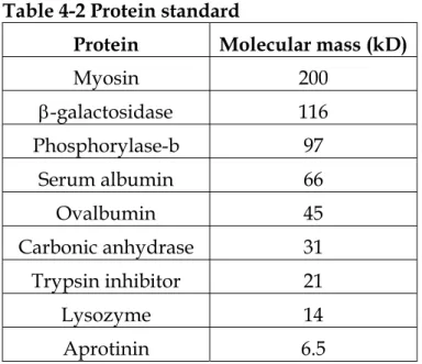
After transfer, the blot was rinsed in water and stained in Ponseau S solutions.
The blot was washed with water until protein bands appear and positions of the
Materials & Methods
standard protein marker were marked (the broad range protein marker from Bio- Rad was used). The size of the protein markers is outlined in Table 4-2.
The blot was blocked with 50 ml of 5% non-fat milk in PBS for 1 hr at room temperature or overnight at 4°C. The blot was rinsed for a while in PBS to remove milk and incubated with primary antibodies appropriately diluted in 1%
BSA/PBST (for anti-FLAG antibodies, 0.05 ng ml-1) for 1 hr at room temperature.
The blot was washed 3 times each in about 100 ml of PBST for 15 min. The blot was incubated with the HRP-conjugated secondary antibodies diluted in 50 ml of 3% non-fat milk in PBST (generally 1:5, 000) for 1 hr at room temperature. The blot was washed as above. The signal was subsequently visualized using ECL® western blotting detection system (Amersham).
For western-blot using anti-FolR antibodies, the sample buffer contains less SDS (0.1% final concentration) and no DTT or β-ME. Samples were directly loaded on normal SDS-PAGE gel without prior boiling.
Table 4-2 Protein standard
Protein Molecular mass (kD) Myosin 200
β-galactosidase 116
Phosphorylase-b 97
Serum albumin 66
Ovalbumin 45 Carbonic anhydrase 31
Trypsin inhibitor 21
Lysozyme 14 Aprotinin 6.5
5.2.3.10 Aerolysin Overlay assay
Protein samples (2 µg of GICs and 50 µg of isolated Golgi membranes or total membranes) were first resolved on SDS-PAGE. After electrophoresis, blotting
Materials & Methods
was subsequently carried out as standard western-blot procedures. All next steps were basically similar to western-blot, including blocking and washing. Instead of primary and secondary antibodies, the blot was incubated with radio-labeled aerolysin probes in a binding buffer (50 mM NaH2PO4 pH7.5 and 0.3% Tween- 20) and subsequently autoradiographed (Abrami et al. 2001).
5.2.4 Sequence analysis by use of web-based programs
DNA sequencing data revealed that all 4 inserts in the isolated clones (4.2.2.5.3) contained the same ORF. The translated protein sequence was analyzed by web- based programs. A hydrophilicity plot was drawn at Weizmann Instittue (http://bioinformatics.weizmann.ac.il/hyd-bin/plotfft_hydroph.pl) using Kyte- Doolittle method, coiled coil structures were predicted by SMART at EMBL (http://smart.embl-heidelberg.de/) and by COILS at Swiss EMBnet node (http://www.ch.embnet.org/software/COILS_form.html). Homology searching was performed by Blast2 or the Retrieve Short Nearly Exact Matches program at NCBI (http:// www.ncbi.nlm.nih.gov). Multiple sequence alignment was done at EBI (http://www.ebi.ac.uk/clustalw/) and graphically represented using BoxShade 3.21 at Swiss EMBnet node (http//
www.ch.embnet.org/software/BOX_form.html). GPI anchoring prediction was carried out using big-PI predictor at University of Vienna (http://mendel.imp.univie.ac.at/sat/gpi/gpi_sever.html).