Original article:
TCRVβ CLONOTYPIC ANALYSIS OF VAGINAL T LYMPHOCYTES DURING EXPERIMENTAL VAGINAL CANDIDIASIS IN THE
MURINE SYSTEM
Mawieh Hamad,1, 2* Enas M. El-Younis,1 Salem R. Yasin,1 Khaled Abu-Elteen1
1 Department of Biology & Biotechnology, Hashemite University, Zarqa, Jordan;
2 JMS-Medicals, Amman, Jordan
* Corresponding author: e-mail: taqiwmohanad@yahoo.com; Tel.: 00966 6 488 5895 ABSTRACT
To further understand the role of localized T cells in protection against vaginal candidiasis (VC), vaginal T lymphocyte TCRVβ repertoir was evaluated during experimental VC. RNA was extracted from lymphocytes of naïve and estrogen-treated Candida albicans-infected mice and RT-PCRed using TCRVβ primers corresponding to 24 common TCRVβ genes. All TCRVβ rearrangements were detected in vaginal T cells and thymocytes of naive mice. Al- though, the full complement of TCRVβ repertoire was detectable in vaginal T cells at days 14 and 21 post-infection, there was overrepresentation of TCRVβs 1, 6, 10 and 15 at day 14 and TCRVβs 1, 4, 6, 8.3, 10, 13, 14 and 18 at day 21. Expression of all TCRVβ rearrangements in vaginal T cells raises the possibility of an extrathymic developmental origin. Time-dependent overrepresentation of specific TCRVβs during VC may indicate infection-related specific T cell clonal expansion.
Keywords: Candida albicans, extrathymic T cell development, TCRVβ, vaginal candidiasis, vaginal T lymphocytes
INTRODUCTION
Vaginal candidiasis (VC) represents a real health problem to women of childbear- ing age worldwide (Ferrer, 2000; Enoch et al., 2006; Pfaller et al., 2007). C. albicans, a commensal of the genitourinary tract, is en- countered in the greater majority of VC cases (Pfaller et al., 2007). The occurrence of VC and recurrent vulvovaginal candidi- asis (RVVC) has been attributed to com- promised immunity and increased levels of estrogen in the reproductive tract milieu (Larsen et al., 1984; Hamad et al., 2004). A significant percentage of RVVC cases also occurs in pre-menopausal women with competent immunity (Giraldo et al., 2000;
Fidel, 2002; Cassone et al., 2007). Al- though T cell-mediated immunity (CMI), specifically Th1-type response, is consid-
ered to be the major defense mechanism against VC, systemic CMI plays a minor role (Cassone et al., 2007; Fidel et al., 1995a; Fidel, 2002). Systemic T cell re- sponses generated following the induction of VC failed to provide significant protec- tion against subsequent localized C. albi- cans infection in mice with experimental VC (Fidel, 2002 and references therein).
Deletion of systemic CD4+ or CD8+ T cells did not significantly influence the progres- sion of VC in mice (Fidel et al., 1995b).
Furthermore, vaginal, but not peripheral, T cells undergo significant kinetic changes during experimental VC in mice (Ghaleb et al., 2003).
Vaginal T cells exhibit a phenotypic profile distinct from that of peripheral T cells (Hamad, 2008; Fidel et al., 1996; Jo- hanson & Lycke, 2003; Ibraghimov et al.,
1995; Fidel et al., 1999). The majority (>80 %) of vaginal T lymphocytes are of the CD3+CD4+ phenotype. Unlike periph- eral T cells, the majority of CD4+ vaginal T cells react with the 2B6 anti-CD4 but not the GK1.5 anti-CD4 antibody (Fidel et al., 1999). The percentage of CD8+ T cells was reported at 1 % (Cassone et al., 2007;
Hamad, 2008). This notwithstanding other findings that have indicated that CD8+ T cells predominate the vaginal mucosa dur- ing VC (Ghaleb et al., 2003). A wealth of literature seems to suggest that vaginal T cells exhibit phenotypic and developmental properties similar to that of intestinal intra- epithelial lymphocytes (IELs) and other lo- calized T cell subsets (Hamad, 2008; Ishi- kawa et al., 2007). For example, reminis- cent of the IEL T cell population, vaginal T cells contain a significant percentage of TCR-γδ+ T cells (Hamad, 2008; Fidel et al., 1996; Johanson & Lycke, 2003; Ibraghi- mov et al., 1995; Fidel et al., 1999). Few CD4+CD8+ cells were reported to populate the vaginal mucosa (Ghaleb et al., 2003;
Hamad, 2008 and some references therein).
A unique population of TCRαβ+CD3+B220lowCD4-CD8-NK1.1- T cells was detected in the vaginal mucosa of nude mice and mice deficient for MHC-II, β2-microglobulin or CD1 (Johanson & Ly- cke, 2003). Furthermore, evidence in sup- port of lymphocyte trafficking between the periphery and the vaginal mucosa are yet to be sufficiently established (Cassone et al., 2007; Rakasz et al., 1998).
To further understand the development and function of vaginal T cells, TCRVβ repertoire was examined during experimen- tal vaginal candidiasis. Kinetics of TCRVβ+ T cell subsets were evaluated in C. albicans infected mice at different time points post- infection. The developmental pathway of vaginal T cells was addressed by comparing the patterns of TCRVβ gene usage between vagina- and thymus-derived T cells.
MATERIALS AND METHODS
Mice and microorganisms
12-14 week-old female Balb/c mice raised at the Hashemite University vivarium were used in this study. Handling of ex- perimental animals was in full compliance with relevant animal welfare policies and procedures as specified in the 1996 ILAR Guide for the care and use of laboratory animals. ATCC C. albicans strain 36082 is a kind gift from Dr. MA Ghannoum (My- cology Reference Laboratory, University Hospital of Cleveland, OH, USA). The fun- gus was maintained on Sabouraud Dextrose Agar (SDA) (Difco, MI; USA), stored at 4oC and subcultured at 3 months intervals.
For inoculation, overnight cultures of C.
albicans were grown at 37 oC in SDA broth as described previously (Abu-Elteen et al., 1997). Prior to use, cells were harvested and washed twice in sterile physiological saline (SPS). Estrogen (estradiol valerate;
Schering AG, Germany) was administered subcutaneously at 0.5 mg per 0.1 ml sesame oil 72 hrs prior to C. albicans inoculation and at weekly intervals thereafter. Vaginal C. albicans inoculate consisted of 50 µl containing 2x10⁷ viable stationary-phase blastoconidia.
Evaluation of C. albicans colonization Mice were sacrificed at days 7, 14 and 21 post-infection; vaginal tissues were iso- lated, examined for the presence of white lesions characteristic of C. albicans infec- tion, trimmed and homogenized in 10ml SPS in a sterile glass homogenizer (Ystral GmbH, Göttingen, Germany). Serial 10- fold dilutions were prepared from the ho- mogenate; 1ml aliquots of the appropriate dilution were added into culture plates con- taining 10 ml SDA and chloramphenicol at 50 mg/L, plates were left to solidify and then incubated at 37 oC; each sample dilu- tion was cultured in triplicate. Yeast colo- nies were counted 48 hrs after plating and colonization results were expressed as the mean colony-forming unit (CFU) per mouse based on data from three animals per group.
Isolation and flow cytometric analysis of vaginal T lymphocytes
Isolation and immunostaining of vaginal T cells was done according to previously published procedures (Ghaleb et al, 2003).
Briefly, 5-6 mice were sacrificed per group per time point, vaginas were isolated and flushed with normal saline, opened up lon- gitudinally and cut into 2 mm pieces. Tis- sue pieces were placed in 50 ml of warm RPMI-1640 (Sigma Chemicals, MO; USA) solution containing 10 mM EDTA plus 1 mM DTT and stirred for 30 min at 37 oC.
The suspension was filtered through a 1 gm nylon wool column moisturized with warm HBSS (Sigma), the filtrate was then centri- fuged and the cell pellet was suspended in 1 ml HBSS. Lymphocytes were counted manually; a total of 104-105 cells in 100 µl HBSS were used per sample for flow cytometric analysis. Biotin-labeled anti- CD3 (KT3), phycoerythin (PE)-labeled anti-CD4 (YTS191.1) and fluorescein isothiocyanate (FITC)-labeled anti-CD8 (KT15) antibodies (Serotec Ltd., Oxford, UK) were used for T cell phenotypic characterization. For single-color analysis, biotin-labeled anti-CD3 was added at 1µl per sample, left to react for 30 minutes on ice, cells were then centrifuged, washed with 100 µl HBSS, cell pellet was then resuspended in 100 µl HBSS and reacted with PE-labeled streptavidin. For dual-color analysis, each sample was reacted with 2 µl FITC-labeled anti-CD8 and 1 µl PE-labeled anti-CD4 for 30 minutes on ice prior to washing. Flow cytometric analysis was performed on a PARTEC-PASflow cyto- meter (Partec, Münster, Germany); 104-105 cells were analyzed per sample and data were collected and analyzed using flowmax software (Partec).
PCR-spectratype analysis
Total cellular RNA was extracted from lymphocytes using the SV Total RNA Iso- lation System Kit (Promega, WA, USA).
Synthesis of cDNA from extracted/purified RNA was performed using the Reverse Transcription System Kit (Promega). Gene segments of rearranged TCRVβ were PCR
amplified across the VDJ junctional site using Vβ and Cβ primers (Pannetier et al., 1993). Amplification reactions were sepa- rately carried out each in a total reaction volume of 100 µl including 10 µl of each synthesized cDNA, 50 µl PCR Master Mix (50 U/ml Taq DNA polymerase, 3 mM MgCL2, and 400 µM of each dNTP; pH 8.5) (Promega) and 1 µl Cβ primer. The mixture was equally aliquoted into 24 tubes, each containing 1 µl of Vβ-specific primer solu- tion. The volume was brought up to 100 µl by adding 38 µl nuclease-free water per tube. PCR amplification was conducted us- ing MyCycler Thermal Cycler (BioRad Laboratories Inc., Life Science Group, Cali- fornia, USA) following the protocol: dena- turation for 1min at 94 ºC followed by 40 cycles consisting of 70 sec at 94 ºC, 1min at 60 ºC, and 4 min at 72 ºC, followed by 10 min incubation at 72 ºC to complete product extension. PCR amplification prod- ucts were separated on a 3 % agarose gel in TBE buffer (Promega). 12 µl of the PCR product of each sample was mixed with 2 µl of Blue/Orange 6X loading dye (Promega), prior to loading. 1.5 µl of loading dye was mixed with 1.5 µl of the 25 Kb DNA ladder (Promega) consisting of 12 bands ranging in size from in 25-300 bp as a control. Gels were run at 50 volts for 3hrs, destained and photographed using a 312 nm UV transil- luminator (Uvitec, EEC) equipped with a gel documentation System (UVIsave, EEC). Amplification of cDNA samples pre- pared from RNA of C. albicans cells was carried out as negative controls of the PCR reaction.
RESULTS
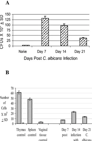
Persistent vaginal candidiasis was evi- dent in estrogen-treated C. albicans- infected mice throughout the infection pe- riod as compared with the minimal C. albi- cans CFU counts detected in naïve mice (Figure 1A). Vaginal fungal burden in mice infected with C. albicans without receiving estrogen declined to background levels within 1 week post-infection (data not shown). Numbers of lymphocytes isolated
from the vaginal tract epithelium of estro- gen-treated C. albicans-infected mice at days 7, 14 and 21 following the inoculation of C. albicans are shown in Figure 1B.
Overall, the number of vaginal lymphocytes was significantly higher in experimental mice than that in control mice (P<0.05).
The peak value of vaginal lymphocyte number (23x106 cells) was reached at day 14 post-infection; which represents an in- crease of 9-fold as compared with that in control mice.
A
B
Figure 1: (A) Vaginal C. albicans burden during experimental vaginal candidiasis. Levels of C.
albicans colonization in naïve untreated mice, untreated infected mice and treated infected mice were assessed at days 7, 14 and 21 post- infection. Mean C. albicans CFU count per va- gina + SD was calculated based on three sepa- rate experiments per group. (B) Kinetics of vaginal lymphocytes during experimental vagi- nal candidiasis. Relative numbers of total vagi- nal lymphocytes isolated from the naïve un- treated mice and estrogen-treated C. albicans- infected were evaluated at days 7, 14 and 21 post-infection. At each time point, 5-6 mice per group were sacrificed, pooled cell preparations were enumerated and the average number of cells per vagina was calculated. Data shown is the mean + SD of three separate experiments.
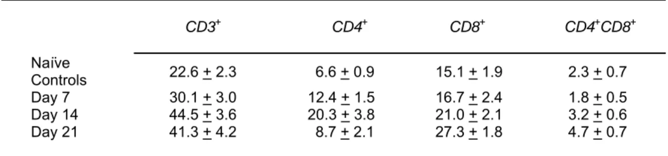
To establish the general phenotypic pro- file of this cell population, flow cytometric analysis was carried out on vaginal cell iso- lates from naïve mice and experimental mice at different time points during the in- fection (Table 1 and Figure 2). Consistent with previous studies (Ghaleb et al., 2003;
Fidel et al., 1996), the percentage of CD3+ T cells in vaginal cell isolates ranged be- tween 20 % at the start of the infection pe- riod to about 45 % at day 21 post-infection;
possibly indicative of T cell proliferation as part of the incessant anti-fungal immune response. Although the predominance of CD8 in the vaginal T cell pool was evident, a significant percentage of CD4+ T cells was present at all time points. Interestingly, a minor T cell population (<5 % of total vaginal T cells) expressing both CD4 and CD8 was also detected.
Figure 2: Expression of CD3, CD4 and CD8 T cell markers on vaginal T cells during experimental vaginal candidiasis. Vaginal T cells were isolated from C. albicans infected – estrogen-treated mice at week 3 post-infection, were pooled from 5-6 mice per group and reacted with anti-CD3 or with anti-CD8 and anti- CD4. Data presented are representative of three separate experiments.
0 25 50 75 100 125 150
Naïve Day 7 Day 14 Day 21 Days Post C. albicans Infection CFUs X 103 + SD
0 10 20 30 40 50 60 70
Thymus control
Spleen control
Vaginal tissue control
Day 7 post
Day 14 infection
with Day 21
C.
albicans Number
of Cells X 106
+ SD
Table 1: Percentage expression of CD3, CD4 and CD8 on murine vaginal T lymphocytes during experimental vaginal candidiasis
Animal group Mean % positive cells + SD*
CD3+ CD4+ CD8+ CD4+CD8+
Naïve
Controls 22.6 + 2.3 6.6 + 0.9 15.1 + 1.9 2.3 + 0.7
Day 7 30.1 + 3.0 12.4 + 1.5 16.7 + 2.4 1.8 + 0.5
Day 14 44.5 + 3.6 20.3 + 3.8 21.0 + 2.1 3.2 + 0.6
Day 21 41.3 + 4.2 8.7 + 2.1 27.3 + 1.8 4.7 + 0.7
* Mean percentages of vaginal T cells expressing the specific marker were calculated based on three separate experiments
The patterns of TCRVβ rearrangements in vaginal and thymus-derived T cells iso- lated from naïve mice were evaluated by RT-PCR clonotypic analysis (Figure 3). As shown in the top panel of Figure 3, thymo- cytes contained all TCRVβ rearrangements except for TCRVβs 19 and 20 which were either absent or very faint. As for peripheral T cells (spleen and lymph nodes), TCRVβs 1, 4, 10, 11, 13, 15, 16 and 17 were strongly represented perhaps indicative of increased relative cell numbers expressing these rear- rangements. In cells from both tissues, TCRVβs 3.1, 5.1, 5.3, 8.1, 8.2, 8.3, 9, 19 and 20 were consistently low or absent (data not shown). With regard to vaginal T cells in naïve mice, the expression of TCRVβs 5.3 and 20 was consistently weak
while that of TCRVβs 4, 6, 7, 8.1, 8.2, 13, 14 and 15 was very prominent perhaps re- flecting increased relative cell number of clones expressing the corresponding rear- rangements (lower panel of Figure 3).
Therefore, the pattern of TCRVβ gene us- age in vaginal T lymphocytes seems to be similar, though not completely identical, to that observed in thymocytes. Amplification reactions using the same primers set and PCR conditions were carried out on cDNA samples prepared from C. albicans cells as negative controls. As expected, no PCR products were observed in this series of ex- periments indicating that the PCR products detected are lymphocyte-specific (data not shown).
Figure 3: PCR- spectraype analysis of Vβ gene rearrange- ments in T cells iso- lated from the thymus (A) and vaginal tract T cells (B) of naïve con- trol mice. Numbers above panels indicate Vβ gene number and the 25-bp DNA ladder is indicated by L on top of the first lane from left. Data shown are representative of three separate experiments.
Vaginal T lymphocytes isolated from estrogen-treated C. albicans–infected mice exhibited an interesting TCRVβ repertoire (Figure 4). Clear shifts in the TCRVβ gene usage seems to occur between the naïve state versus the early stages of infection and between the early stages of infection versus those when the infection was well estab- lished. At day 7 post-infection (Figure 4, top panel), TCRVβs 2, 3, 6 and 7 were missing while TCRVβs 5.1 and 10 were strongly represented. At day 14 post- infection (Figure 4, middle panel), the whole TCRVβ gene complement reap- peared with TCRVβs 1, 6, 10 and 15 being overrepresented in comparison with the rest of the TCRVβ complement. At day 21 post- infection (Figure 4, lower panel) extensive rearrangement of TCRVβs 1, 4, 6, 8.3, 10, 13, 14 and 18 was evident. TCRVβs 5.1, 8.1 and 8.3 were well represented in vaginal T cells of experimental mice at day 14 and day 21 post-infection as compared with that in vaginal T cells of naïve mice. These find- ings were consistently observed in 3 sepa- rate experiments.
DISCUSSION
Findings presented here should help in further understanding the development and function of vaginal T cells. The important role of vaginal T cells in protection against vaginal candidiasis is discernable from sev- eral findings. First, there was a significant increase in the number of vaginal lympho- cytes during the infection which is consis- tent with previous studies (Ghaleb et al., 2003; Fidel et al., 1996; Al-Sadeq et al., 2008). In agreement with previous reports (Ghaleb et al., 2003; Al-Sadeq et al., 2008), the majority of T cells that undergo expan- sion during the course of VC infection are of the CD8+ phenotype (Figure 2). Second, there was consistent overrepresentation of certain TCRVβs (6 and 10 in particular) during vaginal candidiasis. A likely expla- nation of this finding is that T cell clones expressing these rearrangements may be directly involved in responding to C. albi- cans antigens.
Figure 4: PCR- spectratype anal- ysis of V gene
rearrangements in T cells isolated from the vaginal tracts of C.
albicans-infected mice at days 7 (A), 14 (B) and 21 (C). Numbers above panels indicate Vβ gene number and the 25-bp DNA lad- der is indicated by L on top of the first lane from left.
Data shown are representative of three separate experiments.
Third, there were unique time- dependent changes in the pattern of TCRVβ rearrangement dominating the vaginal T cell pool during the course of infection. It is possible that the profile of C. albicans anti- gens being presented to vaginal T cells shifts overtime hence the occurrence of concurrent shifts in the dominating subset of T cell clones expressing the appropriate TCRVβ rearrangements. Based on this ob- servation, it is postulated that T cell clones positive for TCRVβ1, 6, 10 and 15 may represent early T cell responders (Thelper) while those expressing TCRVβs 4, 8.3, 13, 14 and 18 represent late T cell effectors (Tcytotoxic) cell subset (Fgure 4, middle and lower panels). Should these results hold true, there could be significant therapeutic implications in the form of agonistic mono- clonal antibodies to target phase-specific T cells. Refining the rationale of cloning and adoptive T cell transfer into naïve or in- fected hosts as means of treating fungal in- fections is a second envisaged therapeutic avenue (Beck et al., 2006).
Evidence for an extrathymic pathway of vaginal T cell development as presented in this article derives from the finding that the pattern of TCRVβ gene usage in the vaginal T cell pool is more similar to that in thymo- cytes than that in peripheral (spleen or lymph node) T cells. The fact that the vagi- nal tract contained the full complement of TCRVβ provides compelling evidence that this site functions as a primary lymphoid organ in addition to its established role as a secondary lymphoid organ. In other words, it is likely that some T cells resident within the vaginal mucosa may extrathymically develop within the vaginal mucosa. This is further supported by previous findings which have indicated that the vaginal mu- cosa contains a minor subset of immature double positive (CD4+CD8+) T cell precur- sors (Ghaleb et al., 2003; Ibraghimov et al., 1995). Furthermore, the vaginal mucosa of nude mice and mice deficient for MHC-II, β2-microglobulin or CD1 were all shown to house a unique population of TCRαβ+CD3+B220lowCD4-CD8-NK1.1- T cells (Johanson & Lycke, 2003). Evidence
in support of a possible extrathymic devel- opment of similar localized T cell subsets like the IEL subset has been amply fur- nished (Hamad, 2008; Poussier et al., 1992;
Hamad et al., 1995). The close similarities between the small intestinal mucosa and the vaginal mucosa in terms of structure, con- tinuous exposure to pathogens and the need for rapid and effective immunity are to be considered in this regard.
Acknowledgements: This work was funded by research grant MH-2/0406, col- lege of graduate studies and scientific re- search, Hashemite University, Jordan.
REFERENCES
Abu-Elteen K, Abdul-Malek A, Abdul- Wahid NA. Prevalence and susceptibility of vaginal yeast isolates in Jordan. Mycoses 1997;40:179-85.
Al-Sadeq A, Hamad M, Abu-Elteen KH.
Patterns of expression of vaginal T cell activation markers during estrogen-main- tained vaginal candidiasis. Asthma Allergy Clin Immunol 2008;4:156–62.
Beck O, Topp M, Koehl U, Roilides E, Simitsopoulou M, Hanisch M, Sarfati J, Latgé JP, Klingebiel T, Einsele H, Lehrnbecher T. Generation of highly purified and functionally active human Th1 cells against Aspergillus fumigatus. Blood 2006;107:2562-9.
Cassone A, De Bernardis, Santoni G. Anti- candidal immunity and vaginitis: novel op- portunities for immune intervention. Infect Immun 2007;75:4675–86.
Enoch DA, Ludlam HA, Brown NM. Inva- sive fungal infections: a review of epidemi- ology and management options. J Med Mi- crobiol 2006;55:809–18.
Ferrer J. Vaginal candidiasis: epidemiologi- cal and etiological factors. Intl J Gynecol Obstet 2000;71:521-7.
Fidel PL. Immunity to candida. Oral Dis 2002;8:69-75.
Fidel PL, Lynch ME, Conaway DH, Tait L, Sobel JD. Mice immunized by primary vaginal Candida albicans infection develop acquired vaginal mucosal immunity. Infect Immun 1995a;63:547-53.
Fidel PL, Lynch ME, Sobel JD. Circulating CD4 and CD8 T cells have little impact on host defense against experimental vaginal candidiasis. Infect Immun 1995b;63:2403-8.
Fidel PL, Wolf NA, Kukuruga MA. T lym- phocytes in the murine vaginal mucosa are phenotypically distinct from those in the periphery. Infect Immun 1996;64:3793-9.
Fidel PL, Luo W, Steele C, Chabain J, Baker M, Wormley F. Analysis of vaginal cell populations during experimental vagi- nal candidiasis. Infect Immun 1999;67:
3135-40.
Ghaleb M, Hamad M, Abu-Elteen KH.
Vaginal T lymphocyte population kinetics during experimental vaginal candidiasis:
evidence for a possible role of CD8+ T cells in protection against vaginal candidiasis.
Clin Exp Immunol 2003;31:26-33.
Giraldo P, von Nowaskonsk A, Gomes FA, Linhares I, Neves NA, Witkin SS. Vaginal colonization by Candida in symptomatic women with and without a history of recur- rent vulvovaginal candidiasis. Obstet Gyne- col 2000;95:413-6.
Hamad M. The case for extrathymic development of vaginal T lymphocytes. J Reprod Immunol 2008;77:109-16.
Hamad M, Whetsell M, Klein JR. T cell precursors in the spleen give rise to com- plex T cell repertoires in the thymus and the intestine. J Immunol 1995;155:2866-76.
Hamad M, Abu-Elteen KH, Ghaleb M.
Estrogen-dependent induction of vaginal candidiasis in naive mice. Mycoses 2004;47:304-9.
Ibraghimov A Sacco RE, Sandor M, Iakoubov LZ, Lynch RG. Resident CD4+ αβ T cells in the murine female genital tract: a phenotypically distinct T cell line- age that rapidly proliferates in response to systemic T cell activation stimuli. Int Im- munol 1995;7:1763-9.
Ishikawa H, Naito T, Iwanaga T.
Curriculum vitae of intestinal intraepithelial T cells: their developmental and behavioral characteristics. Immunol Rev 2007;215:
154-65.
Johanson M, Lycke N. A unique population of extrathymically derived αβ TCR+CD4- CD8- T cells with regulatory functions dominates the mouse female genital tract. J Immunol 2003;170:1659-66.
Larsen B, Galask RP. Influence of estrogen and normal flora on vaginal candidiasis in the rat. J Reprod Med 1984;29:863-8.
Pannetier C, Cochet M, Darche S, Casrouge A, Zöller M, Kourilsky P. The size of the CDR3 hypervariable regions of the murine T-cell receptor β chains vary as a function of the recombined germ line segments. Proc Natl Acad Sci U S A 1993;90:4319-23.
Pfaller MA, Diekema DJ. Epidemiology of invasive candidiasis: A persistent public health problem. Clin Microbiol Rev 2007;
20:133-63.
Poussier P, Edouard P, Lee C, Binnie M, Julius M. Thymus-independent develop- ment and negative selection of T cells ex- pressing T cell receptor α/β in the intestinal epithelium: evidence for distinct circulation patterns of gut- and thymus-derived T lym- phocytes. J Exp Med 1992;176:187-99.
Rakasz E, Rigby S, De Andres B, Mueller A, Hagen M, Dailey MO, Sandor M, Lynch RG. Homing of transgenic gammadelta T cells into murine vaginal epithelium. Int Immunol 1998;10:1509-17.