Roles of the vagina and the vaginal microbiota in urinary tract infection: evidence from clinical correlations and experimental models
Abstract
Mounting evidence indicates that the vagina can harbor uropathogenic bacteria. Here, we consider three roles played by the vagina and its
Amanda L. Lewis
1,2,3Nicole M. Gilbert
2,3,4bacterial inhabitants in urinary tract infection (UTI) and urinary health.
First, the vagina can serve as a reservoir forEscherichia coli, the most
common cause of UTI, and other recognized uropathogens. Second, 1 Molecular Microbiology, Washington University School several vaginal bacterial species are frequently detected upon urine
of Medicine in Saint Louis, United States
culture but are underappreciated as uropathogens, and other vaginal species are likely under-reported because of their fastidious nature.
Third, some vaginal bacteria that are not widely viewed as uropathogens 2 Obstetrics and Gynecology, Washington University School can transit briefly in the urinary tract, cause injury or immunomodulation,
of Medicine in Saint Louis, United States
and shift the balance of host-pathogen interactions to influence the outcomes of uropathogenesis. This chapter describes the current liter- ature in these three areas and summarizes the impact of the vaginal microbiota on susceptibility to UTI and other urologic conditions.
3 Center for Women's Infectious Disease Research, Washington University School Keywords:bacterial vaginosis, uropathogen, Escherichia coli, Gardnerella
vaginalis, group B streptococcus, lactobacillus, aerococcus,
staphylococcus, ureaplasma, lower urinary tract symptoms, probiotic
of Medicine in Saint Louis, United States
4 Center for Reproductive Health Sciences, Washington University School of Medicine in Saint Louis, United States
Summary of findings
1. Data from clinical studies and model systems highlight the vaginal microbiota as a key factor impacting sus- ceptibility to UTI and other urologic conditions.
2. The vagina can serve as a reservoir for uropathogen colonization.
3. Certain members of the vaginal microbiome are fre- quently detected in urine yet are underappreciated as uropathogens. Other vaginal bacterial species are likely underreported because they are difficult to cul- ture or to identify.
4. The urinary tract can be transiently exposed to vaginal bacteria, some of which can cause injury to the blad- der epithelium and impact pathogenesis of recognized uropathogens.
1 Introduction
It is well known, that women suffer from urinary tract in- fections (UTI) much more frequently than men. Mounting evidence indicates that the vagina can harbor uropatho- genic bacteria. According to the current literature we consider the evidence from clinical correlations and ex- perimental models that the vaginal microbiota impacts susceptibility to UTI and other urologic conditions.
2 Methods
We performed literature searches in PubMed. For our overview of the vaginal microbiome and its impact on the urinary tract, we searched “vaginal microbiome” OR “va- ginal microbiota” AND “bacterial vaginosis” OR “BV” OR
“dysbiosis”. To find articles relating to vaginal colonization by uropathogens, we searched “Escherichia coli” OR
“Staphylococcus” OR “group BStreptococcus” OR “GBS”
AND “vagina” OR “vaginal”. To identify articles relating to vaginal species that may be underappreciated uropatho-
Figure 1: Examples of Gram-stained slides from women with (right, Nugent score = 9) and without (left, Nugent score = 1) BV.
In women with a lactobacilli-dominated microbiome (left), long purple rods are the main morphotype. In contrast, women with BV tend to have a lot more bacteria overall, with a significant fraction that do not stain as Gram-positive (i.e. pink). Morphotypes in
BV tend not to be long Gram-positive rods. Although not part of the Nugent scoring system, women with BV tend to have large conglomerations of bacteria associated with epithelial cells as seen in the upper right quadrant.
gens, we examined the list of organisms commonly de- tected in vaginal microbiome studies and searched these organisms along with “urinary tract” OR “urine” OR “UTI”.
3 Results and discussion
In order to better understand the role of the vaginal mi- crobiota in UTI, we consider the “healthy” or “normal”
state in reproductive age women and in pregnancy and three roles played by the bacterial inhabitants of the va- gina in UTI and urinary health. First, the vagina can serve as a reservoir for Escherichia coli, the most common cause of UTI, and other recognized uropathogens. Second, several vaginal bacterial species are frequently detected upon urine culture but are underappreciated as uropatho- gens, and other vaginal species are likely under-reported because of their fastidious nature. Third, some vaginal bacteria that are not widely viewed as uropathogens can transit briefly in the urinary tract, cause injury or immuno- modulation, and shift the balance of host-pathogen inter- actions to influence the outcomes of uropathogenesis.
3.1 Vaginal microbiota composition
In reproductive age women, the vaginal microbiota is dominated by a few Lactobacillus species, including L. crispatus,L. gasseri,L. jensenii, andL. iners. These lactobacilli are thought to prevent growth of potential pathogens by maintaining the vagina’s characteristic low pH (by producing lactic acid [1]) and by producing antimi- crobials such as hydrogen peroxide and bacteriocin-like substances [2]. Although aLactobacillus-dominant vaginal microbiota is considered the “healthy” or “normal” state, a large proportion of reproductive age women (up to one- third in the United States) have a more diverse vaginal microbiota that contains no or low levels ofLactobacillus and a mixture of Gram-negative anaerobes, Actinobac- teria, and otherFirmicutes[3]. This apparent dysbiosis
is called bacterial vaginosis (BV) or community state type IV [4]. While fewer studies have examined postmenopaus- al women, these echo what has been observed in younger cohorts, with some women having Lactobacil- lus-dominant and others having a more diverse polymi- crobial vaginal microbiome [5], [6], [7]. A few studies have suggested that pregnancy may favor a stableLactobacil- lus-dominant vaginal microbiome, however, a diverse vaginal microbiome is nonetheless present in some pregnant women [8]. In the clinic, a woman is diagnosed with BV if she has three of the four following signs (Amsel criteria): vaginal pH>4.5, ‘thin’ grayish homogenous va- ginal fluid, vaginal fluid that produces a fishy odor when treated with potassium hydroxide, and the presence of epithelial cells studded with bacteria (i.e., ‘clue-cells’) in wet mount. In the laboratory, BV is diagnosed via the Nugent scoring system [9], which is based on morphotype assessment of Gram-stained slides; a score of 7 or higher on a 10-point scale indicates BV (see Figure 1 for ex- amples of Gram-stained slides from women with and without BV). Women with BV are at higher risk of experi- encing a wide array of negative health outcomes, includ- ing increased risk of a variety of secondary infections and adverse pregnancy outcomes.
3.2 Relationships between the vaginal microbiota, UTI, and urinary health
Data from multiple clinical studies suggest that a woman’s vaginal microbiota affects her susceptibility to UTI [10].
For example, women with BV have higher UTI risk than women with lactobacilli-dominated vaginal microbiota [11], [12], [13]. Furthermore, clinical trials suggest that vaginal interventions that affect the microbiota (e.g., va- ginal probiotic and estrogen treatments) can protect against additional episodes of recurrent UTI (rUTI) [14], [15]. As described later in this chapter, many of the bac- terial genera found in the BV-associated vaginal environ-
ment have been detected, via both culture-dependent and -independent methods, in the urinary tract. Addition- ally, some of these bacteria have been implicated as causes of acute UTI or other urologic conditions. These findings suggest that fastidious BV-associated organisms may be important in the etiology of uropathology and uropathogenesis; however, in many cases additional studies are needed to ascribe causal roles for these or- ganisms in the urinary tract.
Why women with a disrupted vaginal microbiota are at increased risk of UTI is unclear. Below, we describe three ways the vaginal microbiota could influence UTI and other urologic conditions. First, the vaginal introitus appears to be a key reservoir for uropathogenicE. coli. Second, other somewhat less common uropathogens can also be com- monly harbored in the vagina (see Figure 2). Finally, transient exposures of the urinary tract to certain vaginal bacteria may prime the urinary tract for uropathogens or trigger rUTI via other mechanisms.
Figure 2: Schematic illustrating vaginal bacteria with potential to impact the urinary tract.
The vagina can serve as a reservoir for several bacterial species known to be causes of UTI (E. coli, GBS,Staphylococcus) as well as underappreciated potential uropathogens (G.vaginalis,
Aerococcs Ureaplasma) that can cause UTI and have been associated with urological conditions such as urgency
incontinence and “sterile” pyuria.
3.3 The vagina provides a reservoir for uropathogenic E. coli and other
recognized uropathogens
3.3.1 E. coli
The most common cause of UTI in most patient popula- tions is uropathogenicE. coli[16], [17]. The pathogenesis ofE. coliUTI is often described as a series of colonization events, starting in the gastrointestinal tract, followed by the vaginal introitus and urethral meatus, and finally the bladder and possibly kidneys [10], [18]. This progression makes sense given the proximity of the urethra to the vaginal introitus and anus in females and that longitudinal examinations have indicated that introital and urethral colonization precedes UTI symptom onset [19], [20].
Moreover, multiple clinical studies have found that women with a history of rUTI more commonly haveE. coliin the vaginal introitus or vagina than do healthy controls [19], [20], [21], [22]. One such study found thatE. colicoloni- zation of the vaginal introitus reached higher levels in women with a history of UTI (>105 colony forming units/mL) than in controls (<200 cfu/mL) [20]. In a sep- arate study, concurrent colonization of the vaginal introi- tus and urinary tract by the same strain ofE. coli(based on random amplified polymorphic DNA fingerprinting) occurred in 85% of paired isolates from women with a history of rUTI [20]. Together, these results demonstrate that the vagina provides a reservoir for uropathogenic E. coli.
As described above, Lactobacillus in the vagina may prevent colonization by potential pathogens such as E. coli. Indeed, women with low levels of lactobacilli more commonly carry vaginalE. colithan do those with lacto- bacilli-dominated microbiomes [13], [21]. In one study, E. colivaginal (introital) colonization was more common among women whose vaginal introitus was negative for hydrogen peroxide-forming lactobacilli than among those whose introitus was positive for these ‘beneficial’ bacteria (35% vs. 11%, odds ratio [OR], 4.0; P=0.01) [21]. Multiple studies in both nonpregnant and pregnant women suggest that those with BV have increased risk for UTI (odds ratios from 2.21 to 13.75) [12], [23], [24]. In one study, BV was associated with bothE. coliintroital colonization and UTI [13]. In another study, over 50% of women with a history of UTI, but only 13% of those with no UTI history, had ab- normal vaginal microbiota (P=0.03) as defined by a Nu- gent score greater than 3 (mean 4.6 vs. 1.7). Having a Nugent score of 7 or greater (indicative of BV) was also more common in UTI-prone women (6/22 [27%] vs. 1/17 [6%], P=0.095) [25]. This may be because lactic acid, hydrogen peroxide, and other small molecules produced by lactobacilli create a hostile environment for potential pathogens, including uropathogens, in the vagina [26].
Additional studies in experimental models are needed to define the effects of specific small molecules on the va- ginal environment.
One intervention that appears to restoreLactobacillus colonization and may protect against rUTI in some pa- tients is vaginal probiotics. A double-blind placebo-con- trolled trial studied 100 premenopausal women with at least one UTI in the past 12 months. Women received either intravaginal suppositories of powderedL. crispatus (Lactin-V) once daily for 5 days and then weekly for 10 weeks or placebo suppositories at the same interval.
Fewer women who established high-level vaginal L. crispatuscolonization (≥106throughout follow-up) de- veloped rUTI (relative risks were 0.07 for Lactin-V and 1.1 for placebo;P<0.01) [14]. In contrast, a smaller study found that vaginal application of other types of lactobacilli (L. caseiandL. rhamnosus) twice weekly did not reduce the UTI incidence [27]. However, the latter study used a higherE. colithreshold (104vs. 102) to diagnose an rUTI.
Additionally,L. caseiandL. rhamnosus, which are more common in the intestine than in the vagina, failed to col- onize the vagina [27]. Future studies are needed to de- velop effective vaginal prebiotic and probiotic strategies to treat or prevent rUTI.
Another promising intervention is vaginal estrogen. This idea is based on the observation that high levels of vagi- nal lactobacilli occur only in 25% to 30% of postmeno- pausal women (who tend to have low estrogen levels) but in 60% to 70% of women who receive estrogen replace- ment therapy. In a placebo-controlled study of postmeno- pausal women with rUTI, vaginal estrogen restored vaginal Lactobacillus colonization and decreased vaginal En- terobacteriaceaecolonization (67% of women pre-treat- ment vs. 31% post-treatment). Moreover, the UTI inci- dence was lower in the estrogen group than in the placebo group (0.5 vs. 5.9 episodes per patient year;
P<0.001) [15]. In a separate study, women receiving va- ginal estrogen had a 45% cumulative likelihood of remain- ing UTI-free over a 36-week follow up period, whereas those in the control group had only 20% likelihood of re- maining UTI-free [28]. For a more detailed description of the clinical studies examining the role ofLactobacillus andE. colivaginal colonization and vaginal interventions for UTI, please see this recent review [10].
3.3.2 Staphylococcus
The most frequent Gram-positive agent of community- acquired UTI is S. saprophyticus, but S. aureus and S. epidermidis can also cause UTI in certain settings (catheterization, pregnancy) [29], [30], [31].Staphylococ- cus uropathogenesis was recently reviewed [32]. Data fromin vitrostudies and rat UTI models have revealed several factors required forS. saprophyticusvirulence, including the secreted surface-associated proteins Aas (hemagglutinin) and Ssp (lipase); the cell wall proteins UafA, SdrI, SssF, and UafB, which mediate adherence;
and a urease that is associated with urinary stone form- ation. Additionally, data from a mouse model suggest that the nickel ABC-transporters Opp2 and Opp5a contribute toS. aureusuropathogenesis.
Both S. aureusandS. epidermidishave been detected in vaginal or cervical samples [33], [34], [35]. Vaginal S. aureushas been implicated in toxic shock syndrome and aerobic vaginitis [36], [37], [38], a controversial in- flammatory condition often mistaken for BV orCandida yeast infection. One study found that women with vaginal toxicogenicS. aureuswere significantly more likely than those without S. aureusto harbor vaginal E. coli[38].
However, vaginal colonization by staphylococci has not been examined as a risk factor for UTI.
3.3.3 Group B Streptococcus
Streptococcus agalactiae, otherwise known as group B Streptococcus(GBS), is a Gram-positive β-hemolytic chain- forming coccus that commonly inhabits the lower gastrointestinal tract and the vagina. Although GBS col- onization is often asymptomatic, GBS has been implicated in aerobic vaginitis [39]. GBS is also a recognized uro- pathogen, causing 2–3% (or ~160,000 cases annually in the U.S.) of all uncomplicated UTIs [17], [40]. GBS UTIs are more common in certain vulnerable populations. For example, among nursing home residents over 70 years of age, up to 39% of UTI cases involve GBS [41]. Addition- ally, GBS often causes asymptomatic bacteriuria and is found at significant titers in up to 7% of pregnant women [42], [43]. GBS is also frequently found in the urinary tract of people with diabetes, immunocompromised indi- viduals, and those with pre-existing urologic abnormalities, all of whom are at increased risk of ascending pyeloneph- ritis that can progress to bacteremia and/or urosepsis [44], [45]. Although GBS can inhabit both the vagina and the urinary tract, no studies have assessed the associ- ation between vaginal GBS and GBS UTI.
Mouse models of GBS UTI [46], [47], [48], [49] have mainly used serotype III GBS strains, which cause more symptomatic UTIs than most other serotypes [45]. The role of GBS virulence factors has not been as thoroughly examined in the urinary tract as in other niches, such as the bloodstream. However, available data suggest that the β-hemolysin/cytolysin is dispensable, whereas sialic acid residues of the GBS capsular polysaccharide are important for GBS survival in the urinary tract [47], [48].
Further studies are needed to define the bacterial and host mechanisms governing GBS disease in the urinary tract.
3.4 Other vaginal bacterial species that are underappreciated as uropathogens
3.4.1 Gardnerella vaginalis
Gardnerella vaginalis, a facultative Gram-variable Actinobacteria(a class frequently called ‘high-GC Gram- positives’), is best known as a frequent isolate and dom- inant member of the vaginal microbiota in BV. Although G. vaginalisis considered an unusual primary pathogen of the urinary tract, this bacterium can cause acute UTI [50]. The contribution of G. vaginalis to urinary tract
pathology is likely underestimated for two reasons. First, G. vaginalisdoes not grow under the standard (aerobic) culture conditions that most clinical microbiology labs use. Second, when labs identifyG. vaginalisin urine cul- tures, they do not report it as a potential uropathogen (even if present in pure culture at levels exceeding clinical thresholds for UTI diagnosis). Several studies suggest that G. vaginalis should be considered as a potential cause of urinary tract pathology. For example, in one study using appropriate culture conditions to detect it,G. va- ginaliswas isolated from 2.3% of urines from hospitalized patients, often in pure culture and >10,000 colony forming units per milliliter (cfu/ml). Compared to individu- als in whomG. vaginaliswas not detected, patients with G. vaginalisbacteriuria were more likely to have a history of rUTI or current pyelonephritis (kidney infection) [51].
These patients often reported symptoms, and 58% had pyuria (neutrophils in urine) [51]. Another study showed that the frequency ofG. vaginalisin catheterized urine samples was higher in women with urgency urinary incon- tinence than in women with other urologic conditions [52]. Other studies have used suprapubic needle aspira- tion to collect urine from the bladder, bypassing possible vaginal contaminants, concluding that pregnancy in- creases women’s risk of harboringG. vaginalisin their bladders and thatG. vaginaliswas especially common in women with underlying renal disease [53], [54], [55], [56], [57]. In another study,G. vaginaliswas commonly found in suprapubic aspirates from women with reflux scarring and “sterile pyelonephritis” [58]. Bladder washout studies strongly suggested thatG. vaginaliswas present in the kidneys of 75% of these patients. Finally, case reports implicateG. vaginalisin more serious dis- eases. For example,G. vaginalishas been isolated from women's bloodstreams during or after giving birth [59], andG. vaginalisbacteremia has been implicated in sys- temic diseases coinciding with urolithiasis (kidney stones) [60]. In short,G. vaginalishas been implicated in rUTI, urgency incontinence, kidney disease, and systemic infec- tions originating in the genitourinary system.
Thus far, few reports have assessedG. vaginalisin both the vagina and the urinary tract. However, one recent study compared the microbiota of paired midstream urine and vaginal fluid from 42 women with BV [61] and found high levels ofG. vaginalisin urine in a subset of women.
The authors noted an association between urine and va- ginal fluid communities in ~60% of women with urotypes dominated byG. vaginalis,Prevotella amni,Atopobium vaginae, or Sneathia amnii. In contrast, they reported only a 10–30% correlation among other urotypes (E. coli, Lactobacillus, etc.). The authors state that urine and va- ginal microbiota patterns were not correlated in most women, and suggest that the two communities are therefore distinct in many women. However, despite the suggestion that some vaginal bacteria, includingG. va- ginalis, can exist concurrently in the vagina and urinary tract, this manuscript does not appear to have evaluated the co-occurrence of individual taxa per se in the two different compartments. The findings raise a number of
interesting questions, including whether vaginal coloniza- tion withG. vaginalisa risk factor for G. vaginalis in urine or having concurrent or subsequent urologic pathologies.
G. vaginalis isolates have highly diverse genomes and can be divided into at least four clades. In fact, some authors have suggested thatG. vaginalisshould be di- vided into multiple species or even genera [62], raising the question of whether certainG. vaginalissubtypes are more likely to be pathogenic in the urinary tract.
3.4.2 Aerococcus
Aerococcus is an α-hemolytic and microaerophilic or facultatively anaerobic Gram-positive coccus. Several Aerococcusspecies, includingA. urinae,A. viridans, and A. sanguinicola, can cause UTI and urosepsis [63], [64], [65], [66], [67], [68], [69]. Patients withAerococcusUTI often have underlying risk factors, such as urological ab- normalities or older age [66], [67], [70]. Many case re- ports describing significantAerococcustiters in urine also reported isolating the bacterium from blood [66], [71], [72].Aerococcusmust be properly identified and treated to avoid life-threatening systemic infection [68]. However, Aerococcusis difficult to distinguish from viridans-group streptococci with routine phenotypic tests and thus re- quires molecular tools such as amplification and 16S rRNA sequencing, a method not commonly employed in clinical microbiology labs. Additionally, most reported Aerococcusisolates are resistant to sulfonamides [69], [70]. Thus, whenAerococcusis not recognized, ineffective antibiotic treatment can be given, leading to rapid pro- gression to systemic infection [67], [69], [71], [72], [73].
In addition to being implicated in UTI, Aerococcuswas more frequently cultured from urine (collected by transurethral catheterization) from patients with urgency urinary incontinence than from women with other urinary tract conditions [52]. To our knowledge, Aerococcus pathogenesis in the urinary tract has not been examined within vivomodels.
Aerococcus sp. are commonly isolated from the human vagina and urinary tract and from air, dust, and vegeta- tion. More studies are needed to understand how these naturalAerococcusniches affect pathogenesis and the clinical course of infections [74]. Aerococcus vaginal colonization is more common and abundant in women with BV [75], [76], but no studies have examined whether women with BV, or their sexual partners, are at increased risk forAerococcusUTI.
3.4.3 Ureaplasma
Ureaplasma species, including U. urealyticum and U. parvum, are characterized by their ability to hydrolyze urea. U. parvum UTI pathogenesis was examined in Fischer 344 rats, revealing two distinct outcomes. Some animals developed a minimal immune response with limited monocytic and lymphocytic lesions and elevated urinary interferon (IFN)-γ, interleukin (IL)-18, and mono- cyte chemoattractant protein-1. Other animals developed
an exaggerated pro-inflammatory immune response with elevated urinary IL-1α, IL-1β, CXCL1 (a.k.a. KC), neutro- philic lesions with extensive uroepithelial hyperplasia, and struvite (stone) formation [77]. A separate study demonstrated that rat strains differed in their susceptibil- ity toU. parvumUTI and struvite formation [78]. Together, these studies suggest that host factors influenceU. par- vumUTI outcomes.
Between 40% and 80% of sexually active women have vaginal/cervical ureaplasmas [79]. Some studies indicate that Ureaplasmavaginal colonization increases during BV, but others have failed to find associations with BV [80] or vaginal symptoms in general [81]. However, ur- eaplasmas are reproducibly the most common cause of amniotic fluid infection in pregnant women, suggesting the bacteria can invade urogenital tissues and cause serious infection [82], [83]. Although acute cystitis by Ureaplasmais rare, the genus has been implicated in lower urinary tract symptoms [84], [85], including over- active bladder [86], “sterile pyuria” [87], and unexplained chronic voiding symptoms [85]. Further studies are needed to determine whether vaginalUreaplasmais a risk factor for UTI or other lower urinary tract symptoms.
3.5 Covert pathogenesis
Although many consider the bladder to be sterile in the absence of UTI, clinical microbiology laboratories often find that urines have “insignificant” titers of bacteria (the
“significant” threshold is usually ~100,000 cfu/ml). Addi- tionally, numerous reports have described the composi- tion of bacteria in urine (the urinary microbiome), further suggesting that bacteria can exist, at least transiently, in the urinary tract. The urinary microbiome is covered in greater detail elsewhere. Here, it is relevant to note that many of these studies have detected bacterial genera that have been independently found as part of the vaginal microbiota, includingLactobacillus,Streptococcus, and the BV-associated organisms Gardnerella, Prevotella, Bacteroides, and others [88]. Several studies classified urine samples into “urotypes” based on the dominant organisms,L. crispatusandG. vaginalisoften being the most frequent [52]. Although the presence of these bac- teria in urine is often attributed to contamination of urine by vaginal fluid, several studies have identifiedGardner- ellaand lactobacilli in urine collected by transurethral catheterization or suprapubic aspiration, which completely rules out the possibility of contamination by periurethral or vaginal bacteria [89].
Vaginal bacteria can enter the urinary tract by mechanical transfer from nearby sites [10], [18], such as during sexual activity. In support of this idea, sexual activity (and its frequency) is one of the strongest risk factors for UTI and rUTI [15], [16], [90], [91], [92], [93], [94]. In addition to uropathogens such asE. coli, sexual activity likely also transfers numerous other vaginal bacteria. This possibility points to anew idea: that transient urinary tract exposure to certain vaginal bacteria can directly influence UTI pathophysiology even if these vaginal bacteria do not
colonize the bladder or are cleared by the host before UTI diagnosis. This idea has been referred to as “covert pathogenesis” and is supported by findings from mouse models in which the urinary tract was exposed to different common vaginal bacteria within the context ofE. coliUTI.
For example, in one model, mice were exposed to group B Streptococcus (GBS, see above), a known immunomod- ulatory bacterium during acute or chronic bladder lumen infection by E. coli in C3H/HeN mice. The studies demonstrated that the presence of GBS enhancedE. coli survival in the bladder lumen in the early hours of acute infection, despite the fact that GBS was more rapidly cleared by the host duringE. coliinfection. In fact, even attenuated strains ofE.c.lacking the ability to adhere to the bladder epithelium due to mutation of the type I pilus, had higher E. colibladder lumen titers when GBS was present [11]. The presence of GBS also had effects on E. coliinfection during chronic inflammatory infection of the bladder lumen byE. coli. This study provided an initial proof of principle that the composition of bacterial expo- sures to the urinary tract (containingE. coli) may influence initial host-E. coliinteractions and therefore help deter- mine whetherE. colicauses UTI.
In another model, effects of another vaginal bacterium were studied during latentE. coliinfection within intracel- lular epithelial reservoirs in C57/Bl6 mice (from a previ- ous experimental infection), to investigate possible trig- gers of recurrentE. coliUTI arising from such reservoirs [95]. In these mice, two exposures to G. vaginalis triggeredE. coliemergence into the bladder lumen, result- ing in rUTI.G. vaginalisexposure also increased the incid- ence of severe E. coli kidney infections. G. vaginalis caused these effects despite being rapidly cleared from the urinary tract (by 12 hours in most mice). Even in the absence of latent E. coli(but also in the rUTI model), G. vaginalis caused apoptosis and exfoliation of the bladder epithelium (see Figure 3) and also caused kidney damage by an IL-1-receptor-mediated mechanism. These findings suggest thatG. vaginaliscould be an important trigger of rUTI and a risk factor for pyelonephritis in wo- men. If data from clinical studies support this idea, then new treatment options (antibiotics to limit G. vaginalis colonization) could be tested to help prevent rUTI, espe- cially among patients with BV.
4 Further research
We have described multiple mechanisms by which vaginal bacteria can impact UTI incidence or pathogenesis. Vagi- nal interventions, whether to eliminate the uropathogen reservoir or to treat associated vaginal conditions (e.g., BV), should be investigated as means to improve UTI outcomes, particularly in patients with rUTI for whom chronic antibiotic treatment is the only other option.
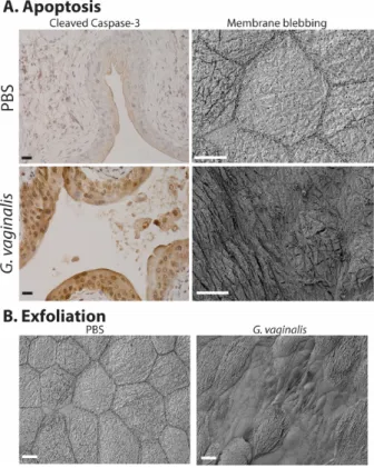
Figure 3: Gardnerella vaginalis induces apoptosis (A) and exfoliation (B) of the mouse bladder epithelium following two transurethral exposures, using our previously reported model
[95].
Our blinded observation showed that ~75% of animals exposed to Gardnerella (compared to only ~25% of controls) exhibited staining for cleaved caspase-3, a marker of apoptotic cell death, in the outermost epithelium (A, left). Membranous protrusions
were another feature we observed by scanning electron microscopy that is consistent with apoptosis (A, right). We also
observed that G.vaginalistriggers exfoliation of superficial umbrella cells, revealing smaller underlying cells of the
transitional epithelium (B). Scale bars = 20 μm.
5 Conclusions
AlthoughE. coliis unquestionably the dominant cause of UTI in young, sexually active women, it is clear that UTI and recurrent UTI stem from a wide array of etiologies.
As outlined here, vaginal bacteria may cause UTI, either themselves (i.e. a traditional uropathogen using the va- gina as a reservoir) or by acting as a “covert pathogens”
to facilitate pathogenesis of another organism. Clinical microbiology labs commonly designate vaginal bacteria – whether identified in pure culture or detected alongside an accepted uropathogen that is present at levels above the usual clinical threshold for UTI – as being of “ques- tionable clinical significance”. We emphasize that, rather than meaning that vaginal bacteria in urine are not im- portant, this simply means that we do not fully understand their effects. UTI caused or encouraged by vaginal bac- teria represent significant etiologies that should not be overlooked, as evidenced by findings that vaginal inter- ventions to increase lactobacilli colonization have prom- ising effects in certain patient subsets, as well as experi-
mental models showing that certain vaginal bacteria can be “covert pathogens” (i.e. encourage virulence by recog- nized uropathogens despite their own rapid clearance).
Note
This article is also to be published as a chapter of the Living Handbook “Urogenital Infections and Inflamma- tions“ [96].
Acknowledgements
We thank Deborah Frank for editorial assistance and thoughtful feedback on the manuscript.
Competing interests
The authors declare that they have no competing in- terests.
References
1. Tachedjian G, Aldunate M, Bradshaw CS, Cone RA. The role of lactic acid production by probiotic Lactobacillus species in vaginal health. Res Microbiol. 2017 Nov-Dec;168(9-10):782-92. DOI:
10.1016/j.resmic.2017.04.001
2. Borges S, Silva J, Teixeira P. The role of lactobacilli and probiotics in maintaining vaginal health. Arch Gynecol Obstet. 2014 Mar;289(3):479-89. DOI: 10.1007/s00404-013-3064-9 3. Allsworth JE, Lewis VA, Peipert JF. Viral sexually transmitted
infections and bacterial vaginosis: 2001-2004 National Health and Nutrition Examination Survey data. Sex Transm Dis. 2008 Sep;35(9):791-6. DOI: 10.1097/OLQ.0b013e3181788301 4. Gajer P, Brotman RM, Bai G, Sakamoto J, Schütte UM, Zhong X,
Koenig SS, Fu L, Ma ZS, Zhou X, Abdo Z, Forney LJ, Ravel J.
Temporal dynamics of the human vaginal microbiota. Sci Transl Med. 2012 May;4(132):132ra52. DOI:
10.1126/scitranslmed.3003605
5. Hummelen R, Macklaim JM, Bisanz JE, Hammond JA, McMillan A, Vongsa R, Koenig D, Gloor GB, Reid G. Vaginal microbiome and epithelial gene array in post-menopausal women with moderate to severe dryness. PLoS ONE. 2011;6(11):e26602.
DOI: 10.1371/journal.pone.0026602
6. Brotman RM, Shardell MD, Gajer P, Fadrosh D, Chang K, Silver MI, Viscidi RP, Burke AE, Ravel J, Gravitt PE. Association between the vaginal microbiota, menopause status, and signs of vulvovaginal atrophy. Menopause. 2018 Nov;25(11):1321-30.
DOI: 10.1097/GME.0000000000001236
7. Burton JP, Reid G. Evaluation of the bacterial vaginal flora of 20 postmenopausal women by direct (Nugent score) and molecular (polymerase chain reaction and denaturing gradient gel electrophoresis) techniques. J Infect Dis. 2002 Dec;186(12):1770-80. DOI: 10.1086/345761
8. Romero R, Hassan SS, Gajer P, Tarca AL, Fadrosh DW, Nikita L, Galuppi M, Lamont RF, Chaemsaithong P, Miranda J,
Chaiworapongsa T, Ravel J. The composition and stability of the vaginal microbiota of normal pregnant women is different from that of non-pregnant women. Microbiome. 2014 Feb;2(1):4. DOI:
10.1186/2049-2618-2-4
9. Nugent RP, Krohn MA, Hillier SL. Reliability of diagnosing bacterial vaginosis is improved by a standardized method of gram stain interpretation. J Clin Microbiol. 1991 Feb;29(2):297-301.
10. Stapleton AE. The Vaginal Microbiota and Urinary Tract Infection.
Microbiol Spectr. 2016 Dec;4(6). DOI:
10.1128/microbiolspec.UTI-0025-2016
11. Sumati AH, Saritha NK. Association of urinary tract infection in women with bacterial vaginosis. J Glob Infect Dis. 2009 Jul;1(2):151-2. DOI: 10.4103/0974-777X.56254
12. Hillebrand L, Harmanli OH, Whiteman V, Khandelwal M. Urinary tract infections in pregnant women with bacterial vaginosis. Am J Obstet Gynecol. 2002 May;186(5):916-7. DOI:
10.1067/mob.2002.123987
13. Hooton TM, Fihn SD, Johnson C, Roberts PL, Stamm WE.
Association between bacterial vaginosis and acute cystitis in women using diaphragms. Arch Intern Med. 1989
Sep;149(9):1932-6.
14. Stapleton AE, Au-Yeung M, Hooton TM, Fredricks DN, Roberts PL, Czaja CA, Yarova-Yarovaya Y, Fiedler T, Cox M, Stamm WE.
Randomized, placebo-controlled phase 2 trial of a Lactobacillus crispatus probiotic given intravaginally for prevention of recurrent urinary tract infection. Clin Infect Dis. 2011 May;52(10):1212- 7. DOI: 10.1093/cid/cir183
15. Raz R, Stamm WE. A controlled trial of intravaginal estriol in postmenopausal women with recurrent urinary tract infections.
N Engl J Med. 1993 Sep;329(11):753-6. DOI:
10.1056/NEJM199309093291102
16. Foxman B. Urinary tract infection syndromes: occurrence, recurrence, bacteriology, risk factors, and disease burden. Infect Dis Clin North Am. 2014 Mar;28(1):1-13. DOI:
10.1016/j.idc.2013.09.003
17. Foxman B. The epidemiology of urinary tract infection. Nat Rev Urol. 2010 Dec;7(12):653-60. DOI: 10.1038/nrurol.2010.190 18. Beerepoot M, Geerlings S. Non-Antibiotic Prophylaxis for Urinary
Tract Infections. Pathogens. 2016 Apr;5(2). pii: E36. DOI:
10.3390/pathogens5020036
19. Pfau A, Sacks T. The bacterial flora of the vaginal vestibule, urethra and vagina in premenopausal women with recurrent urinary tract infections. J Urol. 1981 Nov;126(5):630-4. DOI:
10.1016/s0022-5347(17)54661-3
20. Navas-Nacher EL, Dardick F, Venegas MF, Anderson BE, Schaeffer AJ, Duncan JL. Relatedness of Escherichia coli colonizing women longitudinally. Mol Urol. 2001;5(1):31-6. DOI:
10.1089/109153601750124285
21. Gupta K, Stapleton AE, Hooton TM, Roberts PL, Fennell CL, Stamm WE. Inverse association of H2O2-producing lactobacilli and vaginal Escherichia coli colonization in women with recurrent urinary tract infections. J Infect Dis. 1998 Aug;178(2):446-50.
DOI: 10.1086/515635
22. Stamey TA, Sexton CC. The role of vaginal colonization with enterobacteriaceae in recurrent urinary infections. J Urol. 1975 Feb;113(2):214-7. DOI: 10.1016/s0022-5347(17)59447-1 23. Amatya R, Bhattarai S, Mandal PK, Tuladhar H, Karki BM. Urinary
tract infection in vaginitis: a condition often overlooked. Nepal Med Coll J. 2013 Mar;15(1):65-7.
24. Harmanli OH, Cheng GY, Nyirjesy P, Chatwani A, Gaughan JP.
Urinary tract infections in women with bacterial vaginosis. Obstet Gynecol. 2000 May;95(5):710-2. DOI: 10.1016/s0029- 7844(99)00632-8
25. Kirjavainen PV, Pautler S, Baroja ML, Anukam K, Crowley K, Carter K, Reid G. Abnormal immunological profile and vaginal microbiota in women prone to urinary tract infections. Clin Vaccine Immunol. 2009 Jan;16(1):29-36. DOI:
10.1128/CVI.00323-08
26. Kovachev S. Defence factors of vaginal lactobacilli. Crit Rev Microbiol. 2018 Feb;44(1):31-9. DOI:
10.1080/1040841X.2017.1306688
27. Baerheim A, Larsen E, Digranes A. Vaginal application of lactobacilli in the prophylaxis of recurrent lower urinary tract infection in women. Scand J Prim Health Care. 1994 Dec;12(4):239-43. DOI: 10.3109/02813439409029247 28. Eriksen B. A randomized, open, parallel-group study on the
preventive effect of an estradiol-releasing vaginal ring (Estring) on recurrent urinary tract infections in postmenopausal women.
Am J Obstet Gynecol. 1999 May;180(5):1072-9. DOI:
10.1016/s0002-9378(99)70597-1
29. Muder RR, Brennen C, Rihs JD, Wagener MM, Obman A, Stout JE, Yu VL. Isolation of Staphylococcus aureus from the urinary tract: association of isolation with symptomatic urinary tract infection and subsequent staphylococcal bacteremia. Clin Infect Dis. 2006 Jan;42(1):46-50. DOI: 10.1086/498518
30. Baraboutis IG, Tsagalou EP, Lepinski JL, Papakonstantinou I, Papastamopoulos V, Skoutelis AT, Johnson S. Primary Staphylococcus aureus urinary tract infection: the role of undetected hematogenous seeding of the urinary tract. Eur J Clin Microbiol Infect Dis. 2010 Sep;29(9):1095-101. DOI:
10.1007/s10096-010-0967-2
31. Gilbert NM, O'Brien VP, Hultgren S, Macones G, Lewis WG, Lewis AL. Urinary tract infection as a preventable cause of pregnancy complications: opportunities, challenges, and a global call to action. Glob Adv Health Med. 2013 Sep;2(5):59-69. DOI:
10.7453/gahmj.2013.061
32. Kline KA, Lewis AL. Gram-Positive Uropathogens, Polymicrobial Urinary Tract Infection, and the Emerging Microbiota of the Urinary Tract. Microbiol Spectr. 2016 Apr;4(2). DOI:
10.1128/microbiolspec.UTI-0012-2012
33. Kazi YF, Saleem S, Kazi N. Investigation of vaginal microbiota in sexually active women using hormonal contraceptives in Pakistan.
BMC Urol. 2012 Aug;12:22. DOI: 10.1186/1471-2490-12-22 34. Huang Y, Merkatz RB, Hillier SL, Roberts K, Blithe DL, Sitruk-Ware
R, Creinin MD. Effects of a One Year Reusable Contraceptive Vaginal Ring on Vaginal Microflora and the Risk of Vaginal Infection: An Open-Label Prospective Evaluation. PLoS ONE.
2015;10(8):e0134460. DOI: 10.1371/journal.pone.0134460 35. Akhi MT, Esmailkhani A, Sadeghi J, Niknafs B, Farzadi L, Akhi A,
Nasab EN. The Frequency of Isolated from Endocervix of Infertile Women in Northwest Iran. Int J Fertil Steril. 2017 Apr- Jun;11(1):28-32. DOI: 10.22074/ijfs.2016.4969
36. MacPhee RA, Miller WL, Gloor GB, McCormick JK, Hammond JA, Burton JP, Reid G. Influence of the vaginal microbiota on toxic shock syndrome toxin 1 production by Staphylococcus aureus.
Appl Environ Microbiol. 2013 Mar;79(6):1835-42. DOI:
10.1128/AEM.02908-12
37. Tansarli GS, Kostaras EK, Athanasiou S, Falagas ME. Prevalence and treatment of aerobic vaginitis among non-pregnant women:
evaluation of the evidence for an underestimated clinical entity.
Eur J Clin Microbiol Infect Dis. 2013 Aug;32(8):977-84. DOI:
10.1007/s10096-013-1846-4
38. Chow AW, Bartlett KH, Percival-Smith R, Morrison BJ. Vaginal colonization with Staphylococcus aureus, positive for toxic-shock marker protein, and Escherichia coli in healthy women. J Infect Dis. 1984 Jul;150(1):80-4. DOI: 10.1093/infdis/150.1.80 39. Donders GG, Vereecken A, Bosmans E, Dekeersmaecker A,
Salembier G, Spitz B. Definition of a type of abnormal vaginal flora that is distinct from bacterial vaginosis: aerobic vaginitis.
BJOG. 2002 Jan;109(1):34-43. DOI: 10.1111/j.1471- 0528.2002.00432.x
40. Tan CK, Ulett KB, Steele M, Benjamin WH Jr, Ulett GC. Prognostic value of semi-quantitative bacteruria counts in the diagnosis of group B streptococcus urinary tract infection: a 4-year retrospective study in adult patients. BMC Infect Dis. 2012 Oct;12:273. DOI: 10.1186/1471-2334-12-273
41. Beyer I, Mergam A, Benoit F, Theunissen C, Pepersack T.
Management of urinary tract infections in the elderly. Z Gerontol Geriatr. 2001 Apr;34(2):153-7. DOI: 10.1007/s003910170080 42. Muller AE, Oostvogel PM, Steegers EA, Dörr PJ. Morbidity related to maternal group B streptococcal infections. Acta Obstet Gynecol Scand. 2006;85(9):1027-37. DOI:
10.1080/00016340600780508
43. Persson K, Bjerre B, Elfström L, Polberger S, Forsgren A. Group B streptococci at delivery: high count in urine increases risk for neonatal colonization. Scand J Infect Dis. 1986;18(6):525-31.
DOI: 10.3109/00365548609021657
44. Edwards MS, Baker CJ. Group B streptococcal infections in elderly adults. Clin Infect Dis. 2005 Sep;41(6):839-47. DOI:
10.1086/432804
45. Ulett KB, Benjamin WH Jr, Zhuo F, Xiao M, Kong F, Gilbert GL, Schembri MA, Ulett GC. Diversity of group B streptococcus serotypes causing urinary tract infection in adults. J Clin Microbiol.
2009 Jul;47(7):2055-60. DOI: 10.1128/JCM.00154-09 46. Kline KA, Schwartz DJ, Gilbert NM, Lewis AL. Impact of host age
and parity on susceptibility to severe urinary tract infection in a murine model. PLoS ONE. 2014;9(5):e97798. DOI:
10.1371/journal.pone.0097798
47. Kulkarni R, Randis TM, Antala S, Wang A, Amaral FE, Ratner AJ.
β-Hemolysin/cytolysin of Group B Streptococcus enhances host inflammation but is dispensable for establishment of urinary tract infection. PLoS ONE. 2013;8(3):e59091. DOI:
10.1371/journal.pone.0059091
48. Kline KA, Schwartz DJ, Gilbert NM, Hultgren SJ, Lewis AL. Immune modulation by group B Streptococcus influences host susceptibility to urinary tract infection by uropathogenic Escherichia coli. Infect Immun. 2012 Dec;80(12):4186-94. DOI:
10.1128/IAI.00684-12
49. Sullivan MJ, Carey AJ, Leclercq SY, Tan CK, Ulett GC. Increased Age, but Not Parity Predisposes to Higher Bacteriuria Burdens Due to Streptococcus Urinary Tract Infection and Influences Bladder Cytokine Responses, Which Develop Independent of Tissue Bacterial Loads. PLoS ONE. 2016;11(12):e0167732.
DOI: 10.1371/journal.pone.0167732
50. Pedraza-Avilés AG, Zaragoza MC, Mota-Vázquez R, Hernández- Soto C, Ramírez-Santana M, Terrazas-Maldonado ML. Treatment of urinary tract infection by Gardnerella vaginalis: a comparison of oral metronidazole versus ampicillin. Rev Latinoam Microbiol.
2001 Apr-Jun;43(2):65-9.
51. Josephson S, Thomason J, Sturino K, Zabransky R, Williams J.
Gardnerella vaginalis in the urinary tract: incidence and significance in a hospital population. Obstet Gynecol. 1988 Feb;71(2):245-50.
52. Pearce MM, Hilt EE, Rosenfeld AB, Zilliox MJ, Thomas-White K, Fok C, Kliethermes S, Schreckenberger PC, Brubaker L, Gai X, Wolfe AJ. The female urinary microbiome: a comparison of women with and without urgency urinary incontinence. mBio. 2014 Jul;5(4):e01283-14. DOI: 10.1128/mBio.01283-14
53. Lam MH, Birch DF, Fairley KF. Prevalence of Gardnerella vaginalis in the urinary tract. J Clin Microbiol. 1988 Jun;26(6):1130-3.
54. Gilbert GL, Garland SM, Fairley KF, McDowall DM. Bacteriuria due to ureaplasmas and other fastidious organisms during pregnancy: prevalence and significance. Pediatr Infect Dis. 1986 Nov-Dec;5(6 Suppl):S239-43. DOI: 10.1097/00006454- 198611010-00007
55. Savige JA, Gilbert GL, Fairley KF, McDowall DR. Bacteriuria due to Ureaplasma urealyticum and Gardnerella vaginalis in women with preeclampsia. J Infect Dis. 1983 Sep;148(3):605. DOI:
10.1093/infdis/148.3.605
56. McDowall DR, Buchanan JD, Fairley KF, Gilbert GL. Anaerobic and other fastidious microorganisms in asymptomatic bacteriuria in pregnant women. J Infect Dis. 1981 Aug;144(2):114-22. DOI:
10.1093/infdis/144.2.114
57. McFadyen IR, Eykyn SJ. Suprapubic aspiration of urine in pregnancy. Lancet. 1968 May;1(7552):1112-4. DOI:
10.1016/s0140-6736(68)90185-2
58. Fairley KF, Birch DF. Unconventional bacteria in urinary tract disease: Gardnerella vaginalis. Kidney Int. 1983 Jun;23(6):862- 5. DOI: 10.1038/ki.1983.107
59. Boggess KA, Watts DH, Hillier SL, Krohn MA, Benedetti TJ, Eschenbach DA. Bacteremia shortly after placental separation during cesarean delivery. Obstet Gynecol. 1996 May;87(5 Pt 1):779-84. DOI: 10.1016/0029-7844(96)00037-3 60. Babics A, Roussellier P. Gardnerella vaginalis: An overlooked
pathogen in male patients? Med Mal Infect. 2015 Oct;45(10):423-4. DOI: 10.1016/j.medmal.2015.09.007 61. Gottschick C, Deng ZL, Vital M, Masur C, Abels C, Pieper DH,
Wagner-Döbler I. The urinary microbiota of men and women and its changes in women during bacterial vaginosis and antibiotic treatment. Microbiome. 2017 Aug;5(1):99. DOI:
10.1186/s40168-017-0305-3
62. Ahmed A, Earl J, Retchless A, Hillier SL, Rabe LK, Cherpes TL, Powell E, Janto B, Eutsey R, Hiller NL, Boissy R, Dahlgren ME, Hall BG, Costerton JW, Post JC, Hu FZ, Ehrlich GD. Comparative genomic analyses of 17 clinical isolates of Gardnerella vaginalis provide evidence of multiple genetically isolated clades consistent with subspeciation into genovars. J Bacteriol. 2012
Aug;194(15):3922-37. DOI: 10.1128/JB.00056-12
63. Sierra-Hoffman M, Watkins K, Jinadatha C, Fader R, Carpenter JL. Clinical significance of Aerococcus urinae: a retrospective review. Diagn Microbiol Infect Dis. 2005 Dec;53(4):289-92. DOI:
10.1016/j.diagmicrobio.2005.06.021
64. Senneby E, Petersson AC, Rasmussen M. Clinical and microbiological features of bacteraemia with Aerococcus urinae.
Clin Microbiol Infect. 2012 Jun;18(6):546-50. DOI:
10.1111/j.1469-0691.2011.03609.x
65. Murray TS, Muldrew KL, Finkelstein R, Hampton L, Edberg SC, Cappello M. Acute pyelonephritis caused by Aerococcus urinae in a 12-year-old boy. Pediatr Infect Dis J. 2008 Aug;27(8):760- 2. DOI: 10.1097/INF.0b013e318170af46
66. Ibler K, Truberg Jensen K, Ostergaard C, Sönksen UW, Bruun B, Schønheyder HC, Kemp M, Dargis R, Andresen K, Christensen JJ. Six cases of Aerococcus sanguinicola infection: clinical relevance and bacterial identification. Scand J Infect Dis.
2008;40(9):761-5. DOI: 10.1080/00365540802078059 67. de Jong MF, Soetekouw R, ten Kate RW, Veenendaal D.
Aerococcus urinae: severe and fatal bloodstream infections and endocarditis. J Clin Microbiol. 2010 Sep;48(9):3445-7. DOI:
10.1128/JCM.00835-10
68. Cattoir V, Kobal A, Legrand P. Aerococcus urinae and Aerococcus sanguinicola, two frequently misidentified uropathogens. Scand J Infect Dis. 2010 Oct;42(10):775-80. DOI:
10.3109/00365548.2010.485576
69. Zhang Q, Kwoh C, Attorri S, Clarridge JE 3rd. Aerococcus urinae in urinary tract infections. J Clin Microbiol. 2000 Apr;38(4):1703- 5.
70. Schuur PM, Kasteren ME, Sabbe L, Vos MC, Janssens MM, Buiting AG. Urinary tract infections with Aerococcus urinae in the south of The Netherlands. Eur J Clin Microbiol Infect Dis. 1997 Dec;16(12):871-5. DOI: 10.1007/bf01700552
71. Gritsch W, Nagl M, Hausdorfer J, Gschwendtner A, Pechlaner C, Wiedermann CJ. Septicaemia and endomyocarditis caused by Aerococcus urinae. Wien Klin Wochenschr. 1999
Jun;111(11):446-7.
72. Christensen JJ, Jensen IP, Faerk J, Kristensen B, Skov R, Korner B. Bacteremia/septicemia due to Aerococcus-like organisms:
report of seventeen cases. Danish ALO Study Group. Clin Infect Dis. 1995 Oct;21(4):943-7. DOI: 10.1093/clinids/21.4.943 73. Heilesen AM. Septicaemia due to Aerococcus urinae. Scand J
Infect Dis. 1994;26(6):759-60. DOI:
10.3109/00365549409008648
74. Rasmussen M. Aerococcus: an increasingly acknowledged human pathogen. Clin Microbiol Infect. 2016 Jan;22(1):22-7. DOI:
10.1016/j.cmi.2015.09.026
75. Srinivasan S, Hoffman NG, Morgan MT, Matsen FA, Fiedler TL, Hall RW, Ross FJ, McCoy CO, Bumgarner R, Marrazzo JM, Fredricks DN. Bacterial communities in women with bacterial vaginosis: high resolution phylogenetic analyses reveal relationships of microbiota to clinical criteria. PLoS ONE.
2012;7(6):e37818. DOI: 10.1371/journal.pone.0037818 76. Ling Z, Kong J, Liu F, Zhu H, Chen X, Wang Y, Li L, Nelson KE, Xia
Y, Xiang C. Molecular analysis of the diversity of vaginal microbiota associated with bacterial vaginosis. BMC Genomics.
2010 Sep;11:488. DOI: 10.1186/1471-2164-11-488 77. Reyes L, Reinhard M, Brown MB. Different inflammatory
responses are associated with Ureaplasma parvum-induced UTI and urolith formation. BMC Infect Dis. 2009 Jan;9:9. DOI:
10.1186/1471-2334-9-9
78. Reyes L, Reinhard M, O’donell LJ, Stevens J, Brown MB. Rat strains differ in susceptibility to Ureaplasma parvum-induced urinary tract infection and struvite stone formation. Infect Immun.
2006 Dec;74(12):6656-64. DOI: 10.1128/IAI.00984-06 79. Taylor-Robinson D. Mollicutes in vaginal microbiology:
Mycoplasma hominis, Ureaplasma urealyticum, Ureaplasma parvum and Mycoplasma genitalium. Res Microbiol. 2017 Nov - Dec;168(9-10):875-81. DOI: 10.1016/j.resmic.2017.02.009 80. Cox C, Watt AP, McKenna JP, Coyle PV. Mycoplasma hominis and
Gardnerella vaginalis display a significant synergistic relationship in bacterial vaginosis. Eur J Clin Microbiol Infect Dis. 2016 Mar;35(3):481-7. DOI: 10.1007/s10096-015-2564-x
81. Marovt M, Keše D, Kotar T, Kmet N, Miljković J, Šoba B, Matičič M. Ureaplasma parvum and Ureaplasma urealyticum detected with the same frequency among women with and without symptoms of urogenital tract infection. Eur J Clin Microbiol Infect Dis. 2015 Jun;34(6):1237-45. DOI: 10.1007/s10096-015-2351- 8
82. Sweeney EL, Dando SJ, Kallapur SG, Knox CL. The Human Ureaplasma Species as Causative Agents of Chorioamnionitis.
Clin Microbiol Rev. 2017 Jan;30(1):349-79. DOI:
10.1128/CMR.00091-16
83. Revello R, Alcaide MJ, Abehsera D, Martin-Camean M, Sousa E Faro Gomes M, Alonso-Luque B, Bartha JL. Prediction of chorioamnionitis in cases of intraamniotic infection by ureaplasma urealyticum in women with very preterm premature rupture of membranes or preterm labour. J Matern Fetal Neonatal Med. 2018 Jul;31(14):1839-44. DOI:
10.1080/14767058.2017.1330407
84. Humburg J, Frei R, Wight E, Troeger C. Accuracy of urethral swab and urine analysis for the detection of Mycoplasma hominis and Ureaplasma urealyticum in women with lower urinary tract symptoms. Arch Gynecol Obstet. 2012 Apr;285(4):1049-53. DOI:
10.1007/s00404-011-2109-1
85. Baka S, Kouskouni E, Antonopoulou S, Sioutis D, Papakonstantinou M, Hassiakos D, Logothetis E, Liapis A.
Prevalence of Ureaplasma urealyticum and Mycoplasma hominis in women with chronic urinary symptoms. Urology. 2009 Jul;74(1):62-6. DOI: 10.1016/j.urology.2009.02.014
86. Lee YS, Kim JY, Kim JC, Park WH, Choo MS, Lee KS. Prevalence and treatment efficacy of genitourinary mycoplasmas in women with overactive bladder symptoms. Korean J Urol. 2010 Sep;51(9):625-30. DOI: 10.4111/kju.2010.51.9.625 87. Nassar FA, Abu-Elamreen FH, Shubair ME, Sharif FA. Detection
of Chlamydia trachomatis and Mycoplasma hominis, genitalium and Ureaplasma urealyticum by polymerase chain reaction in patients with sterile pyuria. Adv Med Sci. 2008;53(1):80-6. DOI:
10.2478/v10039-008-0020-1
88. Domann E, Hong G, Imirzalioglu C, Turschner S, Kühle J, Watzel C, Hain T, Hossain H, Chakraborty T. Culture-independent identification of pathogenic bacteria and polymicrobial infections in the genitourinary tract of renal transplant recipients. J Clin Microbiol. 2003 Dec;41(12):5500-10. DOI:
10.1128/jcm.41.12.5500-5510.2003
89. Wolfe AJ, Toh E, Shibata N, Rong R, Kenton K, Fitzgerald M, Mueller ER, Schreckenberger P, Dong Q, Nelson DE, Brubaker L. Evidence of uncultivated bacteria in the adult female bladder.
J Clin Microbiol. 2012 Apr;50(4):1376-83. DOI:
10.1128/JCM.05852-11
90. Nicolle LE, Harding GK, Preiksaitis J, Ronald AR. The association of urinary tract infection with sexual intercourse. J Infect Dis.
1982 Nov;146(5):579-83. DOI: 10.1093/infdis/146.5.579 91. Stapleton A, Latham RH, Johnson C, Stamm WE. Postcoital
antimicrobial prophylaxis for recurrent urinary tract infection. A randomized, double-blind, placebo-controlled trial. JAMA. 1990 Aug;264(6):703-6.
92. Stamatiou C, Bovis C, Panagopoulos P, Petrakos G, Economou A, Lycoudt A. Sex-induced cystitis – patient burden and other epidemiological features. Clin Exp Obstet Gynecol.
2005;32(3):180-2.
93. Hooton TM, Scholes D, Hughes JP, Winter C, Roberts PL, Stapleton AE, Stergachis A, Stamm WE. A prospective study of risk factors for symptomatic urinary tract infection in young women. N Engl J Med. 1996 Aug;335(7):468-74. DOI:
10.1056/NEJM199608153350703
94. Scholes D, Hooton TM, Roberts PL, Stapleton AE, Gupta K, Stamm WE. Risk factors for recurrent urinary tract infection in young women. J Infect Dis. 2000 Oct;182(4):1177-82. DOI:
10.1086/315827
95. Gilbert NM, O’Brien VP, Lewis AL. Transient microbiota exposures activate dormant Escherichia coli infection in the bladder and drive severe outcomes of recurrent disease. PLoS Pathog. 2017 Mar;13(3):e1006238. DOI: 10.1371/journal.ppat.1006238 96. Lewis AL, Gilbert NM. Roles of the vagina and the vaginal
microbiota in urinary tract infection: Evidence from clinical correlations and experimental models.In: Bjerklund Johansen TE, Wagenlehner FME, Cho YH, Matsumoto T, Krieger JN, Shoskes D, Naber KG, editors. Urogenital Infections and Inflammations.
Duesseldorf: GMS; 2017-. DOI: 10.5680/lhuii000055
Corresponding authors:
Amanda L. Lewis
Department of Molecular Microbiology, Campus Box 8230, Washington University School of Medicine, 660 S.
Euclid Avenue, St. Louis, MO 63110, United States, Phone: +1 314 286 0016
allewis@wustl.edu
Nicole M. Gilbert
Department of Obstetrics and Gynecology, Campus Box 8230, Washington University School of Medicine, 660 S.
Euclid Avenue, St. Louis, MO 63110, United States, Phone: +1 314 286 0019
gilbert@wustl.edu
Please cite as
Lewis AL, Gilbert NM. Roles of the vagina and the vaginal microbiota in urinary tract infection: evidence from clinical correlations and experimental models. GMS Infect Dis. 2020;8:Doc02.
DOI: 10.3205/id000046, URN: urn:nbn:de:0183-id0000466
This article is freely available from
https://www.egms.de/en/journals/id/2020-8/id000046.shtml Published:2020-03-26
Copyright
©2020 Lewis et al. This is an Open Access article distributed under the terms of the Creative Commons Attribution 4.0 License. See license information at http://creativecommons.org/licenses/by/4.0/.