Function of the actin nucleator
mitoSPIRE1 in mitochondrial dynamics
DISSERTATION
ZUR ERLANGUNG DES DOKTORGRADES DER NATURWISSENSCHAFTEN (DR. RER. NAT.) DER FAKULTÄT FÜR BIOLOGIE UND VORKLINISCHE MEDIZIN
DER UNIVERSITÄT REGENSBURG
vorgelegt von Felix Straub
aus Weingarten
im Jahr 2020
Das Promotionsgesuch wurde eingereicht am:
20 Februar 2020
Die Arbeit wurde angeleitet von:
Prof. Dr. rer. nat. Eugen Kerkhoff
Unterschrift:
Datenblatt
Titel der Arbeit: Function of the actin nucleator mitoSPIRE1 in mitochondrial dynamics
Institut: Neurologie, Molekulare Zellbiologie, Universitätsklinikum Regensburg
Name, Vorname: Straub, Felix
Erstgutachter: Prof. Dr. rer. nat. Eugen Kerkhoff Zweitgutachter: Prof. Dr. rer. nat. Christian Wetzel Drittprüfer: Prof. Dr. rer. nat. Veronica Egger Ersatzprüfer: Prof. Dr. rer. nat. Stephan Schneuwly Prüfungsvorsitzender: Prof. Dr. rer. nat. Oliver Bosch
Eidesstattliche Erklärung
Ich, Felix Straub, geboren am 21.05.1991 in Weingarten,
versichere hiermit, dass ich die vorliegende Arbeit ohne unzulässige Hilfe Dritter und ohne Benutzung anderer als der angegebenen Hilfsmittel angefertigt habe. Ergebnisse, Abbildungen und Beschreibungen, die im Rahmen einer Kollaboration entstanden sind, habe ich entsprechend gekennzeichnet.
Die Stellen, die anderen Werken dem Wortlaut oder dem Sinn nach entnommen sind, habe ich durch Angabe der Quelle kenntlich gemacht. Insbesondere habe ich nicht die entgeltliche Hilfe von Vermittlungs- bzw. Beratungsdiensten (Promotionsberater oder andere Personen) in Anspruch genommen.
Ich erkläre hiermit weiterhin, dass ich meine wissenschaftlichen Arbeiten nach den Prinzipien der guten wissenschaftlichen Praxis gemäß der gültigen „Grundsätze zur Sicherung guter wissenschaftlicher Praxis“ der Universität Regensburg angefertigt habe.
Die Arbeit wurde bisher weder im In- noch im Ausland in gleicher oder ähnlicher Form einer anderen Prüfungsbehörde vorgelegt.
Figure 2, 11 und 13, sowie Abschnitte des Kapitels „Material und Methoden“ der hier vorliegenden Arbeit enthalten Inhalte bereits veröffentlichter Publikationen (Alzahofi, Robinson, Welz et al., 2018; Kollmar, Welz, Straub et al., 2019). Die besagten Inhalte wurden ausschließlich durch mich generiert / illustriert und sowohl für die Dissertation als auch für die genannten Publikationen erstellt.
Im Kapitel „Material und Methoden“ wurden Standardformulierungen (wie beispielsweise der Satzbaustein „heating and shaking“) der Arbeitsgruppe Kerkhoff zur Beschreibung der verwendeten Methodik benutzt. Diese Standardformulierungen finden sich in Publikationen sowie Protokollen der AG Kerkhoff wieder und wurden in der vorliegenden Arbeit nicht gekennzeichnet.
_________________________ _________________________
Ort, Datum Felix Straub
Table of contents
Datenblatt ... II Eidesstattliche Erklärung ... III Abstract ... VIII Zusammenfassung ... IX
1. Introduction ... 1
1.1 Preamble ... 1
1.2 Mitochondrial dynamics ... 1
1.2.1 Mitochondrial fission ... 3
1.2.2 Mitochondrial fusion ... 4
1.2.3 Mitochondrial motility ... 5
1.3 Actin nucleators facilitate actin polymerization ... 6
1.3.1 SPIRE proteins ... 7
1.3.2 mitoSPIRE1 ... 10
1.4 Cooperation of SPIRE and formin proteins ... 10
1.5 The SPIRE and myosin 5 interaction ... 14
1.6 Cellular functions of SPIRE proteins ... 16
1.6.1 SPIRE actin nucleators drive oocyte maturation ... 16
1.6.2 SPIRE actin nucleators drive vesicular transport processes ... 17
1.6.3 SPIRE1 / FMN1 / MYO5A cooperate in cytoplasmic melanosome dispersion ... 18
1.7 Knockout mouse models of the actin nucleators SPIRE and FMN subgroup formins .. 21
1.7.1 The SPIRE1 mutant mouse ... 21
1.7.2 The FMN1 knockout mouse ... 22
1.7.3 The FMN2 knockout mouse ... 22
1.8 Mutations of the human FMN2 protein ... 23
1.9 Aim of the thesis ... 23
2. Materials and methods ... 25
2.1 Primer design ... 25
2.2 Agarose gel electrophoresis ... 25
2.3 Agarose gel clean-up ... 26
2.4 RNA isolation ... 26
2.4.1 RNA isolation of mouse organs ... 26
2.4.2 Total RNA isolation from eukaryotic cultured cells ... 26
2.4.3 Analysis of RNA / DNA purity and concentration ... 26
2.4.4 RNA quality ... 27
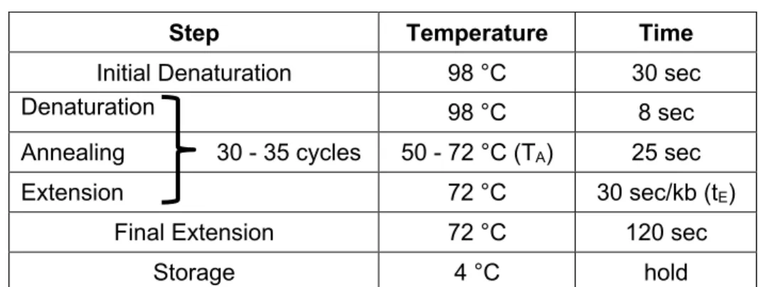
2.5 PCR techniques ... 27
2.5.1 Reverse transcription (synthesis of cDNA) ... 27
2.5.2 Amplification of cDNA fragments ... 28
2.5.3 Amplification of genomic DNA fragments (genotyping of mice) ... 29
2.5.4 Amplification of cDNA fragments for cloning ... 30
2.5.5 Quantitative real-time PCR ... 31
2.6 Cloning ... 32
2.6.1 Restriction digest and ligation of DNA vectors ... 32
2.6.2 Transformation of Escherichia coli bacterial cells ... 33
2.6.3 Plasmid DNA extraction and purification from bacterial cells ... 33
2.6.4 Control digests and DNA sequencing of plasmids ... 34
2.7 Cell culture techniques ... 34
2.7.1 Thawing cells ... 35
2.7.2 Freezing cells ... 35
2.7.3 Seeding cells ... 36
2.7.4 Staining of mitochondria in eukaryotic cells ... 36
2.7.5 Transfection of eukaryotic cells using Lipofectamine2000 reagent ... 37
2.8 Colocalization studies ... 37
2.9 Immunostaining ... 37
2.10 Microscopy ... 38
2.10.1 Microscopy of in situ hybridization experiments ... 38
2.10.2 Fluorescence microscopy of fixed and immunostained cells ... 38
2.10.3 Fluorescence microscopy of viable cells ... 38
2.11 Quantification of mitochondrial length using microscopy and ImageJ ... 39
2.12 Quantification of mitochondrial motility ... 39
2.13 Analysis of MYO5A positive mitochondria and mitochondrial sizes using FAMS .... 40
2.14 Software ... 41
2.14.1 Statistical analysis ... 41
2.14.2 Graphs and cartoons ... 42
2.14.3 Written work ... 42
2.14.4 Identification of protein domains ... 42
2.14.5 DNA and cDNA sequences ... 43
2.15 Mitochondrial respiration ... 43
2.16 Isolation of pMEFs from mouse embryos ... 43
2.17 In situ hybridization ... 44
2.18 Protein work ... 46
2.18.1 Recombinant protein expression and purification ... 46
2.18.2 GST-pulldown from wild type and mitoSPIRE1 knockout fibroblast lysates ... 47
2.18.3 Immunoblotting ... 47
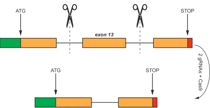
2.19 Generation of the mitoSPIRE1 knockout mouse ... 48
2.19.1 Overview of generating the mitoSPIRE1 knockout mouse ... 49
2.19.2 Guide selection and cloning of CRISPR constructs ... 50
2.19.3 Embryonic Stem Cell targeting ... 50
2.19.4 Genotyping and targeting efficiency ... 50
2.19.5 Off target screening ... 51
2.19.6 Chimeric mice generation and breeding ... 51
3. Results ... 52
3.1 Expression of SPIRE splice variants in mouse tissues ... 54
3.2 Similar expression patterns of SPIRE1 and mitoSPIRE1 in the mouse brain ... 56
3.3 Subcellular localization of SPIRE proteins ... 57
3.4 Interaction of mitoSPIRE1 with the outer mitochondrial membrane ... 59
3.5 mitoSPIRE1 targets FMN subfamily formin proteins towards mitochondria ... 63
3.6 mitoSPIRE1 targets MYO5 actin motor proteins towards mitochondria ... 67
3.7 Generation of a mitoSPIRE1 knockout mouse model ... 71
3.8 Mitochondria integrity in mouse SPIRE knockout models ... 74
3.8.1 Influence of SPIRE absence on mitochondrial morphology ... 74
3.8.2 Influence of SPIRE1 absence on mitochondrial respiration ... 77
3.8.3 Function of mitoSPIRE1 in mitochondrial motility ... 78
3.8.4 Overexpression of mitoSPIRE1 influences mitochondrial motility ... 81
4. Discussion ... 84
4.1 Expression of SPIRE splice variants in mouse tissues ... 85
4.2 Similar expression patterns of SPIRE1 and mitoSPIRE1 in the mouse brain ... 87
4.3 Subcellular localization of SPIRE proteins ... 88
4.4 Interaction of mitoSPIRE1 with the outer mitochondrial membrane ... 89
4.5 mitoSPIRE1 targets FMN subfamily formin proteins towards mitochondria ... 90
4.6 mitoSPIRE1 targets MYO5 actin motor proteins towards mitochondria ... 91
4.7 Generation of a mitoSPIRE1 knockout mouse model ... 92
4.8.1 Influence of SPIRE absence on mitochondrial morphology ... 94
4.8.2 Influence of SPIRE1 absence on mitochondrial respiration ... 96
4.8.3 Function of mitoSPIRE1 in mitochondrial motility ... 97
4.9 Model for mitoSPIRE1 function in mitochondrial transport processes ... 99
5. References ... 103
6. Supplement ... 116
6.1 Overview of used materials ... 116
6.1.2 Chemicals and reagents ... 116
6.1.3 Cell culture media, reagents and supplements ... 118
6.1.4 Buffers, solutions and media ... 119
6.1.5 Disposable materials ... 122
6.1.6 Kits ... 123
6.1.7 Antibodies ... 124
6.1.7.1 Primary antibodies ... 124
6.1.7.2 Secondary antibodies ... 124
6.1.8 Enzymes ... 124
6.1.8.1 Restriction endonucleases ... 124
6.1.8.2 DNA polymerases ... 125
6.1.8.3 Additional enzymes ... 125
6.1.9 Primer (PCR / qPCR) ... 125
6.1.10 Mouse lines ... 126
6.1.11 Machines and equipment ... 127
6.1.12 Eukaryotic expression vectors ... 128
6.1.12.1 pAcGFP1-C1 (Clontech) ... 128
6.1.12.2 pcDNA3 (Invitrogen) ... 128
6.1.12.3 pEGFP-C1 (Clontech) ... 128
6.1.12.4 pEGFP-C2 (Clontech) ... 129
6.1.12.5 pmStrawberry-C1 ... 129
6.1.12.6 List of used constructs ... 129
6.1.13 Sequence related data ... 130
6.2 Amino acids, one letter code ... 131
7. List of abbrevations ... 132
8. List of Figures ... 135
9. List of Tables ... 137
10. Acknowledgements / Danksagung ... 138
Abstract
Cells of all known living organisms require a constant energy supply in the form of adenosine triphosphate (ATP) for their survival and function. The intracellular transport and diffusion of ATP molecules are rather limited and thus, ATP producing mitochondria are directly transported to their site of action such as synaptic densities to provide all cellular compartments with an adequate amount of ATP. Fast and long-range transport of mitochondria along microtubule tracks via kinesin and dynein motor proteins is well established, whereas only little is known about actin / myosin functions in mitochondrial transport. There is growing evidence that actin filaments and myosin motor proteins play an essential role in the transport of mitochondria. A SPIRE formin actin nucleator complex facilitates actin filament generation. In addition, SPIRE proteins coordinate myosin 5 (MYO5) motor protein activation at vesicle and organelle membranes. Due to alternative splicing of the SPIRE1 gene, mammalian cells generate four SPIRE proteins from two SPIRE genes. The SPIRE1 actin / myosin organizer is targeted by the alternatively spliced exon 13 towards mitochondrial membranes and the corresponding protein is named ‘mitoSPIRE1’. The present thesis quantitatively addressed the expression of all SPIRE splice variants including mitoSPIRE1 in distinct organs, showing that the brain has the majority of SPIRE expression. In a previous study it was shown that SPIRE1 mutant mice lacking the expression of all SPIRE1 proteins, including mitoSPIRE1, have increased fear in both cued and contextual fear conditioning experiments. To dissect the vesicular and mitochondrial SPIRE functions in fear behavior and to analyze the function of the mitoSPIRE1 protein in more detail, we generated a novel knockout model by targeted deletion of the mouse SPIRE1 exon 13 - the mitoSPIRE1 knockout mouse. Fibroblasts of the novel mitoSPIRE1 knockout mouse and the SPIRE1 mutant mouse show increased mitochondrial motility in live cell fluorescence microscopy analysis. Colocalization studies unraveled that mitoSPIRE1 colocalizes with FMN subfamily formins and MYO5 proteins at mitochondria. Subsequent fluorescence-activated mitochondria sorting (FAMS) experiments confirmed that SPIRE proteins contribute to target MYO5 actin motor proteins towards mitochondrial membranes. Furthermore, FAMS and fluorescence microscopy revealed an influence of mitoSPIRE1 on mitochondrial morphology. SPIRE proteins did not influence mitochondrial oxygen consumption rate, which was analyzed by a Seahorse Mito Stress Test assay. The mentioned function of mitoSPIRE1 in the regulation of mitochondrial motility led us to speculate that mitoSPIRE1 might be involved in targeting mitochondria towards synaptic terminals and thereby influencing synaptic transmission and possibly fear behavior.
Zusammenfassung
Sämtliche Zellen lebender Organismen benötigen für ihr Überleben und die erfolgreiche Ausführung der zellulären Funktion eine ausreichende Menge an Energie in Form von Adenosintriphosphat (ATP). Die Diffusion und der Transport von einzelnen Molekülen wie ATP, welches hauptsächlich von Mitochondrien synthetisiert wird, ist intrazellulär stark eingeschränkt. Aus diesem Grund werden Mitochondrien über ein Transportsystem an ihren Einsatzort, beispielsweise synaptische Kontakte, bewegt. In der Literatur ist bereits detailliert beschrieben, dass Kinesin- und Dynein-Motoren einen schnellen, weitreichenden und bidirektionalen Mitochondrientransport entlang von Mikrotubuli ermöglichen. Gleichzeitig ist über einen möglichen mitochondrialen Transport entlang von Aktinfilamenten mittels Myosin-Motoren bisher nur wenig bekannt. Dennoch häufen sich die Hinweise, dass Aktinfilamente sowie Myosin-Motoren den mitochondrialen Transport beeinflussen. Die intrazelluläre Aktinfilament-Bildung wird unter anderem durch den SPIRE / Formin Aktinnukleations-Komplex ermöglicht. Zusätzlich koordinieren SPIRE Proteine die Aktivierung von Myosin 5 Motoren an Vesikel- und Organell-Membranen. Durch alternatives Spleißen entstehen in einer Säugetier-Zelle aus zwei SPIRE Genen insgesamt vier SPIRE Proteine. Hierbei wird durch das alternative Spleißen der SPIRE1 mRNA ein Protein synthetisiert, welches das alternative Exon 13 des SPIRE1 Genes enthält. Das zusätzliche Exon 13 lokalisiert das eigentlich vesikuläre SPIRE1 Protein an die mitochondriale Membran, weshalb dieses dementsprechend als „mitoSPIRE1“ bezeichnet wird. In der hier vorliegenden Arbeit wurde die Expression aller SPIRE Spleißvarianten inklusive mitoSPIRE1 in verschiedenen Organen der Maus quantitativ adressiert. Hierbei konnte gezeigt werden, dass das Gehirn der Hauptexpressionsort für SPIRE ist. In der Literatur wurde bereits ein Maus-Modell beschrieben, welches keinerlei funktionelle Proteine des SPIRE1 Gens exprimiert und ein erhöhtes Angstverhalten in kontextuellen und reiz-induzierten Angstkonditionierungs-Experimenten aufweist. Um die Funktion des vesikulären und mitochondrialen SPIRE auf das Angstverhalten zu analysieren und die generelle mitoSPIRE1 Funktion zu untersuchen wurde in dieser Arbeit mittels Deletion des SPIRE1 Exon 13 ein neues Knock-out Model erstellt - die mitoSPIRE1 Knock-out Maus. In nachfolgenden Fluoreszenz-Mikroskopie-Analysen lebender Zellen zeigen Fibroblasten der neuen mitoSPIRE1 Knock-out Maus und der SPIRE1 mutant Maus, welche keinerlei funktionsfähige Proteine des SPIRE1 Genes besitzt, eine erhöhte mitochondriale Motilität. Außerdem konnte durch eine umfassende Kolokalisations-Studie gezeigt werden, dass das mitochondriale
mitoSPIRE1 mit FMN und MYO5 Proteinen an Mitochondrien kolokalisiert. Hierbei wurde durch fluorescence-activated mitochondria sorting (FAMS) Experimente bestätigt, dass unter anderem SPIRE für die Translokation von MYO5 Proteinen auf mitochondriale Membranen benötigt wird. Zusätzlich wurde mittels FAMS und mikroskopischen Analysen gezeigt, dass mitoSPIRE1 die mitochondriale Morphologie beeinflusst. Im Gegensatz dazu haben SPIRE Proteine des SPIRE1 Genes keinen Einfluss auf die oxygen consumption rate der Mitochondrien, was mit einem Seahorse Mito Stress Test Assay festgestellt wurde. Die hier gezeigte Funktion von mitoSPIRE1 in der mitochondrialen Motilität impliziert, dass mitoSPIRE1 die mitochondriale Verankerung an synaptischen Nervenenden und aus diesem Grund die synaptische Transmission und das Angstverhalten beeinflussen könnte.
1. Introduction 1.1 Preamble
Evolutionary adaption over millions of years generated animals which can rapidly react in response to external cues and are able to solve complex tasks. A perfect combination of energy consuming organs is required to recognize, evaluate and react to distinct events in time and space. In this respect, the analysis of changing environmental allurements is a continuous challenge for the organism and desires an adaptable processing system. To process complex novel information, neuronal networks of animal brains undergo structural and functional alterations induced by external and internal stimuli (Sehgal et al., 2013). In this context, the energy consuming structural rearrangement of synapses and dendrites is the cellular correlate for the process of learning, memory and cognition (Hamdan et al., 2011; van Bokhofen, 2011).
To facilitate such adaptive alterations on a molecular level, the eukaryotic cytoskeleton plays a fundamental role, with the cytoskeleton serving as a transport system and being used together with mitochondria to provide distinct cell areas with energy in the form of ATP (Hollenbeck and Saxton, 2005; Mandal and Drerup, 2019). The filamentous components of the cytoskeleton build a highly dynamic system, which constantly undergoes alterations and facilitates the cell adaptation to distinct environmental influences (Fletcher and Mullins, 2010).
1.2 Mitochondrial dynamics
Eukaryotic cells require a constant energy supply in the form of adenosine triphosphate (ATP) for their survival and proper function (Nicholls and Budd, 2000). Mitochondria work as cellular power plants and provide all cell areas with an adequate amount of ATP (Sheng, 2014). In addition, these organelles function as Ca2+ storage for neurotransmission (Medler and Gleason, 2002), produce precursors for macromolecules, such as proteins or lipids, and generate metabolic byproducts like reactive oxygen species (Spinelli and Haigis, 2018). Overall mitochondrial function is very complex and is facilitated by the specific organelle structure, as mitochondria are enveloped by an inner and an outer membrane resulting in the intermembrane space and the mitochondrial matrix (Pagliuso et al., 2018). However, mitochondria are highly dynamic organelles undergoing processes of fission, fusion and controlled recycling called mitophagy (Chen and Chan, 2009). Furthermore, mitochondria migrate along the cytoskeleton to distinct cell areas where energy is needed. Fission, fusion and motility are the main components of mitochondrial dynamics, and allow these organelles to provide the demanded
energy in space and time. As a consequence of these dynamic processes, mitochondria are able to adapt to environmental and physiological requirements (Chen and Chan, 2009). In this context, mitochondrial fission and fusion events, as well as mitochondrial motility highly depend on the cellular subtype and its metabolic activity (Pagliuso et al., 2018). Mitochondrial dynamics influence the structure, function and the overall morphology of the organelle, and therefore, their functional integrity is of great importance for cellular homeostasis (Figure 1).
Abnormalities in mitochondrial dynamics often lead to cellular dysfunction and can be lethal or result in diseases, particularly neurodegenerative diseases (Chen and Chan, 2009). These severe consequences for the organism show the importance of mitochondrial integrity by itself and the associated mitochondrial dynamics.
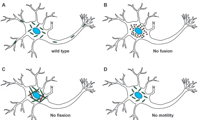
Figure 1 - Mitochondrial dynamics is characterized by fission, fusion and motility. A change of mitochondrial dynamics influences the function of mitochondria and can alter the supply of energy in distinct subcellular areas. (A) In wild type neurons, mitochondria are transported along the cytoskeleton to distinct cell areas and are well distributed to provide energy all over the cell. Mitochondria are functional and not fragmented or elongated. (B) A loss of mitochondrial fusion leads to fragmented mitochondria and a subpopulation of fragmented mitochondria becomes dysfunctional (red). In addition, fragmented mitochondria show transport defects to distal cell areas. (C) In the absence of mitochondrial fission, the mitochondrial population shows extremely elongated mitochondria and a subpopulation is not functional anymore (red). The elongated mitochondria show dysfunction in transport and cluster around the nucleus. (D) Without functional transport machinery, the mitochondria are not distributed throughout the cell but rather cluster in the perikaryon (modified from Chen and Chan, 2009).
1.2.1 Mitochondrial fission
The ratio of mitochondrial fission and fusion events determines the length of mitochondria.
Mitochondrial fission is characterized by the division of a mitochondrion into two daughter mitochondria. In the case of excessively frequent fission events, the balance between fission and fusion is shifted, resulting in a large number of fragmented mitochondria (van der Bliek et al., 2013). Mitochondrial fission is probably required to ensure that a population of organelles is available for segregation, to produce smaller organelles that are easier to transport along the cytoskeleton, to maintain a healthy population of organelles, and to transfer mitochondrial DNA between organelles (Kraus and Ryan, 2017). Mice without effective mitochondrial fission die shortly after birth, which shows the necessity of mitochondrial fission in Mammalia (Ishihara et al., 2009). Mitochondrial fission is a complex process that requires the interplay of many proteins and actin filaments (Korobova et al., 2013, 2014; Hatch et al., 2014; Li et al., 2015;
Manor et al., 2015; Lee et al., 2016). Mitochondrial fission can be separated into two consecutive / sequential stages; in the first stage, the mitochondrial diameter is significantly reduced at fission sites. This beginning step involves the endoplasmic reticulum (ER) wrapping around the mitochondrial membranes, which marks the fission zones and initiates constriction (Friedman et al., 2011). Along these fission zones, actin polymerization occurs and exerts pressure on the mitochondrial outer membrane to perform mitochondria constriction, because the floppy ER tubules fail to do so (Korobova et al., 2013, 2014; Hatch et al., 2014; Manor et al., 2015). The ER associated actin nucleator Inverted formin 2 (INF2) and the mitochondria located actin nucleator mitoSPIRE1 (also known as SPIRE1C or SPIRE1-E13) are thought to interact, and thus to facilitate actin polymerization at constriction sites (Manor et al., 2015).
The second stage of mitochondrial fission describes the final mitochondrial membrane scission.
The scission event is initiated by the GTPase dynamin-related protein 1 (Drp1), which forms a polymeric ring after being recruited to fission zones at the outer mitochondrial membrane (Smirnova et al., 2001; Lackner and Nunnari, 2009; Archer, 2013). The Drp1 ring is not large enough to enclose tubules of human mitochondria, which often have a diameter of ∼ 300 nm.
For the generation of a Drp1 polymeric ring the mitochondrial diameter should not be larger than ∼ 150 nm (Friedman et al., 2011). That is why the mitochondrial constriction of the first fission stage reduces the diameter at fission zones to the appropriate size so that Drp1 can build the polymeric ring (Lackner and Nunnari, 2009; Friedman et al., 2011; Archer, 2013; Manor et al., 2015). Like it was done in the first fission stage the Drp1 polymeric ring excerts pressure on the mitochondrial fission zone. After the fission zone is narrowed to its maximum,
dynamin-2 is recruited to the constricted area and drives the final membrane scission event (Lee et al., 2016; Kraus and Ryan, 2017).
1.2.2 Mitochondrial fusion
The balance between the opposed processes of mitochondria organelle fission and fusion regulates mitochondria number and size (Liesa et al., 2009). Mitochondrial fusion is characterized by two mitochondria merging into one mitochondrion. An excessive increase of mitochondria fusion events brings the equilibrium of mitochondrial fission and fusion out of balance, resulting in a large number of hyperfused mitochondria (Pagliuso et al., 2018). Fusion of mitochondria is required to mix mitochondrial contents like mitochondrial DNA (mtDNA) to maintain a homogenous population of organelles, and to prevent the permanent loss of essential components. Consequently, a decreased rate of fusion events results in a subpopulation of mitochondria that lack functional mtDNA. Mitochondria with deficient mtDNA show respiration defects and can impair the neuronal outgrowth, thereby promoting neurodegenerative diseases (Chen and Chan, 2010). Mitochondrial fusion can be separated into two stages; The first stage describes the outer mitochondrial membrane (OMM) fusion of two mitochondria mediated by the membrane-anchored dynamin GTPase family members mitofusin 1 (Mfn1) and mitofusin 2 (Mfn2). In the second step, the inner mitochondrial membrane (IMM) fusion is mediated by the optic atrophy 1 (OPA1) protein. In the absence of mitofusins, the fusion of both the inner and the outer mitochondrial membrane is abolished (Chen and Chan, 2009). The need for mitochondrial fusion is demonstrated by two knockout mouse models which lack either Mfn1 or Mfn2. A homozygous knockout of Mfn1 or Mfn2 results in embryonic lethality. In contrast, heterozygous animals demonstrate full viability and fertility (Chen et al. 2003). Interestingly, heterozygous mutations in human Mfn2 cause an axonal peripheral neuropathy, known as Charcot-Marie-Tooth disease type 2A (Amiott et al., 2008). In addition, heterozygous mutations in the human OPA1 gene can cause dominant optic atrophy. Patients who suffer from dominant optic atrophy develop a bilateral degeneration of the optic nerves, and usually lose their visual sense within the first decade of their life (Lenaers et al., 2012).
1.2.3 Mitochondrial motility
Diffusion and transport of intracellular molecules like ATP are rather limited, and therefore simple molecule diffusion is not sufficient to provide all cell areas with energy (Kinsey et al., 2011). A complex transport machinery enables mitochondrial motility, and facilitates transport of these cellular power plants to their sites of operation (Hollenbeck and Saxton, 2005). The mitochondrial transport system is essential, especially in highly polarized cells, such as neurons, to efficiently distribute mitochondria to distal cell compartments (Hollenbeck and Saxton, 2005; Sheng, 2014). A dysfunction in the mitochondrial transport machinery results in the absence of mitochondria in dendrites and axons, leading to neurotransmission defects (Guo et al., 2005). In addition, transport defects of mitochondria can influence mitochondrial morphology resulting in an imbalance of fission and fusion events (Varadi et al., 2004).
Mitochondrial trafficking in neurons and thus their distribution depends on synaptic activity, certain stress conditions, and mitochondria integrity (Miller and Sheetz, 2004; Chang and Reynolds, 2006; Cai et al., 2012; Sheng, 2014). In mature neurons, ∼ 20 - 30 % of axonal mitochondria are motile whereas the remaining mitochondria are stationary. Mitochondria are specifically docked / anchored to synaptic sites in order to provide a stable ATP supply as stationary power plants (Kang et al., 2008). The absence of stationary mitochondria significantly influences the synaptic vesicle release due to fluctuations in synaptic ATP levels, which are induced by mitochondrial movement (Sheng, 2014). Furthermore, spatially stable mitochondrial compartments fuel local translation during synaptic plasticity in neuronal dendrites (Rangaraju et al., 2019). As an effect of altered metabolic requirements, the anchored stationary mitochondria can become motile and motile mitochondria can become stationary (Sheng, 2014).
The molecular transport of mitochondria in mammalian neuronal cells is well researched and allows assumptions to be made about the mitochondrial transport system of other cell types.
However, there is still uncertainty concerning the exact mechanism of mitochondrial targeting.
It is known that mitochondria are transported along cytoskeletal components mediated by motor and adapter proteins (Hollenbeck and Saxton, 2005). The transport of axonal mitochondria is modulated by microtubule- and actin-dependent mechanisms, in which the majority of mitochondria are transported on fast tracks along microtubules via kinesin and dynein motor proteins (Hollenbeck and Saxton, 2005; Pathak et al., 2010; Sheng, 2014). Microtubules are polarized along the axonal processes, which makes it possible to study bidirectional mitochondrial transport (Heidemann et al., 1981; Baas et al., 1988, 1989). Dynein motor
proteins mediate a retrograde (towards the cell body) movement of mitochondria, whereas kinesin motor proteins drive an anterograde (towards axonal termini) mitochondria transport.
KIF5, a member of the kinesin-1 family, is described as a key motor protein of neuronal mitochondrial transport (Hurd and Saxton, 1996; Tanaka et al., 1998; Górska-Andrzejak et al., 2003; Cai et al., 2005; Glater et al., 2006; Pilling et al., 2006; Sheng, 2014). In line with all other mitochondria-associated motor proteins, KIF5 does not bind directly to mitochondria, but rather interacts with the adapter protein complex Miro1-Trak2 to be finally connected to mitochondria and facilitate transport along microtubules (MacAskill et al., 2009; van Spronsen et al., 2013; Sheng, 2014). A large number of adapters between motor proteins and mitochondria have already been identified, but it is often unclear how exactly they influence mitochondrial motility and how they are regulated (Sheng, 2014). However, the adapter protein syntaphilin immobilizes motile axonal mitochondria by anchoring them to microtubules, which results in stationary pools of mitochondria (Sheng, 2014). Compared to the microtubule-associated mitochondrial transport, only little is known about the role of actin / myosin forces in mitochondrial transport. The actin / myosin transport machinery might be used to move mitochondria to cell regions where there is a lack of microtubules or it could be used to recouple mitochondria to microtubules after kinesin or dynein motor proteins are disengaged (Hollenbeck and Saxton 2005). Further research into Drosophila melanogaster shows an increase of mitochondrial movement and velocity following myosin 5 depletion in neuronal cells. These data indicate that myosin 5 activity opposes microtubule-based axonal transport of mitochondria (Pathak et al., 2010). However, the role of actin / myosin in mitochondrial transport has not been investigated sufficiently and is not well understood. In contrast to KIF5, it is still unknown how myosin motor proteins interact with mitochondria, or which proteins might mediate their connection.
1.3 Actin nucleators facilitate actin polymerization
Actin filaments are highly dynamic and function in diverse cellular processes (Rottner et al., 2017). They provide mechanical stability and regulate cell shape, enable cell membrane dynamics and facilitate intracellular transport processes (Dominguez and Holmes, 2011;
Pollard and Goldman, 2016; Rottner et al., 2017). Actin filaments are generated by the polymerization of actin monomers (G-actin) into double helical filaments (F-actin). Actin dimers and trimers are relatively unstable, and therefore a spontaneous polymerization of actin monomers into actin filaments is possible, but insufficient and rare. In addition, actin monomer
binding proteins like profilin and ß-thymosin further inhibit spontaneous actin polymerization in cells (Sept and McCammon, 2001; Pollard und Cooper, 2009; Xue and Robinson, 2013). A well-regulated initiation of actin polymerization in cells in space and time is thus essential and is controlled by a large set of actin nucleators. These actin nucleators bind actin monomers and stabilize an actin nucleus (dimer, trimer, tetramer), which enables further polymerization (Firat-Karalar and Welch, 2011). There are three major classes of actin nucleators; namely the Arp2/3 complex, the formin superfamily and the Wiskott-Aldrich-Syndrome protein homology 2 (WH2) domain containing nucleators, including the SPIRE proteins. These actin nucleators differ in their molecular mechanisms to initiate actin polymerization (Robinson et al., 2001;
Quinlan et al., 2005; Kerkhoff, 2006; Schönichen and Geyer, 2010; Carlier et al., 2011).
1.3.1 SPIRE proteins
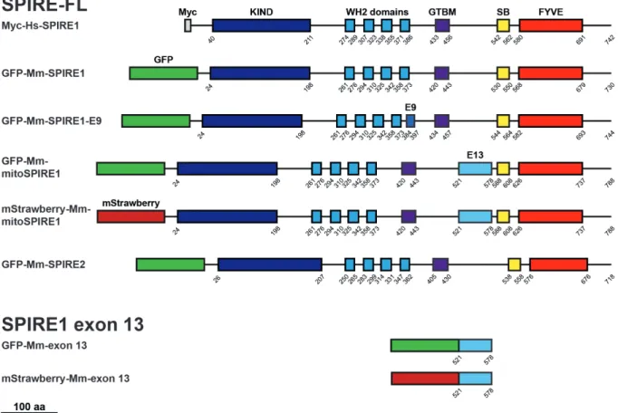
SPIRE proteins were first identified in 1999 / 2000 and were shown to play a role in Drosophila oocyte maturation (Wellington et al., 1999; Otto et al., 2000). Twenty years later, modern technology traced back the origin of SPIRE to be at least a holozoan invention, because SPIRE proteins were found in Ichthyosporea, Choanoflagellida, and Metazoa (Kollmar, Welz, Straub et al., 2019). Mammals have two SPIRE genes, namely SPIRE1 and SPIRE2. The mammalian SPIRE1 gene locus contains two alternatively spliced exons - exon 9 and exon 13. In contrast, the SPIRE2 gene locus encodes only a single transcript (Figure 2A). As a result, the two mammalian SPIRE genes encode a total of four different SPIRE proteins: SPIRE1, SPIRE1-E9, mitoSPIRE1 and SPIRE2, which are highly similar proteins regarding their domain organization. The primary structure of the mouse SPIRE1 and SPIRE2 proteins show an identity of 37 % and a similarity of 50 % (Kerkhoff, 2006). SPIRE1 and SPIRE2 proteins do not contain alternatively spliced exons and have similar subcellular localization at vesicular membranes (Kollmar, Welz, Straub et al., 2019). The vesicle located SPIRE1-E9 contains the alternatively spliced exon 9, a unique 14 amino acid spanning sequence. The alternatively spliced exon 13, which encodes a sequence of 58 amino acids, is part of the mitoSPIRE1 protein, which is localized at mitochondria. A SPIRE protein containing both the exon 9 and the exon 13 was not detected (Kollmar, Welz, Straub et al., 2019).
All SPIRE proteins contain highly conserved regions in their structural protein organization (Figure 2, B, C). The SPIRE proteins consist of an N-terminal kinase non-catalytic C-lobe domain (KIND) for FMN subgroup formin interaction (Pechlivanis et al., 2009), and a cluster
of four Wiskott-Aldrich-Syndrome protein homology 2 (WH2) domains for actin monomer binding and nucleation (Quinlan et al., 2005). The globular tail domain binding motif (GTBM) in the central linker region of SPIRE proteins mediates a direct interaction between SPIRE and myosin 5 (MYO5) proteins (Pylypenko, Welz et al., 2016). The C-terminal SPIRE-box (SB) shows sequence homology to the Slp homology domain (SHD) of RAB GTPase interacting proteins like rabphilin 3A (Kerkhoff, et al. 2001; Fukuda 2013). In this context, the C-terminal SPIRE SHD-like sequences mediate the interaction between SPIRE1 and RAB27A/B (Alzahofi, Robinson, Welz et al., 2018). Furthermore, SPIRE1 and SPIRE2 interact with comparable affinity with RAB8A (Kollmar, Welz, Straub et al., 2019). This indicates the opportunity of the SPIRE-box to interact with different RAB family GTPases. The C-terminal FYVE-type zinc-finger (FYVE) domain contains a hydrophobic turret loop which integrates into lipid bilayers, enabling intracellular membrane localization of the whole protein (Misra and Hurley, 1999; Dumas et al., 2001; Kerkhoff et al., 2001; Tittel et al., 2015).
In situ hybridization in mouse brains revealed a high expression of SPIRE1 in the hippocampus, dentate gyrus, and the cerebellum (Schumacher et al., 2004; Pleiser et al., 2010). Expression analysis in mice show that SPIRE mRNA is most abundant in oocytes and in the brain (Pfender et al., 2011; Pleiser, 2012). As a conclusion of the expression studies, the overall SPIRE expression levels seem to be low, which complicates the study of endogenous SPIRE functions.
Nevertheless, in several studies, the function of SPIRE has been addressed. SPIRE nucleated actin filaments mediate long-range transport of vesicles in mouse oocytes (Schuh, 2011) and are essential for asymmetric oocyte cell division (Pfender et al., 2011). In addition, SPIRE1 function influences fear expression (Pleiser et al., 2014) and contributes to the regulation of melanosome transport in melanocytes (Alzahofi, Robinson, Welz et al., 2018).
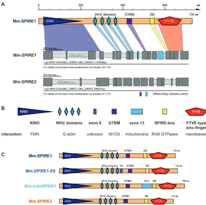
Figure 2 - Structure of mouse SPIRE genes and proteins. (A) Schematic drawing of the mouse SPIRE1 protein and the predicted gene structure of mouse SPIRE1 and SPIRE2.
Conserved domains of the SPIRE1 protein are highlighted in color and are assigned to their respective exon sequence. Dark and light grey boxes indicate exons and introns, respectively.
Introns have been scaled down for a better presentation of the gene structures as denoted. (B) Overview on functional SPIRE protein domains and the specific interactions they mediate.
KIND, kinase non-catalytic C-lobe domain; WH2, Wiskott-Aldrich-Syndrome protein homology 2; GTBM, globular tail domain binding motif; FYVE, FYVE-type zinc-finger. (C) Overview on mouse SPIRE proteins and their respective domain organizations. Numbers indicate amino acids. The SPIRE1 gene encodes for three different proteins, SPIRE1, SPIRE1-E9 and mitoSPIRE1. The SPIRE2 gene encodes for one protein, SPIRE2. E9, exon 9;
E13, exon 13; SB, SPIRE-box; aa, amino acids. Figure 2 was already published in Kollmar, Welz, Straub et al., 2019.
1.3.2 mitoSPIRE1
The alternatively spliced actin nucleator protein mitoSPIRE1 was first described in 2015. Until recently, the protein was named SPIRE1C, because it includes the alternatively spliced exon 13 also known as exon C. Unfortunately, Drosophila already encodes a SPIRE protein isoform which is called SPIREC. To avoid misunderstandings, in 2019 we renamed the mammalian SPIRE1C protein in agreement with its discoverer Uri Manor into mitoSPIRE1. We chose this name, because mitoSPIRE1 is localized at mitochondria, and is a splice variant of the SPIRE1 gene. The 58 amino acid sequence of the alternatively spliced exon 13 binds to the outer mitochondrial membrane, and targets the mitoSPIRE1 protein towards mitochondria (Manor et al., 2015). Manor and colleagues describe a role of mitoSPIRE1 in mitochondrial fission. In their current model, a dimer of mitoSPIRE1 is expected to interact with the endoplasmic reticulum (ER)-localized inverted formin 2 (INF2) to form an actin nucleation complex during ER initiated mitochondrial constriction. The subsequent nucleation and elongation of actin filaments along fission zones by the mitoSPIRE1 / INF2 complex might exert pressure on the mitochondrial outer membrane, and therefore cause constriction of the mitochondrial membrane. The mitoSPIRE1 / INF2 complex could be necessary for mitochondrial fission (Manor et al., 2015). Additional functions of mitoSPIRE1 remain unknown thus far.
1.4 Cooperation of SPIRE and formin proteins
Initial findings of the association between the two actin nucleators SPIRE and formin were already provided in the late 1980s. A mutation in the gene locus of the Cappuccino protein (a Drosophila melanogaster FMN2 ortholog) revealed the same phenotype in oocytes of Drosophila melanogaster as a mutated SPIRE gene (Manseau and Schüpbach, 1989). Years later, the identical oocyte phenotype was explained. SPIRE and Cappuccino cooperate to assemble a cytoplasmic mesh of actin filaments that controls a distinct microtubule organization during oocyte development (Dahlgaard et al., 2007). In addition to this functional cooperation, further studies also revealed a physical interaction between formins and SPIRE proteins (Quinlan et al., 2007; Pechlivanis et al., 2009). The mammalian FMN subgroup formins (FMN1, FMN2) and SPIRE proteins (SPIRE1, SPIRE2) interact directly with each other in order to form a functional actin nucleator complex (Pechlivanis et al., 2009; Pfender et al., 2011). The very C-terminal end of FMN1/2 contains a basic amino acid cluster in the so called formin-SPIRE Interaction (FSI) sequence, which binds to an acidic cluster in the N-terminal KIND of SPIRE proteins (Pechlivanis et al., 2009; Zeth et al., 2011).
FMN subfamily formins contain, like all formins, two formin homology domains which are termed formin homology domain 1 (FH1) and formin homology domain 2 (FH2). Formin proteins dimerize which results in a dimeric ring of the two FH2 domains that encircles and processively moves with the growing plus end of an actin filament, therefore, facilitating its elongation (Schönichen and Geyer, 2010). The FH1 domains of the formin dimer contain multiple clusters of poly-proline stretches, which enable the binding of profilin-actin and providing it to the FH2 dimer for actin filament elongation (Romero et al., 2004; Otomo et al., 2005; Paul and Pollard, 2008; Schönichen and Geyer, 2010). The actin nucleation activity of FMN formins and the binding of FH2 to barbed ends of actin filaments is inhibited by the formation of the SPIRE-FMN complex. However, the formation of the SPIRE-FMN actin nucleator complex significantly increases the overall actin nucleation in comparison to the single nucleator proteins acting alone (Vizcarra et al., 2011; Montaville et al., 2014). The SPIRE-formin complex facilitates both actin nucleation via the WH2 domains of SPIRE and actin filament elongation via FMN (Figure 3; Quinlan and Kerkhoff, 2008; Kerkhoff, 2011).
It has been shown that the four WH2 domains of SPIRE are sufficient to nucleate actin polymerization in vitro, but these domains are not able to nucleate actin polymerization of profilin bound actin, being the most abundant source of actin monomers in cells (Quinlan et al., 2005; Montaville et al., 2014; Pollard, 2016). Thus, for SPIRE actin nucleation actin monomers have to be provided as a non-coupled actin monomer directly or by other proteins, which are able to process profilin-actin (Quinlan et al., 2005; Montaville et al., 2014). A present model of the SPIRE-formin complex explains that SPIRE WH2 domains nucleate actin monomers and passes over to an FMN dimer which elongates the newly synthesized SPIRE nucleated filaments (Quinlan and Kerkhoff, 2008; Kerkhoff, 2011). This model accumulates evidence for the theory that FMN subfamily formins provide SPIRE with actin monomers, because FMN processes profilin-actin with its FH1 domains and as a consequence, could supply actin monomers to SPIRE (Figure 3). However, in a current model, the building of the SPIRE-formin complex leads to the dimerization of SPIRE proteins at the membrane and initiates actin nucleation (Quinlan and Kerkhoff, 2008). The increase of SPIRE WH2 domains could be an explanation for higher actin nucleation activity of SPIRE in the complex.
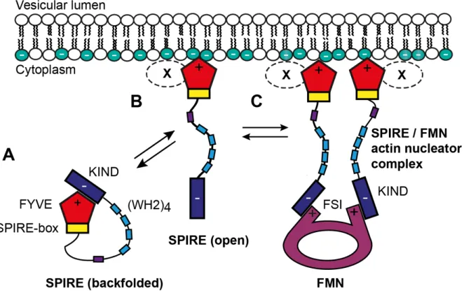
A further model shows that SPIRE proteins exist in two distinct states: a potentially auto-inhibited inactive state and an unfolded, membrane-bound active state (Figure 4). In the inactive state, the SPIRE protein is cytosolic and might adopt a backfolded structure by an intramolecular interaction between the C-terminal FYVE domain and the N-terminal KIND. In
this state membrane-binding of the FYVE domain is inhibited and KIND is not accessible for binding to FMN subfamily formins. In the active state, the SPIRE protein gets unfolded, is targeted to intracellular membranes and is now able to recruit FMN subfamily formins through the accessible KIND. The intramolecular FYVE / KIND and the trans-regulatory FMN-FSI / KIND interactions are competitive and couples the membrane binding of SPIRE to the recruitment of FMN subfamily formins (Tittel et al., 2015). A distinct mechanism which drives SPIRE protein activation and membrane targeting is still not completely understood. In this context, the interaction of SPIRE proteins and myosin 5 actin motors might contribute to the SPIRE activation process (Pylypenko, Welz et al., 2016).
Figure 3 - Model of SPIRE / FMN subfamily formin cooperation in efficient actin nucleation and filament elongation. For actin nucleation SPIRE proteins need monomeric unbound actin molecules, whereas FMN subfamily formins can process profilin bound actin monomers which are the most abundant form of actin monomers in eukaryotic cells. In the current model SPIRE WH2 domains nucleate actin monomers and passes over to an FMN dimer which elongates the novel synthesized SPIRE nucleated filaments. (A) Activated SPIRE proteins are targeted at subcellular membranes by the interaction of the SPIRE FYVE-type zinc-finger with negatively charged lipids of the membrane. Specific subcellular localization of SPIRE proteins could be determined by the interaction of the SPIRE-box with small G-proteins like RAB27 and the localization might be influenced by dimerization of SPIRE proteins. After SPIRE dimerization FMN subfamily formins are translocated to SPIRE and FH2 domains of FMN formins form a dimer as well, whereas each FSI motif interacts directly with a single N-terminal SPIRE KIND. In addition, each WH2 domain of the SPIRE dimer is used to interact with an unbound actin monomer to facilitate actin nucleation. It is not verified but in the
following functional cooperation of SPIRE and FMN formins, the proline rich region of FMN FH1 could process profilin bound actin monomers to provide SPIRE WH2 domains with non-bound actin monomers for actin nucleation (indicated by the arrow). If FMN does not provide unbound actin monomers to the WH2 domains, the SPIRE dimer is mainly dependent on cytosolic unbound actin monomers. (B) After SPIRE mediated actin nucleation, the novel actin filament is released from WH2 domains and the dimerized donut shaped FH2 domains of FMN elongate the growing actin filament by processive addition of actin monomers. Actin monomers for the FH2 mediated elongation of the actin filament are provided by the FH1 domains, processing profilin bound actin monomers. The FSI domain is highlighted in green (modified from Kerkhoff, 2011).
Figure 4 - Model for the different molecular states of the SPIRE / FMN actin nucleation complex at vesicle membranes. In this model cytosolic SPIRE proteins are primarily inactive (closed conformation) as backfolded monomers, but after transient membrane binding SPIRE proteins are activated (open conformation), able to dimerize and interact with FMN subfamily formins to build a functional unit as SPIRE / FMN actin nucleator complex. (A) The cytosolic backfolded SPIRE monomer is inactive due to interaction by FYVE / KIND with itself. The KIND is blocked by FYVE which is why the protein is not able to interact with FMN subfamily formins. (B) After transient interaction of the SPIRE C-terminus in a non-specific pattern with negatively charged intracellular membranes, the protein is unfolded. The accessible KIND is able to interact with FMN-FSI to initiate formation of a functional SPIRE / FMN actin nucleator complex. (C) A donut shaped FMN subfamily dimer interacts with two SPIRE proteins. The FMN subfamily formin and SPIRE build a functional unit to nucleate and elongate actin filaments. Additional factors (X) may specify targeting of SPIRE at vesicle membranes (modified from Tittel et al., 2015).
1.5 The SPIRE and myosin 5 interaction
Myosin actin motor proteins are molecular motors that are involved in a variety of cellular and sub-cellular mechanisms including cell migration and adhesion, muscle contraction, cytokinesis and signal transduction (Vicente-Manzanares et al., 2009; Hartman and Spudich, 2012). Furthermore, motor proteins are essential for intracellular transport of distinct cargos (Hartman and Spudich, 2012). In particular, neurons depend on these transport mechanisms as they synthesize most axonal and synaptic proteins in the cell body. These proteins need to be transported to their respective places of action for instance in large neurites and therefore, they require a molecular transport system (Hirokawa et al., 2010). In this context, myosin motors transport cargos along actin filaments, which facilitate a slow but precise transport system to subcellular target regions (Ross et al., 2008; Hirokawa et al., 2010; Welz et al., 2014).
Myosin superfamily motor proteins are actin binding ATPases. ATP hydrolysis induces an intramolecular conformational change within the motor. This conformational change is used to generate forces along actin filaments in order to induce morphological changes of cells, to mediate muscle contractions or to transport distinct cargos along actin tracks (Hartman and Spudich, 2012; Houdusse and Sweeney, 2016). Myosin motor proteins are divided into conventional myosins (myosin 2 family) and unconventional myosins (all other myosins;
Venkatesh et al., 2019). To date, 38 different myosin genes have been identified in humans, which are grouped into distinct classes. In general, myosins consist of three different regions:
An N-terminal motor domain, a neck region, and a tail region (Hammer and Sellers, 2011;
Figure 5). The motor domain, which is also known as the head, harbors the ATPase activity and binds to actin filaments (Hammer and Sellers, 2011).
In the present thesis, myosin 5 motor proteins are of great interest because these motor proteins serve as cargo transporters (Hammer and Sellers, 2011). In humans, there are three different myosin 5 genes: myosin 5A, 5B and 5C, encoding myosin 5A (MYO5A), myosin 5B (MYO5B) and myosin 5C (MYO5C) proteins, respectively (Mercer et al., 1991; Zhao et al., 1996; Berg et al., 2001; Rodriguez et al., 2002). In gene expression studies, MYO5A, MYO5B and MYO5C are found in the brain (Espreafico et al., 1992; Cheney et al., 1993; Rodriguez et al., 2002;
Tilelli et al., 2003; Lisé et al., 2006). Myosin motor proteins like MYO5 assemble as a dimer to form a two-headed motor and work as a unit to allow the dimer to ‘walk’ along actin filaments (Hammer and Sellers, 2011). The neck region of MYO5 contains six IQ motifs and each IQ repeat binds to calmodulin or a related light chain (Espindola et al., 2000). As a consequence,
the MYO5 neck region builds the lever arm required for processive movement of the whole protein. In the case of MYO5, the tail region contains a proximal coiled coil domain to form a dimerization of heavy chains and a C-terminal globular tail domain (GTD). The GTD of MYO5 has an important role in cargo binding because it enables the motor protein to bind to adapter proteins, which mediate vesicle or organelle targeting (Wu et al., 1998; Reck-Peterson et al., 1999; Hammer and Sellers, 2011; Syamaladevi et al., 2012). In 2016, it was demonstrated that the GTDs of MYO5A, 5B and 5C, interact directly with an evolutionarily conserved sequence motif termed globular tail domain binding motif (GTBM) of the SPIRE actin nucleator.
Through the interaction of MYO5 with RAB11, the SPIRE GTBM facilitates the formation of a tripartite complex at vesicle membranes made of SPIRE, MYO5 and RAB11. In summary, this complex could explain how RAB11 vesicles coordinate actin filament nucleation and myosin force generation at RAB11 vesicle membranes to regulate transport processes (Pylypenko, Welz, et al., 2016). Human homozygous mutation of MYO5A is known as Griscelli syndrome type 1, which is characterized by partial albinism and primary neurological defects (Thomas et al., 2009; Cağdaş et al., 2012). Mutations of MYO5B are also found in humans, and cause microvillus inclusion disease, a rare genetic disorder of the small intestine (Knowles et al., 2014).
Figure 5 - Schematic overview of a vertebrate myosin 5 motor protein. The myosin 5 motor protein is organized into three different functional units, facilitating processive movement and consequently transport of cargos along actin filaments. The N-terminal motor domain provides ATPase activity and binds to actin filaments. The following lever arm / neck carries six calmodulin binding IQ motifs. Given the elongated nature of the neck region compared to other myosin motors the ATP hydrolysis induced conformational change is translated into a large step size of the motor which allows for processive movements. The C-terminal tail is divided into two functional and structural parts:
the coiled-coil region necessary for dimerization of two myosin 5 heavy chains and at the very end of the protein the globular tail domain (GTD) which mediates as cargo binding domain cargo interaction of the motor and direct interaction with SPIRE proteins (modified from Pylypenko, Welz et al., 2016; Welz, 2018).
1.6 Cellular functions of SPIRE proteins
As mentioned previously, SPIRE proteins are expressed at very low levels but still have a significant impact on several cell biological mechanisms, including oocyte maturation, transport of RAB11 vesicles and peripheral transport of melanosomes in melanocytes (Pfender et al., 2011; Schuh et al., 2011; Pleiser, 2012; Alzahofi, Robinson, Welz et al., 2018; Kollmar, Welz, Straub et al., 2019). The ability of SPIRE to directly interact with FMN subfamily formins and MYO5 actin motors suggests a much wider spectrum of SPIRE function in vesicle and organelle transport (Pechlivanis et al., 2009; Pylypenko, Welz, et al., 2016). Further investigations are necessary to unravel the diversity of SPIRE functions.
1.6.1 SPIRE actin nucleators drive oocyte maturation
The maturation of oocytes into eggs is a complex differentiation program that is initiated early on in embryonic development. During early meiosis, the diploid precursor cells termed
‘oocytes’ are arrested at prophase I and wait for their chance to mature into pregnable eggs.
During sexual maturity, mostly a single oocyte per menstrual cycle matures into a haploid egg and becomes ovulated from its surrounding follicle. The maturation of an oocyte into an egg is dependent on asymmetric cell division, including a reduction to half of the oocyte chromosomes (Mogessie et al., 2018). The process of asymmetric meiotic cell division can be described by two different stages. In the first stage, an asymmetric positioning of the meiotic spindle takes place in order to move excessive chromosomes for a haploid egg into a small cell termed the polar body. In this context, the dense chromosomes must first be aligned to the spindle apparatus, which is necessary to separate chromosomes in space (Kirschner et al., 1986;
Kline-Smith et al., 2004). After chromosomes are aligned to the spindle, the whole spindle has to be moved to the cell cortex to initiate the second stage of asymmetric cell division. Spindle positioning is dependent on a dynamic actin meshwork, which is generated by FMN2 and SPIRE actin nucleators (Leader et al., 2002; Dumont et al., 2007; Pfender et al. 2011). For that matter, SPIRE and FMN interact as a functional unit in which both actin nucleators depend on each other. Interestingly, a single depletion of either SPIRE1 or SPIRE2 compared to a double depletion does not affect spindle positioning, indicating that the protein similarity allows a mutual rescue of both proteins. Finally, the extrusion of polar bodies describes the second stage of the asymmetric meiotic division. A loss of SPIRE proteins not only inhibits proper spindle positioning, but consequently impairs polar body extrusion as well. In summary, SPIRE proteins are important for flawless oocyte maturation (Pfender et al., 2011). Interestingly the
corresponding Drosophila melanogaster (Dm) SPIRE and Cappuccino (a Dm FMN2 ortholog) produce a similar actin meshwork in fly oocytes, which is necessary to facilitate the maturation of the oocyte into an egg (Dahlgaard et al., 2007). Errors in the maturation of oocytes may result in aneuploidy, hypofertility, and genetic disorders in offspring (Leader et al., 2002; Zhai et al., 2018).
1.6.2 SPIRE actin nucleators drive vesicular transport processes
Intracellular transport of cargo and organelles is essential for communication, organization and polarity of mammalian cells (Pylypenko, Welz et al., 2016). In this respect, cargo, such as proteins, lipids, or even RNA molecules, have to be transported to their target areas, and especially proteins are therefore wrapped into membrane enclosed carriers termed vesicles.
Transport of intracellular cargo is generally mediated by microtubules and actin filaments.
Polarized microtubules serve as long bidirectional transport tracks and provide a fast but constricted path used by kinesin and dynein motor proteins (Schliwa and Woehlke, 2003;
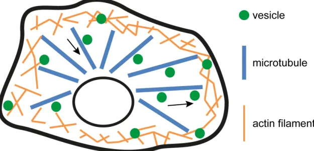
Hirokawa et al., 2009; Welz et al., 2014). Actin filament-based transport processes are much slower but more dynamic than microtubule-based transports and are mostly important for precise short-range movements of vesicles, facilitated by myosin motor proteins (Hirokawa et al., 2009; Kneussel and Wagner, 2013; Welz et al., 2014). Typical intracellular transport processes including microtubule- and actin-based transport systems for cargo are commonly described as the highways and local roads model (Figure 6). In this controversial discussed model, vesicles are transported quickly along microtubules to the cell periphery. Here, vesicles are detached from microtubules and begin to move slowly along actin filaments to their final destinations (Langford, 1995; Ross et al., 2008; Woolner and Bemet, 2009; Welz et al., 2014).
The dynamic actin cytoskeleton facilitates transport of vesicles to areas which are not reached and therefore supplied with cargos by the coarse microtubule network (Welz et al., 2014).
Already in 2001, it was found that the actin nucleator SPIRE is targeted to intracellular membranes and involved in vesicle transport processes (Kerkhoff et al., 2001). Years later, in mouse metaphase oocytes, SPIRE was identified to contribute to a microtubule-independent long-range transport of RAB11 vesicles (Schuh et al., 2011). In this context, SPIRE and FMN proteins cooperate in nucleating actin filaments originating from vesicle membranes to facilitate the formation of a cytoplasmic actin mesh and to allow a myosin 5B (MYO5B)-dependent RAB11 vesicle transport. A lack of SPIRE1 and SPIRE2 results in a loss of directed transport of RAB11 vesicles to the oocyte cortex (Schuh et al., 2011). SPIRE