Berichterstatter: Prof. Dr. Ulrich Baumann Prof. Dr. Peter Nürnberg
Tag der mündlichen Prüfung: Februar 19, 2019
Dedicated to the three most important women in my life; my dear mother Dekah Yusuf, sister Asha Anab and wife Halima Sonny Juma
“The scientist is not a person who gives the right answers, he is one who asks the right questions”
Claude Lévi-Strauss
I
Summary
Various stimuli that perturb neuronal homeostasis and cause neurodegeneration trigger microglia reactivity. The microglia reaction and the increased production of pro-inflammatory mediators is intended to protect the central nervous system, including the retina, from noxious insults and facilitate a rapid return to normal homeostasis. However, sustained and dysregulated microglia inflammatory responses can lead to robust neuropathological changes that contribute to the severity and progression of neurodegenerative diseases. In the retina, pathologically activated microglia have been shown to not only contribute to neurodegenerative processes indirectly via the release of neurotoxic substances, but also directly via the indiscriminate phagocytosis of stressed but living photoreceptors. Consistently, innumerous studies have demonstrated that microglial modulation represents an attractive therapeutic avenue to alleviate or delay neurodegenerative diseases of the retina. Translocator protein (18kDa) (TSPO) located in the outer mitochondrial membrane is acutely and specifically expressed in activated microglia during retinal pathology. Importantly, specific TSPO ligands have been shown to potently modulate microglia inflammatory responses and ameliorate light-induced photoreceptor degeneration in mice.
However, understanding the regulation of TSPO expression in microglia during health and disease is paramount prior to any utilization of this protein as a therapeutic target to modulate chronic microglia inflammatory responses and alleviate retinal degeneration. The aim of the current study therefore was to carry out a detailed functional characterization of the TSPO promoter and identify genetic elements and transcription factors responsible for maintaining basal and mediating induced TSPO expression in microglia.
To this end, a 2.812Kb murine Tspo promoter sequence was amplified by PCR and cloned into the promoterless pGL4.10-Basic luciferase reporter vector.
Plasmids containing 5’ unidirectional deletions of the promoter were then generated by PCR. BV-2 microglia cells were transfected with the generated reporter plasmids for 24 h, and an additional 6 h in the case of lipopolysaccharide (LPS) stimulation before measuring luciferase activity. Sequence analysis was performed using Matinspector software and site-directed substitution
II
mutagenesis was used to confirm the functional status of the putative transcription factor binding sites. Chromatin immunoprecipitation (ChIP) was used to investigate binding of transcription factors to the endogenous promoter.
The effect of transcription factor knockdown on Tspo promoter activity was assessed using RNAi mediated gene silencing followed by transfection with the reporter constructs and measurement of luciferase activity 24 h later.
Additionally, effects of RNAi mediated gene silencing on endogenous Tspo protein levels were determined using western blot analysis.
Deletion mutagenesis indicated that −845 bp upstream of the transcription initiation site was sufficient to reconstitute near maximal promoter activity in BV- 2 microglia cells. Deletion of sequences extending -593 to -520, which harbour an Ap-1, Ets.2 and Nkx3.1 site which also serves as a non-canonical binding site for Sp1-family transcription factors, led to a dramatic decrease in both basal as well as LPS induced promoter activity. Further deletion of sequences extending - 168 to -39, which contains four GC boxes, also significantly decreased Tspo promoter activity. Site-directed mutagenesis of Ap-1, Ets.2, Nkx3.1/Sp1/3/4 and the proximal GC boxes led to significant decreases in promoter activity. ChIP- qPCR revealed that Pu.1, Ap-1(cJun/cFos), Stat3, Sp1, Sp3 and Sp4 bind the endogenous Tspo promoter. Notably, upon LPS stimulation, the binding of these factors, with the exception of Stat3, was significantly enhanced. RNAi mediated silencing of Pu.1, cJun, cFos, Stat3, Sp1, Sp3 and Sp4 gene expression significantly diminished Tspo promoter activity while Ap1(cJun/cFos) silencing effectively blocked LPS-induced increase in Tspo protein levels. Taken together, these findings demonstrate that the consensus binding sequences for Ap-1, Ets.2, distal as well as proximal Sp1/3/4 sites must be intact for maximal basal and LPS induced Tspo promoter activity in microglia. Furthermore, the current findings indicate that LPS-mediated increase in Tspo expression is mediated, at least in part, at the transcriptional level and is accompanied by an enhanced recruitment of Pu.1, Ap-1(cJun/cFos), Sp1, Sp3, and Sp4 factors to the Tspo promoter.
III
Zusammenfassung
Verschiedenste Stimuli, welche die neuronale Homöostase stören und neurodegenerative Erkrankungen verursachen, führen zur inflammatorischen Aktivierung von Mikrogliazellen. Mikrogliaaktivierung und die erhöhte Freisetzung von pro-inflammatorischen Signalmolekülen dienen ursprünglich dem Schutz des zentralen Nervensystems, inklusive der Netzhaut, vor schädlichen Einflüssen und ermöglichen ein rasches Wiederherstellen der gesunden Homöostase.
Dauerhafte und fehlregulierte inflammatorische Reaktivität von Mikrogliazellen führt hingegen zu auffälligen neuropathologischen Veränderungen, welche die Schwere und den Verlauf neurodegenerativer Erkrankungen negativ verstärken können. In der Netzhaut tragen pathologisch aktivierte Mikroglia nicht nur indirekt, durch die Ausschüttung neurotoxischer Substanzen, sondern auch direkt, durch die fehlgesteuerte Phagozytose von geschädigten aber noch lebenden Photorezeptorzellen, zum verschlechterten Krankheitsverlauf bei. Auf diesen Beobachtungen basierend, demonstrierten bereits zahlreiche Studien die Modulation der Mikrogliaaktivität als vielversprechenden therapeutischen Weg um neurodegenerative Erkrankungen der Netzhaut zu mildern oder zu verzögern.
Das Translokatorprotein (18 kDa) (TSPO) befindet sich in der äusseren Mitochondrienmembran und ist selektiv in reaktiven Mikrogliazellen während verschiedenen Netzhautdegenerationen verstärkt exprimiert. Interessanterweise konnte bereits nachgewiesen werden, dass spezifische TSPO Liganden in der Lage sind die pro-inflammatorische Mikrogliaaktivierung zu modulieren und dadurch die lichtinduzierte Photorezeptordegeneration im Mausmodell zu reduzieren. Das klare Verständnis der Regulation der TSPO Expression in Mikrogliazellen während Homöostase und Krankheit stellt eine Grundvoraussetzung für die Nutzung des Proteins als therapeutisches target im Kontext der Mikrogliaaktivierung und Netzhautdegeneration dar. Daher war das Ziel der vorliegenden Studie eine detaillierte funktionelle Charakterisierung des TSPO Promotors sowie die Identifikation von genetischen Elementen und Transkriptionsfaktoren, welche für die basale sowie induzierbare TSPO Expression in Mikrogliazellen verantwortlich sind. Zu diesem Zwecke wurde eine 2,812 kb grosse murine Tspo Promotorsequenz mittels PCR amplifiziert und in
IV
den promotorlosen pGL4.10-Basic Luciferase Reporter Vektor kloniert. Als nächstes wurden durch PCR einzelne Plasmide mit 5` Deletionen verschiedener Längen generiert. BV-2 Mikrogliazellen wurden mit den so erhaltenen Reporterplasmiden für 24 h transfiziert und nach weiterer sechsstündiger Inkubation mit Lipopolysaccharid (LPS) die Luciferaseaktivität gemessen.
Sequenzanalyse wurde mittels der Software MatInspector ausgeführt und zielgerichtete Mutagenese PCR wurde angewandt um die Funktionalität der putativen Transkriptionsfaktorbindestellen nachzuweisen. Die tatsächliche Bindung verschiedener Transkriptionsfaktoren an den endogenen Promotor wurde mittels Chromatin Immunpräzipitation (ChIP) experimentell belegt. RNAi- vermittelte Gen-Silencing Experimente wurden zur Untersuchung des Effekts von Transkriptionsfaktor Knock-Down auf die Luziferaseaktivität der Reporterkonstrukte durchgeführt. Zusätzlich wurde die endogene Tspo Protein Menge nach diesen RNAi Gen-Silencing Experimenten mittels Western Blot analysiert. Unsere Mutageneseexperimente belegten, dass ein Abschnitt von - 845 bp upstream der Transkriptionsinitiationsstelle für nahezu maximale Promotoraktivität in BV-2 Mikrogliazellen ausreichend ist. Die Deletion des Sequenzbereichs zwischen -593 und -520, welcher sowohl eine Ap-1, Ets.2 als auch Nkx3.1 Bindestelle - die auch als Bindestelle für Transkriptionsfaktoren der Sp1-Familie fungiert – enthält, führte zu einem drastischen Verlust sowohl der basalen als auch LPS induzierten Promotoraktivität. Des Weiteren führte die Deletion des Sequenzbereichs von -168 bis -39, welcher vier GC Boxen enthält, ebenfalls zu einem signifikanten Abfall der Tspo Promotoraktivität. Analog dazu führte die zielgerichtete Mutagenese der AP-1, Ets.2, Nkx3.1/Sp1/3/4 und proximalen GC Boxen ebenfalls zu starken Abschwächungen der Promotoraktivität. ChiP-qPCR Experimente belegten anschliessend die tatsächliche physikalische Bindung der Faktoren Pu.1, Ap-1(cJun/cFos), Stat3, Sp1, Sp3 und Sp4 am endogenen Tspo Promotor. Bemerkenswerterweise war die Bindung aller dieser Faktoren (mit Ausnahme von Stat3) nach LPS- Stimulation siginifikant verstärkt. RNAi-vermitteltes Gen-Silencing von Pu.1, cJun, cFos, Stat3, Sp1, Sp3 und Sp4 reduzierte die Tspo Promotoraktivität signifikant, wohingegen ein Silencing von Ap1(cJun/cFos) effektiv die LPS vermittelte Tspo Induktion blockierte. Zusammenfassend demonstrieren unsere
V
Ergebnisse, dass die Konsensusbindesequenzen für Ap-1, Ets.2 und der distalen wie proximalen Sp1/3/4 Bindestellen für maximale basale und LPS induzierte Tspo Promotor Aktivität in Mikrogliazellen vorhanden und intakt sein müssen.
Zusätzlich deuten unsere Ergebnisse darauf hin, dass der LPS induzierte Anstieg der Tspo Expression zumindest teilweise auf dem transkriptionellen Weg vermittelt wird und mit einer erhöhten Rekrutierung der Transkriptionsfaktoren Pu.1, Ap-1(cJun/cFos), Sp1, Sp3, und Sp4 zum Tspo Promotor einhergeht.
VI
Contents
Summary ... I Zusammenfassung ... III List of Tables ... VIII List of Figures ... IX List of abbreviations and acronyms ... XI
1.0 Introduction ... 1
1.1 The Retina ... 1
1.2 Retinal microglia ... 5
1.2.1 Origin and maintenance of microglia in the retina ... 6
1.2.2 Microglia in the healthy retina ... 10
1.2.3 Microglia in the diseased retina ... 14
1.2.4 Transcriptional control of microglia phenotypes in health and disease ... 21
1.3 Translocator protein (18 kDa) (TSPO) ... 29
1.4 Immunomodulatory and neuroprotective effects of TSPO ligands ... 33
1.5 Hypothesis and Specific Aims of the Thesis ... 35
2.0 Materials and Methods ... 37
2.1 Materials ... 37
2.1.1 Mammalian and bacteria cells ... 37
2.1.2 Culture media ... 37
2.1.3 Enzymes ... 38
2.1.4 Antibodies ... 39
2.1.5 Buffers and solutions ... 40
2.1.6 Agarose and SDS-PAGE gels ... 41
2.1.7 Kits ... 41
2.1.8 Chemicals and reagents ... 42
2.1.9 Devices ... 43
2.1.10Software ... 44
2.2 Methods ... 44
2.2.1 Cell culture ... 44
2.2.2 Plasmid construction ... 45
VII
2.2.3 Site directed mutagenesis ... 48
2.2.4 Transient transfections, luciferase and β-Gal assays ... 49
2.2.5 Chromatin immunoprecipitation (ChIP) ... 50
2.2.6 RNA-Isolation and reverse transcription ... 52
2.2.7 Quantitative RT-PCR ... 52
2.2.8 siRNA-mediated gene silencing ... 54
2.2.9 Western Blots ... 55
2.2.10Statistical analysis ... 56
3.0 Results ... 58
3.1 Basal activity of the Tspo promoter in BV-2 microglia ... 58
3.2 LPS stimulation increases Tspo promoter activity ... 60
3.3 Ap-1, Ets and Sp binding sites are strong positive elements regulating the expression of Tspo in BV-2 microglia ... 61
3.4 Pu.1, cJun, cFos, Stat3, Sp1, Sp3 and Sp4 binds the endogenous Tspo promoter in BV-2 microglia ... 65
3.5 LPS induces Pu.1, cJun, cFos, Sp1, Sp3 and Sp4 recruitment to the Tspo promoter in BV-2 microglia ... 66
3.6 Pu.1, Sp1, Sp3, Sp4, cJun, cFos and Stat3 siRNA significantly reduce Tspo promoter activity in BV-2 microglia ... 68
3.7 Ap-1 mediates LPS-induced increase in Tspo protein expression in BV-2 microglia ... 69
3.8 Differential utilization of the Tspo promoter between BV-2 microglia and ARPE-19 cells ... 71
3.9 Trichostatin A (TSA) can induce Tspo promoter activity in ARPE-19 cells but not in BV-2 microglia ... 74
4.0 Discussion ... 76
5.0 Conclusion and future perspectives ... 82
References ... 85
Acknowledgement ... 110
Erklärung... 111
VIII
List of Tables
Table 1: List of all cell lines and bacteria strain used in the study ... 37
Table 2: Reagents and recipes for cell culture and LB media/plates ... 37
Table 3: List of all enzymes and their corresponding buffers used in the study 38 Table 4: List of all antibodies used in the study ... 39
Table 5: Recipes for buffers and solutions used in this study ... 40
Table 6: Recipes for agarose and SDS-PAGE gels ... 41
Table 7: Commercially available kits used in this study ... 41
Table 8: List of chemicals and reagents used in this study ... 42
Table 9: List of devices used in the study ... 43
Table 10: List of software used in the study ... 44
Table 11: PCR recipe to amplify Tspo promoter from mouse BAC clone ... 45
Table 12: PCR temperature profile ... 45
Table 13: Restriction digest protocol ... 46
Table 14: Ligation protocol ... 46
Table 15: Test digest protocol ... 47
Table 16: Primers used to generate reporter plasmids ... 47
Table 17: List of sequencing primers used in this study ... 47
Table 18: Site directed mutagenesis PCR temperature profile ... 48
Table 19: Primer sequences (5' → 3') used in ChIP experiments ... 51
Table 20: qRT-PCR recipe... 52
Table 21: qRT-PCR cycling conditions ... 52
Table 22: Primer sets used for qRT-PCR ... 53
Table 23: ON-TARGETplus SMARTpool siRNA sequences... 54
IX
List of Figures
Figure 1: Cellular organization of the mammalian retina. ... 4
Figure 2: Irradiation weakens the integrity of the blood brain barrier ... 8
Figure 3: Microglia morphology in health and disease. ... 15
Figure 4: Schematic representation of microglia reactivity in the retina. ... 20
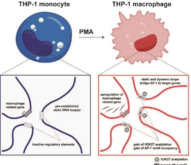
Figure 5: Enhancer-promoter interactions drive cell specific gene expression 22 Figure 6: Multi-loop activation hubs that form at key macrophage genes during differentiation are AP-1 enriched. ... 27
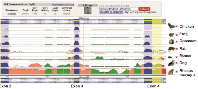
Figure 7: TSPO gene is highly conserved throughout evolution. ... 30
Figure 8: Constitutive and inducible Tspo expression in RPE and microglia cells ... 32
Figure 9: Immunomodulatory effects of endogenous and synthetic TSPO ligands. ... 35
Figure 10: Functional characterization of the mouse Tspo promoter ... 59
Figure 11: LPS significantly induces Tspo promoter activity ... 60
Figure 12: Regulatory DNA sequences within the Tspo promoter. ... 61
Figure 13: Sanger sequencing validation of the introduced mutations ... 62
Figure 14: Ap-1, Ets.2, Nkx3.1/Sp1/3 and the conserved GC boxes in the proximal promoter are crucial for TSPO promoter activity. ... 64
Figure 15: Pu.1 binds to the endogenous Tspo promoter in BV-2 microglia. .. 66
Figure 16: LPS stimulates recruitment of transcription factors Pu.1, Ap-1 (cJun/ cFos) Sp1, Sp3 and Sp4 to the endogenous Tspo promoter in BV-2 microglia 67 Figure 17: Pu.1, cJun, cFos, Stat3, Sp1, Sp3 and Sp4 regulate Tspo promoter activity in BV-2 microglia cells ... 69
Figure 18: LPS-induced expression of Tspo in BV-2 cells is mediated by Ap-1 ... 70
Figure 19: Functional characterization of the Tspo promoter in ARPE-19 cells ... 72
Figure 20: GC box binding proteins Sp1, Sp3 and Sp4 regulate TSPO transcriptional activity in ARPE-19 cells. ... 74
Figure 21: TSA induces TSPO promoter activity in ARPE-19 but not in BV-2 cells ... 75
X
Figure 22: Schematic representation of cis-elements and transcription factors regulating Tspo gene transcription in BV-2 microglia during physiological and LPS-induced pathophysiological conditions ... 84
XI
List of abbreviations and acronyms
AMD Age-related macular degeneration
ANOVA Analysis of variance
AP-1 Activator protein-1
BAC Bacteria artificial chromosome
BDNF Brain derived neurotrophic factor bFGF Basic fibroblast growth factor
BM Bone marrow
BRB Blood retinal barrier
ChIP Chromatin immunoprecipitation
CSF1R Colony stimulating factor-1 receptor
CTNF Ciliary neurotrophic factor
DBI Diazepam Binding Inhibitor protein
DMEM Dulbecco’s modified eagle’s medium
DR Diabetic retinopathy
EAE Experimental autoimmune encephalomyelitis eGFP Enhanced green fluorescent protein
GCL Ganglion cell layer
GDNF Glial cell-line derived neurotrophic factor Iba1 Ionized calcium-binding adapter molecule 1
INL Inner nuclear layer
IPL Inner plexiform layer
IRF8 Interferon regulatory factor 8
IS Inner segments
JAK Janus kinase
LDTF Lineage determining transcription factor
LIF Leukemia Inhibitory factor
LPS Lipopolysaccharide
NEMO NF-κB essential modulator
NF-κB Nuclear factor kappa-light-chain-enhancer of B cells
ONL Outer nuclear layer
OPL Outer plexiform layer
XII
OS Outer segments
PU.1 PU box binding-1
RGCs Retinal ganglion cells
RNAi Ribonucleic acid interference
RONS Reactive oxygen and nitrogen species
RPE Retinal pigment epithelium
RPMI Roswell Park Memorial Institute RUNX1 Runt-related transcription factor 1 SDTF Signal dependent transcription factor siRNA Small interfering ribonucleic acid SOCS Suppressor of cytokine signaling
SP Specificity protein
STAT Signal transducer and activator of transcription TFBS Transcription factor binding site
TNF-α Tumor necrosis factor-α
TSA Trichostatin A
TSPO Translocator protein (18 kDa)
TSS Transcription start site
Introduction
1
1.0 Introduction
1.1 The Retina
The sensory retina is a subtle, 200 µM thick central nervous tissue lining the back of the eye and whose structure is evolutionary conserved (Masland, 2012). It consists of more than 60 different functional cell types that come in a wide collection of shapes and sizes (Masland, 2001). Retinal cells are highly ordered into anatomical layers where cellular nuclei and neuronal processes are distinctly segregated (Forrester et al., 2015). The innermost layer of the retina near the vitreous is the ganglion cell layer (GCL) which contains the cell bodies of the retinal ganglion cells (RGCs). Electrical signals are transmitted from photoreceptors to RGCs via bipolar cells in the inner plexiform layer (IPL) (Wässle, 2004). However, some bipolar cells do not transmit signals directly to RGCs, but rather form synapses with amacrine cells which subsequently transmit these visual signals to dendrites of RGCs within the IPL (Masland, 2012).
Amacrine cells which are inhibitory interneurons function to modulate signals reaching ganglion cells (Forrester et al., 2015). RGCs then relay the collected visual signals from bipolar and amacrine cells to the brain via their axons which bundle together to form the optic nerve (Tian and Copenhagen, 2003). Thus, the RGCs make up the primary output units of the retina and inform the brain everything it knows about the content of the visual world (Dhande and Huberman, 2014).
Adjacent to the GCL is the IPL, one of the two major synaptic layers found in the vertebrate retina (Koontz and Hendrickson, 1987). This layer is composed of multiple strata where intricate synaptic connections between processes of different types of bipolar cells, RGCs and amacrine cells can be found (Wässle, 2004). The IPL is also broadly subdivided into two functionally discrete sublaminae termed as sublamina “a” and “b” (Wässle, 2004). The axons of “OFF”
bipolar cells terminate in sublamina “a” (outer half) of the IPL where they make contact with “OFF” retinal ganglion cell arbors, whereas “ON” bipolar cells
Introduction
2
terminate in sublamina “b” (inner half) and make contact with “ON” ganglion cells (Wässle, 2004). This bisublaminar organization of the IPL is designed to separate
“ON” and “OFF” channels in retinal ganglion cells (Kolb and Famiglietti, 1974).
The next layer is the inner nuclear layer (INL), composed of nuclei from bipolar, amacrine and horizontal cells. The nuclei are oriented such that those of horizontal cells are located next to the outer plexiform layer (OPL) whereas those of the amacrine cells are located next to the inner plexiform layer (Forrester et al., 2015). Such an organization allows amacrine and horizontal cells to make synaptic contacts with RGCs and photoreceptors respectively with ease (Forrester et al., 2015). The bipolar cells in the INL pass their dendrites outwards to synapse with the rod spherules and cone pedicles in the OPL and relay their signals to the RGCs and amacrine cells in the IPL (Masland, 2012). Similarly, the horizontal cells in the INL, which are second order neurons in the retina, extend wide spreading lateral processes to the OPL where they make synaptic connections with the axon terminals of cone and rod photoreceptors (Strettoi and Masland, 1995). These horizontal cells in most vertebrate retinas can be categorized into two morphological forms; a Type A which is a large axon-less cell with stout dendritic processes that makes contact exclusively with cones, and a Type B which is smaller in size and bears, in addition to dendritic ends that contact cones, an extensive 300 µM axon that contacts exclusively rods (Boije et al., 2016). Horizontal cells, like amacrine cells, function as inhibitory neurons to modulate synaptic transmission between photoreceptors and bipolar cells (Chaya et al., 2017). Therefore, the outer plexiform layer can be termed as a as a layer of synapses where glutamatergic neurotransmission occurs between photoreceptors and bipolar cells, with the horizontal cells playing an integrative role in photoreceptor signal processing via the release of the inhibitory neurotransmitter γ-aminobutyric acid (GABA) (Wu, 2010).
The photoreceptor layer lies in the outermost part of the retina. The two types of photoreceptor cells, rods and cones, have a basic structure composed of four morphologically distinguishable compartments namely outer segment, inner segment, a nucleus and a synaptic terminal that contacts bipolar and horizontal
Introduction
3
cells in the OPL (Sung and Chuang, 2010). The outer segment (OS) lies adjacent to the retinal pigment epithelium (RPE) and is composed of cylindrically shaped membranous disks that are synthesized at the base of the OS (Forrester et al., 2015). Once synthesized, the disks travel over a course of around 10 days to the OS tips where they are enclosed by microvilli present on the apical side of the RPE (Forrester et al., 2015; Wright et al., 2010). Older disks are continuously shed from the OS tips in a circadian fashion and phagocytosed by the RPE (Forrester et al., 2015; Wright et al., 2010). The inner segment (IS), connected to the OS via a modified cilium, contains the major metabolic and biosynthetic machinery of the cell including the mitochondria, endoplasmic reticulum, Golgi complex and lysosomes (Molday and Moritz, 2015). The OS are incapable of synthesizing membrane proteins required for phototransduction, and therefore once produced in the IS, these proteins have to be trafficked to the OS via a connecting cilium (Molday and Moritz, 2015). Failure of connecting cilium function has been implicated in many retinal diseases including retinitis pigmentosa and Leber congenital amaurosis (Adams et al., 2007). The nuclei of the photoreceptors are tightly stacked in the ONL, with cone nuclei forming just a single row beneath the outer limiting membrane and rod nuclei making up the remainder (Molday and Moritz, 2015; Mustafi et al., 2009). Of note is that out of the different photoreceptor compartments, the light-sensitive photopigments are contained in the outer segments which lie furthest from the path of incoming light (Figure 1) (Masland, 2001). Rods, responsible for scotopic vision, contain rhodopsin in their OS as the light sensitive pigment whereas cones posses one of three different types of cone opsins in their OS to mediate photopic vision (Mustafi et al., 2009). Light must pass through vitreous humor and the different layers of the retina before it can be captured and converted into chemical signals by the photoreceptors (Masland, 2001, 2012).
Introduction
4
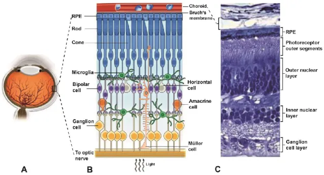
Figure 1: Cellular organization of the mammalian retina. A) Schematic representation of the human eye showing the posterior location of the retina. B) Simple schematic diagram of the cellular and synaptic organization of the vertebrate retina C) Representative cross-section of the human neural retina, the retinal pigment epithelium and the choroidal vessels showing the highly ordered arrangement of retinal layers. A) modified from (Rajala and Gardner, 2016), (B) schematic representation of the retina modified from (Reyes et al., 2017), choroid adopted from (Akhtar-Schäfer et al., 2018) (C) modified from (Wright et al., 2010).
In addition, and similar to other parts of the nervous system, neuronal cells in the retina are found in intimate apposition to glia cells (Reichenbach and Robinson, 1995). Three morphologically distinct glia cells types can be identified in the mammalian retina including Müller cells, astrocytes and microglia (Vecino et al., 2016). Müller cells, first described by Heinrich Müller in 1851 as radial fibres, are the predominant glial cells in the retina representing approximately 90% of the retinal glia population (Forrester et al., 2015; Vecino et al., 2016). They are the only cells that span the entire depth of the neural retina, radiating from the outer limiting membrane where they form adherens junctions with photoreceptor inner segments to the inner limiting membrane where their expanded end-foot terminate (Forrester et al., 2015; Newman and Reichenbach, 1996). They are seen to wrap neuronal somata and axons in an insulating sheath, helping maintain neuronal function by providing neuronal termini with nutrients and neurotransmitter precursors (Vecino et al., 2016). Moreover, Müller cells support
Introduction
5
neuronal survival and function by releasing trophic factors, recycling neurotransmitters and controlling ionic balance in the extracellular space (Bringmann et al., 2009; Goldman, 2014; Pow and Crook, 1996). Retinal astrocytes on the other hand are predominantly located in the nerve fibre layer where their processes join to form a honeycomb structure (Ramírez et al., 1998).
They perform similar functions to Müller cells including extracellular ion balance and neurotrophic, metabolic and mechanical support of neurons (Vecino et al., 2016). They also constitute a key functional component of the blood-retina barrier, which is essential in maintaining retinal homeostasis (Ramírez et al., 1998; Vecino et al., 2016). The third group of retinal glial cells, microglia, constitute the primary immune cell type in the retina and morphologically resemble those found throughout the CNS with their highly branched and dynamic protrusions (Langmann, 2007; Silverman and Wong, 2018). They are regarded as immunological watchdogs in the healthy retina, where they fulfil diverse tasks of surveillance (Karlstetter et al., 2015).
1.2 Retinal microglia
Microglial cells, distributed in the GCL, IPL and OPL layers, form an integral part of the innate immune defense system in the retina (Chen and Xu, 2015). They are equipped with a host of surface receptors and filopodia-like processes that constantly extend and retract to allow them efficiently monitor the surrounding microenvironment (Karlstetter et al., 2015). In addition, microglia housekeeping activities such as phagocytic removal of cellular corpses and debris and sculpting of synaptic circuits helps maintain a conducive retinal environment for the healthy functioning and survival of neurons (Linnartz and Neumann, 2013; Schafer et al., 2012; Wong, 2013). In the presence of tissue damaging stimuli, microglia cells respond by undergoing rapid proliferation and migration to the site of injury where they release a host of proinflammatory mediators aimed at neutralizing the noxious insult (Rashid et al., 2018c; Scholz et al., 2015c). However, while much is known about the major pathways involved in microglia activation and proinflammatory responses, it was not until recently when the expansion and
Introduction
6
renewal mechanisms of microglial cells in the retina were uncovered (Elmore et al., 2014; Huang et al., 2018).
1.2.1 Origin and maintenance of microglia in the retina
Over several decades, questions lingered as to whether myeloid cell populations in the CNS including retina were renewed by in-situ proliferation of resident microglia or whether bone marrow (BM) derived macrophages could also migrate to the healthy CNS tissue and readily differentiate into functional microglia (Simard and Rivest, 2004). To address this question, earlier studies used bone marrow (BM) chimeras obtained by transplantation of labelled BM derived cells from a transgenic donor animal to a recipient that has been lethally irradiated to ablate BM cells (Ransohoff, 2007). Xu and colleagues used this protocol to adoptively transfer BM derived cells expressing enhanced green fluorescent protein (eGFP) from eGFP-transgenic mice into lethally irradiated normal adult mice and waited for 8, 14- and 26-weeks prior to sacrificing the animals (Xu et al., 2007). Retinal flatmounts from the recipient mice revealed that BM derived eGFP+ cells could be detected in the retina as early as 8-weeks post transplantation, and by the 6th month, the entire retinal myeloid cell population was eGFP+ (Xu et al., 2007). A separate study using Cx3cr1-gfp/+ knock-in mice as BM donors, where eGFP protein expression is restricted to cells of the monocyte lineage, observed recruitment of eGFP+ monocyte-derived cells into recipient retinas just 4 weeks post-transplantation (Kezic and McMenamin, 2008).
The authors attributed the earlier appearance of eGFP+ donor cells observed in their study, as opposed to that of Xu et al., to differences in chimera models (global eGFP mice vs. Cx3cr1-gfp/+) and blood retinal barrier (BRB) integrity between the mouse strains (Kezic and McMenamin, 2008). Of note is that both studies observed very little in-situ proliferation of the resident microglia under steady state conditions and therefore concluded that a majority of the microglia/macrophages in the retinal tissue were replenished from BM derived monocyte precursor cells (Kezic and McMenamin, 2008; Xu et al., 2007). In contrast, findings from a different study where irradiated mice were injected with
Introduction
7
eGFP+ BM derived cells showed that a very small number of eGFP+ cells accessed the uninjured healthy retina up to 12 months post transplantation, despite a large number of them accumulating in surrounding ocular tissues such as the ciliary body, RPE, choroid and the optic nerve head (ONH) (Kaneko et al., 2008). However, in response to the neurotoxic agent N-methyl-N-nitrosourea that induces retinal degeneration, numerous eGFP+ BM derived cells could be detected in the retina, implying that the recruitment of BM-derived microglia in the retina occurs almost exclusively under retinal damage (Kaneko et al., 2008).
Interestingly, the authors argued that the enhanced migration of BM-derived cells to the retina observed in the previous studies might have been prompted by an unintentional damage of retinal neurons and vasculature as a result of not shielding the eyes and heads of experimental animals during irradiation (Kaneko et al., 2008). This line of thought was in fact consistent with previous findings which had demonstrated that rats exposed to X-ray doses between 200-500 cGy (compared to 800-1000 cGy used in the aforementioned studies) exhibited structural abnormalities in the retina characterized by damaged rod photoreceptor outer segment membranes and swollen or ruptured mitochondria in the RPE (Amoaku et al., 1989). Nonetheless, while the arguments from Kaneko et al. were accurate, they were somehow understated and failed to capture other flaws of the irradiation experimental protocol that might have biased the system towards a non-physiological transmigration of eGFP+ cells to the CNS: Firstly, irradiation induced bone marrow ablation is associated with enormous fluxes of cytokines through circulation and in tissue which subsequently cause disruptive changes to the blood brain barrier (BBB) (Ransohoff, 2007; Varatharaj and Galea, 2017); Secondly, irradiation of the CNS induces vascular perturbations that affect the competence of the BBB (Figure 2) (Greene-Schloesser et al., 2012;
Ransohoff, 2007).
Introduction
8
Figure 2: Irradiation weakens the integrity of the blood brain barrier. To investigate whether bone marrow (BM) derived microglia precursors are recruited to the healthy CNS, animals are exposed to irradiation to kill their bone-marrow cells. These are then replaced with labeled ones derived from a transgenic animal. Irradiation induced apoptosis of BM cells elicits a strong immune response characterized by enormous fluxes of cytokines through the circulation and in tissues. In addition, CNS exposure to irradiation causes vascular changes that compromise the integrity of the blood-brain barrier. Such experimental confounds associated with irradiation, and in particular, high amounts of circulating cytokines, can lead to the non-physiological transmigration of BM cells into the CNS which can give rise to microglia-like cells. Figures adopted and modified from (Ginhoux and Garel, 2018; Ransohoff, 2007).
Therefore, to further assess whether BM-derived microglia precursors are recruited to the healthy CNS while circumventing the problems associated with irradiation, a subsequent study used parabiosis experiments (Ajami et al., 2007).
Parabiosis, a surgical technique that fuses the vasculature of two living
Introduction
9
organisms, allowed for the sharing of the blood circulation between wild type and GFP transgenic mice without affecting their blood-brain nor the blood-retinal barrier (Ajami et al., 2007; Kamran et al., 2013). Five months post-surgery, no GFP+ microglia could be detected in sections obtained from the brain, brainstem or the spinal cord, indicating that under steady-state conditions, CNS microglia were a closed system with the capacity to self-renew and that their maintenance was independent of BM-derived circulating progenitors (Ajami et al., 2007).
Furthermore, the authors observed that when facial motoneuron injury was induced in both parabiotic pairs and irradiated/transplanted mice, exogenous GFP+, Iba-1+, BM-derived microglia cells could only be detected in the irradiated/transplanted groups, strongly corroborating earlier findings that the replacement of microglia by circulating precursors can be induced by experimental manipulations associated with irradiation (Ajami et al., 2007). In a subsequent landmark study, Ginhoux et al. established that microglia residing in the adult CNS in the steady state are derived from primitive myeloid progenitors that migrate from the yolk sac into the brain by embryonic day 9.5, after which the BBB forms and effectively blocks postnatal hematopoietic progenitors from contributing to microglia maintenance (Ginhoux et al., 2010).
To further examine whether microglia renewal and expansion occurred solely from local resident cells, Elmore et al. used an inhibitor of colony stimulating factor 1 receptor (CSF1R), PLX3397, to deplete microglia cells in the brain without compromising the integrity of the BBB (Elmore et al., 2014). Findings from the study demonstrated that the brain harbored latent nestin+ microglial progenitors, which following microglia depletion in the brain, undergo rapid proliferation and differentiation to repopulate the entire brain within 1 week of inhibitor cessation (Elmore et al., 2014). Notably, the observed regeneration of microglia in the depleted brain mirrored some aspects of normal development, as embryonic stem cells similarly require a nestin+ stage on their way to becoming microglia (Elmore et al., 2014; Hughes and Bergles, 2014). These findings raised the question whether retinas depleted of microglia could be repopulated in similar fashion from nestin+ precursors (Elmore et al., 2014). To this end, Huang et al.
Introduction
10
used a selective CSF1R inhibitor, PLX5622, to deplete microglia in the CNS including retina and assess the origin of the repopulated microglia (Huang et al., 2018). In contrast to the brain, the repopulated retinal microglia were not derived from nestin+ progenitors, but rather from dual extra retinal origins; residual microglia in the optic nerve which repopulated the retina along the center-to- periphery axis, and macrophages in the ciliary body/iris which repopulated the retina along the periphery and accounted for around 15% of the repopulated microglia (Huang et al., 2018). Of note is that findings from the study uncovered novel radial migratory routes of retinal microglia and demonstrated the presence of peripheral macrophage-derived microglia which were significantly less ramified than their central counterparts (Huang et al., 2018).
1.2.2 Microglia in the healthy retina
Microglia cells play active roles in maintaining normal retinal tissue homeostasis.
During the course of embryonic development, amoeboid cells immunopositive for microglia/macrophage markers invade the retina by crossing the vitreal surface and by migrating from nonneural ciliary regions (Santos et al., 2008). These cells are largely confined to the inner half of the retina in close proximity to dying RGCs which are excessively produced during development (Santos et al., 2008). As professional phagocytes, these strategically located amoeboid microglia engage in extensive phagocytic clean-up of apoptotic cellular corpses of the RGCs, thereby preventing leakage of cellular contents that promote inflammation and tissue necrosis (Ravichandran, 2003). Moreover, during the first postnatal days when RGC neurons make exuberant synaptic connections to the lateral geniculate nucleus (LGN) of the thalamus, microglia cells actively engage in pruning of transient, intact retinogeniculate synapses (Schafer et al., 2012). This process is dependent upon neural activity, where microglia preferentially engulf weak synaptic connections (Schafer et al., 2012). Furthermore, microglia mediated synaptic pruning occurs via a complement C3-CR3-dependent mechanism, where activated C3 (iC3b/C3b) selectively labels the weak RGCs terminals triggering a C3-receptor dependent phagocytosis pathway (Schafer et
Introduction
11
al., 2012). Indeed, C3 or CR3 knockout mice exhibit striking defects in synaptic pruning during development (Schafer et al., 2012).
Microglia cells in the developing retina also play an essential role in shaping vascular development (Silverman and Wong, 2018). Consistent with this role, microglia have been demonstrated in rodents and humans to populate the retina before developmental vascularization commences (Checchin et al., 2006; Rymo et al., 2011). During retinal vascularization, microglial cells are commonly associated with endothelial tip-cells at the vascular front and have been shown to play an active role in promoting angiogenic sprout anastomosis formation (Checchin et al., 2006; Rymo et al., 2011). This process is however independent of a direct contact between microglia cells and the vessels, and instead occurs via soluble factors secreted by both microglia and vascular endothelial cells (Rymo et al., 2011). Soluble factors released by the vasculature attracts microglial cells and promotes their release of angiogenic factors (Rymo et al., 2011). Consequently, the absence of microglia has been demonstrated to alter CNS vascularization (Arnold and Betsholtz, 2013). In neonatal rats for example, pharmacologic depletion of microglia using clodronate liposomes results in pronounced decreases in the retinal vascular area and density (Checchin et al., 2006). Intriguingly, replenishment of microglia-depleted retinas with exogenous microglia restored vascularity in the developing tissue, strongly indicating a prominent role for microglia in normal retinal blood vessel formation (Checchin et al., 2006). This notwithwithstanding, retinal myeloid cells have also been demonstrated to suppress angiogenic branching of deep retinal vessels via a Wnt-Flt1 pathway, implying that microglia cells can be either anti-angiogenic or pro-angiogenic depending on context (Stefater III et al., 2011).
The outer retina, from the ONL to the RPE, is an immune privileged zone that remains consistently devoid of microglia cells under normal physiological conditions (Silverman and Wong, 2018). RPE cells play a prominently role in development and maintenance of this immunosuppressive microenvironment (Karlstetter et al., 2015; Zamiri et al., 2007). They secrete inhibitory factors such as transforming growth factor-β (TGF-β), thrombospondin-1 (TSP-1) and
Introduction
12
somatostatin (SOM) into the subretinal space that actively blocks the undesirable infiltration of microglia and other mononuclear phagocytes into this region (Miyajima-Uchida et al., 2000; Pfeffer et al., 1994; Zamiri et al., 2006). During adulthood, microglia cells are found distributed as horizontal arrays of cells in the IPL and OPL where they exhibit a quiescent phenotype characterized by very small somata and extensively ramified processes that are highly dynamic and motile in nature (Langmann, 2007). They form a mosaic network of evenly distributed non-overlapping cells that allow the neural retina to be continuously sampled every few hours by the continuous movement of the microglial processes (Damani et al., 2011; Karlstetter et al., 2015). The dynamic movement of the processes also serves other housekeeping functions including homeostatic regulation of neuronal activity and the elimination of accumulated metabolic waste products and cellular debris in the retinal microenvironment (Li et al., 2012;
Nimmerjahn et al., 2005). In addition, microglia processes which intimately intercalate with neuronal axons and dendrites in the healthy retina plexiform layers are required for the maintenance of synaptic structure and physiology (Wang et al., 2016). Indeed, long-term absence of microglia in the retina results in the degeneration of synapses with concomitant progressive deficits in retina’s functional response to light (Wang et al., 2016).
To effectively patrol the retinal environment and limit unnecessary microglia activation, microglia cells are equipped with a versatile subset of different cell- surface proteins that they use to communicate with neighboring neuronal and glial cells (Karlstetter et al., 2015; Kierdorf and Prinz, 2013). CD200R is an important example of an inhibitory receptor expressed predominantly on retinal microglia that actively maintains microglia in a quiescent state when ligated (Broderick et al., 2002; Copland et al., 2007). Its ligand, CD200 (previously known as OX2), is a membrane glycoprotein that is widely expressed in the retina, including in ganglion cells, photoreceptors, vascular endothelium and RPE (Horie et al., 2013; Karlstetter et al., 2015; Langmann, 2007). Previous studies have demonstrated that CD200 deficient mice exhibit augmented pro-inflammatory responses in experimental animal models of uveoretinitis and wet form of age
Introduction
13
related macular degeneration (AMD) (Broderick et al., 2002; Copland et al., 2007;
Horie et al., 2013). In contrast, pharmacological activation of CD200R using an agonist monoclonal rat anti-mouse CD200R (DX109) antibody ameliorates pathological outcome in optic nerve injury and experimental autoimmune uveoretinitis animal models, suggesting that CD200-CD200R interaction could be harnessed for therapeutic purposes (Copland et al., 2007; Horie et al., 2013).
Another inhibitory factor secreted by neurons to inhibit the inflammatory capacity of microglia is CX3CL1 (fractalkine), a constitutively expressed cleavable chemokine which binds the Gi-protein coupled receptor CX3CR1 on the surface of microglia (Wolf et al., 2013). Numerous studies have demonstrated the neuroprotective and immunomodulatory role of CX3CL1-CX3CR1 interaction in the CNS (Wolf et al., 2013; Zieger et al., 2014). Transplanting mesenchymal stem cells engineered to secrete CX3CL1 in the subretinal space of rats exposed to high-intensity light to induce retinal degeneration inhibited microglia activation and migration to the ONL with concomitant reduction in neuronal demise (Huang et al., 2013). Conversely, in retinal degeneration (rd10) mutant mice deficient of CX3CR1, significantly greater numbers of reactive microglia were shown to infiltrate the ONL and induce a pro-inflammatory milieu that accelerated photoreceptor apoptosis and atrophy when compared with CX3CR1-sufficient rd10 littermates (Zabel et al., 2016). Of note is that delivery of exogenous CX3CR1 to the rd10 mouse eye significantly decreased the density of reactive microglia infiltrating the ONL and slowed the rate of neuronal loss, further underscoring CX3CL1-CX3CR1 signaling axis as an important regulator of microglial reactivity (Zabel et al., 2016).
Microglia also establish important interactions with Müller cells by exchanging functionally significant signals, and this bidirectional communication can act as a mediator of neuron-microglia crosstalk during both physiological and pathophysiological conditions (Madeira et al., 2015; Wang and Wong, 2014).
Certain microglia-derived neurotrophic factors such as BDNF and CNTF are consistently shown to be neuroprotective for photoreceptor cells despite these cells not expressing their receptors (Kirsch et al., 2002; Ugolini et al., 1995; Wen
Introduction
14
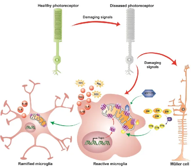
et al., 2008; Wilson et al., 2007). It was later revealed that microglia derived neurotrophic factors interact with Müller cells and induce or inhibit the release of secondary factors including basic fibroblast growth factor (bFGF), leukemia inhibitory factor (LIF) and glial cell line-derived neurotrophic factor (GDNF) that could act directly on photoreceptors and mediate survival or apoptosis during pathophysiological conditions (Harada et al., 2000, 2002; Shen et al., 2013; Wang et al., 2011; Wenzel et al., 2005). Additionally, microglia-Müller cell cross-talk can occur via the translocator protein (TSPO; 18kDa) signaling axis, where Müller cells release an endogenous TSPO ligand, diazepam binding inhibitor (DBI) protein, which binds microglial TSPO and suppresses microglial activation during retinal pathology (Wang et al. 2014).
1.2.3 Microglia in the diseased retina
When the retina suffers from a noxious insult, an immune response is launched by a local defense system that involves the resident microglia cells and the complement system (Chen and Xu, 2015). Microglia cells respond to noxious stimuli by retracting their filopodia-like processes (Figure 3) and upregulating a variety of cell surface molecules including major histocompatibility complex (MHC) class I and II antigens and receptors for cytokines and chemokines (Jurgens and Johnson, 2012).
Introduction
15
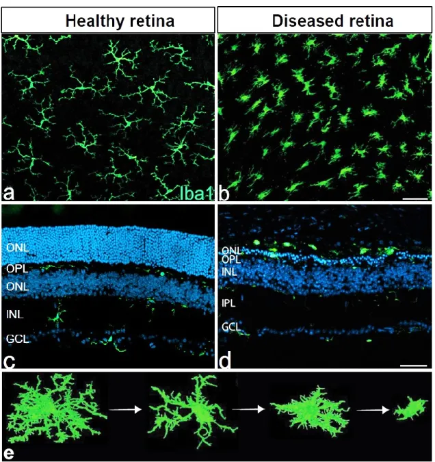
Figure 3: Microglia morphology in health and disease. Flat-mounts (a & b) and cross- section (c & d) images of Iba1-positive microglial cells in retinas from wild-type (a & c) andFam161a-deficient mouse model of Retinitis Pigmentosa (b & d). In the healthy retina (a & c), microglia cells are found in the IPL, OPL and GCL where they form a mosaic network of evenly distributed non-overlapping cells and exhibit a resting but surveillant phenotype characterized by very small somata and extensively ramified processes. In the diseased retina (b & d), microglia adopt an amoeboid morphology that is either completely devoid of processes or has very few unbranched processes. They are found in the degenerating ONL and the subretinal space where they not only engage in the phagocytic clearance of cellular corpses and debris, but also actively contribute to the degenerative processes. Panel (e) 3D images of Iba-1 stained brain sections generated using Fiji software (NIH, USA) showing the progressive changes in microglial morphology in response to bacterial lipopolysaccharide. Figures a, b & d adopted and
Introduction
16
modified from (Dannhausen et al., 2018), figure (e) adopted and modified from (Martyanova and Tishkina, 2015).
In addition, microglia alter their transcriptional profiles and undergo a metabolic switch from oxidative phosphorylation (OXPHOS) to glycolysis for the rapid production of energy necessary to fuel inflammatory events (Tannahill et al., 2015). Inflammation-induced metabolic rewiring is also crucial for microglial cellular proliferation since biosynthetic pathways for nucleotides, amino acids and lipid synthesis branch out from glycolysis (Orihuela et al., 2016; Rashid et al., 2018c). Reactive microglia cells subsequently migrate to the site of injury where they produce multiple pro-inflammatory factors including interleukin IL-1β, IL-6, IL-15, interferon gamma (IFN-γ), tumor necrosis factor alpha (TNF-α) and inducible nitric oxide synthase (iNOS) as well as reactive oxygen and nitrogen species (RONS) (Crotti and Ransohoff, 2016; Kierdorf and Prinz, 2013).
Microglia mediated inflammatory processes are aimed at the rapid return of a perturbed tissue back to normal homeostasis. However, if the insults persists, such as in aging and degenerative diseases of the retina, microglia transition from a well-balanced activation state to a hyperreactive state and produce exaggerated levels of pro-inflammatory mediators that contribute to tissue damage and exacerbate disease severity (Chen and Xu, 2015; Karlstetter et al., 2015). In AMD for instance, the widespread accumulation of drusen components provides a prominent chemoattractant stimulus that attracts microglia to the subretinal space (Indaram et al., 2015; Penfold et al., 2001; Rodriguez et al., 2014). Once in the outer retina, reactive microglia or microglia-derived factors induce NLRP3 inflammasome activation in RPE with the concurrent secretion of IL-1β and the degeneration of RPE cells via caspase-1-mediated pyroptosis (Ma et al., 2009; Madeira et al., 2018; Nebel et al., 2017; Tseng et al., 2013). The downstream effects of RPE loss are deleterious, and include choriocapillaris attenuation and secondary photoreceptor demise which ultimately result in loss of visual function (Ambati and Fowler, 2012; Kurihara et al., 2016). Accumulating subretinal microglia and other mononuclear phagocytes can also directly instigate death of photoreceptors in the vicinity by engaging in indiscriminate phagocytosis
Introduction
17
of stressed but living photoreceptors (Zhao et al., 2015a). Furthermore, the pro- inflammatory milieu induced by reactive subretinal mononuclear phagocytes can also promote photoreceptor cell death (Scholz et al., 2015c). We have indeed demonstrated in our previous research that conditioned medium obtained from reactive human and murine microglial cells triggers photoreceptor apoptosis (Madeira et al., 2018; Wiedemann et al., 2018).
The presence of reactive microglia in the outer retina is not a phenomenon unique to AMD but is also observed in other retinal degenerative pathologies such as Retinitis pigmentosa (RP) (Gupta et al., 2003). RP is the most prevalent and severe form of inherited retinopathies and is brought about by the primary degeneration of mutation containing rods and the subsequent degeneration of cones (Hartong et al., 2006). Microglia in human RP patients migrate to the photoreceptor layer following cues from apoptotic rods and engage in the phagocytic clearance of cellular corpses and debris (Gupta et al., 2003). Bloated microglia containing rhodopsin-positive cytoplasmic inclusions have indeed been demonstrated in the outer retina of RP patients using immunocytochemistry with microglia and rod cell-specific markers (Gupta et al., 2003). In addition to phagocytic clearance of degenerate cells, bloated infiltrating microglia secrete high levels of pro-inflammatory molecules such as TNF-α, IL-1β, CCL5 (alias RANTES) and CCL2 (alias MCP-1) that kill normal neurons and accentuate the ongoing degenerative process (Zeng et al., 2005). Intriguingly, using the retinal degeneration 10 (rd10) mouse model of RP, Guo et al. reported that mice deficient in ccr2 exhibited significantly reduced number of microglia infiltrating the photoreceptor layer compared to ccr2+/+ rd10 controls, indicating that the ccr2/ccl2 signaling pathway plays a key role in mobilizing microglia into the degenerating photoreceptor layer (Guo et al., 2012). Importantly, the reduced microglial infiltration observed in ccr2−/− rd10 mice was associated with increased retinal thickness and function, confirming microglia’s involvement in inducing degenerative changes during retinal pathology (Guo et al., 2012).
In diabetic retinopathy (DR), which is the most common ocular complication of diabetes mellitus, hypertrophic and amoeboid microglia are present at different
Introduction
18
stages of the disease and are mainly associated with retinal vasculature, cotton- wool spots and microaneurysms in the inner retina (Grigsby et al., 2014; Zeng et al., 2000). In the non-proliferative form of the disease, there is a moderate increase in the number of reactive microglia which are mostly clustered around perivascular region involving arterioles, venules and capillaries as well as around fresh hemorrhages in microaneurysms (Zeng et al., 2008). In the intermediate pre-proliferative disease form, there is a dramatic increase in reactive microglia which cluster around peripheral regions of cotton-wool spots and dilated vessels (Zeng et al., 2008). Lastly, in the proliferative form of the disease characterized by pathological neovascularization, there is a marked increase in the number of reactive microglia in the GCL which heavily surround the new vessels in the nerve fibre layer and the optic nerve head where the proliferative process is most prominent (Zeng et al., 2008). This close topological association between reactive microglia and the perivascular compartments in human DR is postulated to exacerbate vascular permeability via pro-inflammatory mechanisms (Grigsby et al., 2014; Zeng et al., 2008).
Similarly in the human glaucomatous eyes, clusters of large amoeboid microglia are found in the compressed prelaminar and lamina cribrosa regions where they surround blood vessels and form concentric rings (Neufeld, 1999). In addition, reactive microglia occurring either singly or in clusters are found in the parapapillary chorioretinal region (where the RPE and the bruch’s membrane terminate) of glaucomatous optic nerve heads (Neufeld, 1999). Of note is that microglia activation in the glaucomatous retina occurs prior to overt RGCs neurodegeneration, suggesting that microglial neuroinflammatory responses play a critical role in the onset and perpetuation of RGC loss (Bosco et al., 2011; Inman and Horner, 2007). (Bosco et al., 2011; Inman and Horner, 2007). The precise mechanisms involved in microglia mediated RGCs neurodegeneration remains poorly understood, but involves, at least in-part, the upregulation of Toll-like receptor (TLR) 4 and the secretion of high amounts of pro-inflammatory mediators TNF-α, IL-6 and nitric oxide (NO) (Echevarria et al., 2017; Neufeld et al., 1997; Sappington and Calkins, 2006; Tezel et al., 2004; Vidal et al., 2006).
Introduction
19
Consistently, the depletion of TNF-R1 or IL-6 or the pharmacological inhibition of TLR4 or NO synthesis significantly abrogates the loss of RGCs associated with experimental animal models of glaucoma (Echevarria et al., 2017; Tezel et al., 2004; Vidal et al., 2006).
In summary, ample evidence generated from experimental animal models to human tissue shows unequivocally that microglia play a central role in the pathophysiology of retinal neurodegenerative disorders and thus cannot be simply regarded as “innocent” bystanders of disease. Moreover, microglia responsiveness to noxious stimuli during retinal pathology suggests that these cells have the potential to act as diagnostic biomarkers for predicting disease onset, disease exacerbation, as well as response to treatment (Karlstetter et al., 2015). It is therefore imperative that we clearly understand the mechanisms underlying the establishment, maintenance and regulation of microglia responses during normal and pathological conditions. Such advances will provide an opportunity to better pinpoint candidate molecules and signal-transduction pathways that are dysregulated in overreactive neurotoxic microglia and set the stage for better-informed immunomodulatory strategies.
Introduction
20
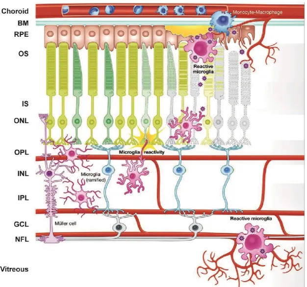
Figure 4: Schematic representation of microglia reactivity in the retina. Under steady state conditions, microglia reside in the plexiform layers where they contribute constitutively to maintaining neuronal synaptic structures, engage in phagocytosis of cell debris and constantly surveil their microenvironment for any disturbance in retinal homeostasis. When retinal neurons or the RPE suffer from noxious insults that lead to alteration in normal cellular function and/or degeneration, microglia become rapidly alerted, transform into amoeboid phagocytes and migrate to the lesion sites in an attempt to restore homeostasis. However, if the insults persist, such as in aging and degenerative diseases of the retina, microglia become pathologically activated and produce exaggerated levels of pro-inflammatory mediators that contribute to tissue damage and exacerbate disease severity. Figure adopted and modified from (Akhtar-Schäfer et al., 2018).
Introduction
21
1.2.4 Transcriptional control of microglia phenotypes in health and disease
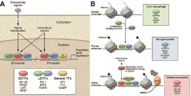
Much of our understanding of cell-type specific transcriptional regulation has come from detailed analysis of promoter (regulatory) regions which harbor sequence elements that are targets for signal dependent regulation (Holtman et al., 2017). Promoters are primarily occupied by widely expressed transcription factors such as SP1 and GABP which by themselves are incapable of orchestrating cell-type specific gene expression (Holtman et al., 2017). To generate cell-type specific programs of gene expression, distant gene regulatory sequences known as enhancers are required (Holtman et al., 2017; Smale and Natoli, 2014). A classical feature of enhancers is the presence of heterotypic clusters of binding sites for one or more lineage determining transcription factors (LDTF) and for broadly expressed stimulus dependent transcription factors (SDTF) (Figure 5) (Ghisletti et al., 2010; Holtman et al., 2017). In myeloid derived cells, such LDTFs include PU box binding-1 (PU.1), Runt-related transcription factor 1 (RUNX1) and Interferon regulatory factor 8 (IRF8) (Kierdorf et al., 2013).
These factors play fundamental roles in microgliogenesis (Ginhoux et al., 2010;
Jin et al., 2012; Kierdorf and Prinz, 2013; Satoh et al., 2014). In addition, they have been shown to be constitutively bound to enhancers where they collaborate with other lineage determining factors to induce histone modifications associated with a primed state of activity (Smale and Natoli, 2014).
In macrophages for instance, enhancers controlling endotoxin-stimulated gene expression are almost invariably bound by the lineage dependent Ets transcription factor Pu.1 (Ghisletti et al., 2010). Collaborative DNA binding of Pu.1 with other partner TFs such as C/EBPβ leads to the deposition of monomethyl groups on lysine 4 of histone 3 (H3K4) and the displacement of nucleosomes to expose enhancer DNA sequences (Figure 5) (Ghisletti et al., 2010; Heinz et al., 2010). Notably, the ectopic expression of Pu.1 in NIH-3T3 mouse fibroblast cells results in the depletion of nucleosomes in Pu.1 bound regions as well as several- fold increase in the histone modification H3K4me1 in regions corresponding to
Introduction
22
macrophage enhancers (Ghisletti et al., 2010). High levels of the histone mark H3K4me1 is a core chromatin signature of primed enhancers and serves as a beacon for signal-dependent effectors of signaling pathways such as nuclear factor-κB (NFκB), interferon responsive factors (IRFs) and activator protein 1 (AP- 1) (Ghisletti et al., 2010; Heinz et al., 2010; Smale and Natoli, 2014). The subsequent binding of SDTFs in the presence of stimulus promotes the recruitment of the ubiquitously expressed histone acetyltransferase (HAT) p300 to the nucleosome depleted enhancer regions with concomitant increase in the acetylation states of various residues of histones H3 and H4 (Heintzman et al., 2007; Smale and Natoli, 2014; Visel et al., 2009). Indeed, p300-mediated histone 3 lysine 27 acetylation (H3K27Ac) has been reported to be a distinct epigenetic mark for active enhancers (Raisner et al., 2018). The observations that SDTFs bind to enhancer landscapes predetermined by LDTFs such as Pu.1 in the presence of stimulus partially explains how broadly expressed TFs can regulate tissue-resident macrophage-specific gene expression (Crotti and Ransohoff, 2016; Holtman et al., 2017)
Figure 5: Enhancer-promoter interactions drive cell specific gene expression. (A) Promoters are primarily occupied by broadly expressed transcription factors which by themselves are incapable of orchestrating cell-type specific gene expression. Enhancers occupied by LDTFs and SDTFs interact with promoters to achieve cell type-specific expression and response profiles. (B) Initial steps of enhancer selection in closed chromatin regions containing regularly positioned nucleosomes involves the binding of
Introduction
23
LDTFs. This results in the depletion/sliding of nucleosomes to expose enhancer DNA sequences. LDTFs also deposit stereotypical histone marks such as H3K4me1 that act as beacons for SDTFs. The net effect of LDTFs binding is the maintenance of inducible genes in a silent, yet primed state which can be rapidly induced in the presence of stimuli.
Figures A and B adopted from (Holtman et al., 2017).
Several LDTFs have been shown to play roles in maintaining the unique resting but surveilling phenotype of microglia cells that is characteristic of a healthy CNS (Holtman et al., 2017). RUNX1, which is expressed as early as embryonic day 6.5 in erythromyeloid precursors, has been shown to promote the transition of amoeboid microglia to the ramified deactivated state during brain development (Kierdorf et al., 2013; Zusso et al., 2012). Consistently, mouse primary microglia cells infected with GFP-expressing adenovirus was shown to express significantly high levels of the proinflammatory enzyme iNOS when compared with adenovirus infected primary cultures transduced with Runx1 (Zusso et al., 2012). SALL1, a zinc finger transcriptional repressor whose expression in the CNS is virtually exclusive to adult microglia, is another factor involved in the maintenance of microglia homeostasis (Buttgereit et al., 2016). Ablation of Sall1 results in the conversion of microglia from a ramified phenotype to reactive phenotype characterized by shorter, thicker processes and a larger cell soma (Buttgereit et al., 2016). In addition, Sall1 deletion in microglia induces alterations in neurogenesis that perturb CNS homeostasis (Buttgereit et al., 2016). Another important transcription factor expressed by microglia during their transition from pre-microglia to mature microglia is MAFB (Matcovitch-Natan et al., 2016). Loss of MafB expression in microglia leads to the disruption of developmental genes and the upregulation of interferon and inflammation-related pathways in adult mice (Matcovitch-Natan et al., 2016).
In response to inflammatory stimuli, several SDTFs also play crucial roles in limiting microglia mediated inflammatory responses and inducing neuroprotective behavior. NR4A2 (Nuclear receptor subfamily 4, group A, member 2), also known as Nurr1, is an orphan nuclear receptor that is crucial for the development and homeostasis of dopaminergic neurons (Zetterström et al., 1997). NR4A2 has been shown to protect against loss of dopaminergic neurons by suppressing pro-
Introduction
24
inflammatory signaling in microglia and astrocytes (De Miranda et al., 2015; Saijo et al., 2009). The anti-inflammatory mechanism involves the recruitment of NR4A2 to NF-κB-p65 on inflammatory gene promoters and the subsequent recruitment of corepressor for repressor element 1 silencing transcription factor (CoREST) and the nuclear receptor corepressor 2 (NCOR2) (De Miranda et al., 2015; Saijo et al., 2009). The recruitment of CoREST and NCOR2 to the inflammatory gene promoters results in the clearance of NF-κB-p65 and restoration of activated pro-inflammatory gene transcription to a basal state (De Miranda et al., 2015; Saijo et al., 2009).
Ligation of estrogen receptors alpha and beta (ERα & ERβ) by endogenous and synthetic estrogen ligands has also been shown to exert potent immunomodulatory effects in reactive microglia (Arevalo et al., 2015; Bruce- Keller et al., 2000; Saijo et al., 2011). 17β‑estradiol (estradiol), produced both in the CNS and in peripheral organs by aromatase-mediated conversion of testosterone, is the most potent estrogen and has been shown in a multitude of studies to attenuate microglia inflammatory responses through both ERα and ERβ activation (Bruce-Keller et al., 2000; Liu et al., 2005; Vegeto et al., 2003;
Zhu et al., 2015). Similarly, Androstenediol, an ERβ subtype-specific ligand produced in the brain, has been shown to inhibit the magnitude and duration of microglia inflammatory responses by recruiting C-terminal binding protein (CtBP) corepressor complexes to AP-1 dependent promoters (Saijo et al., 2011).
Interestingly, the genetic ablation of ERα leads to a spontaneous reactive phenotype of microglia in the brains of adult ERα-null mice, demonstrating a critical role for this receptor in the maintenance of microglia homeostasis (Vegeto et al., 2003).
NF-E2-related factor-2 (NRF2) is an essential transcription factor for protection against oxidative/xenobiotic stress and inflammation (Kobayashi et al., 2016).
NRF2 suppresses inflammation by the upregulation of a battery of antioxidant cytoprotective proteins including hemeoxygenase-1 (HO-1), NAD(P)H quinone oxidoreductase-1 (NQO1), thioredoxins (TRXs) and enzymes of glutathione metabolism (Kobayashi et al., 2016; Lastres-Becker et al., 2014; Li et al., 2004;