Towards a method for the simultaneous quantification of
Gα
qactivation and β-arrestin2
recruitment
DISSERTATION
ZUR ERLANGUNG DES DOKTORGRADES DER NATURWISSENSCHA FTEN (DR. RER. NAT.) AN DER FAKULTÄT FÜR CHEMIE UND PHARMAZIE
DER UNIVERSITÄT REGENSBURG
Vorgelegt von Timo Littmann aus Wüsten
im Jahr 2018
Diese Arbeit entstand von Oktober 2014 bis November 2018 unter der Anleitung von Prof. Dr. Armin Buschauer, der leider viel zu früh verstorben ist, und Prof. Dr. Günther Bernhardt an der Fakultät für Chemie und Pharmazie der Universität Regensburg
Promotionsgesuch eingereicht am: 14.12.2018 Tag der mündlichen Prüfung: 29.04.2019
Vorsitzender des Prüfungsausschusses: Prof. Dr. Jens Schlossmann
Erstgutachter: Prof. Dr. Günther Bernhardt
Zweitgutachter: Prof. Dr. Carsten Hoffmann
Drittprüfer: Prof. Dr. Joachim Wegener
Acknowledgments
First and foremost, I would like to thank Prof. Dr. Armin Buschauer, who died far too early and could therefore not witness the finalisation of this thesis, for giving me the opportunity to work on this very interesting project and for his continuous scientific advice.
Another very important person for the realisation of this thesis was Prof. Dr. Günther Bernhardt. I would like to express my gratitude to him for his continuous scientific support and input, and especially for being always in the mood to give spontaneous ideas a try.
Furthermore, I would like to thank Prof. Dr. Takeaki Ozawa from the University of Tokyo for hosting my research stay abroad and his scientific support, not only during my time in Tokyo, but also during the entire course of this thesis. At this point, I would also like to thank Dr. Osamu Takenouchi for his help with all kinds of issues in the lab in Tokyo, his scientific input and for taking his time to show and explain the Japanese culture to me. Beyond that, I want to thank all lab members of the Ozawa group for the pleasant working atmosphere and scientific input.
Thanks are due to Prof. Dr. Carsten Hoffmann and the input he gave to this thesis, especially for chap- ter 3, and for being the second examiner of this thesis.
I would like to thank Prof. Dr. Joachim Wegener for inspiring scientific conversations on various occa- sions during my time at the University of Regensburg and for taking part in the evaluation committee of this thesis.
I would like to thank Prof. Dr. Jens Schlossmann, for being part of the evaluation committee of this thesis and for the fruitful collaboration in the proteinkinase G project. In this context, I also want to thank Dr. Andrea Schramm, Philip Müller-Thümen and the other co-authors.
For the productive collaboration on the comparison of the different label-free and label-dependent assays at the human histamine H1 receptor, I want to thank Dr. Nicole Plank, Dr. Sebastian Lieb and the other co-authors.
I want to thank Maria Beer-Krön for help in many situations during my time in the lab, Elvira Schreiber for showing me how to operate ancient technologies (Ca2+ assay) and Denise Mönnich for excellent technical assistance with some of the experiments presented in chapters 5 and 6.
Beyond that, I thank all colleagues in the lab for the excellent working atmosphere and the members of the international doctoral programme “Receptor Dynamics – Emerging Paradigms of Novel Drugs”
for many interesting conferences, symposia and workshops.
For their financial support I would like to thank the Elite Network of Bavaria for awarding me with a PhD-position within the international doctoral programme “Receptor Dynamics – Emerging Paradigms of Novel Drugs” and the Deutsche Forschungsgemeinschaft for the association to the research training group 1910 “Medicinal Chemistry of Selective GPCR Ligands”.
I want to express my gratitude to my parents for their continuous support during my education.
Zu guter Letzt möchte ich noch meiner geliebten Edith danken. Für ihre Liebe und den Rückhalt den sie mir tagtäglich gibt.
Publications, presentations and professional training
Peer-reviewed journal articles
(published prior to the submission of this thesis)
Littmann, T., Ozawa, T., Hoffmann, C., Buschauer, A. & Bernhardt, G. A split luciferase-based probe for quantitative proximal determination of Gαq signalling in live cells. Sci Rep 8, 17179 (2018).
Schramm, A., Müller-Thümen, P., Littmann, T., Harloff, M., Ozawa, T. & Schlossmann, J. Establishing a Split Luciferase Assay for Proteinkinase G (PKG) Interaction Studies. Int J Mol Sci 19 (2018).
Yang, Z., Han, S., Keller, M., Kaiser, A., Bender, B. J., Bosse, M., Burkert, K., Kögler, L. M., Wifling, D., Bernhardt, G., Plank, N., Littmann, T., Schmidt, P., Yi, C., Li, B., Ye, S., Zhang, R., Xu, B., Larhammar, D., Stevens, R. C., Huster, D., Meiler, J., Zhao, Q., Beck-Sickinger, A. G., Buschauer, A. & Wu, B. Structural basis of ligand binding modes at the neuropeptide Y Y1 receptor. Nature 556, 520-524 (2018).
Kuhn, K. K., Littmann, T., Dukorn, S., Tanaka, M., Keller, M., Ozawa, T., Bernhardt, G. & Buschauer, A.
In Search of NPY Y4R Antagonists: Incorporation of Carbamoylated Arginine, Aza-Amino Acids, or D- Amino Acids into Oligopeptides Derived from the C-Termini of the Endogenous Agonists. ACS Omega 2 (2017).
Dukorn, S., Littmann, T., Keller, M., Kuhn, K., Cabrele, C., Baumeister, P., Bernhardt, G. & Buschauer, A. Fluorescence- and Radiolabeling of [Lys4,Nle17,30]hPP Yields Molecular Tools for the NPY Y4 Receptor.
Bioconjug Chem 28, 1291-1304 (2017).
Lieb, S.*, Littmann, T.*, Plank, N.*, Felixberger, J., Tanaka, M., Schäfer, T., Krief, S., Elz, S., Friedland, K., Bernhardt, G., Wegener, J., Ozawa, T. & Buschauer, A. Label-free versus conventional cellular as- says: Functional investigations on the human histamine H1 receptor. Pharmacol Res 114, 13-26 (2016).
(*equal contribution)
Oral presentations
Littmann, T., Ozawa, T., Hoffmann, C., Buschauer, A. & Bernhardt, G. Split-luciferase complementation enables visualization and quantification of Gαq-dependent signaling. Frontiers in Medicinal Chemistry (2018, Jena, Germany)
Littmann, T., Ozawa, T., Hoffmann, C., Bernhardt, G. & Buschauer, A. Split-luciferase complementation enables quantification of Gαq activation and live cell imaging. 1st Joint Meeting of the European and Japanese Histamine Research Societies (2017, Amsterdam, Netherlands)
Littmann, T., Ozawa, T., Hoffmann, C., Bernhardt, G. & Buschauer, A. Split-luciferase complementation enables quantification of the Gαq/PLC-β3 interaction. 8th Summer School in Medicinal Chemistry (2016, Regensburg, Germany)
Poster presentations
(only contributions as presenting author are listed)
Littmann, T., Ozawa, T., Hoffmann, C., Buschauer, A. & Bernhardt, G. Simultaneous quantification of Gαq activation and β-arrestin2 recruitment. 9th International Summer School in Medicinal Chemistry (2018, Regensburg, Germany)
Littmann, T., Ozawa, T., Hoffmann, C., Buschauer, A. & Bernhardt, G. Split-luciferase complementation enables visualization and quantification of Gαq-dependent signalling. Frontiers in Medicinal Chemistry (2018, Jena, Germany)
Littmann, T., Ozawa, T., Hoffmann, C., Bernhardt, G. & Buschauer, A. Split-luciferase complementation enables quantification of the Gαq/PLC-β3 interaction. 8th International Summer School in Medicinal Chemistry (2016, Regensburg, Germany)
Littmann, T., Ozawa, T., Hoffmann, C., Bernhardt, G. & Buschauer, A. Using split-luciferase comple- mentation for quantification of Gαq signalling. XXIV EFMC International Symposium on Medicinal Chemistry (2016, Manchester, United Kingdom)
Littmann, T., Ozawa, T., Hoffmann, C., Bernhardt, G. & Buschauer, A. Quantification of Gαq protein activation by split-luciferase complementation. COST CM1207: GLISTEN Meeting (2016, Erlangen, Ger- many)
Professional training
Since October 2014 member of the international doctoral programme “Receptor Dynamics – Emerging Paradigms of Novel Drugs” funded by the Elite Network of Bavaria
Since December 2016 associated member of the research training group 1910 “Medicinal Chemistry of Selective GPCR Ligands” funded by the Deutsche Forschungsgemeinschaft
Index
Acknowledgments ...V
Publications, presentations and professional training ... VII
Peer-reviewed journal articles ... VII Oral presentations ... VIII Poster presentations ... VIII Professional training ... IX
1. Introduction ... 1
1.1 G protein-coupled receptors: a historical perspective ... 2
1.1.1 G protein-dependent signalling ... 2
1.1.2 Discovery of mechanisms curbing signalling ... 4
1.1.3 β-Arrestin-dependent signalling ... 6
1.1.4 Three-dimensional structure of GPCRs ... 6
1.1.5 Functional selectivity ... 8
1.1.6 GPCRs as drug targets ... 8
1.2 Functional characterisation of GPCR ligands ... 10
1.2.1 Resonance energy transfer-based techniques ... 10
1.2.2 Split luciferase complementation ... 11
1.3 References ... 14
2. Objective and aim ... 28
3. A split luciferase-based probe for quantitative proximal determination of Gαq
signalling in live cells ... 33
3.1 Introduction ... 34
3.2 Material & Methods ... 36
3.2.1 Materials ... 36
3.2.2 Cell cultivation ... 36
3.2.3 Generation of plasmids ... 37
3.2.4 Identification of the best pair of Gαq and PLC-β3 fusion proteins ... 37
3.2.5 Generation of stable expression cell lines ... 37
3.2.6 Characterisation of standard agonists and antagonists using the developed probe ... 38
3.2.7 Fura-2 Ca2+ assay ... 38
3.2.8 Live cell luminescence microscopy ... 38
3.3 Results and discussion ... 40
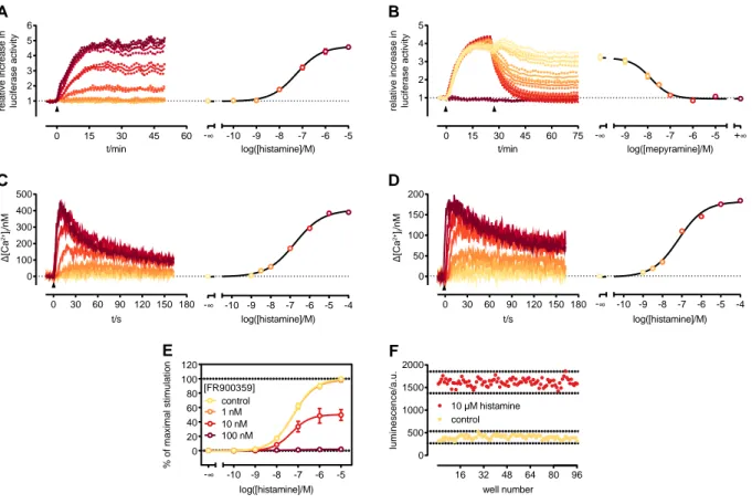
3.3.1 Development of the Gαq activation sensor ... 40
3.3.2 Characterization of the new probe ... 41
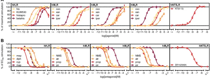
3.3.3 Characterization of reference ligands at five different GPCRs ... 43
3.3.4 Live cell luminescence ... 46
3.4 Conclusion ... 47
3.5 References ... 48
4. Towards probing the interactions of Gαs and Gαi with adenylyl cyclases using split-luciferase complementation ... 54
4.1 Introduction ... 55
4.2 Materials & Methods ... 57
4.2.1 Materials ... 57
4.2.2 Cell cultivation ... 57
4.2.3 Generation of plasmids ... 57
4.2.4 Detection of interaction between Gαs/i and AC2/6 fusion proteins ... 57
4.2.5 Fluorescence immunostaining of AC2 and AC6 fusion proteins ... 57
4.2.6 Identification of the best pair of Gαs/i and AC5 fusion proteins ... 58
4.3 Results and discussion ... 59
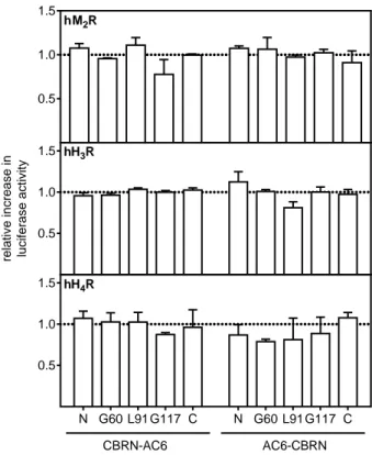
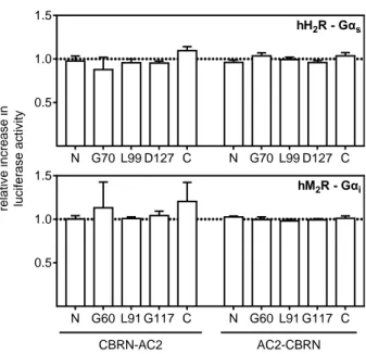
4.3.1 Interaction between Gαi and AC6 fusion proteins ... 59
4.3.2 Interaction between AC2 and either Gαs or Gαi fusion proteins ... 60
4.3.3 Cellular localisation of AC2 and the AC6 fusion proteins ... 60
4.3.4 Interaction of AC5 with Gαs and Gαi fusion proteins ... 61
4.4 Conclusion ... 64
4.5 References ... 65
5. A split luciferase-based assay for simultaneous analyses of the ligand concentration- and time-dependent recruitment of β-arrestin2 ... 68
5.1 Introduction ... 69
5.2 Material & Methods ... 71
5.2.1 Materials ... 71
5.2.2 Cell cultivation ... 71
5.2.3 Generation of plasmids ... 71
5.2.4 Generation of stable transfectants ... 71
5.2.5 Characterisation of standard agonists and antagonists using the developed probes .... 71
5.2.6 Fura-2 Ca2+ assay ... 72
5.3 Results and discussion ... 73
5.3.1 Assay characteristics ... 73
5.3.2 Influence of the luciferase fragment position on β-arrestin2 recruitment and downstream signalling ... 74
5.3.3 Time course of β-arrestin2 recruitment ... 78
5.3.4 Influence of exogenous GRK2 co-expression on β-arrestin2 recruitment ... 78
5.4 Conclusion ... 80
5.5 References ... 81
6. Simultaneous quantification of G protein activation and β-arrestin2
recruitment ... 86
6.1 Introduction ... 87
6.2 Materials & Methods ... 89
6.2.1 Materials ... 89
6.2.2 Cell cultivation ... 89
6.2.3 Generation of stable transfectants ... 89
6.2.4 Determination of spectral cross-talk ... 89
6.2.5 Quantification of agonistic potencies and antagonistic activities in the developed multiparametric assay ... 89
6.3 Results and discussion ... 91
6.3.1 Spectral cross-talk ... 91
6.3.2 Characteristics of the multiparametric assay ... 91
6.3.3 Analysis of standard agonists and antagonists in the multiparametric assay ... 95
6.4 Conclusion ...100
6.5 References ...101
7. Summary ... 105
8. Appendix ... 108
8.1 Appendix 1 ...109
8.2 Appendix 2 ...115
8.3 References ...116
8.4 List of Abbreviations ...119
1. Introduction
1.1 G protein-coupled receptors: a historical perspective
G protein-coupled receptors (GPCRs) are proteins responsible for transducing signals across plasma membranes1. Although, Paul Ehrlich was the first one describing the principle of a receptor/ligand inter- action already at the end of the 19th century with the proposal of his side-chain theory2, it was John Langley, who formally introduced the term “receptor”, or “receptive substance”3 a few years later. He investigated the effects of nicotine and curare on the neuromuscular junction of frogs and proposed the existence of “receptive substances” in muscles, which are activated through an endogenous agonist, secreted by neurons. We now know that muscle contraction is mainly regulated by ionotropic receptors, but the principle of the receptor/ligand interaction also applies to GPCRs.
1.1.1 G protein-dependent signalling
First work in context with GPCRs started in the 1950s and 1960s with Earl Sutherland and co-workers, who investigated the regulation of glycogenolysis in liver homogenates. They found out that a key regu- lator enzyme in this process, namely glycogen phosphorylase, was activated by administration of gluca- gon or epinephrine, not in a direct manner, but via the formation of an unknown “factor”4. Several years later, this factor was identified as 3’,5’-cyclic adenosine monophosphate (cAMP), which is formed from adenosine-5’-triphosphate (ATP) by an enzyme designated adenylyl cyclase (AC)5,6. This finding led Suth- erland to the proposal of the concept of “second messengers”5, which are molecules such as cAMP. The formation of second messengers is a direct consequence of an extracellular stimulation of cells by, e.g.
hormones5. Although at that time, it was unclear, if hormones act directly via an allosteric binding site on the AC itself, or if additional proteins are involved in the process6,7.
In the following years, research could profit from developments made in the pharmaceutical industry8. In search of compounds to treat asthma and hay fever, for example, antagonists like propranolol9 and mepyramine10 were developed. Antagonists are ideal pharmacological tools to characterise the function of receptors by blocking the action of (endogenous) agonists.
Some twenty years later, evidences began to accumulate that guanosine-5’-triphosphate (GTP) and a
“transducer” protein11 are playing an important role in the AC signalling cascade12-14. The hypothesis arose that the so-called “β adrenoceptor-regulated adenylyl cyclase”, which was the most widely used model system to study cAMP-dependent effects at that time, actually consists of three different pro- teins, composed of the AC, the “transducer” and the receptor itself. Rodbell et al., also working on ACs in liver lysates stimulated through glucagon, discovered that the formation of cAMP is drastically en- hanced when GTP is added to the lysates together with glucagon, compared to the stimulation with glucagon alone12. Cassel and co-workers, investigating catecholamine effects on erythrocyte mem- branes, showed that the hydrolysis of [32P]-GTP was drastically enhanced after addition of isoproterenol,
an epinephrine analogue13. At about the same time, radioligand competition binding experiments, also on erythrocyte membranes, using the tritiated β-blocker dihydroalprenolol, revealed substantially dif- ferent characteristics of agonists and antagonist, when displacing the radioligand. Antagonists displaced the radioligand in a uniform manner15,16. By contrast, agonists showed biphasic displacement curves with two inflection points, reflecting a high and a low binding constant. The addition of GTP converted the biphasic character into a monophasic one, and the corresponding binding constant was then similar to the lower one of the biphasic curve16. These findings inspired De Lean and co-workers to propose the ternary complex model, providing an explanation for the two affinity states of the receptor17. The au- thors postulated in addition to receptor and ligand a third interaction partner, which is only present, when an agonist binds to the receptor and which can be uncoupled from the receptor/ligand complex by addition of GTP. The latter then leads to a reduced affinity between agonist and receptor17.
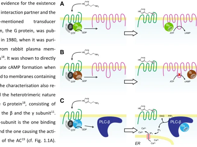
Direct evidence for the existence of this interaction partner and the above-mentioned transducer protein, the G protein, was pub- lished in 1980, when it was puri- fied from rabbit plasma mem- branes18. It was shown to directly stimulate cAMP formation when titrated to membranes containing ACs. The characterisation also re- vealed the heterotrimeric nature of the G protein18, consisting of the α, the β and the γ subunit11. The α-subunit is the one binding GTP and the one causing the acti- vation of the AC19 (cf. Fig. 1.1A).
Upon agonist binding, the GPCR/Gαβγ complex dissociates into the GPCR, the α subunit and the βγ complex19. Both compo- nents of the G protein are then capable of activating different downstream effector pro-
Fig. 1.1: Schematic illustration of the activation and effectuation of Gαs, Gαi and Gαq proteins. Binding of an agonist to a GPCR results in a GDP/GTP exchange within the α subunit. The subsequent influence on effector proteins depends on the type of α subunit activated. GPCRs usually have a canonical G protein, to which they pre- dominantly couple to. In case of the H2 histamine receptor (hH2R) (A), for example, it is the Gαs protein, which translocates to ACs and activates their enzymatic activ- ity, leading to the formation of cAMP from ATP. Histamine H3 and H4 receptors (hH3,4R) (B) couple to Gαi proteins, which also interact with ACs. By contrast, inter- action with Gαi proteins leads to a decrease in AC activity. A third major Gα protein is the Gαq (C), which is activated, for example, through histamine H1 receptors (hH1R). Their effector proteins are from the PLC-β class and catalyse the formation of IP3 and DAG from PIP2. The second messenger IP3 then promotes the release of
γ β γ β γ β
PLC-β
NH2 HN
N
NH2 HN
N
PLC-β
GTP
αq
αq
GTP GDP
PIP2
IP3 DAG
ATP cAMP
αs
GTP GDP
NH2 HN
N
GTP
αs
NH2 HN
N
ATP cAMP
αi
GTP GDP
NH2 HN
N
GTP
αi
NH2 HN
N
X
A
B
C
Ca2+
Ca2+
Ca2+
Ca2+
IP3
The G protein, purified by Northup et al, and the one responsible for the stimulation of ACs is what we now call the Gαs protein11. Its counterpart, the Gαi protein was discovered during investigations on the mode of action of the whooping cough-causing toxin secreted by Bordetella pertussis, the pertussis toxin21. Activation of Gαi leads to an inhibition of the enzymatic activity of the AC, as illustrated in Fig.
1.1B. Pertussis toxin inhibits these Gαi proteins, which is the reason that it is still widely used as a phar- macological tool, especially for investigations on the coupling specificity of a GPCR22,23.
A third major Gα subtype, the Gαq, was discovered during investigations on receptor-promoted Ca2+ ion influx into the cytosol. It was known that Ca2+ influx and the formation of inositol trisphosphate (IP3) from phosphatidylinositol 4-phosphate (PIP2) by phospholipases C-β (PLC-β) was interconnected, but it was not known, if the influx of Ca2+ caused IP3 formation, or vice versa24. However, in 1983 Streb et al.
showed that the addition of exogenous IP3 to permeabilised cells caused a Ca2+ efflux from intracellular stores, which gave insight into the order of events within this signalling cascade25. Although it was al- ready known that hormones can cause Ca2+ influx into the cytosol and that treatment of specific mem- brane preparations with the non-hydrolysable GTP analogue GTPγS leads to the production of IP319, it took until the 1990s to identify and purify the regulator of PLC-βs26,27. By using affinity chromatography with immobilized βγ-subunits, novel Gα subtypes were purified26 of which Gαq was identified as the reg- ulator of PLC-β proteins27 (cf. Fig. 1.1C). Both, IP3 and Ca2+ are also referred to as second messengers and the other product formed by PLC-βs from PIP2, diacylglycerol (DAG), is a third one involved in this signal- ling pathway. It was shown that DAG, which diffuses in the cellular membrane after formation, due to its lipophilic nature, directly stimulates the activity of, e.g. protein kinase C24.
Also, βγ-subunits were identified as signal transducers. Several publications described them as modula- tors of PLCs28, ACs29, and different types of ion channels30,31.
With the cloning of GPCRs, beginning with the β2 adrenoceptor32, and especially the human genome project, a new era in receptor research began. On one hand, recombinant expression of receptors in e.g.
cancer cell lines became possible making research more convenient, but on the other hand the complex- ity increased tremendously7: multiple different receptors and their splice variants were identified, amounting to approx. 2000 different proteins33. Furthermore, differences in sequence of the same re- ceptor between species became apparent, which, in some cases, also put the translational validity of animal models into question34,35.
1.1.2 Discovery of mechanisms curbing signalling
Already in 1968, the existence of a regulatory mechanism of unknown function was described that causes a reduction of cAMP concentration after continuous incubation of rat cerebellar slices with nore- pinephrine to the basal level within several minutes36. But it took until the mid-1980s, when Mahan and
co-workers demonstrated by radioligand binding studies that receptors disappear from the cellular sur- face after stimulation with an agonist37. This was the first direct evidence that cells prevent themselves from an over-stimulation via down-regulation of receptors and not of the G proteins or effector pro- teins38. In addition, it was demonstrated that phosphorylation of the activated receptors, e.g. by GPCR kinases (GRKs)39, is linked to the internalisation of the receptors14,40 and that it also impairs their ability to activate G proteins41. However, GPCR desensitisation by GRKs was only observable with crude kinase preparations. The purer the preparation was, the less efficacious was the desensitisation, which led to the assumption that an additional protein had to be involved in the process42.
This protein was found to be β-arrestin43, which binds to phosphorylated receptors with high affinity39. Therefore, it is the key protein responsible for receptor desensitisation (cf. Fig. 1.2). On one hand, it provokes a steric hindrance when bound to the receptor, making G protein activation impossible38. On the other hand, it functions as an adaptor for clathrins, facilitating the formation of pits at the mem- brane44. From these so-called clathrin-coated pits vesicles are formed that internalise together with the receptors45. After internalisation, the receptors undergo different ways of processing. They can be rap- idly recycled back to the plasma membrane, or degradation46, mediated by ubiquitinilation47, can oc- cur48. The receptors that get recycled back to the plasma membrane to the most part, such as e.g. the β2 adrenoceptor45, are categorised as class A GPCRs49. Receptors are considered class B receptors, such as e.g. the neurotensin NTS1 receptor (hNTS1R)46, when they are preferentially degraded in lysosomes after internalisation49.
Fig. 1.2: Schematic illustration of β-arrestin-mediated internalisation of receptors and G protein-independent signalling.
Upon agonist binding, a GDP/GTP exchange occurs within the α subunit, leading to the dissociation of the receptor/G protein complex. The agonist-bound receptor is now accessible for phosphorylation through, e.g. GRKs at its C-terminus and intracellu- lar loop regions. This process results in an increased affinity of the receptor for β-arrestins, which bind the receptor and mediate its internalisation through clathrin-coated pits. Before and during the internalisation process, arrestin acts as a scaffolding pro- tein. It brings proteins, which are part of a common signalling cascade, in close proximity, so that they can activate each other.
After internalisation and gathering in vesicles, the receptors can undergo two different pathways. On one hand, recycling, back to the plasma membrane, can occur, or the receptors can be degraded by the ubiquitin/proteasome machinery.
NH2 HN
N
NH2 HN
N
β-arr
NH2 HN
N
β-arr
NH2 HN
N
GTP
αq
ATP ADP
GRK
NH2 HN N
β-arr
NH2 HN N
β-arr ERK c-Src
ERK c-Src
Downstream signalling Receptor
degradation
NH2 HN
N
β-arr γ β
GDP
αq
GTP
γβ
Two different isoforms of β-arrestin are known, namely β-arrestin1 and β-arrestin2. Both isoforms are ubiquitously expressed in the human body38,50,51. Studies with knock-out mice suggest that both isoforms serve a similar function33, since mice lacking only one variant, appear normal, provided that they are not pharmacologically challenged52-54. The latter might be explained by reports on higher affinities of class A receptors for β-arrestin2 than for β-arrestin146. A double knock-out, however, is prenatally lethal33.
1.1.3 β-Arrestin-dependent signalling
Besides blocking the interaction site of G proteins and mediating the internalisation of receptors, β-ar- restins are involved in intracellular signalling33. They act as scaffolding proteins within signalling cas- cades38,55. For example, kinases, which activate the enzymatic function of other proteins by phosphory- lation, directly bind to β-arrestin. Prominent representatives are the extracellularly-regulated kinases 1 and 2 (ERK1/2), which are mitogen-activated protein (MAP) kinases56,57. Both bind to β-arrestins, and their downstream signalling partners do the same. In this way ERK1/2 and e.g. c-Src58 are brought into close proximity, leading to phosphorylation and therefore, to the activation of the latter33. In addition to ERK1/2 and c-Src, there are numerous interaction partners33 such as p38 MAP-59 or Akt kinases60 and signalling pathways61 described that are activated through β-arrestins. By contrast, through the binding of E3 ubiquitin ligases, β-arrestins convey the degradation of receptors46,47,62, whereas phosphodiester- ases as interaction partners are responsible for the cleavage of cAMP63.
1.1.4 Three-dimensional structure of GPCRs
Although at that time scientists were oblivious to that64, structural investigations on GPCRs began inde- pendently with the work of two research groups, determining the amino acid sequence of rhodopsin by Edman degradation65,66. Direct evidence that rhodopsin and GPCRs share the same overall structure was reported, when the first GPCR, the β2 adrenoceptor, was cloned32. It became obvious that both proteins consist of seven transmembrane helices with their N-terminus facing the extracellular space and their C-terminus the inside32,65,66.
The first three-dimensional structure was reported in 2000, again of rhodopsin67, with the up to now most commonly used technique, i.e. X-ray diffraction analysis of proteins crystals68. Seven years later, the first crystal structure of a ligand-activated GPCR was reported by the Kobilka and Stevens groups.
The authors were able to crystallise the β2 adrenoceptor in complex with the β-blocker carazolol69. These reports demonstrated that crystallisation of a GPCR is challenging, requiring specialised proce- dures, which must be individually optimised for each receptor70: for reconstitution, some kind of syn- thetic membrane environment is needed, e.g. lipid bicelles, or the so-called lipidic cubic phase. To en- hance crystal packing, a fusion protein of the receptor together with a well-crystallising protein, such as lysozyme, is generated. Furthermore, crystallisation of the receptor alone is not possible, a stabilisation
of the inactive conformation is needed and achieved by adding an antagonist. Finally, since the formation of crystals can take a long time, the receptor´s primary structure must be mutated, to increase its long- term stability.
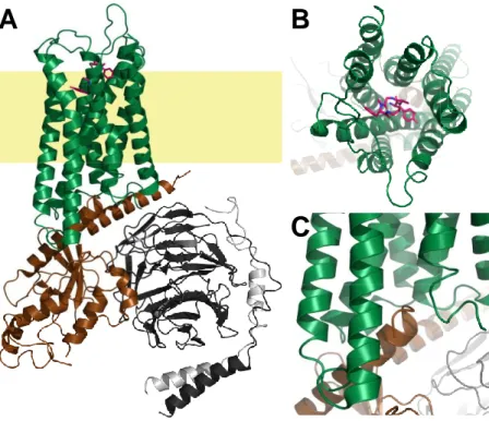
These findings paved the way for the crystallisation of a plethora of different receptors, such as the histamine H1 receptor (hH1R)71, the dopamine D3 receptor72, the chemokine receptor CXCR473, or the neuropeptide Y Y1 receptor74. Another milestone was reached soon with the description of the first structure of a GPCR in com- plex with its canonical G protein, the β2 adrenoceptor/Gs com- plex75. Until now, over 200 struc- tures of approx. 30 different GPCRs have been solved68, which help not only pharmacologists to understand the activation of such receptors, but also medicinal chemists to design compounds with enhanced binding and functional properties. In addition to X-ray diffraction, advances in the field of cryo-electron microscopy (EM) begin to influence receptor structure determination more and more68. Although the size of a GPCR alone is insufficient for structure determination using cryo-EM76, a receptor in complex with intracellular signalling partners, such as G proteins or arrestins, is. The structure of the μ-opioid receptor in complex with Gαiβγ and the ligand DAMGO, obtained by Koehl et al. through cryo- EM77, is shown in Fig. 1.3.
The GPCR consists of seven bundled α-helices within the plasma membrane. The ligand binds on the extracellular side, in a cave formed by the helix bundle, to the receptor (Fig. 1.3B). Through the types of amino acids, their localisation and their orientation inside the binding cleft, the unique ligand specificity of each GPCR is determined. From several studies, a general activation mechanism, which starts with an outward movement of the sixth α-helix of the receptor (the foremost helix in Fig. 1.3A and C) upon ago- nist binding, became clear1. This movement generates space for the C-terminal helix of the Gα subunit,
Fig. 1.3: Cartoon representation of the three-dimensional structure of the μ-opi- oid receptor in complex with Gαiβγ as a general example the structure of a GPCR.
The coordinates of the µ-opioid receptor/Gi-complex77 were obtained from pdb (6ddf) and processed using PyMOL. The receptor is represented in green, the Gαi is brown, the Gβ is black and the Gγ is depicted in light-grey. The putative localisation of the membrane is represented in light-yellow. A is a full-size representation of the entire complex. B is a close-up on the ligand, bound in the middle of the seven-helix bundle. The interaction site between GPCR and Gαi is shown in C, where the C-ter- minus of Gαi points inside the helix bundle after an outward movement of helix 6.
A B
C
GPCR/G protein complexes suggest that this intracellular binding pocket functions analogously to the extracellular one. The amino acids interacting with the C-terminal α5-helix of the Gα subunit determine to which Gα subtype the GPCR couples1,77. This intracellular interface is also one of the key interaction sides of arrestins with GPCRs, and by occupying the very same space as the C-terminus of Gα does, it directly prevents the receptor from activating the latter1,78,79.
1.1.5 Functional selectivity
The classical ternary complex model of receptor activation takes only one intracellular binding partner of the GPCR, the G protein, into account17. However, as mentioned under 1.1.2, there are at least GRKs and β-arrestins also interacting with GPCRs39,51, and it was shown that certain ligands preferentially ac- tivate (or are biased towards the activation of) Gα proteins over the recruitment of β-arrestins80, or vice versa81. Furthermore, agonists were reported that led to the selective activation of one Gα subtype over another82, which shows that GPCRs not always signal exclusively through their primarily-attributed G protein subtype. Taken together, these findings imply that each agonist can stabilise its own distinct receptor conformation, each of which can differ in its ability to activate downstream signalling part- ners83. Evidence for these agonist-dependent receptor conformations was found using X-ray crystallog- raphy84 and was demonstrated multiple times with different ligands at different receptors84-87. There- fore, the classical ternary complex model of receptor activation is inadequate, suggesting that a GPCR is more than a simple on/off-switch88. Rather than having an inactive and only one active conformation, a receptor has multiple active conformations differing in their ability to signal downstream89. To further increase complexity, allosteric modulators of receptor activation, which unveil their effects only in the presence of an orthosteric ligand90-92, have been described. Moreover, multiple receptors were reported to exist as homo-93 or heterodimers94 within the cellular membrane, capable of e.g. transactivating each other94. To conclude, a reductionist approach in functional ligand characterisation can be misleading and sensitive techniques for the characterisation of multiple pathways, ideally in a simultaneous manner, are needed, because important effects might otherwise be overlooked.
1.1.6 GPCRs as drug targets
GPCRs have been a drug target long before their existence was even known9,10,95. First compounds ad- dressing GPCRs with a therapeutic purpose were antihistamines and β-blockers, which were the first so- called blockbuster medications. As mentioned above, these compounds helped researchers to elucidate the function of GPCRs8. To date, approx. 27% of the global market share are drugs that target GPCRs96 with a multitude of different indications, such as asthma, diabetes type 2 or various autoimmune dis- eases96. The mode of action of the drugs or drug candidates varies from classical agonists and antago- nists96 to more recently identified compounds, which are allosteric modulators96.
Especially certain biased agonists are considered to be promising drug candidates, because side-effects are seen in connection with the activation of unwanted signalling pathways at several receptors97,98. An example is the µ-opioid receptor, at which the induction of β-arrestin recruitment is presumably respon- sible for the severe side-effects99,100 associated with opioid analgesics and for which recently a G protein- biased agonist (PZM21) was reported80. However, it should be mentioned that in a very recent study G protein bias of PZM21 and lacking respiratory depression were not confirmed101. Hill et al. 101 point out the difficulty to quantify bias, in particular of low efficacyagonists, and to compare bias between differ- ent cell types and assays, a problem, which is increasingly being acknowledged102-104. These contradictory studies demonstrate that there is a need for techniques enabling a reliable quantification of agonist bias.
1.2 Functional characterisation of GPCR ligands
As mentioned in the beginning, functional investigations on GPCRs began with pharmacology in living animals95 or isolated organs3, which is an approach that has been used until to date105. Due to structural differences between the receptor orthologues, data obtained by organ pharmacology from, e.g. guinea- pigs, might not always apply in the exact same way to the corresponding human tissue34,106,107. Hence, cancer cell lines of human origin with endogenous expression of a certain receptor108, or recombinant expression of the receptor under study in immortal(-ised) cell lines have become the most prominent systems for investigations on GPCRs109,110.
First in vitro experiments were focused on the formation of second messengers, such as cAMP6, IP spe- cies111,112, or Ca2+ ions113,114 and these are still the most often assessed effects provoked by receptor activation, because they can be determined using assays with moderate to high throughput. By deter- mining the activity of the GTPase of the Gα subunit, receptor activation can be probed more proximally.
This is beneficial for precise determinations of agonist efficacies, or agonist bias, since substantial ampli- fication occurs the more distally the activation of the signalling cascade is probed. The latter can result in an over-interpretation of agonistic effects101,115. Classical assays for a proximal determination of GPCR activation are the steady-state GTPase assay13, in which the hydrolysis of a [32,33P]-labelled γ-phosphate of GTP is determined, or the [35S]GTPγS assay116,117, which utilises a non-hydrolysable GTP analogue. The readouts of these two assays are both based on the radioactive decay of phosphorus or sulfate isotopes.
There are several major drawbacks of the aforementioned techniques: most of them are lysis-based or require the preparation of membranes, and, if this is not the case, throughput is often limited. Thus, sensitive assays for the proximal quantification of GPCR-mediated activation of various pathways in live cells are needed118. Most promising techniques that should meet the demands are resonance energy transfer techniques (RET), or split luciferase complementation (SLC), employed to probe protein/protein interactions (PPIs) within signalling cascades activated through GPCRs.
1.2.1 Resonance energy transfer-based techniques
Förster, or “fluorescence” resonance energy transfer (FRET) was initially described by Theodor Förster119 and describes a phenomenon that applies to two fluorophores, the excitation and emission spectra of which overlap. If the first fluorophore (donor) with the shorter excitation and emission maxima is excited by an external light source, it can transfer its energy (a radiationless process) to the other (acceptor) fluorophore, provided that both are in close proximity (≤ 10 nm) and in correct orientation120. Therefore, resonance energy transfer can be used for investigations on PPIs. It was successfully applied using pro- teins labelled with organic fluorophores121, but with the discovery and cloning of the green-fluorescent
protein (GFP)122,123 and the subsequent engineering of its colour variants124, it became possible between fluorescent proteins and also in intact cells125.
In addition, RET can also occur between luciferases, which are enzymes capable of emitting light when their substrates are provided, and appropriate fluorescent proteins126,127. This variant of RET is called bioluminescence resonance energy transfer (BRET) and is, although rarely, occurring in nature. The jel- lyfish Aequorea victoria, for example, from which GFP originates, uses its blue light-emitting luciferase aequorin as donor to excite GFP122. In modern molecular biology the most prominent luciferases used for BRET studies are the renilla luciferase (RLuc) from the sea pansy Renilla reniformis128, the firefly lu- ciferase from the firefly Photinus pyralis129 and more recently, genetically optimised luciferases such as the NanoLuc (NLuc)130,131, derived from a luciferase excreted by the deep-sea shrimp Oplophorus gracil- irostris.
On one hand, FRET has the advantage that the overall light-intensities are high, which means that only very short integration times of the detectors are needed, enabling a high temporal resolution during measurements120. On the other hand, since no external excitation light is needed, BRET often has better signal-to-noise ratios.
With respect to the functional characterisation of GPCRs, FRET and BRET have been successfully applied multiple times to probe, e.g. second messenger formation125,132-134, dissociation and rearrangements of G protein subunits135-138, β-arrestin recruitment139,140 and even conformational changes of the GPCR it- self141-144. The high temporal resolution that can be achieved using FRET141,145, helped to unravel the temporal order of the events taking place after agonist binding to the receptor120,141, some of which happening on the µs time-scale.
However, a major shortcoming of FRET- and BRET-based assays is the fact that multiparametric meas- urements of the activation of two or more signalling pathways at once are very challenging. A large frac- tion of the visible part of the light spectrum is already covered by the excitation light and the emitted light of the two luminescent proteins used, which would make meticulous spectral unmixing neces- sary146-148.
1.2.2 Split luciferase complementation
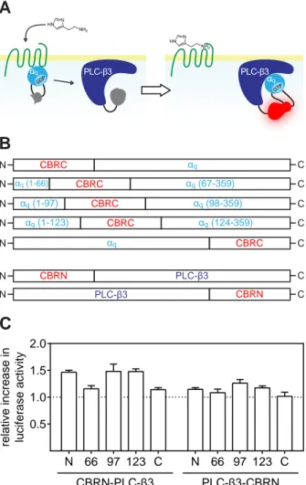
A better option for probing multiple pathways simultaneously is SLC, belonging to the overall group of split-reporter assays. The underlying principle is the “dissection” of a protein into two complementing fragments, which can restore the catalytic function of the original protein when brought into close prox- imity149,150. By means of molecular biology techniques, fusion proteins, consisting of the proteins for which a specific interaction is expected and reporter protein fragments, must be generated150-152. The
induction of the interaction of the two host proteins brings the reporter fragments in close proximity, leading to the restoration of the enzymatic function (Fig. 1.4).
The first described approach, using split-reporter complementation, involved ubiquitin. Upon inter- action of the two host proteins, a reporter enzyme becomes cleaved off the complex by a ubiquitin- specific protease, which can be detected by SDS- PAGE153. The first assays that did not depend on cell lysis were based on split β-galactosidase or split β-lactamase. Both make use of synthetic sub- strates, which change their optical properties (e.g.
formation of a fluorescent dye) after processing by the enzymes154,155.
Imaging was already possible using the split β-galactosidase assay, due to the formation of the fluores- cent dye. The localisation of the fluorophore, however, did not resemble the exact site of the probed PPI within the cell. Precise localisation became possible when Ozawa et al. and Ghosh et al. reported inde- pendently that also GFP can be used in a split reporter assay156-158. Shortly after, this was also achieved in a multiplexed manner using colour variants of GFP159. However, a major disadvantage of the split GFP technique are the long maturation times, which are needed for GFP to fully refold and rebuilt its fluoro- phore158. This makes kinetic analyses nearly impossible. Furthermore, the interaction of the two frag- ments is not reversible once the full-size GFP has been restored151,156.
Therefore, a very attractive option are luciferases as reporter proteins. Although first approaches used intein-assisted luciferase complementation, causing an irreversible reaction160, newer assays are not de- pendent on inteins anymore, which provides a dynamic, fully reversible, system161. The relatively short maturation time periods, compared to split GFP, enable kinetic analyses of the probed PPI, and the in- dependence of excitation light results in a low background and therefore, higher sensitivities161,162. In the context of GPCR research, SLC has been applied multiple times e.g. for the detection of second messenger formation163,164, or β-arrestin recruitment152,165-167.
Fig. 1.4: Schematic illustration of the split-reporter principle. A reporter protein is split into two catalytically-inactive frag- ments, which are capable of restoring the enzymatic function, when brought into close proximity. These two fragments are then fused to two proteins through flexible linkers, from which a specific interaction is expected. When applying a stimulus, which induces the interaction of the host proteins, the enzy- matic activity of the reporter is restored and can be detected.
stimulus
Luciferases used in these assays are mainly the same as men- tioned under 1.2.1. On one hand, glow-type luciferases, such as the firefly luciferase, or lucifer- ases cloned from click-beetles, which utilise D-luciferin, ATP and O2 as substrate (Scheme 1.1), have the advantage that longer time periods can be analysed, due to the continuous light emis- sion152,163,164. On the other hand, flash-type luciferases, like RLuc or NLuc, which consume coelenterazine and O2 (Scheme 1.1) have the advantage that their initial bright- ness is very high130,168.
NLuc, when split into two fragments, has a short maturation time, and the two fragments have a very low auto-affinity, making the measurement of faster kinetics possible165. However, the duration of the measurements is limited by the flash kinetics of the luminescence reaction, which causes a rapid decline in light output, reaching the lower detection limit of most devices, already after a few hours and makes a precise baseline correction necessary to unveil the true kinetics of the PPI.
Another advantage of luciferases is the availability of enzymes, emitting light of different wavelengths169, enabling the application of different luciferases in the same assay to probe two or more PPIs simultane- ously170,171. On one hand, the emission spectrum depends on the amino acids surrounding the catalytic site of the protein172-174. On the other hand, derivatives of the native substrates can shift the emission peak175,176. Most flash-type luciferases emit light of shorter wavelength, with peaks in the blue range of the visible spectrum128,130. The glow-type luciferases are available in “different colours”169. For example, the firefly luciferase emits light with an intensity peak in the orange range173,177, whereas luciferases from click-beetles, and genetically-engineered variants thereof, emit green, or red light169,174. Therefore, SLC harbours the highest potential with respect to easy and reliable multiparametric measurements of PPIs at a proximal stage within signalling cascades activated by GPCRs with high throughput.
Scheme 1.1: Reactions catalysed by luciferases, which are most-commonly used in molecular biology. Both luciferase types catalyse an oxidative decarboxylation un- der consumption of molecular oxygen. In addition, firefly-type luciferases utilize ATP as reactant, which is dephosphorylated into AMP and pyrophosphate.
O2 +
ATP
AMP + PPi
CO2
+ + hv
S N
HO N
S O HO D-Luciferin
S N
HO N
S O
Oxyluciferin
O2 + NH
N N
O OH
HO
Coelenterazine
CO2
+ + hv
NH H N
HN
HO
O
Coelenteramide Luciferases from deep-sea organisms (flash kinetics)
Firefly-type luciferases (glow kinetics)