Mitochondrial Aspartyl-tRNA Synthetase (DARS2) Deficiency and Tissue-Specific Consequences of
Defective Mitochondrial Translation
Inaugural–Dissertation
zur
Erlangung des Doktorgrades
der Mathematisch-Naturwissenschaftlichen Fakultät der Universität zu Köln
vorgelegt von
ŞÜKRÜ ANIL DOĞAN
aus Malatya, Türkei
Berichterstatter: Prof. Dr. Aleksandra Trifunovic
Prof. Dr. Elena Rugarli
To My Dear Family and Beloved Ones…
Table of Contents
Table of Contents ... iv
List of Figures ... vii
List of Tables ... ix
Abbreviations ... x
Abstract ... xiii
Zusammenfassung ... xv
1. Introduction ... 1
1.1. Mitochondria ... 2
1.2. Mitochondrial Diseases ... 4
1.2.1 Mitochondrial genetics ... 4
1.2.2 Mitochondrial diseases caused by mitochondrial DNA (mtDNA) mutations ... 6
1.2.3 Mitochondrial diseases caused by nuclear DNA (nDNA) mutations ... 10
1.3. (Mitochondrial) Aminoacyl-‐tRNA synthetases and -‐related diseases ... 12
1.3.1 Aminoacyl-‐tRNA synthetases ... 12
1.3.2 Mitochondrial aminoacyl-‐tRNA synthetase-‐related diseases ... 16
1.4. Mitochondrial Stress Signaling ... 19
1.4.1 Mitochondrial retrograde signaling ... 20
1.4.2 Mitochondrial anti-‐oxidative response ... 22
1.4.3 Mitochondrial unfolded protein response (UPR
mt) ... 23
1.4.3.1 ‘Mitokines’ and Fibroblast growth factor 21 (FGF21) ... 26
1.4.4 (Macro)Autophagy and Mitophagy ... 27
1.5. Objectives ... 29
2. Materials and Methods ... 32
2.1 Mouse Experiments ... 32
2.1.1 Animal Care ... 32
2.1.2 Mouse handling and breeding ... 32
2.1.3 Mice ... 32
2.1.4 Blood collection and determination of blood glucose, non-‐esterified fatty acids and FGF21 levels ... 33
2.1.5 Analysis of body composition (NMR) ... 34
2.1.6 Perfusion ... 34
2.2 Molecular biology ... 35
2.2.1 Isolation of genomic DNA from mice tails ... 35
2.2.2 Isolation of genomic DNA from mice tissues ... 35
2.2.3 Isolation of total RNA from mice tissues ... 36
2.2.4 Quantification of nucleic acids ... 36
2.2.5 Polymerase chain reaction (PCR) ... 36
2.2.6 Southern blot analysis for mitochondrial DNA (mtDNA) quantification ... 38
2.2.7 Northern blot analysis for mRNA and tRNA levels ... 39
2.3.3 Mitochondria isolation from skeletal muscle ... 42
2.3.4 Blue Native polyacrylamide gel electrophoresis (BN-‐PAGE) and in-‐gel activity of respiratory chain complexes I and IV ... 43
2.3.5 Western blot analysis ... 44
2.3.6 Citrate synthase activity and respiratory chain complex activity assays ... 46
2.3.7 Oxygen consumption rates ... 46
2.3.8 Analyses of de novo transcription and translation in isolated mitochondria . 46 2.3.9 tRNA aminoacylation assay ... 47
2.4 Histological Analyses ... 48
2.4.1 Vibratome and cryostat sections ... 48
2.4.2 Transmission electron microscopy ... 48
2.4.3 Nissl Staining ... 48
2.4.4 COX-‐SDH staining ... 49
2.4.5 Hemotoxylin and Eosin staining (H&E Staining) ... 49
2.4.6 Masson’s trichrome staining ... 49
2.4.7 TUNEL assay ... 50
2.4.8 Immunohistochemical and immunofluorescence analyses ... 50
2.5 Statistical analyses ... 51
2.6 Chemicals and biological material ... 51
3. Results ... 54
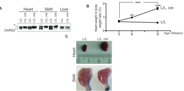
3.1. Mitochondrial aspartyl-‐tRNA synthetase (DARS2) is essential for embryonic development in the mouse ... 54
3.2. DARS2
+/-‐mice are haplosufficient ... 55
3.3. Tissue-‐specific disruption of Dars2 ... 57
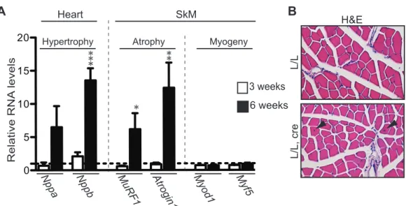
3.4. DARS2 deficiency leads to early pathological changes in heart and skeletal muscle ... 57
3.5. Defective mitochondrial translation gives rise to strong respiratory chain deficiency ... 62
3.6. Mitochondrial stress responses are activated exclusively in DARS2-‐ deficient heart ... 68
3.7. Early disturbance in mitochondrial proteostasis triggers stress responses in heart independent of MRC deficiency ... 72
3.8. DARS2 deficiency in forebrain neurons, hippocampus and striatum ... 79
3.9. DARS2 deficiency in forebrain cause respiratory chain deficiency ... 81
3.10. DARS2 deficiency causes progressive neuronal degeneration ... 86
3.11. Corticohippocampal nerve cell loss was highly likely to be caused by apoptosis ... 88
3.12. Increased immune response and gliosis in DARS2-‐deficient mice ... 91
4. Discussion ... 97
4.1. Mitochondrial aspartyl-‐tRNA synthetase (DARS2) is essential for embryonic development and one copy of the gene is enough for survival in mouse ... 99
4.2. DARS2 deficiency in heart and skeletal muscle causes comparable mitochondrial dysfunction in both tissues but activates mitochondrial stress responses exclusively in heart ... 100
4.3. DARS2 deficiency in forebrain neurons, hippocampus and striatum causes progressive neuronal degeneration accompanied by an activation of inflammatory responses and reactive astrogliosis in an age-‐ and region-‐ dependent manner ... 110
4.4. Summary and perspectives ... 117
Erklärung ... 140
Teilpublikationen ... 141
Curriculum Vitae ... 142
List of Figures
Figure 1.1 Human mitochondrial DNA and related diseases. ... 7 Figure 1.2 Aminoacylation reaction. ... 13 Figure 1.3 The mitochondrial unfolded protein response (UPR
mt) in
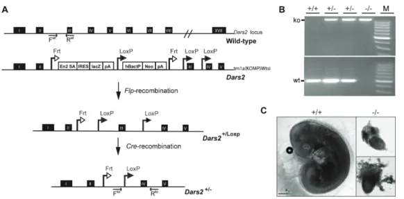
Caenorhabditis elegans. ... 25 Figure 3.1 Disruption of Dars2 in the germline. ... 55 Figure 3.2 Respiratory Chain Complexes in Heart, Skeletal Muscle (SkM) and
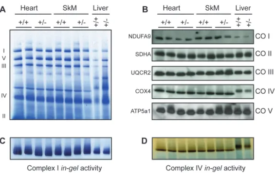
Liver Mitochondria of 104-Week-Old wild type (+/+) and heterozygous (+/-) Dars2 mice. ... 56 Figure 3.3 Phenotypic characterization of tissue-specific DARS2-deficiency in
heart and skeletal muscle. ... 58 Figure 3.4 Molecular characterization of tissue-specific DARS2-deficiency in
heart and skeletal muscle. ... 59 Figure 3.5 Immunohistochemical characterization of heart and skeletal muscle. 60 Figure 3.6 Increased mitochondrial mass was observed only in DARS2-deficient
cardiomyocytes. ... 61 Figure 3.7 Characterization of mitochondrial dysfunction in 6-week-old DARS2-
deficient heart and skeletal muscle. ... 63 Figure 3.8 Deregulated protein synthesis and steady-state levels of mitochondrial
ribosomal subunits. ... 65 Figure 3.9 RNA-related assays (Northern blot, aminoacylation and in organello
transcription). ... 67 Figure 3.10 Antioxidant responses in DARS2-deficient heart and skeletal muscle.
... 69 Figure 3.11 Mitochondrial unfolded protein response and autophagy in 6-week-
old DARS2-deficient heart and skeletal muscle. ... 70 Figure 3.12 Characterization of mitochondrial dysfunction and stress responses in
3-week-old DARS2-deficient heart and skeletal muscle. ... 72 Figure 3.13 Proof on perturbed mitochondrial proteostasis in DARS2-deficient
hearts. ... 74 Figure 3.14 Fibroblast growth factor 21 (FGF21) levels and related adaptive
systemic changes in DARS2 knockout mice. ... 76 Figure 3.15 Western blots analysis of 1-week-old hearts. ... 78 Figure 3.16 Phenotypic characterization of tissue-specific DARS2-deficiency in
forebrain neurons, hippocampus and striatum. ... 80 Figure 3.17 Characterization of mitochondrial dysfunction in 28- (and 23-) week-
old DARS2-deficient cortex and unaffected cerebellum. ... 82
Figure 3.18 Further characterization of mitochondrial dysfunction by COX-SDH staining and TEM in 28/30-week-old mice. ... 85 Figure 3.19 Neuronal degeneration in DARS2-deficient cortex and hippocampus.
... 87 Figure 3.20 TUNEL staining in DARS2-deficient cortical and hippocampal
regions. ... 90 Figure 3.21 IBA1 staining in DARS2-deficient cortical and hippocampal regions.
... 94 Figure 3.22 GFAP staining in DARS2-deficient cortical and hippocampal regions.
... 96 Figure 4.1 Proposed model for the heart-mediated stress responses to perturbed
protein homeostasis caused by DARS2 deficiency. ... 110
List of Tables
Table 1.1 Diseases and affected organs due to the mutations in mitochondrial
aminoacyl-tRNA synthetases. ... 18
Table 2.1 Genotyping PCR primer sequences ... 37
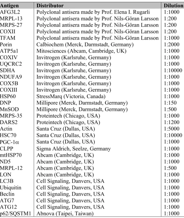
Table 2.2 Primary antibodies used for Western blot analysis ... 45
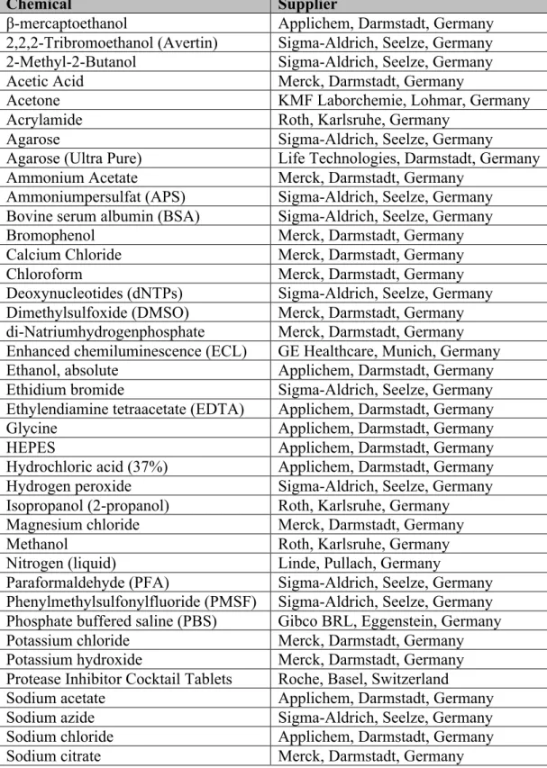
Table 2.3 Chemicals used and suppliers ... 52
Table 3.1 Length of mitochondrial-encoded MRC subunits and the number,
percentage and positions of aspartate residues ... 66
Abbreviations
3’ three prime end of DNA sequence 5’ five prime end of DNA sequence
A adenosine
ADP adenosine diphosphate
ARS2 mitochondrial aminoacyl-tRNA synthetase ATP adenosine triphosphate
Avertin tribromoethyl alcohol and tert-amyl alcohol BAT brown adipose tissue
bp base pairs
BN blue native
C cytosine
CA cornu ammonis (hippocampus)
CaMKIIα calcium/calmodulin-dependent kinase II α cDNA complementary DNA
CNS central nervous system
Cre bacteriophage P1 derived site-specific recombinase COX cytochrome c oxidase
Da Dalton
DAPI 4,6-diamidino-2-phenylindole
DARS2 mitochondrial aspartyl-tRNA synthetase ddH
2O double distilled water
DG dentate gyrus (hippocampus) DNA desoxyribonucleic acid
dNTP desoxyribonucleotide-triphosphate
EC enzyme commission number
EDTA ethylendiamine tetraacetate EGTA ethylene glycol tetraacetic acid ELISA enzyme-linked immunosorbent assay EtBr ethidium bromide
ETC Electron transport chain EtOH ethanol
g gram
G guanine
GFAP glial fibrillary acidic protein
h hour
H&E hematoxylin/eosin H
2O
2hydrogen peroxide HCl hydrochloric acid
HEPES N-2-hydroxyethylpiperazine-N-2-ethansulfonic acid i.e. id est
i.p. intraperitoneal
IBA1 ionized calcium-binding adapter molecule IRES internal ribosomal entry site
k kilo
KCl potassium chloride
ko knockout
KOH potassium hydroxide
l liter
L loxP flanked
lacZ gene encoding β-galactosidase
m milli
M molar
MgCl
2magnesium chloride min minute
mtDNA mitochondrial DNA
mRNA messenger RNA NaCl sodium chloride NaF sodium fluoride
NAH
2PO
4monosodium phosphate NaHCO
3sodium bicarbonate NaOH sodium hydroxide
PAGE polyacrylamide gel electrophoresis PBS phosphate buffered saline
PCR polymerase chain reaction RNA ribonucleic acid
RNase ribonuclease
Rpm revolutions per minute RT room temperature
rtPCR reverse transcription polymerase chain reaction SDS sodiumdodecylsulfate
sec second
SEM standard error of the mean TBE tris-borate-EDTA buffer TE tris-EDTA buffer
Tris 2-amino-2-(hydroxymethyl)-1,3-propandiole tRNA transfer RNA
TWEEN polyoxethylene-sorbitan-monolaureate
U units
V volt
v/v volume per volume w/v weight per volume WAT white adipose tissue
WT wild type
β-me β-mercaptoethanol
Abstract
Cells try to counteract mitochondrial respiratory chain deficiencies via various kinds of largely unknown compensatory mechanisms, which play a central role in determining the extent of tissue-specific defects leading to disease phenotypes. In this study, we directly disrupted mitochondrial protein synthesis in mice by deleting the mitochondrial aspartyl-tRNA synthetase (Dars2) gene in a tissue- specific manner. We generated DARS2 deficiency in three different tissues (heart, skeletal muscle and forebrain neurons) and followed the dynamics and extent of pathological changes that occurred.
Deficiency of this essential protein leads to severe deregulation of mitochondrial
protein synthesis in both heart and skeletal muscle. Yet, mitochondrial stress
responses, like increased biogenesis, decreased autophagy, upregulation of
mitochondrial unfolded protein response and mitokine FGF21, are only observed
in DARS2-deficient cardiomyocytes. Surprisingly, the initiation of these stress
responses is stemming from perturbed mitochondrial proteostasis, rather than the
respiratory deficiency. Skeletal muscle, on the other hand, has intrinsic protective
mechanisms that make it better equipped for folding and turnover of
mitochondrial proteins, as well as slow turnover of mitochondrial transcripts that
is coupled with possible upregulation of muscle regeneration. As a result, skeletal
muscle is able to cope with increased levels of unassembled proteins better.
Although DARS2 depletion leads to very strong, deleterious respiratory
deficiency in heart and skeletal muscle, causing animals to die within 7-8 weeks,
its deficiency in forebrain neurons seems to have a milder effect that takes much
longer time to develop. Defective mitochondrial translation in forebrain neurons
caused abnormal behavior, and severe forebrain atrophy, which is caused by
neuronal cell apoptosis and accompanied by activation of inflammatory responses
such as microgliosis and reactive astrogliosis. Surprisingly, neurodegeneration
occurred in an age-dependent manner and affected cortex and hippocampal
regions differently.
Zusammenfassung
Zellen kompensieren Defekte der mitochondrialen Atmungskette mit Hilfe verschiedener oftmals noch weitgehend unbekannter Mechanismen, welche eine zentrale Rolle dabei spielen, in welchem Ausmaß gewebe-spezifische Defekte zu einem Krankheitsphänotyp beitragen.
In dieser Arbeit wurde mit Hilfe eines konditionalen Mausmodells gewebe- spezifisch die mitochondriale Protein-Synthese durch die Deletion des mitochondrialen Asprtyl-tRNA-Synthase (Dars2) Gens zerstört. Die Dynamik und das Ausmaß der hierdurch verursachten pathologischen Veränderungen wurden dabei einerseits im Herz- und Skelettmuskel sowie den Neuronen des Vorderhirns untersucht.
Der Verlust dieses essenziellen Proteins führt zu einer schweren Dysregulation der mitochondrialen Proteinsynthese sowohl im Herzen wie auch im Skelettmuskel. In DARS2-defizienten Kardiomyozyten konnten mitochondriale Stressreaktionen wie vermehrte Biogenese, verminderte Autophagie, die Hochregulierung der mitochondriale unfolded protein response und des Mitokins FGF21 nachgewiesen werden. Überraschenderweise werden diese Stressreaktionen jedoch weniger durch die auftretende respiratorische Fehlfunktion als vielmehr durch eine gestörte Protein-Homöostase hervorgerufen.
Im Skelettmuskel wiederum scheinen intrinsisch protektive Mechanismen zu existieren, welche die Faltung und Stabilität mitochondrialer Proteine erhöhen.
Darüberhinaus weist der Skelettmuskel eine geringere Abbaurate mitochondrialer
Transkipte auf, was möglicherweise in Zusammenhang mit einer erhöhten
Muskel-Regeneration steht.
Im Gegensatz zu dem starken Phänotyp in Herz- und Skelettmuskel, bei dem die
Mäuse innerhalb der ersten 7-8 Lebenswochen sterben, hat der Verlust von
DARS2 spezifisch in Vorderhirn-Neuronen einen milderen Effekt, der deutlich
länger für eine Entwicklung braucht. Defekte in der mitochondrialen Translation
in diesem Gewebe verursacht abnormales Verhalten der Mäuse, eine schwere
Atrophie des Vorderhirns, welche durch Apoptose der Neuronen hervorgerufen
wird und die Aktivierung inflammatorischer Prozesse wie Mikrogliose and
reaktive Astrogliose. Erstaunlicherweise tritt diese Neurodegeneration in alters-
abhängiger Weise auf und betrifft den Cortex und Hippocampus Regionen in
unterschiedlicher Wiese.
1. Introduction
Chinese philosophy explains the basis of nature through yin and yang, opposing
forces, interdependent and able to exist only in relation to each other. The cell
and the mitochondria have a very similar story. It is a tale of two enemies, who
later became friends and allies. On this particular day, an ancient bacterium has
invaded a single cell organism and instead of killing its host -as it happened many
times before- it has found a safe haven and decided to stay within. Why not? The
available nutrients were more than sufficient and the host offered shelter from the
hostile environment of the ancient world. As for the single cell, beside the fact that
it has survived the attack, benefits have also been great. Finally, it was granted a
way to fight the poisonous oxygen in the surrounding and moreover, a new, more
efficient energy form was suddenly available. This random encounter, which
happened around 1-2 billion years ago to give rise to a synergically superior
eukaryote, is described by the ‘endosymbiotic theory’ (Margulis, 1975). We now
believe that the bacterium became integrated into the recipient cell and evolved
into an organelle, the mitochondrion. Whether the random encounter was an
invasion, infection or an unwilling indigestion event, the yin -the anaerobic cell-
and the yang -the aerobic mitochondrion- left their differences behind and
cooperated. The story, which started almost as a horror movie, turned into a
romantic comedy in the end; or did it? Does the initial invasion of the bacteria
continue even if it lost its ability to live independently? Is the mitochondrion
abusing its powers in the cell and waiting a suitable time for revenge like a smart
serial killer? A number of different disorders and diverse disease manifestations,
as well as mitochondrial involvement in age-associated diseases, seem to prove
this (Dogan and Trifunovic, 2011).
1.1. Mitochondria
Mitochondria are small organelles found in almost every eukaryotic cell. They form a very dynamic network, with constant fusion and fission, and occupy roughly one fifth of its total volume (McBride et al., 2006). The mitochondrion comprises of two membranes, the outer (OMM) of which is separated from the inner (IMM) one by the intermembrane space (IMS). The inner membrane exhibits a folded structure, named cristae, to maximize its surface. The innermost compartment of the mitochondria is called the mitochondrial matrix, and contains the mitochondrial genome, ribosomes, transfer RNAs (tRNAs), and various proteins and enzymes required for mitochondrial function. Mitochondria are unique because they are the only organelles in animal cells containing their own DNA, mitochondrial DNA (mtDNA).
The main function of the mitochondria is to use oxygen to generate the cell’s major energy source, adenosine triphosphate (ATP). Thus, mitochondria are the powerhouses of the cell, generating ATP through the process of oxidative phosphorylation (OXPHOS). The proteins mediating electron transport and OXPHOS reside in the inner mitochondrial membrane. Moreover, mitochondria are very important for other cellular processes such as the first step of iron-sulfur (Fe-S) cluster biosynthesis, pyruvate decarboxylation and tricarboxylic acid cycle (TCA cycle), programmed cell death (apoptosis), steroid synthesis, calcium homeostasis and reactive oxygen species (ROS) formation.
The redox reactions in the cell feed the electron transport chain (ETC), which
couples the electron transfer between an electron donor (NADH and FADH
2) and
acceptor (oxygen) with the transfer of protons across the inner membrane. ETC
has four macromolecular complexes: complex I (CO I - NADH:ubiquinone
oxidoreductase, EC 1.6.5.3), complex II (CO II - Succinate dehydrogenase, EC
1.3.5.1), complex III (CO III - Ubiquinol cytochrome-c reductase, EC 1.10.2.2) and complex IV (CO IV or COX - cytochrome c oxidase, COX, EC 1.9.3.1). ETC catalyzes the electron transfer from reducing equivalents to molecular oxygen.
Electrons are carried from CO I and CO II to CO III by coenzyme Q (CoQ or ubiquinone), linking TCA cycle to the process. Other sources of electrons, such as glycolysis, fatty acid oxidation, pyrimidine biosynthesis, choline and amino acid oxidation, exist that can donate electrons to CoQ (Vafai and Mootha, 2012). The electron transport from CO III to CO IV is mediated by soluble electron carrier cytochrome c. The synthesis of ATP from ADP and P
iin mitochondria is catalyzed by Complex V (or ATP synthase) (CO V, EC 3.6.3.14), which is powered by the proton gradient generated.
The organization of the OXPHOS system is more intricate than separately
assembled complexes that are arranged in sequence in the inner mitochondrial
membrane. Two models have been proposed for the organization of the
mitochondrial respiratory chain: (i) the “fluid-state” or “random collision” model,
where all OXPHOS complexes diffuse individually in the membrane and electron
transfer depends on the random collision of the complexes and electron carriers
(Hackenbrock et al., 1986); (ii) the “solid-state” model, which was proposed over
50 years ago, where the complexes together form large, rigid supramolecular
structures termed respirasomes (Hatefi et al., 1962). The most plausible scenario,
however, is a combination of these two models: the “plasticity” model. In this
model, single complexes (“fluid-state” model) and different types of
supercomplexes (“solid-state” model) coexist in the inner membrane. Complex I,
for instance, is mainly found in association with complex III in various
supercomplexes that additionally contain the electron carriers coenzyme Q and
cytochrome c, complex IV, and sometimes complex II or V, and are able to
respire. On the other hand, most of the complexes II and IV are present as
individual entities. How the supercomplexes are assembled is currently not
known, but the significance of this arrangement for the stability of the different complexes is certain (Acin-Perez et al., 2008).
1.2. Mitochondrial Diseases
Mitochondrial diseases are one of the most common inborn errors of metabolism with a frequency of ~ 1 in 5000 (Schaefer et al., 2004). The term “mitochondrial encephalomyopathies” is often used since the affected organs/tissues are mostly brain and skeletal muscle (Shapira et al., 1977). Nowadays, the term
“mitochondrial diseases” is almost exclusively used to describe diseases caused by defects in mitochondrial oxidative phosphorylation (OXPHOS) and not regarding the defects in numerous other cellular processes within mitochondria.
Even within these boundaries, the classification of the mitochondrial diseases became quite complicated because mutations in either mtDNA or nuclear DNA (nDNA) genes coding for mitochondrial proteins lead to major and catastrophic diseases in humans. The first patient suffering from a mitochondrial disorder was identified in 1962 (Luft et al., 1962). Since then, thousands of patients have been diagnosed with different kinds of mitochondrial diseases. Due to the complexity of mitochondrial diseases, a new way of classification is embraced, one that is using the diseases’ genetic defect rather then clinical manifestation.
1.2.1 Mitochondrial genetics
Human mitochondrial DNA (mtDNA) is a small (16,569 basepair long), circular,
double-stranded molecule (Figure 1.1). mtDNA only encodes 13 respiratory chain
subunits, 22 tRNAs and 2 rRNAs, which are essential for mitochondrial
translation (Larsson, 2010). During the course of evolution, mtDNA lost more
than 99% of its original genes, thus now depends on nDNA for all its basic
synthesis of the phospholipids of the inner mitochondrial membrane and the necessary aids for its DNA’s replication, transcription, and translation (Vafai and Mootha, 2012).
A couple of unique features of mtDNA genetics and inheritance make it very difficult to predict the course of the disease, prenatal diagnosis and/or genetic counseling in everyday clinical practice (Dogan and Trifunovic, 2011):
• mtDNA does not follow the Mendelian rules of inheritance while it is maternally inherited. Therefore, a mother carrying an mtDNA mutation can transmit it to her children, but only her daughters can further transmit it to the next generation. As each cell contains ∼10,000 copies of mtDNA, a pathogenic mutation could be present in all of them or just few of copies of the molecule. Existence of two or more different populations of mtDNA in a single cell is called ‘heteroplasmy’, in contrast to ‘homoplasmy’
where all mtDNA molecules are identical.
• Threshold effect represents the minimal critical level of a pathogenic mutation in mtDNA that should be present in the cell or tissue to have a deleterious effect. A certain proportion of mutant mtDNA must be present before reduction in OXPHOS activity is observed, and the threshold seems to be lower in tissues that are more dependent on oxidative metabolism. It has been shown that there are different thresholds for different types of mtDNA mutations, 60% for large mtDNA deletions (Bourgeron et al., 1993) to 90% for some tRNA mutations (Chomyn et al., 1992; Hanna et al., 1995).
• The last but not least problem of mtDNA genetics is the mitotic
segregation. Random distribution of mtDNA molecules during cell
division can lead to increased amounts of mutant mtDNA molecules in one of the daughter cells. This can lead to a cell carrying low levels of mutated molecules giving rise to one of relatively high levels, which in turn will affect OXPHOS in that cell.
1.2.2 Mitochondrial diseases caused by mitochondrial DNA (mtDNA) mutations
About 200 mtDNA point mutations and numerous single large-scale partial deletions have been associated with human diseases, most of which affect the nervous system (Wallace, 2005). Although genetically distinct, most mtDNA diseases share common features such as lactic acidosis, mosaic pattern of cells deficient in cytochrome c oxidase (COX) activity and massive mitochondrial proliferation in muscle resulting in ragged-red fibers (DiMauro et al., 1985). The more mtDNA-related diseases are identified, the more it became clear that mitochondrial diseases commonly have a delayed onset and progressive course.
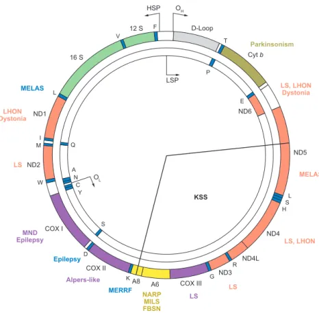
Mutations in mtDNA are divided into two groups: (i) mtDNA point mutations and (ii) mtDNA rearrangements. A depiction of human mitochondrial DNA and the most common diseases can be found in Figure 1.1 (DiMauro and Schon, 2008).
Diseases and the genes, the mutations in which cause the disease, are labeled with
the same color.
Figure 1.1 Human mitochondrial DNA and related diseases.
Human mitochondrial DNA. Names of the common mitochondrial diseases -caused by point mutations and/or rearrangements- are indicated on the figure. The disease manifestations and the related mutations are color-coded (DiMauro and Schon, 2008).
LS, Leigh syndrome; MELAS, mitochondrial myopathy, encephalopathy, lactic acidosis, and stroke-like episodes; MERRF, myoclonic epilepsy and ragged red fibres; MILS, maternally inherited Leigh syndrome; NARP, neurogenic weakness, ataxia, and retinitis pigmentosa; PS, Pearson syndrome.
(i) mtDNA point mutations:
Most human mtDNA point mutations occur in tRNA genes, thus the most common mtDNA-related disorders are caused by mutations in those genes:
ANRV346-NE31-05 ARI 14 May 2008 7:23
HSP
LSP
D-Loop T
P
E Cyt b
ND5 ND6
ND4L KSS
S A Q
OL N
C Y
ND4
ND3 ND2
ND1 L
V
12 S F
16 S
R COX III G
COX II COX I
W M
I
D
A8 A6 K
L HS Parkinsonism
DystoniaLHON
LS, LHON Dystonia
LS, LHON
LS NARP LS
FBSNMILS MERRF Epilepsy
Alpers-like EpilepsyMND
LS
MELAS MELAS
OH
Figure 1
The human mitochondrial genome. The mtDNA-encoded gene products for the 12S and 16S ribosomal RNAs, the subunits of NADH-coenzyme Q oxidoreductase (ND), cytochromecoxidase (COX), cytochromeb(Cyt b), and ATP synthase (A), and 22 tRNAs (1-letter amino acid nomenclature) are shown, as are the origins of heavy- and light-strand replication (OHand OL) and the promoters of heavy- and light-strand transcription (HSP and LSP). Some pathogenic mutations (for expanded versions of all the key terms in this article, seeSupplemental Term List; follow theSupplemental Material linkfrom the Annual Reviews home page at
http://www.annualreviews.org) that affect the nervous system in particular are indicated (colors correspond to those of the affected genes).
but with no evidence of paternal transmis- sion is strongly suggestive of an mtDNA point mutation.
About 200 mtDNA point mutations and innumerable single large-scale (kilobase-sized)
partial deletions have been associated with hu- man diseases, most of which affect the central and peripheral nervous system, especially if my- opathies are considered—as they should—the domain of peripheral neurology. This concept 94 DiMauro
·
SchonSupplemental Material Annu. Rev. Neurosci. 2008.31:91-123. Downloaded from www.annualreviews.org by WIB6386 - Universitat zu Koln - Und Stadbibliothek on 11/04/10. For personal use only.
Mitochondrial encephalomyopathy, lactic acidosis and stroke-like episodes (MELAS) is a multisystem disorder that is often fatal in childhood or in young adulthood. The disease principally affects muscle, brain and the endocrine system.
Stroke-like episodes are experienced before the patients are 40 years old. Most people with MELAS have a buildup of lactic acid in their bodies. Less commonly, people with MELAS may experience involuntary muscle spasms (myoclonus), impaired muscle coordination (ataxia), hearing loss, heart and kidney problems, diabetes, and hormonal imbalances (Kaufmann et al., 2011). Although the most common mutation is A3243G in tRNA
Leu(UUR), other mutations (in protein coding genes, as well as tRNAs) have been found to be associated with the disease.
Myoclonus epilepsy and ragged red fibres (MERRF) is almost exclusively a result of mutations in tRNA
Lys. MERRF is characterized by muscle twitches, myopathy, and spasticity. Affected individuals sometimes have short stature and heart abnormalities (cardiomyopathy) (Moraes et al., 1993). Gomorri Trichrome staining of the muscle cells reveals clumps of diseased mitochondria accumulation in the subsarcolemmal region of the muscle fiber, which appear as
‘rough’ or so-called ‘Ragged Red Fibers’. As in MELAS, the disease affects translational efficiency via failure in tRNA modification with taurine (Suzuki et al., 2011).
Additionally, missense mutations in mtDNA protein coding genes can also result in an array of clinical manifestations:
A mutation in the mtDNA ATP6 gene is associated with neurogenic muscle
weakness, ataxia, and retinitus pigmentosum (NARP) when present at lower
percentages of mutant (~40%) (Holt et al., 1990) and lethal childhood Leigh
syndrome when present at higher percentages (~95%) of mutant (Tatuch et al.,
1992).
Leber’s hereditary optic neuropathy (LHON), the most common mtDNA-related disease, causes severe visual loss in both eyes. LHON, mostly an early-onset disease, is usually caused by homoplasmic mutations in one of three genes encoding complex I subunits (G11778A in NADH dehydrogenase 4 (ND4), G3460A in ND1 and T14484C in ND6). A significant percentage of people with a mutation that causes LHON do not develop any features. Specifically, more than 50 percent of males with a mutation and more than 85 percent of females with a mutation never experience vision loss or related medical problems (Man et al., 2002).
(ii) mtDNA rearrangements:
Systemically distributed mtDNA rearrangement mutations, mostly deletions, can be either inherited or spontaneous. Deletions usually result in a spectrum of symptoms. The nature and severity of the symptoms from mtDNA deletion rearrangements is usually due to the tissue distribution of the rearranged mtDNAs (Wallace, 2005).
Kearns–Sayre syndrome (KSS) is defined by the onset before age 20 of
ophthalmoplegia (paralysis of the muscles that move the eyeballs), ptosis (droopy
eyelids), pigmentary retinopathy (Kearns and Sayre, 1958). In this multisystemic
disorder, partially deleted mtDNAs are present in all examined tissues. In chronic
progressive external ophthalmoplegia (CPEO) deleted mtDNAs are found only in
muscle (Moraes et al., 1989). In Pearson’s syndrome, which is characterized by
sideroblastic anaemia and exocrine pancreas dysfunction, deleted mtDNAs are
initially abundant in haematopoietic cells (Pearson et al., 1979).
1.2.3 Mitochondrial diseases caused by nuclear DNA (nDNA) mutations
Mitochondrial diseases caused by a mutation in nuclear encoded genes are a very heterogeneous group. Not only are most of the ~80 structural proteins of the OXPHOS system encoded by nDNA, but all the proteins needed for their import from the cytoplasm and assembly in mitochondria are also encoded by the nucleus. Defects in any of these proteins could lead to functionally impaired OXPHOS and therefore to mitochondrial disease. Furthermore, defects in any protein affecting stability, expression and/or integrity of mtDNA could lead to the same deleterious effect.
Mitochondrial diseases caused by mutations in nDNA can be divided into four categories (DiMauro and Schon, 2008):
1) Mutations in genes encoding respiratory chain subunits 2) Mutations in genes encoding ancillary proteins
3) Mutations in genes affecting the lipid milieu of respiratory chain
4) Mutations in genes encoding for mtDNA maintenance, replication, transcription and translation
1) Mutations in genes encoding respiratory chain subunits: A severe autosomal-
recessive neurological disease, clinical features of which includes psychomotor
retardation with extrapyramidal signs, restlessness, global dementia, severe
defects in verbal communication, and mild axial hypotonia has been found in
2008 (Barel et al., 2008). Mutations in nuclear-encoded UQCRQ, encoding
ubiquinol-cytochrome c reductase, CO III subunit VII, are found to be the main
reason for the observed phenotypes. In most of the cases, the mitochondrial-
encoded subunit of CO III (cytb) is the main cause of disease phenotypes (CO III
is composed of ten nuclear-encoded subunits and one mitochondrial-encoded
subunit). So far, one other nuclear mutation in protein coding genes of respiratory
binding protein), encoding subunit VI of CO III (De Meirleir et al., 2003). One patient with this mutation is shown to have CO III dysfunction with clinical hypoglycemia and lactic acidosis (Haut et al., 2003).
2) Mutations in genes encoding ancillary proteins: Leigh syndrome (LS), or subacute necrotizing encephalomyelopathy, is a neurodegenerative disorder characterized by predominant involvement of the central nervous system (CNS) (Leigh, 1951). LS is an early-onset and progressive disease. This condition is characterized by progressive loss of mental and movement abilities (psychomotor regression) and typically results in death within a couple of years, usually due to respiratory failure (Finsterer, 2008). Mutations in both nuclear and mitochondrial genes have been identified in LS patients. However, mutations in the nuclear Surf1 gene, coding for a putative CO IV assembly factor are one of the main cause of LS. In these patients, SURF1p mutations or depletions cause a reduction in the fully assembled COX (Tiranti et al., 1999).
3) Mutations in genes affecting the lipid milieu of respiratory chain: Cardiolipin
is the major component of the inner mitochondrial membrane and it has been
shown to participate in the formation of supercomplexes in yeast (Zhang et al.,
2005). This fact exemplifies the importance of cardiolipin in proper respiratory
function. Barth syndrome, an X-linked recessive disease manifesting
cardiomyopathy, causes underdeveloped skeletal musculature and muscle
weakness, growth delay, 3-methylglutaconic aciduria, and and altered
composition of cardiolipin. Mutations in the TAZ gene, encoding for tafazzin, is
found to be responsible for the disease (Schlame and Ren, 2006). Tafazzin, a
phospholipid acyltransferase, is involved in cardiolipin remodelling and mutations
in the TAZ gene cause defects in mitochondrial architecture and function.
4) Mutations in genes encoding for mtDNA maintenance, replication, transcription and translation: The most common nuclear mutations associated with mitochondrial diseases are found in the gene encoding mitochondrial DNA polymerase ϒ. Around 70 disease manifestations related to the mutations in the gene coding for the polymerase have been reported: such as progressive external ophthalmoplegia (PEO) (Van Goethem et al., 2001), Alpers syndrome (Naviaux and Nguyen, 2004), mitochondrial neurogastrointestinal encephalomyopathy (MNGIE) (Van Goethem et al., 2003) or sensory ataxic neuropathy, dysarthria and ophthalmoparesis (SANDO) (Van Goethem et al., 2003).
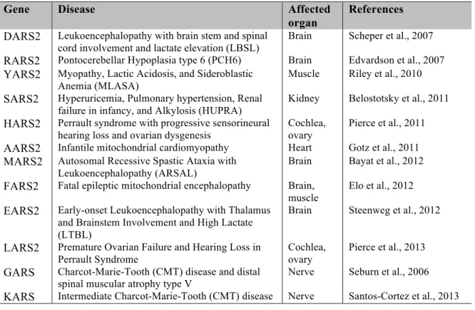
One class of housekeeping genes encoded by nuclear genome, namely mitochondrial aminoacyl-tRNA synthetases, has also been implicated in various diseases. Before going deeper with the disease phenotypes caused by aminoacyl- tRNA synthetases, we will have a closer look to these fascinating enzymes.
1.3. (Mitochondrial) Aminoacyl-tRNA synthetases and -related diseases
1.3.1 Aminoacyl-tRNA synthetases
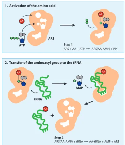
One of the first steps during translation is aminoacylation: the covalent ‘charging’
of a tRNA with its cognate amino acid, a two-step process that uses ATP
(Delarue, 1995). tRNA charging is performed by a highly specialized subgroup of
enzymes, the aminoacyl-tRNA synthetases (ARSs). ARSs are ubiquitiously
expressed and highly conserved enzymes, which can be found in a range of
species. In the first step, the amino acid and a molecule of ATP is bound via a
specific ARS for that amino acid (Figure 1.2). Following the formation of an
aminoacyl adenylate intermediate, a pyrophosphate molecule is released. The
second step starts with the binding of the cognate tRNA molecule to the ARS.
After the binding of tRNA molecule, the amino acid is transferred to the tRNA and an adenosine monophosphate (AMP) molecule is released. After the releasing of the charged tRNA molecule and ARS is free for another aminoacylation reaction. Typically, ARSs have a catalytic domain and an anticodon-binding domain but some also have an editing domain for deacylating mischarged amino acids. The anticodon binding domain as well as the editing domain, which only some ARSs contain, is very important for specificity of the ARSs, as well as serves as a quality control mechanism of protein synthesis (Ling et al., 2009).
Figure 1.2 Aminoacylation reaction.
The two-step aminoacylation reaction catalyzed by aminoacyl-tRNA synthetases (ARSs) (Antonellis and Green, 2008). AA: aminoacid; PPi: pyrophosphate; ATP: adenosine triphosphate; AMP: adenosine monophosphate.
ANRV353-GG09-05 ARI 25 July 2008 12:20
tRNA: transfer ribonucleic acid Aminoacyl-tRNA synthetases (ARSs):
the family of enzymes responsible for performing aminoacylation reactions in the cytoplasm and mitochondria
BACKGROUND ON AMINOACYL- tRNA SYNTHETASES
The transfer of biological information from DNA to RNA to protein is critical for the sur- vival and propagation of cells, tissues, and or- ganisms. One key component of this central dogma is protein translation, which involves using the genetic code to translate genetic in- formation (in the form of messenger RNA) to
AA
P P P
AA AA
P P
AA AA AA AA P AA
1. Activation of the amino acid
2. Transfer of the aminoacyl group to the tRNA
AA
AA AA AA AA
P P
AA AA AA AA AA AA
AA AA AA P AA
ARS + AA + ATP ARS(AA-AMP) + PPi Step 1
ARS(AA-AMP) + tRNA AA-tRNA + AMP + ARS Step 2
ARS ATP
AMP tRNA
+
Figure 1
The two-step aminoacylation reaction. Each aminoacyl-tRNA synthetase (ARS) charges a specific tRNA molecule with its cognate amino acid (AA) via a two-step enzymatic reaction. In the first step, the ARS binds the AA and an ATP molecule to form the aminoacyl adenylate (AA-AMP) intermediate, and a pyrophosphate molecule (PPi) is released. In the second step, a tRNA molecule binds the ARS via the anticodon binding domain (white space on ARS with three extensions), and the AA is transferred to the tRNA. An AMP molecule is then released, followed by the charged tRNA. The ARS is then free to charge another tRNA molecule. The chemical equation for each step is provided in