Bur J Clin Chem Clin Biochem 1995; 33:59-63
© 1995 Walter de Gniyter & Co.
Berlin · New York
Adaptation of the Troponin T ELISA Test to a Microplate Immunoassay Reader
By L Penttil
1'
2, K. Hirvonen
3, A. Julkunen
2, K. Penttil
2and T. Rantanen
11
Department of Clinical Chemistry, Kuopio University Hospital, Kuopio, Finland
2
Department of Clinical Chemistry, Kuopio University, Kuopio, Finland
3
Oriola Oy Prolab, Espoo, Finland
(Received July 1 I/October 5, 1994)
Summary: Troponin T concentration in serum is usually measured by the automated method developed by Boeh- ringer Mannheim for the ES-series of analysers. These instruments need at least 140 μΐ of serum and 700 μΐ of reagents for a single analysis, which takes 90 min. We describe an alternative procedure, using streptavidin-coated microtitre plates, troponin T reagents of Boehringer Mannheim and an ELIS Α-reader to measure the concentration of troponin T. The present assay needs only 30 μΐ of sample and 200 μΐ of reagents, and it takes 75 min; the detection limit is 0.10 μg/l. We also assessed the microtitre plate method for sensitivity and precision and compared the results with those measured by an ES-300 automatic analyser. Both methods have the same measurement range for troponin T of 0.1 to 15 μg/l. For daily routine use of the microtitre plate method we recommend duplicate determinations.
Introduction
Troponin T belongs to the troponin family of muscle proteins (1,2). Troponins are located in the thin filament of the myocyte. Troponin T has the highest relative mo- lecular mass of 37000, other members of the family be- ing troponin I (M
T= 24 000) and troponin C (M
T= 20 000) (2). The function of troponin T is to bind the other troponins to the tropomyosin filament of the myocyte. Initially the cytosolic troponin T, but later also the coniplexed troponin T are released from damaged muscle cells into the circulation (3). This release de- pends on the early reperfusion of the damaged area, and it can be utilized to follow the progress of a myocardial infarction (3,4). Troponin T is found in both myocardial and in skeletal muscle. Differences in the amino acid contents and structures of myocardial and skeletal tro- ponin T (5) are sufficient to permit the preparation of
!) Enzymes
Isoenzyme MB of creatine kinase (EC 2.7.3.2) Isoenzyme l of lactate dehydrogenase (EC 1.1.1.27)
highly specific antibodies against myocardial troponin T (3, 5, 6). By using two selected monoclonal antibodies, Katus and coworkers (7) developed a new and highly specific enzyme-linked immunosorbent assay (ELISA) for the measurement of serum cardiac troponin T, in which skeletal troponin T in serum shows only minimal cross reactivity. Many recent studies have shown that the new troponin T method is very useful in the diagnosis of acute myocardial infarction and for following the time course of cardiac muscle perfusion in patients with un- stable angina pectoris (4, 7, 8, 9).
In this article we describe a modification of the original method for measuring troponin T in serum, using a stan- dard microplate immunoassay reader and the commer- cial reagents of Boehringer Mannheim (Mannheim, Ger- many) for the ES-series of analysers. A very close agreement was obtained between the results from the ES-300 analyser and those from the microtitre plate method. The microtitre plate method for troponin T can therefore be used in place of the ES-300 analyser (10,
Π).
Eur J Clin Chem Clin Biochem 1995; 33 (No 1)
60
Penttil et al.: Microplate ELISA method for serum troponin TMaterials and Methods
Samples
For the present study, surplus serum from patients in the cardiac care unit of the Kuopio University Hospital, taken for ordinary serum enzyme assays, was used and stored at -70°C (10, 11) until analysed for serum troponin T concentration. Venous blood samples for enzyme determinations were collected with minimal stasis and centrifuged within 60 min to obtain serum for analyses.
Haemolysed samples were replaced with new samples. For measur- ing the variations within and between series, two serum pools were made and divided into 1.0 ml portions and stored at -70 °C until analysed. To evaluate the present method, patients with acute myo- cardial infarction, with typical clinical signs, ECG-patterns and se- rum enzyme1) activities (12) were monitored by the serial measure- ment of serum troponin T concentration.
Methods and reagents
We measured serum troponin T with an ES-300 analyser using the reagent kit of the ELISA Troponin-T Test of Boehringer Mannheim (Cat. No. 1289055) for the ES-series of analysers (10, 11). The reagents of the kit were used as recommended by the manufacturer.
The basic method uses 140 μΐ of serum (standard or control) and 700 μΐ of the mixture of two antibodies, the first complexed with biotin and the second with peroxidase. After incubation of the mix- ture for 60 min at 25 °C, the tubes were washed, substrate was added (di-ammonium 2,2'-azino-bis[3-ethylbenzothiazoline-6- sulphonate] and H2O2), and the tubes were incubated for 30 min at 25 °C for colour formation. The colour intensity was then mea- sured at 405 nm (tab. 1) and the troponin T concentration obtained from the standard curve as calculated by the instrument.
Unmodified reagents produced by Boehringer Mannheim were used for the determination of serum troponin T concentration as a microtitre plate immunoassay method. Instead of the original tubes, the reaction was performed in the 250 μΐ wells of a microtitre plate after coating the wells with streptavidin. The wells of the microtitre plate were coated for 24 h with streptavidin from Boehringer Mannheim (14 U/mg) at a concentration of 10 mg/1 in 10 mmol/1 phosphate buffer, pH 7.2 containing 150 mmol/1 NaCl at + 4 °C.
The binding capacity of streptavidin for biotin on the walls of the wells was at least 2 ng of biotin per well, which was found to be sufficient. Commercial microtitre plates coated with streptavidin (Boehringer Mannheim and Labsystems Oy, Helsinki, Finland) could also be used. Coated microtitre plates were stored at + 4 °C until used. Otherwise, the method was similar to that used with the ES-300 analyser, with minor modifications of the ratios of sample and reagent volumes (tab. 1). Sera, standards and controls were dispensed into the wells of the plate with a manual pipette (Proline 5-50, Biohit Oy, Helsinki, Finland). The tip of the pipette was changed after each operation. The mixture of antibodies to troponin T was then added with a multitip automated pipette (Proline Electronic 250, Biohit Oy). The plate was incubated for 60 min at 25 °C in a Wellwarm l shaker (Denley Instruments Ltd, Bill- ingshurst, England) then washed twice with the washing solution using a Multiwash instrument (Labsystems Oy, Helsinki, Finland).
The substrates (di-ammonium 2,2'-azino-bis[3-ethylbenzothiazo- line-6-sulphonate] and H2O2) were then added and the microplate was incubated on a Multiscan MCC/340 reader (Labsystems Oy) for 15 min, after which the concentration of the colour in the wells was read at 405 nm. The concentrations of troponin T were then obtained from a standard curve made with the Multiscan reader.
The standard curve for troponin T was constructed by using the following kit standards: 0.0, 0.9, 2.4, 5.2, 10.7 and 14.5 μβ/1. The controls purchased with the kit were low (0.19 ± 0 06 ug/1) and high (5.45 ± 0.82 μβ/1). The fitting of the standard curve was studied by different modes of the Multiscan reader and also by using a microcomputer.
For comparison of the clinical usefulness of the microtitre plate troponin Τ method, the activity of serum creatine kinase1), its iso-
enzyme MB, the activity of lactate dehydrogenase and its isoen- zyme 1 as well as the mass concentration of creatine kinase-MB were measured. The activity of creatine kinase in serum was mea- sured using a commercial assay by Boehringer Mannheim accord- ing to the ECCLS recommendation (13) and Hitachi 717 Analyser (Tokyo, Japan). The activity of serum creatine kinase-MB was measured with the Boehringer Mannheirn kit which incorporates an antibody for inhibition of the M-subunit; the value obtained for the creatine kinase-B subunit was then doubled. The mass concen- tration of creatine kinase-MB in serum was measured with a com- mercial IMx® creatine kinase-MB assay (Abbott Laboratories, Ab- bott Park, Chicago, 111., USA); the method uses a monoclonal anti- creatine kinase-MB antibody bound to latex microparticles; the se- cond antibody is a polyclonal anti-creatine kinase-MM antibody coupled with alkaline phosphatase (14). After inhibition of the M- containing isoenzymes with urea and qumidine at pH 10, lactate dehydrogenase and its isoenzyme lactate dehydrogenase-1 were measured using the lactate dehydrogenase-method of Boehringer Mannheim and a Hitachi 717 analyser in accordance with the Scan- dinavian Recommendation (15).
Statistics
The coefficients of variation and the correlation coefficients be- tween the different methods were calculated using the StatView 4.0 software package (Abacus Concepts Inc., Berkeley, U. S. A.).
The results are expressed as the mean values with corresponding standard deviations. The curve fittings by a microcomputer were performed using SPSS/PC+ Statistical Data Analysis package (SPSS Inc., Chicago, USA). v *_
Results and Discussion
Evaluation of the microtitre plate ELISA method for the determination of troponin T in serum
The purpose of the present investigation was to study the possibility of adapting the commercial troponin Τ test of Boehringer Mannheim to a standard microtitre plate ELISA method using the reagents of the manufac- turer as supplied for the ES-series of analysers. Each step of the method was tested, and the results of serum analyses were compared with those obtained with an ES-300 analyser. In table 1 both procedures are pre- sented.
The linearity of the Multiscan ELISA-reader was studied by using 10 mmol/1 K-dichromate solution. It was found that the instrument was linear at 405 nm up to an absor- bance value of 2.400. Using solutions of K-dichromate with absorbance values of 0.387 or 1.561, we pipetted 30 μΐ of the colour red solution with a single-tip manual pipette (Proline 5-50) and 200 μΐ of H
2O with a multitip automated pipette (Proline Electronic 250) into the 250 μΐ wells of microtitre plates (from Labsystems). The co- efficients of variation were 0.96 and 0.74%, respec- tively.
The optimal streptavidin coating of the microtitre plate wells was checked by using the highest troponin T stan- dard and steptavidin-c ated plates from Labsystems (the
Eur J Clin Chem Clin Biochem 1995; 33 (No 1)
Tab. l Protocols for the determinations of serum troponin T with the microplate immunoassay and with an ES-300 analyser. ABTS®
= diammonium 2,2'-azino-bis(3-ethylbenzothiazoline-6-sulpho- nate).
Step in assay Microplate ES-300
Sample/Standard
Anti-troponin T antibody, biotinylated
Anti-troponin T antibody peroxidase conjugated
30 μΐ 200 μΐ
140 μΐ 700 μΐ Microtitre wells or tubes coated with streptavidin. The plates or tubes were incubated for 60 min at 25 °C with continuous mixing.
Washing the samples and extra reagents one/two times with wash- ing solution.
Microplate ES-300 Addition of reagents ABTS®
and H2O2 Volume
Incubation of the mixture at 25 °C
U
200 μΐ 15 min
700 μΐ 30 min Measurement of the colour directly from the microtitre plate wells or after transferring the coloured liquid to the cuvettes (ES-300) at 405 nm.
Calculation of the results from the standard curves made by the in- struments.
binding capacity was stated to be at least 2 ng biotin per well). By varying the sample size (from 10 to 50 μΐ) it was found that 30 μΐ or less resulted in similar values to those obtained with an ES-300 analyser, but with sample volumes of 40 μΐ and 50 μΐ the results were too low (-12 and —32%, respectively). We also coated the wells of a microtitre plate with different concentrations of streptavidin (Boehringer Mannheim) and found that a 250 μΐ well should bind at least 2 ng of biotin, based on the analysis of 30 μΐ of the highest troponin Τ standard (the streptavidin tubes for the ES-series of analysers have a binding capacity of approximately 14 ng of biotin per tube).
Using an incubation temperature of 25 °C, it was shown that the incubation times can be the same as in the origi- nal method. However, the reaction time for substrate- chromogen solution could be shortened from 30 min to 15 min in the microtitre plate technique (tab. 1). It was also found that the incubation and the wash steps in the microtitre plate method should be performed mechani- cally rather than manually, in order to obtain constant results. After checking the conditions, the standard curve of the microtitre plate method was curvilinear (fig. 1) as is the standard curve of the ES-300 analyser. The measurement range was the same for both methods (0.1
'to 15 μg/l). The point-to-point calculation mode and the second degree regression mode gave the best results with the present method.
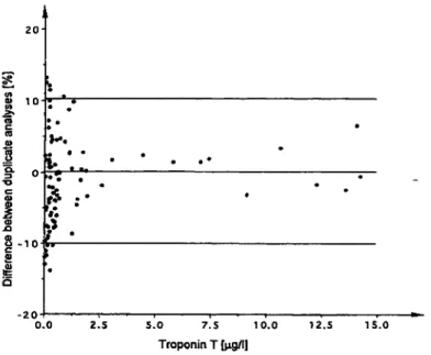
The difference between duplicate analyses were studied over the whole measuring range by using human serum samples. Figure 2 shows clearly that the percentage dif- ferences increase at lower and higher troponin T concen- trations. The situation was somewhat better when greater amounts of streptavidin were used for coating the micro- plate wells (data not shown). At concentrations below the lowest standard (0.9 μg/l), the relative differences were so high that duplicate analyses are recommended for routine laboratory practice. In contrast, with an ES- 300 analyser, single determinations seem to fulfil the
10 is
Troponin T in serum [μς/Ι]
20
Fig. 1 A typical standard curve of the troponin Τ method for the microplate immunoassay method.
20
10
ι - 1 0
-20
S
0.0 2.5 5.0 7.5 10.0 Troponin Τ [μο/1]
12.5 15.0
Fig. 2 Percentage differences between duplicate analyses for se- rum troponin Τ measured by the microplate immunoassay method.
Eur J Clin Chem Clin Biochem 1995; 33 (No 1)
62
Penttil et al.: Microplate ELISA method for serum troponin TTab. 2 The within and between series variation of the troponin T microplate method, using low and medium serum pools, and controls from the manufacturer. The assayed values for the tro- ponin T control sera were: low 0.19 ± 0.06 μ§/1 and high 5.2
± 0.82 Mg/l.
Coefficient of variation
of troponin Τ method within series Sample level
Mean (μ§/1) CV (%) No. of analyses
Serum pool Low High
0.237 2.28 1.52 1.92 10 10
Control Low
0.229 2.44 10
serum High
4.602.23 10 Coefficient of variation
of troponin Τ method between series Sample level Serum pool Control serum
Low High Low High
CV (%)Mean No. of analyses
0.242 12.98
10.02.13 8
0.234 12.7 4.35
7.678
criteria of acceptance even in the lower concentration range (11).
Variations within and between series were studied by using frozen serum pools and control samples (tab. 2).
At the troponin Τ concentrations of 0.2 and 2.2 μg/l in serum pools or 0.2 and 4.5 μg/l in control samples, the within series variations were between 1.52 and 2.44%.
The variations between series were higher and ranged from 7.67 to 12.9% (tab. 2). It is possible that small variations in the streptavidin binding capacity can increase the coefficient of variation at the lower range, or that the streptavidin binding capacity of the wells should be greater.
The highest standard concentration in the kits is 14.5 μ§/1. In daily laboratory practice, several values are higher than this, suggesting that the sample should be diluted. The only obvious problem of standardisation is the absence of a standard concentration near the critical value of the measurement (between 0 and 0.9 μg/l). Such a standard would help to increase the accuracy of the measurement of low serum troponin Τ values.
A very close agreement was found between the meth- ods when the values from the microtitre plate tech- nique were compared with those obtained with an ES- 300 analyser (fig. 3). The mean values for 42 human sera did not significantly differ for the two methods (r = 0.99).
Detection limit, reference values and discrimination limit of the microtitre plate assay
The detection limit for troponin T in serum with the present microtitre plate method was about 0.10 μg/l when defined as the mean of 14 non-specific binding values + 3 SD. In the sera of 63 healthy laboratory per- sonnel, more than 95% of all troponin Τ values were below 0.10 μ§/1, the highest value being 0.19 μg/l. By doubling the value of 0.10 μg/l it can be stated that tro- ponin Τ values in serum exceeding 0.20 μg/l should be regarded as abnormal. This limit corresponds well with those presented earlier (4, 10, 11, 16).
20
5 10 I S Troponin T (ES-300 analyser)
Fig. 3 The comparison of the results for serum troponin Τ mea- sured with the microplate immunoassay method and with an ES- 300 analyser, y = -0.059 + 0.988x (x = ES-300, y = microplate method). Ν = 42, r = 0.99.
1000
100 ISO
Time after onset of symptoms [h)
zoo
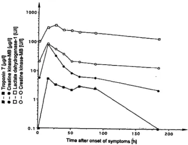
Fig. 4 Aetypical time-curve of serum troponin Τ (Β) compared with the catalytic activity concentrations of creatine kinase-MB (o) and lactate dehydrogenase isoenzyme 1 (a) and the mass concen- tration of creatine kinase-MB (·) in the serum of a patient with acute myocardial infarction. » »
Eur J din Chem Clin Biochern 1995; 33 (No 1)
In figure 4, typical changes of serum troponin T and enzyme values in a patient with acute myocardial infarc- tion with early reperfiision are presented. This clinical result obtained with our modified method corresponds well to previous observations made with automated as- says for troponin T in serum (3, 4, 7, 11, 16).
Conclusion
A comparison of the values obtained with the microtitre plate immunoassay method with those from an ES-300 analyser shows that the microtitre plate method can be
dised for the measurement of serum troponin T concen- tration. The day-to-day variation still has to be reduced and we are currently working on this problem. From a clinical point of view the present method fulfils the needs of daily clinical laboratory practice for the diagno- sis of acute myocardial infarction, thus confirming the usefulness of the troponin T assay.
Acknowledgement
The authors thank Mr. Tero Hongisto for technical assistance and Pharm. Dr. Even McDonald for revising the English language.
References
1. Saprans I, Takahash H, Rüssel MP, Watanabe S. Skeletal and cardiac troponins and their components. Biochem J 1972;
72:723-35.
2. Greaser ML, Gergeley J. Purification and properties of the components from troponin. J Biol Chem 1973; 248:2125-33.
3. Katus HA, Remppis A, Looser S, Hallermeier K, Scheffold T, Kubier W. Enzyme linked immunoassay of cardiac troponin T for the detection of acute myocardial infarction in patients. J Mol Cell Cardiol 1989; 21:1349-53.
4. Gerhardt W, Katus H, Ravkilde J, Hamm C, Jorgensen PJ, Peheim E, Ljungdahl L, Löfdal P. S-troponin T in suspected ischemic myocardial injury compared with mass and catalytic concentrations of s-creatinine isoenzyme MB. Clin Chem 1991; 37:1405-11.
5. Briggs MM, Schachat F. N-terminal amino acid sequences of three functionally different troponin T isoforms from rabbit skeletal muscle. J Mol Biol 1989; 206:245-9.
6. Scheffold T, Temppis A, Kubier W. Only one troponin T iso- form is expressed in the failing human heart. Eur Heart J
1991; 12(suppl):215.
7. Katus HA, Looser S, Hallermeir K, Remppis A, Scheffold T, Borgya A, Essig U, Geuss U. Development and in vitro char- acterization of anew immunoassay for cardiac troponin T. Clin Chem 1992; 38:386-93.
8. Hamm CW, Ravkilde J, Gerhardt W, J0rgensen P, Peheim E, Ljungdahl L, Goldman B, Katus HA. The prognostic value of serum troponin T in unstable angina. N Engl J Med 1992;
327:146-50.
9. Hamm CW, Katus HA, Ravkilde J, Goldmann BU, Bleifeld W, Gerhardt W. Identification of high-risk patients with unstable angina by troponin T release. Circulation 1992; 82(Supp III):7.
10. Penttilä I, Helin M, Julkunen A, Miettinen M, Rantanen T.
Evaluation of ES-300 ELISA analyzer for the measurement of troponin-T in the diagnosis of acute myocardial infarction.
Clin Chem 1992; 38:965-6.
11. Penttilä I, Helin M, Julkunen A, Miettinen M, Rantanen T.
Evaluation of an ES-300 ELISA analyzer for the measurement of troponin-T in the diagnosis of acute myocardial infarction.
LabMedica International 1993; X:16-9.
12. World Health Organization. Report of the fifth working group on the establishment of ischemic heart disease registers. WHO, Copenhagen 1971.
13. European Committee for Clinical Laboratory Standards. Stan- dards for enzyme determination: creatine kinase, aspartate aminotransferase, alanine aminotransferase, gamma-gluta- myltransferase; 1988 ECCLS Document ISSN 1011-6265, No. 3-4.
14. Brandt DR, Gates RC, Eng KK, Forsythe CM, Korom CK, Notro AS, Koffler PA, Ogunro EA. Quantifying the MB isoen- zyme of creatine kinase with the Abbott "IMx" immunoassay analyzer. Clin Chem 1990; 36:375-78.
15. Keiding P, H0rder M, Gerhardt W, Pitkänen E, Tenhunen R, S fromme JH, Theodorsen L, Waldenström J, Try ding M, Westlund L. Recommended methods for the determination of four enzymes in blood. Scand J Clin Lab Invest 1974;
33:291-306.
16. Ravkilde J, H0rder M, Gerhardt W, Ljungdahl L, Pettersson T, Tryding N, M011er BH, Hamfelt A, Graven T, Äsberg A, Helin M, Penttilä I, Thygesen K. Diagnostic performance and pro- gnostic value of serum troponin T in suspected acute myocar- dial infarction. Scand J Clin Lab Invest 1993; 53:677-85.
Professor Ilkka Penttilä, M. D., Ph. D.
Department of Clinical Chemistry Kuopio University Hospital FIN-70210Kuopio Finland
Eur J Clin Cheni CKn Biochem 1995; 33 (No 1)