Eur J Clin Chem Clin Biochem 1995; 33:553-558
© 1995 Walter de Gruyter & Co.
Berlin · New York
Changes of Ionized Magnesium and Free Fatty Acids in Serum after Acute Myocardial Infarction
By Frank Bertschat
1, Hartmut hing
2, Theodor Günther
3, Allen Jeremias
1and Elisabeth Jeremias
11
Medizinische Klinik und Poliklinik mit Schwerpunkt Nephrologie/Intensivmedizin, Universitätsklinikum Rudolf Virchow, Charlottenburg, Freie Universität Berlin, Berlin, Germany
2
Institutför Wasser-, Boden- und Lufthygiene, Umweltbundesamt, Berlin, Germany
3
Institut für Molekularbiologie und Biochemie, Freie Universität Berlin, Berlin, Germany
(Received January 12/May 29, 1995)
Summary: The most feared early complications after an acute myocardial infarction are ventricular arrhythmias.
These may be initiated by changed concentrations of catecholamines and electrolytes. The present study shows a reduction of total serum magnesium after acute myocardial infarction which is normalized within a few days.
Further, it could be shown that a more significant decrease of ionized Mg
2+(iMg
2+) takes place at the day of acute myocardial infarction in the total group of myocardial infarction patients (n = 36). A closer investigation reveals that iMg
2"
1" was considerably decreased in one third of the patients, whereas two thirds showed minor changes of iMg
2"
4" in both directions. The pronounced decrease of iMg
2"*" in the first sub-group can be explained by the time course of free fatty acids in serum. On the day of the myocardial infarction free fatty acids in serum were increased.
This is probably caused by ß-adrenergicrinduced lipolysis due to catecholamines released by the stressful situation of an acute myocardial infarction. The increased free fatty acids in serum bind Mg
24", thus reducing iMg
2+. As long as a beneficial effect of a general Mg infusion in all acute myocardial infarction patients is controversial,
4
" should be measured and Mg infusion therapy should be applied only in patients with low iMg
2"
1".
Introduction
Lethality after acute myocardial infarction is particu- Several authors described a transient decrease of total larly caused by ventricular arrhythmias (1—3). Re- serum magnesium during the first 24 hours after acute cently, several publications indicated that these early myocardial infarction which reached normal values 6 complications are initiated by changed concentrations to 14 days later (19—23). The fall of serum magne- of catecholamines and electrolytes (4-10), and possi- sium may be explained by increased lipolysis due to bly by increased formation of free radicals and lipid stress-induced catecholamine release. The free fatty peroxidatipn (11, 12). In these mechanisms extracellu- acids bind Mg
2"
1". Thus the free fatty acids in adipo- lar magnesium may be involved. At reduced serum cytes reduce the concentration of intracellular free Mg more catecholamines are released (13) and perme- Mg
2+ in these cells followed by Mg
24" uptake in ability of Na
+and K
+(14) and lipid peroxidation is adipocytes (24, 25). The increased free fatty acids in increased (15). Dyckner et al. (16) and Bigg & Chia serum after acute myocardial infarction bind Mg
2+(17) showed that cardiac fibrillations appeared more and consequently the concentration of free Mg
2"
1" after
frequently in myocardial infarction patients with low acute myocardial infarction may be more reduced than
total serum Mg. Iseri et al. could even improve ther- total serum Mg. Therefore, we investigated the alter-
apy-resistent ventricular arrythmias with Mg
2"*" infu- ation of ionized Mg
2+(iMg
2"
1") in seruni after acute
sions (18). myocardial infarction.
Materials and Methods
Forty-two patients (37 men and 5 women) with an acute myocar- dial infarction who were admitted to the Klinikum R. Virchow, Charlottenburg, Free University Berlin in the time of July 1992 until October 1992 were examined. The average age of the patients was 62.5 years (range: 40-88 years). The diagnosis "acute myo- cardial infarction" was determined by ECG, the rise of creatine kinase and the subjective pain of the patients. At days 1, 2, 3, 5 and 7 after infarction (day 1 = day of infarction) blood was taken in the morning before breakfast with a plain vacutainer. Blood was centrifuged within 60 minutes after collection, the temperature not exceeding 25 °C. iMg2+ in serum was measured with the "Micro- lyte Magnesium" (Kone Instr. Espoo, Finland) within 60 minutes after blood centrifugation. The deviation of pH and the resulting change in iMg2"1" was corrected using the equation:
Ig [iMg2+ (pH)] = Ig [iMg2* (7.4)] + χ (7.4 - pH).
For χ a value of 0.11 was used (26). Total serum magnesium was determined by atomic absorption spectroscopy. Free fatty acids were measured with the enzymatic colour test "NEFA c" (27).
Results
Table 1 shows the mean values and standard deviations of the electrolyte concentrations and free fatty acids in serum of the patients at day 1 (day of infarction) and their changes from day 1 to day 2. Because of missing values at day 2 the number of measurements varies between 28 and 36. iMg2+ was significantly lower at day 1 than at day 2, whereas free fatty acids were significantly higher at day 1 than at day 2. The
Tab. 1 Mean values and standard deviations of electrolytes and free fatty acids in blood samples of acute myocardial infarction patients at day 1 and mean differences of these analyte concentra- tions between day 1 and day 2.
Tab. 2 Rank correlation coefficients (r) between each analysis at day 1 and day 2 of acute myocardial infarction.
Mgtotai, iMg2+, Catotai, iCa2+, iK+, iNa+, free fatty acid concentra- tions in serum at day 1 or day 2.
Analysis
iMg2* Mgtotal
iCa2+
Catotai iNa+iK+
Free fatty acids
Day 1 Mean mmol/1
0.550.79 2.091.15 139.37 4.302.32
SD
0.070.10 0.080.34 3.960.79 1.81
n
3636 3634 3628 30
Day 2-Day 1 Mean mmol/1
+0.04 +0.01 +0.02 +0.07 +0.04 +0.07 -0.69
SD
0.070.08 0.090.31 2.150.81 1.16
Sig-nifi- cance
**
n.s.n.s.
n.s.n.s.
n.s.**
Levels of significance:
ρ < 0.05 ρ < 0.01 ρ < 0.001 non-significant
number of acute myocardial infarction patients : ionized free serum magnesium
: total serum magnesium iCa2+: ionized free serum calcium Cal0tal· total serum calcium iNa+: ionized free serum sodium iK+: ionized free serum potassium
***
***
n.s.n:
iMg2+
Analyses Day 1
Mgtoial iMg2+
Catoiai iCa2+
iK+iNa+
Free fatty acids
Day 2
Mgtotai
iMg2+
Calotai iCa2+
iK+
iNa+
Free fatty acids r 0.540.08 0.400.24 0.410.84 0.55
n 36 *36 ή. s.
36 *36 n. s.
28 *36 ***
31 * For significance see legend of table 1.
Tab. 3 Rank correlations coefficients (r) between differences of analyses at day 1 and day 2 of acute myocardial infarction, e.g.: [AiMg2+ =
Analyses AiMg24- AiMg2+ AiMg2*
AMgtotaj
ΔΪΚ+
ΔΜ&013ι AiK+
AiNa+
AiK+ AiNa+
0.450.77 -0.37 -0.370.46
36 **
28 *36 * 28 *28 * For significance see legend of table 1.
other electrolytes did not change significantly from day 1 to day 2. These results show that iMg24" changes much more from day 1 to 2 after acute myocardial infarction than Mgtotai.
Table 2 presents the rank correlation coefficients of each electrolyte species and free fatty acid concentration be- tween day 1 and day 2. The high correlation coefficient (r = 0.84) between serum Na+ concentration at day 1 (iNaf) and day 2 (iNaJ) indicates that the sodium con- centration in serum does not change significantly after acute myocardial infarction. The concentration of serum potassium from day 1 to day 2 is less constant (r = 0.41) and the ionized fractions of Ca2+ (r = 0.24) and Mg2H~ (r = 0.08) are not even significantly correlated between day 1 and day 2 while Mgtotal (r = 0.54) and Catotal
(r = 0.40) are significantly correlated. These results show that the individual changes of iMg2+ and iCa2+
from day 1 to day 2 after acute myocardial infarction are more pronounced than the individual changes of Mgtotal
and Catotai·
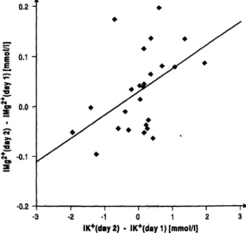
In table 3 the rank correlation coefficients of all signifi- cant correlations between changes of different electro- lyte species and free fatty acids are listed. The correla- tion between the changes of iMg2+ and iK4" (r = 0.45) from day 1 to 2 indicate that potassium and magnesium are concordantly affected. This is .also shown by a plot
of the two analytes together with a linear regression (fig.
1). This result may be of clinical importance (see Dis- cussion). The differences of iNa
4" are negatively corre- lated both with the differences of iMg
2* (r = -0.37) andofiK+(r = -0.37).
It is important to note that there existed no significant correlation between the changes of iMg
24" and the free fatty acids during the first two days after the acute myo- cardial infarction. We therefore investigated whether such a correlation existed in the sub-group with the strongest relative decrease of iMg
24" on the day of the acute myocardial infarction. For this purpose the distri- bution of the relative changes of iMg
2H" from day 1 to day 2 is shown in figure 2. As indicated in figure 2 the patients were divided into three sub-groups with 12 pa- tients each according to the relative changes of their iMg
2* from day 1 to day 2. Thus, in sub-group A iMg
2"
1"
was lower at day 1 by -11.5% to -32%. In sub-group B on the other hand iMg
2* was higher by + 1% to + 20%. Between these two sub-groups lies a sub-group with changes of iMg
2 +by -11% to 0%.
Time courses of iMg
2"
1", Mg
totai> iK
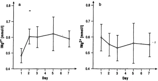
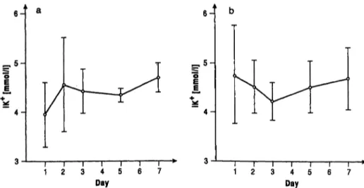
+and free fatty acids were plotted for sub-groups A and B (see figs 3—6). In the patients of sub-group A at the day of infarction iMg
2+(fig. 3 a), Mg
total(fig. 4 a) and iK+ (fig. 5 a) were low, increased to day 2 and remained almost constant thereafter. In these patients free fatty acids (fig. 6 a) were highest at day 1 and decreased thereafter.
In patients of sub-group B, who were selected by their high iMg
2"*" at day 1, Mg
totai
a^d iK
+were also highest at day 1 and decreased later. Free fatty acids remained almost constant throughout day 1 to day 7 (fig. 3 b, 4b,
0.2-
sub-group
0.1 -
0.0-
-0.1 -
• ·
φ
- 3 ^2 - 1 0 1 2 3
IK+(day2) - IK+(day 1) [mmol/l]
Fig. l Scatter plot of the differences of iMg2+ versus the differ- ences of iK"1" from day 1 to day 2 of acute myocardial infarction.
n
4-
3-
2-
1-
0.68
v
p T
S
>l
'\0.84 1
J •1
1.~A
m
»
«".
ί
0
^ ·»
•5 .' '.' '"'
:i . ^
P
.·. ^·,'
·· l·
i'* 'i
1.1
_Ar
-
'·'·'1
1" ,'·
f
6
Π l •i i 1
1ϊ
.32 Fraction of IMg2* on day 2
Fig. 2 Distribution of fractions of iMg2+ on day 2 after myocar- dial infarction: (iMgi* - iMg2*) / iMgi*. n: numbers of patients with given fractions of iMg2+ at day 2. A: sub-group of 12 patients with lowest iMg2"1", B: sub-group of 12 patients with highest iMg2* at day of infarction.
5b, 6b). Time courses of iCa
2*, Ca
totai and iNa
+are not shown, since their changes with time as well as their differences between sub-groups were not significant.
Discussion
Low iMg
2+was observed at the day of infarction in the total group of myocardial infarction patients. A more thorough analysis showed that acute myocardial infarc- tion patients did not behave homogeneously with respect to iMg
2+, Mgtotab iK
+and free fatty acids. Therefore, the acute myocardial infarction patients were divided into 3 sub-groups according to their relative changes of iMg
24" from day 1 to day 2.
Sub-group A consisted of patients with the strongest in- creases of iMg
2"
1" from day 1 to day 2. In this sub-group Mg
totai snd iMg
2"
1" in serum were low immediately after acute myocardial infarction (day 1) and iMg
2"
1" reached normal values at day 2. The changes of iK
+are closely correlated to the changes of iMg
2+. A close relationship was also shown for total Mg and iK
+in serum (19).
The behaviour of total serum Mg is in accordance with earlier results (19-23), which showed that the low total serum Mg after acute myocardial infarction normalized during the following few days. In the present study we additionally measured iMg
2+and found that in the pa- tients of sub-group A, iMg
2+at the day of acute myocar- dial infarction was much more reduced than total Mg.
This result can be explained by the time course of free
fatty acids in serum. At day 1 of acute myocardial in-
farction, free fatty acids were increased. This is probably
caused by -adrenergic-induced lipolysis due to cate-
cholamines released by the stressful situation of acute
0.8-
0.7- ο 1 0.6 Η
0.5-
0.4
0,8-
0.7- ο Ι 0.6 Η
0.5-
I Ι Γ Ι Ι Ι Γ
1 2 3 4 5 6 7
0.4 \^ ι —ι ι l l r
1 2 3 4 5 6 7
Day Day
Fig. 3 Time courses of iMg2"1" concentration in serum of acute myocardial infarction patients:
sub-group A (a) and sub-group Β (b). Mean values ± SD.
1.1- 1.0- 0.9- 0.8-
0.6- 0.5- 0.4 1.1 -
1.0- ίΕ"
ο| 0.8-
1 0.7-
Ο)
0.6- 0.5- 0.4-
a
TU M ΗΊ ^
Ι
JL1 2 3 4 5 6 7
Day
ι ι ι ι ι ι r
1 2 3 4 5 6 7
Day
Fig. 4 Time courses of Mgtotai concentration in serum of acute myocardial infarction patients:
sub-group A (a) and sub-group Β (b). Mean values ± SD.
myocardial infarction (4, 28). The increased free fatty acids in serum bind Mg
2+and Ca
2+, thus reducing iMg
2+. iCa
2+may not be reduced since it can be rapidly regulated by parathormone.
The transient reduction of total serum Mg can be ex- plained by a transient uptake of extracellular Mg
2+by adipocytes (24, 25). Catecholamines increase lipolysis in adipocytes. The released free fatty acids in adipocytes bind intracellular Mg
2+ and thus reduce intracellular free Mg
2+in the adipocytes. This is followed by uptake of extracellular Mg
2+by adipocytes (25), resulting in a reduction of total serum Mg.
Our findings may have clinical consequences. We sug- gest that only acute myocardial infarction patients with decreased iMg
2+and iK
+should be infused with Mg
2+. In the case of reduced iK+, K
+infusion is common clinical practice. However, in two major clinical studies LIMIT 2 (29-31) and ISIS 4 (32, 35) and in 12 small trials controversial effects of Mg
2+infusions in acute
myocardial infarct patients were found and in a few meta-analyses the controversial results were discussed.
For a review of the literature see 1. c. (36).
While LIMIT 2 (29-31) found positive effects, this was not confirmed in ISIS 4 (32, 35). The missing beneficial effect of ISIS 4 was explained and experimentally proven by the late time point of Mg
2+infusion, e. g.
after lysis (33). It was also suggested that the high Mg
2"
1"
dose used in ISIS 4 may have produced negative side effects (34).
As long as it remains controversial whether a general
Mg
2+infusion of all acute myocardial infarction pa-
tients is beneficial, we suggest that iMg
2+should be
measured and Mg
2+should be infused only in acute
myocardial infarction patients with low iMg
2"
1". Since in
most hospitals the equipment for iMg
2"
1" estimation is
not available, hypokalaemia may be taken as indication
for low iMg
2"
1". This compromise can be justified by the
significant correlation between the changes of iK
+and
J
1a
, 5 - E
£
4-1 b
, 5 -
4-
4 5 Day
4 5 Day
Fig. 5 Time courses of iK* concentration in serum of acute myocardial infarction patients:
sub-group A (a) and sub-group B (b). Mean values ± SD.
5-
H
5-
=. 4
a
l §
H
T l \
2 3 4 5 4l l l5 6 7
Day Day
Fig. 6 Time courses of free fatty acid concentration in serum of acute myocardial infarction patients:
sub-group A (a) and sub-group B (b). Mean values ± SD.
after acute myocardial infarction. However, as figure 1 shows, this compromise is by far not optimal.
Therefore, iMg
2+measurements should be installed as clinical routine for acute myocardial infarction pa- tients.
Since reduction of iMg
2+can increase the release of catecholamines (13) and in cooperation with catechola- mines may intensify cardiac arrhythmia (4), Mg
2+in- fusion should be an additional beneficial treatment of acute myocardial infarction patients with low iMg
2+.
References
1. Wolfe CL, Nibley C, Bhandari A, Chatterjee K, Scheinman M.
Polymorphous ventricular tachycardia associated with acute myocardial infarction. Circulation 1991; 84:1543—51.
2. Brugada P, Andries EW, Gursoy S, Willems H, Kaissar S.
Mechanisms of sudden cardiac death. Drugs 1991; 41 Suppl 2:16-23.
3. Späth G. Torsade de pointes oder die andere Ursache des plötz- lichen Herztodes. Wien, Berlin, Ueberreuter Wissenschaftsver- lagG.m.b.H., 1988.
4. Cefemuzynski L. Hormonal and metabolic reactions evoked by acute myocardial infarction. Circ Res 1981; 48:767-76.
5. Dyckner T, Wester PO. Relation between potassium, magne- sium and cardiac arrhythmias. Acta Med Scand 1981; Suppl 647:163-9.
6. Kafka H, Langevin L, Armstrong PW. Serum magnesium and potassium in acute myocardial infarction. Influence on ventri- cular arrhythmias. Arch Intern Med 1987; 147:465-9.
7. Havestadt C, Ising H, Günther T, Feldmann B, Schlüter HJ.
Electrolytes and ventricular arrhythmias. Magnesium 1985;
4:29-33.
8. Joborn H, Hjemdahl P, Larsson PT, Lithell H, Olsson G, Wiede L, et al. Effects of prolonged adrenaline infusion and of mental stress on plasma minerals and parathyroid hormone. Clin Phy- siol 1990; 10:37-53.
9. Flink EB, Brick JE, Shane SR. Alterations of long-chain free fatty acid and magnesium concentrations in acute myocardial infarction. Arch Intern Med 1981; 141:441-4.
10. Solomon RJ. Ventricular arrhythmias in patients with myo- cardial infarction and ischemia. Relationship to serum potas- sium and magnesium. Drugs 1984; 28 Suppl 1:66-75.
11. Tosaki A, Das DK. Reperfusion induced arrhythmias are caused by generation of free radicals. Cardiovasc Res 1994;
28:422.
12. Euler DE. Reperfusion induced arrhythmias are not caused by generation of free radicals. Cardiovasc Res 1994; 28:423.
13. Günther T, Ising H, Merker HJ. Elektrolyt- und Kollagengehalt im Rattenherz bei chronischem Magnesium-Mangel und Streß.
J Clin Chem Clin Biochem 1978; 16:293-7.
14. Günther T. Biochemistry and pathobiochemistry of magnes- ium. Mag Bull 1981; 3:91-101.
15. Günther T, Vormann J, Höllriegl V, Disch G, Classen HG.
Role of lipid peroxidation and vitamin E in magnesium defi- ciency. Mag Bull 1992; 14:57-66.
16. Dyckner T, Helmers C, Lundmann T, Wester PO. Initial serum potassium level in relation to early complications and pro- gnosis in patients with acute myocardial infarction. Acta Med Scand 1975; 197:207-10.
17. Bigg RFC, Chia R. Magnesium deficiency: role in arrhythmias complicating myocardial infarction. Med J Austral 1981;
1:346-8.
18. Iseri LT, Freed J, Bures AR. Magnesium deficiency and cardiac disorders. Am J Med 1975; 58:837-46.
19. Landmark K, Urdal P, Basmo GM. Changes in serum K and Mg during acute myocardial infarction [abstract]. Eur Heart J 1988; 9 Suppl 1:227, 1260.
20. Ising H, Günther T, Bertschat F, Ibe K, Stoboy V, Heldmann E. Alterations of electrolytes in serum and erythrocytes after myocardial infarction. Magnesium 1987; 6:192-200.
21. Chadda KD. Serum, red cell and whole blood magnesium in patients with uncomplicated acute myocardial infarction. Mag- nesium 1986; 5:76-84.
22. Rasmussen HS, Aurop P, Hojberg S, Kehn-Jensen E, McNair P. Magnesium and acute myocardial infarction. Transient hy- pomagnesemia not induced by renal magnesium loss in pa- tients with acute myocardial infarction. Arch Intern Med
1986; 146:872-4.
23. Tsutsui M, Shimokawa H, Yoshihara S, Sobashima A, Hayash- ida K, Higuchi S, et al. Intracellular magnesium deficiency in acute myocardial infarction. Jap Heart J 1993; 34:391-401.
24. Elliott DA, Rizack MA. Epinephrine and adrenocorticotropic hormone-stimulated magnesium accumulation in adipocytes and their plasma membranes. J Biol Chem 1974; 249:3985- 90.
25. Vormann J, Förster R, Günther T, Ebel H. Lipolysis-induced magnesium uptake into fat cells. Mag Bull 1983; 5:39-41.
26. Ising H, Bertschat F, Günther T, Jeremias E, Jerernias A. Meas- urement of free magnesium in blood, serum and plasma with an ion-sensitive electrode. Eur J Clin Chem Clin Biochem 1995; 33:365-71.
27. Mulder C, Schonten JA, Popp-Snijders C. Determination of free fatty acids. A comparative study of the enzymatic versus the gaschromatographic and the colQr^metric method. J Clin Chem Clin Biochem 1983; 21:823-7.
28. Hansen O, Johansson BW. S-Mg does not change inversely to S-FFA during acute stress situations. Angiology 1989;
11:1011-8.
29. Woods KL, Fletcher S, Roffe C, Haider Y. Intravenous mag- nesium sulphate in suspected acute myocardial infarction: re- sults of the second Leicester Intravenous Magnesium Interven- tion Trial (LBvflT-2). Lancet 1992; 339:1553-8.
30. Yusuf S, Teo K, Woods K. Intravenous magnesium in acute myocardial infarction. An effective, safe, simple, and inex- pensive intervention. Circulation 1993; 876:2043-6.
31. Millane TA, Camm AJ. Magnesium and the myocardium. Brit Heart J 1992; 68:441-2.
32. Cascells W. Magnesium and myocardial infarction. Lancet 1994; 343:807-9.
33. Atar D, Serebruany V, Poulton J, Godard J, Schneider A, Her- zog WR. Effects of magnesium supplementation in a porcine model of myocardial ischemia and reperfusion. J Cardiovasc Pharmacol 1994; 24:603-11.
34. Galloe A, Graudal N. Magnesium and myocardial infarction.
Lancet 1994; 343:1286-7.
35. ISIS-4 (Fourth International Study of Wäret Survival) Collab- orative Group ISIS-4: A randomised factorial trial assessing early oral captopril, oral mononitrate, and intravenous magne- sium sulphate in 58050 patients with suspected acute myocard- ial infarction. Lancet 1995; 345:669-85.
36. Egger M, Smith G. Misleading meta-analysis. Br Med J 1995; 310:752-4.
Priv. Doz. Dr. med. Frank Bertschat Virchow Klinikum
der Humboldt Universität zu Berlin Med. Klinik und Poliklinik mit
Schwerpunkt Nephrologie/Intensivmedizin Augustenburger Platz l
13353 Berlin Germany