Capillary Electrophoresis of Serum Proteins
Reproducibility, Comparison with Agarose Gel Electrophoresis and a Review of the Literature
Petal A. H. M. Wijnen and Marja P. van Dieijen-Visser
Department of Clinical Chemistry, Academic Hospital, Maastricht, The Netherlands
Summary: Conditions for serum protein analysis by capillary electrophoresis were optimized and within day, between day and between capillary variations were examined for both migration times and relative peak areas. For the five currently accepted zones, albumin, αϊ, a2, β and γ-globulin, reproducibilities of migration times were in the range of 2.3-3.1% (n = 200 measurements). Although variations in relative peak areas were slightly higher than those obtained by conventional agarose gel electrophoresis, from a resolution perspective, capillary electro- pherograms provided better detail than the densitometric scans of agarose gel electrophoresis. Precise localization of C3 and transferrin in capillary electrophoresis resulted in more accurate detection of the -globulin fraction.
When C3 appeared in the γ-fraction it was not detected as a separate peak in agarose gel electrophoresis, whereas it was in capillary electrophoresis.
In artificially prepared mixtures of highly purified albumin and γ-globulin preparations, best correspondence with theoretical values was found with capillary electrophoresis.
Inter-individual variations and reference values were obtained by measuring 140 samples from healthy controls (59 females, 81 males) with both techniques. For capillary electrophoresis the inter-individual variations of the albumin, αϊ, α2, β and γ fractions were respectively 6, 21, 19, 14 and 18% and for agarose gel electrophoresis 5, 20, 17, 18 and 22%. From these results it can be concluded that the more precise localization of the β- and γ-globulin fraction results in about 4% lower inter-individual variations in capillary electrophoresis compared to agarose gel electrophoresis. For the other fractions, comparable variations were obtained. Differences between males and fe- males were not significant.
For patient samples, a good correlation was found between capillary electrophoresis and agarose gel electrophoresis data for all five protein fractions.
We conclude that separation efficiency of capillary electrophoresis is better than that of agarose gel electrophoresis and even weak monoclonal components can easily be distinguished with the capillary electropherogram. Capillary electrophoresis is a qualitatively good, cheap, fast and easy to perform alternative to agarose gel electrophoresis.
Introduction
Capillary electrophoresis has been suggested as a new detector window. Separation is based on differences in tool for separation and quantification of serum proteins velocities of the charged particles (migration times). The (1 — 14). It combines the separation principles of conven- data obtained in the electropherogram are collected, tional electrophoresis with the advanced instrumental stored and interpreted with an appropriate data acquisi- design of high-performance liquid or gas chromatogra- tion system. For each separation, only nanoliters of sam- phy and capillary technology. The serum sample is intro- pie and microliters of buffer are used. The walls of un- duced into a buffer-filled fused silica capillary (internal treated fused silica capillaries are negatively charged in diameter 20 to 200 μιη and lengths of 10—100 cm), aqueous solution from the ionization of surface silanol either electrokinetically or hydrodynamically with pres- groups (pi = 1.5). The negatively charged silica surface sure (fig. 1). The amount of the sample applied can be attracts positively charged ions and cations from the regulated by changing the injection time. For separation, buffer, creating an electrical double layer (fig. 2). When both ends of the capillary are placed into a buffer solu- a voltage is applied across the capillary, cations in the tion that also contains the electrodes, and high voltage diffuse portion of the double layer migrate in the direc- is applied to the system. The applied voltage causes the tion of the cathode, carrying water with them. The result analytes to migrate through the capillary and past the is a net flow of buffer solution in the direction of the
536 Wijnen and Dieijen-Visser: Serum protein analysis by capillary electrophoresis
negative electrode - electro-osmotic flow. This flow is particularly important at alkaline pH, and a small change of pH can dramatically alter the separation pattern. Li- quid cooling of the capillary allows excellent mainte- nance of temperature control. The" final result of the pro- tein separation is affected by capillary length and dia- meter, buffer composition and pH, sample injection mode, capillary thermostating (Joule heat), separation temperature, the electro-osmotic flow, solute concentra- tion effects, wall-solute interactions and applied field.
Table 1 presents a review of the methods used for serum protein analysis by capillary electrophoresis presented in the literature to date. Although several methods are published, very few data are available on reproducibility of capillary electrophoresis and no data are available on variation between capillaries.
In most clinical laboratories agarose gel electrophoresis is used as a screening method for detection of abnormal- ities of the major proteins in biological fluids like serum, urine and cerebrospinal fluid. The results of serum pro- tein agarose electrophoresis are quantified from peak area determination of the electrophoresis scanning pattern. Comparison of visual inspection of electro- pherograms with agarose electrophoresis has been per- formed by Jenkins et al. (13). They found that capillary electrophoresis was able to detect all monoclonal bands detected by high resolution agarose electrophoresis, and, in particular, better able to detect IgA monoclonal bands occurring in the beta region.
Reference values were determined only by Klein et al.
(8), but exact description of the measurement conditions was not given.
Serum sample:
1 : 39 diluted in phosphate buffered saline of half ionic strength
Pressure injection:
34.5 kP, 2 s
Capillary inlet
Electrolyte buffer 100 mmol/1 borate pH 10.2
5 min, 10 kV P/ACE 5500 system Beckman
Fig. 1 Capillary electrophoresis system with configuration used in the text.
Inlet
S S S S S S S S ^
(?) Off
V- Vo L^ EOF
Untreated fused silica
Outlet
Capillary wall
Fig. 2 Untreated fused silica capillary. The negatively charged the negative electrode, electro-osmotic flow (EOF). V is the migra- silica surface attracts cations from the buffer, creating an electrical tion velocity of the different charged particles,
double layer. The result is a net flow of buffer in the direction of
έ
=3££
•5
•σ c ω
i ε
CO
"to Ο
t
"ά>t I
c'S g. ε
's I I
S t
I g
s:' δ ο?^ '§Si
G#
•δ.|
•^S
o?
i•3 w^
|
c&
|g
^^*
V—'
13
1
g ω
•S
Chen et al (1)
1 ·
CDυ
c
&%
CQ «n
^ j|
1
ό ε~Η
>·*·*·* δ ,
Ό t<* ·Μ ^^
Ο, C*· J2 Ο
< «
t tι
c
}
Μ 2"Ι
tuε
0
_ |
Ι 1
|
Capillary electrophoresis system Beckman
1
£
• l1 4> 5 S
1.1 ο ill
'S. "S ε οχ S ^
S ^ iti it:
·£
£ cS w
β> * Jw
t jil
*- 4, «Λ•S aQ.« °
ζ " S Ί9
co — ja je
1 5
U)c
*c c es 5 w
||
I ^ *i
c8 -5 υc3 «
fi
CX ^g| 7|
.£oo 'Sc 2
C Ov (D
'S u . δ CO — Ό
I ii
Serum 1 : 19 phosphate buffered saline
4) C
"S. ·ϋ
CO (5
_OΛ
"ΪΛ
J
S«•gxt =L§ §
5s
ri*«o
•s ί
55Ix
s εII
ε
ο«« <N
!i
Lu «ns^
3 OoCM
X
ε
ι S
I
sε'55
-σ ε 8 =
L£S
u
\Ou-»
X
ι £
.§·"· CC/3 C Ό 3·
«H ζ3
? X
I I
c 0M
J·™V)
J|
||
! fr
1 a
ω CQ
.£ JS00 0
ll
G «-o 'Sίί
•° §o
CQ
1
v CQo
*co
9
Cu"
a co *··51
U JS 1
&*
«
Beckman propriel buffer
1 1
oo
S oε 81
s
o
1
1
*" *
ll
0 HS S
1 1
co |>
NO ro TT
0ε
m
1
>>
•cJ2
I n
• JDSO
1
εl »O V)
: <*· o c >
, v> ooI S
I ε
§11
ε "i ε
o I
"Z * '
sS·
o rz:c S E B S .S
<N 01 C
g
—1H
O & Λε -σ ao
Ο .ϊί
— -β 5 .£ .£ .£
ε ε ε
04 οι m
α- <Ο
c > S
00 — (N
sS s
Jcx
I
Oc >
'
Hl
§ §
0.ε
It
«CO g
111 iii
c c c"e 'ε 'ε
ϊ. S
Ι
ΟΕ*•α
a
οο
I!
O CQα. J=
2 s
l 8
g t^o ^ m ^J.o '£
H II S2
Ο ί
538 Wijncn and Dieijen-Visscr: Serum protein analysis by capillary electrophoresis
The objective of the present study was to present the state of the art in the literature, to establish the reprodu- cibility of capillary electrophoresis and compare it with results from agarose electrophoresis. Differences be- tween capillaries were also examined. Artificially pre- pared mixtures of purified protein preparations (albumin and γ-globulin) were used to check the quantification of peak areas.
Reference values were measured, especially for peaks that could not be detected separately with agarose elec- trophoresis like transthyretin (pre-albumin), transfemn and C3. The results were compared to results obtained with agarose electrophoresis.
Materials and Methods
Materials
Control sera: Beckman I.D.-Zone normal (BI 015-555985-AR) and abnormal (Bl 015-555983-AP) were used for examination of variation and are indicated as normal control and abnormal control.
Seronorm protein, (Mat no. 1003405, batch no. 305024) obtained from Nycomed Pharma AS, Oslo, Norway and a pooled serum were used as control sera for agarose electrophoresis.
Cartridge coolant for the Beckman P/ACE system 2000 capillary cartridge coolant (No. 359976) was used as cooling fluid.
Albumin purified and essentially globulin-free, electrophoretic pu- rity approximately 99% was obtained from Sigma (lot 109F93041, Zwijndrecht, NL).
γ-Globulins of electrophoretic purity approximately 99% were ob- tained from Sigma (lot 106F9315, Zwijndrecht, NL).
Methods
Capillary electrophoresis was performed with a P/ACE 5500 sys- tem (Beckman Instruments Inc., Mijdrecht, NL) with P/ACE Sys- tem Gold software controlled by an IBM 330-450-DX computer (fig. 1). Post-run data analysis, like data integration or mobility/
area correction, was performed with System Gold software (Beck- man Instrument Inc.). A capillary column of 50 μπι and 27 cm (20 cm to the detector window) was assembled in the P/ACE car- tridge format (100 X 800 μιη aperture) from Beckman.
Samples were placed on the inlet tray of the P/ACE 5500 system and introduced into the capillary by pressure injection (2 seconds, 34.5 kPa). Serum was diluted 1 : 39 in phosphate buffered saline with half ionic strength (diluted l H- 1). Serum protein separations were carried out using an untreated fused silica capillary of 50 μπι,
at an oven temperature of 20 °C. The column was maintained at ambient temperature during electrophoresis with circulating cool- ant surrounding the column. Electrophoresis was performed for 5 minutes at 10 kV at 20 °C. Detection was made at the cathodic end by on-capillary UV absorbance measurements at 200 nm. The sys- tem contains built-in filters that can be changed. Quantification of the various fractions was obtained from the area under the curve by real-time data analysis. Before each run, the capillary was se- quentially rinsed one minute with 0.1 mol/1 NaOH and one minute with distilled water and two minutes with assay running buffer (100 mmol/1 borate buffer pH 10.2). The method we used was a slight modification of the Beckman application (tab. 1).
Agarose gel electrophoresis was performed' with the Paragon Se- rum Protein Electrophoresis kit from Beckman Instruments Inc., Mijdrecht NL (BI 015-556458-J). After electrophoresis, using a barbital buffer of pH 8.6, ionic strength 0.05, and staining with Paragon Blue Stain, the gels were scanned at 600 nm on the Beck- man Appraise System. The fraction of each protein zone was calcu- lated from the area under the curve. Serum protein electrophoresis gels were also visually interpreted for the presence of monoclonal bands or polyclonal gamrnopathy and were quantitated by densi- tometry. Agarose electrophoresis allowed discrimination of five protein fractions, i.e. albumin, ar, a2-, - (in some cases r and
2-) and γ-globulins. Total analysis time, electrophoresis (25 min- utes) and staining, is about 90 minutes for 10 serum protein electro- phoresis's on a gel.
Total protein and albumin were determined on a Synchron CX-7 analyzer (Beckman Instruments Inc, USA, California) using test- kits from Beckman Instruments Inc. For the determination of serum albumin, the bromcresol purple method (testkit 442765) was used.
Determination of serum total protein occurred with a timed end- point biuret-method (testkit 442740). Mean total protein and albu- min content in the Beckman I. D.-zone normal control and abnor- mal control samples were determined by measuring the concentra- tion of 20 different days.
Results
We investigated the within-day, between-day and be- tween-capillary variation of migration times and relative peak areas using Beckman I. D.-Zone control sera nor- mal (BI 015-555985-AR) and abnormal (BI 015- 555983-AP). Within day variation was obtained by measuring the normal normal control and abnormal con- trol ten times during one day and this was performed on five different days, giving a mean within-day variation.
Between-day variation was obtained by measuring the control sera ten times a day on five different days (n = 50). Between capillary variation was obtained by
Tab. 2 Overall reproducibility of migration times for different control sera.
Fraction Mean migration time (n = 100) Mean migration time (n = 200)
Transthyretin Albumin (XrGlobulins a2-Globulins
β ι -Globulins (transfemn)
2-Globulins (C3) γ-Globulins
Minutes (normal control)
4.093.52 3.36 3.162.89 2.732.52
C V % (normal control)
4.1 3.43.3 3.0 2.72.4 2.4
Minutes (abnormal control)
4.04 3.53 3.363.14 2.892.69 2.53
CV%
(abnormal control)
5.12.9 2.72.4 2.3 2.01.8
Minutes (normal control and
abnormal control) 4.07
3.533.36 3.15 2.892.71 2.53
CV%
(normal control and
abnormal control) 4.73.1
3.0 2.72.5 2.22.2
performing the same procedure on two different capillar- ies (n = 100). Table 2 presents the mean migration times for the normal control and for the abnormal control (n = 100) and the mean migration times for all normal control and abnormal control measurements (n = 200) performed. Variation in migration times on different days, using different capillaries and with different con- trol sera appears less than 3.5% for all fractions, except for pre-albumin, where a variation of 4.7% is found.
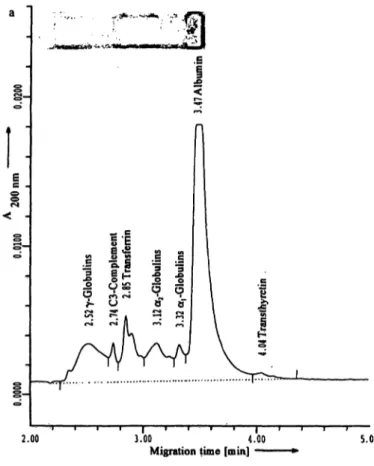
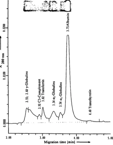
Figure 3a,b presents capillary electropherogram and agarose gel of the normal (fig. 3 a) and abnormal con- trol (fig. 3 b).
Variations in relative peak areas are presented in tables 3 and 4. For the five currently accepted zones, within day variations are below 5% (tab. 3). Overall variation, obtained by measuring relative peak areas on different days, using different capillaries, is higher. The high γ- globulin fraction makes quantification of the C3 less re- liable, but the localization of the peak is precise. In agar- ose gel electrophoresis, C3 cannot be detected as a sepa- rate peak in samples with a high γ-globulin fraction, for instance in patients with polyclonal gammopathy. If in capillary electrophoresis the C3 is counted with the γ- globulin fraction, the CV for the -globulin fraction be- comes 5% instead of 18%, which is comparable to agar- ose gel electrophoresis. Variations for agarose gel elec- trophoresis were obtained by measuring the normal, ab- normal control, Pool serum and Seronorm Protein on an
agarose gel on 30 different days. Although variations in relative peak areas with capillary electrophoresis are a slightly higher than those obtained by conventional agarose gel electrophoresis, capillary electropherograms provided better detail than the densitometric scans of agarose electrophoresis from a resolution perspective.
Table 5 presents capillary electrophoresis and agarose electrophoresis results of artificially prepared mixtures of albumin and γ-globulin preparations dissolved in phosphate buffered saline of half ionic strength to known concentrations. The results are compared with the theoretical values. Data are means of duplicate analysis.
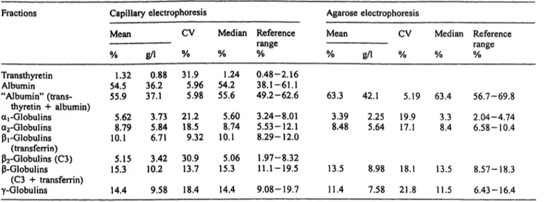
Table 6 presents the inter-individual variations (refer- ence values) obtained by measuring 140 serum samples from normal healthy controls.
Figures 4a—e, present the correlation of capillary elec- trophoresis result (y) with agarose gel electrophoresis (x). A good correlation was obtained for all fractions.
Special examples
Figure 5 shows an example of a serum sample, where Paragon serum protein electrophoresis showed a band on the application slot. This occurs when large mole- cules are kept in the agarose layer and cannot be sepa- rated. In capillary electrophoresis this artifact disappears and the band appears in the γ-globulin fraction.
.ε
<
3.00 4,00 5.00
Migration time [min]
Fig. 3a, b Serum protein analysis by capillary electrophoresis and agarose gel electrophoresis for the normal (fig. 3 a) and abnormal control (fig. 3b). For the abnormal control separate detection of
I-
l
c2.00 3.00 4.00
Migration time (min) — s.oo
C3 is only possible in capillary electrophoresis and not in agarose gel electrophoresis. The figures show the capillary electrophero- gram and the agarose gel.
540 Wijnen and Dieijen-Visser: Serum protein analysis by capillary electrophoresis
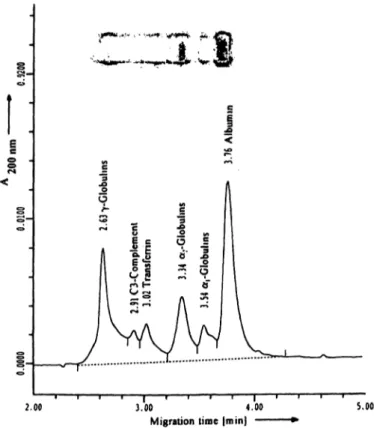
Figure 6 shows that two weak M-components in the γ- globulin fraction can be discriminated even better in the capillary electropherogram, as compared to the visual inspection of the agarose gel.
Figure 7 shows an example of a clear-M-component both in the capillary electropherogram and on the agar- ose gel.
The figures also show that very clear detection, of C3 and transferrin is possible, which allows better discrimi- nation from weak M-components.
Discussion
Klein et al. (8) exhaustively evaluated the variables af- fecting the capillary electrophoresis separation of serum proteins. However, an exact description of the method advocated is not given. Therefore, in table 1 an overview of the methods and the differences between the methods is presented. In the present study, a slight modification of the method described by Chen, Beckman Company (no official reference available) was used. Serum sam- ples were diluted 1 :39 instead of 1 : 19 and capillary
Tab. 3 Reproducibility of relative peak areas by capillary electrophoresis.
Fractions Overall mean relative peak area
Normal control n = 100
Transthyretin Albumin
"Albumin" (trans- thyretin 4- albumin) arGlobulins
a2-Globulins β, -Globulins (transferrin)
2-Globulins (C3) -Globulins
(C3 -f transferrin) γ-Globulins
% 55.141.15 56.29 11.054.29 10.41 5.06 15.47 12.88
g/1 0.68 32.633.3
2.54 6.546.16
3.00 9.16 7.62
Abnormal control n = 100
% 32.340.88 33.21 2.14 6.276.02
7.73 13.75 44.61
g/l 0.70 25.626.3
4.961.69 4.76 6.11 10.88 32.29
Within-day variation cap 1
CV normal control mean of 5 days
% 17.59
1.51 1.31 3.942.35 2.81 4.98 2.67 1.96
Between-day variation cap 1
CV CV abnormal normal control control mean of
5 days
% 17.62
0.860.83
4.82 2.55 2.29 5.10 3.48 1.08
% 21.14
3.453.31
5.47 6.16 6.79 13.08 3.13 7.74
Between-capillary variation
(cap 1 and cap 2) CV CV CV abnormal normal abnormal control control control ή — <n »» — inn « — inn
% 50.57
2.59 1.81 5.98 4.03 4.09 23.97 13.95 4.45
% 25.97
4.16 4.19 13.11 8.427.54
14.75 6.59 8.55
% 63.01
4.28 3.92 7.68 5.124.22
30.70 17.91 6.37 Total protein:
normal control = 59.2 g/l ± 1.8%;
abnormal control = 79.1 g/l ± 1.8%.
Albumin:
normal control = 35.6 g/l ± 1.5%;
abnormal control = 26.0 g/l ± 1.6% (mean over 20 days).
Tab. 4 Comparison of reproducibility of capillary electrophoresis and agarose electrophoresis.
Fraction Capillary electrophoresis
η = 100 Agarose electrophoresis
η = 30
Albumin arGlobulins a2-Globulins -Globulins γ-Globulins
CV%(normal control)
13.14.2 8.46.6 8.6
CV%
(abnormal control)
3.97.7 17.95.1 6.4
CV%(normal control) 2.34.6 3.57.4 7.3
CV%(abnormal control) 2.86.7 4.14.7 1.9
CV%
(pooled serum) 6.35.2 4.65.2 6.9
CV%
(seronorm protein) 2.94.8 4.33.1 7.1
Tab. 5 Recovery of artificially prepared protein mixtures.
Albumin : Globulin mixture
theoretical values Agarose electrophoresis
Albumin (%) Globulins (%) Albumin (%) Globulins (%)
Capillary electrophoresis
Albumin (%) Globulins (%) 8050
20
2050 80
78.454.1 25.7
21.5 45.974.3
78.347.1 19.2
21.7 52.980.8
Tab. 6 Reference values for capillary electrophoresis and agarose gel electrophoresis.
Fractions Capillary electrophoresis Agarose electrophoresis
Mean CV Median Reference Mean CV Median Reference
Transthyretin Albumin
% g/l % % % % 1.32 0.88 31.9 1.24 0.48-2.16
54.5 36.2 5.96 54.2 38.1-61.1
"Albumin" (trans- 55.9 37.1 5.98 55.6 49.2-62.6 63
range gA % % %
3 42.1 5.19 63.4 56.7-69.8 thyretin + albumin)
aj -Globulins a2-Globulins β,-Globulins (transferrin)
5.62 3.73 21.2 5.60 3.24-8.01 3 39 2.25 19.9 3.3 2.04-4.74 8.79 5.84 18.5 8.74 5.53-12.1 8.48 5.64 17.1 8.4 6.58-10.4 10.1 6.71 9.32 10.1 8.29-12.0
p2-Globulins (C3) 5.15 3.42 30.9 5.06 1.97-8.32
-Globulins 15.3 10.2 13.7 15.3 11.1-19.5 13.5 8.98 18.1 13.5 8.57-18.3
(C3 + transferrin) γ-Globulins
Total protein:
14.4 9.58 18.4 14.4 9.08-19.7 11 Albumin:
4 7.58 21.8 11.5 6.43-16.4
66.5 ± 3.5 g/1 (mean ± SD), range 56.5-75.6 g/1. 39.7 ± 2.7 g/1 (mean ± SD), range 32.4-45.3 g/1.
a 90
80 i? 70
07 **
50 - .£ 40 -
1-
< 2 0 - 10 - 0 -0 15
.x HE
·#* 07 1C
•s&ff y.
£&& ~ C
^^* ο
"
J^
• ^r
**ts *
^r*/i *
• ix^^
JC!
0 10 20 30 40 50 60 70 80 90 Q 5 1Q 15
C 3UCTJ
I 40-
07 30 -
¥ I
ο2 0e> 101
ο J
Albumin (AGE) [%]
X
^ 5007 40| 20CDcs 10Qg 300 10 20 30 40 50 a2 -Globulins (AGE) [%]
I
40"X^
1 Μ · ;&*
1 si$
*-x
0 10 20 30 40α,-Globulins (AGE) [%]
• yr.^
~/^^
.S
*^f*
\1$: ·,
^'"
0 10 20 30 40 50 60 -Globulins (AGE) [%]
^
50
7-Globulins (AGE) [%]
Fig. 4a-^e Correlation between capillary electrophoresis data and fractions, albumin, αϊ-, ct2-, β- and γ-globulin (n = 61). Fractions agarose gel electrophoresis data for the five currently accepted are indicated as fractions of the total protein concentration.
Regression lines: a. y = 0.96x - 3.34, r = 0.95 c. y = l.lOx - 0.02, r = 0.99 e. y = 1.07 + 0.84, r = 0.97 b. y = 1.21x + 0.62, r = 0.90 d. y = 0.98x + 0.76, r = 0.93
542 Wijnen and Dicijen-Visser: Serum protein analysis by capillary electrophoresis Albumin
y-Globulins Albumin
_0<
-J..V
\ T r a n s t h y retinMigration time
Fig. 5 Capillary electrophoresis (a) and agarose electrophoresis (b) of a sample containing large proteins that remain at the applica- tion slot after agarose gel electrophoresis separation (both
diameter was 50 μιη. At 10 kV we obtained a separation time of five instead of six minutes. We obtained slightly better results with this procedure. The most important factors affecting the final electropherogram shapes are discussed below.
Capillary length and diameter
Shorter separation times can be obtained with small and thin capillaries. Higher detection limits can be obtained
2.00 3.00 4.00
Migration time |min) — 5.00
Fig. 6 Identification of gammopathies is possible both by capil- lary electrophoresis and by agarose gel electrophoresis. The figure shows two weak M-components that can be detected with both techniques, capillary electropherogram and agarose gel.
Electrophoretic mobility
electropherogram and gel are shown) (b) and not after separation with capillary electrophoresis (a).
with larger bore capillaries due to an increase in the absorbing path length. Our analysis time could be short- ened by using a 20 μιη thick capillary, but this can give more technical problems like obstruction of the capil- lary. A 50 μιη thick capillary gave reliable results within 10 minutes and therefore we did not change to a smaller capillary diameter.
Voltage effects
The applied field seems to affect only the migration times up to a point of adverse Joule heating. High volt- age gives short separation times, but has repercussions for the quality of the separation (7).
Sample injection
Sample injection can be performed electrokinetically or hydrodynamically. For charged particles like proteins, hydrodynamic injection is preferred to obtain optimal peak resolution (8, 10).
Capillary temperature control
Adequate temperature control is of utmost importance in quantitative analysis of serum proteins by capillary electrophoresis. This is primarily due to thermal change of the pH of the buffers, small changes in pH can have noticeable effect on pattern shape (8). In some of the papers published until now, temperature control was inadequate (1, 6, 13). Liquid cooling of the capillary in "the cartridge gives excellent cooling results, which is of importance for reproducibility of migration times.
Detection
Detection of proteins by absorbance ranges from 190 to 280 nm. If possible, UV detection should be performed
2.00 5.00 Migration time |min)
Fig. 7 Capillary electropherogram and agarose gel of a serum sample with a clear M-component.
at low wavelengths where sensitivity is much higher (10).
Buffer ionic strength effects and sample dilution
A borate buffer is used because of its transparency, especially in the far-ultraviolet (UV) spectral regions.
An increase of the ionic strength of the separation buffer gives a decrease of the mobility. The mobility of the proteins is retarded by the surrounding buffer ions. With respect to analysis time and resolution, we obtained the best results with a buffer with an ionic strength of 100 mmol/1. At 75 mmol/1, analysis time was within four minutes, but peak resolution was in- sufficient. At 150 mmol/1 analysis time was six min- utes, but differentiation between γ-globulins and C3 was no longer possible.
Serum samples were diluted in phosphate buffered saline with half the ionic strength, resulting in a lower conductivity and proportionally higher field strength in the sample plug as compared to the separation buffer. A higher field strength in the diluted sample plug causes sample stacking (sample concentration) at the border between the sample and separation buffers.
However, dilution in distilled water resulted in a higher signal to noise ratio. Therefore, phosphate buf- fered saline with half the ionic strength was used.
Moreover, the 1 : 19 dilution in phosphate buffered saline gave a higher background, baseline, compared to the 1 :39 dilution.
Effect of pH
A higher pH of the buffer gives an increase in the electro-osmotic flow. At high pH (approximately 10) the pH of the protein is typically less than that of the buffer.
Therefore, both the proteins and the capillary wall are negatively charged and the adsorption process is mini- mized as a result of a charge repulsion effect. However, at too high pH a relatively high electro-osmotic flow is generated, which has negative consequences for the peak resolution.
Wall-solute interactions
Even though the pH of the running buffer was selected to induce a strong negative charge on all proteins, re- gions of the protein with net positive charges remained.
Experiments involving successive runs without the NaOH rinse showed progressive lengthening of retention times and a loss of resolution. After the NaOH rinse, the capillary returned to normal (8). In our case a pH of 10.2 gave optimal results.
Determination of electro-osmotic flow
A higher pH of the running buffer results in an increase of the electro-osmotic flow and, as a consequence, of migration times. To ensure that the system is properly controlled it is necessary to measure the electro-osmotic flow. In contrast to Chen (2), we did not add a solution of dimethylformamide (0.1 ml/I) to the sample diluent as a neutral marker of electroendoosmotic flow, because the dime thy Iformamide-peak appeared in the γ-globulin peak. We diluted the serum samples in phosphate buf- fered saline of half ionic strength. Therefore, the electro- osmotic flow could be determined from the negative peak occurring at the transition of the sample to run- ning buffer.
Migration time
Migration time of a protein is the result of its electropho- retic mobility plus the buffer's electro-osmotic flow.
Changes in viscosity, temperature, pH, ion depletion etc.
can influence the mobility and thus the migration time.
Changes in mobility can lead to false identification of peaks and improper quantification. In the abnormal con- trol, a higher variation is observed, especially for the - globulin fraction (tabs. 3 and 4).
Variation in peak areas
Reif et al. (9, 10) reported standard deviations of peak areas of less than 6% (migration times less than 1%).
No further information is given on how this value was obtained. Stob et al. (7) reported variations for the peak areas between 1.2 and 6.2% for the different fractions. Variation was obtained by measuring one sample six times. Variation in migration times was
544 Wijnen and Dicijen-Visser: Scrum protein analysis by capillary electrophoresis
less than 0.4% for all fractions. In the present study variation in peak areas was obtained from at least 200 measurements and gave slightly larger variation in peak areas for capillary electrophoresis than for agar- ose gel electrophoresis. The high -globulin fraction makes quantification of the C3 less reliable, but the localization of the peak is precise (fig. 3). In agarose gel electrophoresis, C3 cannot be detected as a sepa- rate peak in samples with a high -globulin fraction, for instance in patients with polyclonal gammopathy.
If in capillary electrophoresis the C3 is counted with the -globulin fraction, the CV for the ß-globulin frac- tion becomes 5% instead of 18%, which is comparable to agarose gel electrophoresis.
Colour correction
Quantification of stained agarose gels is subject to a number of factors that can influence stain binding. In contrast, the capillary electrophoresis profiles represent the direct measurement of protein via the peptide bonds, a more accurate method for quantifying their relative concentrations.
Peak resolution
From the standpoint of resolution, the smaller the capil- lary internal diameter and the shorter the separation time, the better the separation. However, a smaller dia- meter results in increased heat production. Therefore, a sufficient cooling system is required.
Capillary electrophoresis is generally characterized by a higher peak resolution as compared to agarose electro- phoresis. In general, agarose electrophoresis is not suit- able for identification of a separate transthyretin (pre- albumin) fraction. However, in capillary electrophoresis it is possible to quantify transthyretin because of its clear resolution. Transthyretin occurs in a concentration range that allows quantification (12). However, we found an inter-individual variation of 31%, making capillary elec- trophoresis not suitable for reliable quantitative pre-albu- min determinations.
In addition to transthyretin, a clearer separation is also possible for the ß-globulin fraction, giving separate C3 and transferrin peaks. Precise localization of C3 and transferrin in capillary electrophoresis results in a more accurate detection of the ß-globulin fraction.
When C3 appears in the -globulin fraction it is not detected as a separate peak in the agarose electro- pherogram, whereas it is in capillary electrophoresis.
Generally the ß-globulin peak is overestimated in agarose gel electrophoresis because it overlaps with the -globulin fraction. Chen et al. (2) showed a sepa- rate ß-lipoprotein peak and differentiation between a2- macroglobulin and haptoglobin. In later studies a com- parable resolution was not shown.
Agarose electrophoresis sometimes shows the phenom- enon that large proteins remain at the application slot.
In capillary electrophoresis this problem is fully solved.
In capillary electrophoresis reliable quantification of transferrin is possible, inter-individual variation about 10%, this is not the case for C3, where an inter-indivi- dual variation of 41% is found.
Correlation with agarose gel electrophoresis We found a good correlation of capillary electrophoresis with agarose gel electrophoresis and even better com- pared to the correlation found by Kim et al. (6).
We conclude that capillary electrophoresis is a very useful technique, suitable for reliable quantification and separation of serum proteins. Total variation for capillary electrophoresis seems slightly higher com- pared to agarose electrophoresis. For capillary electro- phoresis of the ß-globulin fraction, a clear separation of the complement C3 and transferrin fraction is pos- sible. The same holds for the separation of transthyre- tin. Generally, a better peak resolution is obtained with capillary electrophoresis. Some artifacts of gel electrophoresis are eliminated when using capillary electrophoresis.
References
1. Chen FA, Liu CM, Hsieh YZ, Sternberg JC. Capillary electro- phoresis - a new clinical tool [overview]. Clin Chem 1991;
37:14-9.
2. Chen FA. Rapid protein analysis by capillary electrophoresis.
J Chromatogr 1991; 559:445-53.
3. Chen FA. High-resolution protein analysis by automated capil- lary electrophoresis. Clin Chem 1992; 38:1651-2.
4. Shihabi ZK. Clinical application of capillary electrophoresis.
Ann Clin Lab Sei 1992; 22:398-405.
5. Hiraoka A, Miura I, Hattori M, Tominaga I, Machida S. Capil- lary-zone electrophoretic analyses of the proteins and amino acid components in cerebrospinal fluid of central, nervous sys- tem diseases. Biol Pharm Bull 1993; 16:949-52.
6. Kim JW, Park JH, Park JW, Doh HJ, Heo GS, Lee KJ. Quanti- tative analysis of serum proteins separated by capillary electro- phoresis. Clin Chem 1993; 39:689-92.
7. Stob S, Lauer HH, Swart A. Capillaire zone elektroforese in de klinische chemie. Tijdschr NVKC 1993; 18:299-305.
8. Klein G, Jolliff K. Capillary electrophoresis for the routine clinical laboratory. In: Landers JP, editor. Handbook of capil- lary electrophoresis. Boca Raton, FL: CRC Press, 1993:419- 58.
9. Reif O W, Lausch R, Freitag R. Application of CE to the quan- titative and qualitative analysis of serum proteins. International Laboratory 1994; 10:11-14.
10. Reif OW, Lausch R, Freitag R. High performance capillary 14. Wang HP, Liu CM. Separation and identification of human electrophoresis of human serum and plasma proteins. Adv serum proteins with capillary electrophoresis. Beckman Instru- Chromatogr 1994; 34:1-56. ments Inc. Brea, California 92621. (Internal note).
11. Chen FA, Sternberg JC. Characterization of proteins by capil-
1^ electrophoresis in fused-silica columns. Electrophoresis ^/w ^^ ^^ ^ jm 17bf ^TJ ID: 1.5 Z . I . .
12. Landers JR Clinical capillary electrophoresis. Clin Chem Corresponding author: Prof. Dr. M. P. van Dieijen-Visser, 1995; 41:495-509. Department of Clinical Chemistry, Academic Hospital Maastricht, 13. Jenkins MA, Guerin MD. Quantification of serum proteins P.O. Box 5800, NL-6202 AZ Maastricht, The Netherlands
using capillary electrophoresis. Ann Clin Biochem 1995;
32:493-7.


