electrophoresis – development of a
two-dimensional separation and a dual detection system
Dissertation
zur Erlangung des Doktorgrades der Naturwissenschaften (Dr. rer. nat.) der Fakultät für Chemie und Pharmazie
der Universität Regensburg
vorgelegt von Andrea Beutner
aus Etzenricht
im Jahr 2018
Promotionsgesuch eingereicht am: 08.06.2018 Kolloquiumstermin: 24.07.2018
Prüfungsausschuss
Vorsitzender: Prof. Dr. Alkwin Slenczka
Erstgutachter: Prof. Dr. Frank-Michael Matysik
Zweitgutachter: PD Dr. Hans-Heiner Gorris
Drittprüfer: Prof. Dr. Werner Kunz
An dieser Stelle möchte ich mich bei allen bedanken, die mich auf dem Wege zur Promotion unterstützt und begleitet haben.
1.3.2. Two-dimensional CE ... 15
1.4. Selectivity enhancement by dual detection concepts ... 24
1.4.1. C
4D in dual detection approaches ... 25
1.4.2. MS in dual detection approaches ... 30
1.4.3. Amperometry in further dual detection approaches ... 34
1.4.4. Dual optical detection approaches ... 36
1.5. Conclusion ... 38
1.6. References ... 39
2. Scope of this thesis ... 48
3. Comprehensive hyphenation of capillary ion chromatography and capillary electrophoresis ... 49
3.1. Two-dimensional separation of ionic species by hyphenation of capillary ion chromatography × capillary electrophoresis - mass spectrometry ... 49
3.1.1. Abstract ... 49
3.1.2. Introduction ... 51
3.1.3. Experimental ... 52
3.1.4. Results and discussion ... 53
3.1.5. Conclusion ... 58
3.1.6. Further experimental details ... 58
3.1.7. References ... 59
3.2. Methodical studies of the simultaneous determination of anions and cations by ICxCE-MS using arsenic species as model analytes ... 62
3.2.1. Abstract ... 62
3.2.2. Introduction ... 64
3.2.3. Materials and methods ... 66
3.2.4. Results and discussion ... 72
3.2.5. Conclusion ... 79
3.2.6. References ... 80
4. Dual detection combining capacitively coupled contactless conductivity detection and mass spectrometry ... 84
4.1. Combining C
4D and MS as a dual detection approach for capillary electrophoresis ... 84
4.1.1. Abstract ... 84
4.1.2. Introduction ... 86
4.1.3. Materials and methods ... 87
4.1.4. Results and discussion ... 90
4.1.5. Concluding Remarks ... 93
4.1.6. References ... 94
4.2. Dual detection for non-aqueous capillary electrophoresis combining contactless conductivity detection and mass spectrometry ... 97
4.2.1. Abstract ... 97
4.2.2. Introduction ... 99
4.2.3. Experimental ... 101
4.2.4. Results and discussion ... 104
4.2.5. Concluding remarks ... 111
4.2.6. References ... 112
5. Surfactant-free microemulsion electrokinetic chromatography (SF-MEEKC) with UV and MS detection - a novel approach for the separation and ESI-MS detection of neutral compounds ... 115
5.1. Abstract ... 115
5.2. Introduction ... 117
5.3. Experimental ... 119
5.3.1. Chemicals and materials ... 119
5.3.2. Experiments ... 120
5.4. Results and discussion ... 123
5.4.1. Composition and structural investigations of the surfactant-free microemulsion ... 123
5.4.2. SF-MEEKC-UV/VIS ... 126
5.4.3. SF-MEEKC-ESI-TOF-MS ... 129
5.5. Conclusion ... 131
5.6. References ... 131
6. Summary ... 134
1. Selectivity enhancement in capillary electrophoresis by means of dual
detection or two-dimensional separation
1. Selectivity enhanceme nt in CE
1.1. Abstract
For the identification and quantification of analytes in complex samples highly selective
analytical strategies are required. The selectivity of single separation techniques such as
gas chromatography (GC), liquid chromatography (LC), or capillary electrophoresis (CE)
with common detection principles can be enhanced hyphenating orthogonal separation
techniques but also complementary detection systems. In this review, two-dimensional
systems containing CE in at least one dimension are described, namely LC-CE or 2D CE
systems. Particular attention is paid to the selectivity enhancement due to the
orthogonality of the different separation mechanisms. As an alternative concept, dual
detection approaches are reviewed using the common detectors of CE such as UV/VIS,
laser-induced fluorescence, capacitively coupled contactless conductivity (C
4D),
electrochemical detection, and mass spectrometry. Special emphasis is given to dual
Author contributions
AB wrote the manuscript and did the literature research. Both authors revised the
manuscript. FMM is the corresponding author.
1.2. Introduction
Separation and determination of analytes in complex samples is a challenging task in analytical chemistry. Especially biological and environmental samples consist of a complex matrix and a variety of different analytes rendering their analytical determination difficult. Thus, high selectivity is needed to identify and quantify all components. In common analytical strategies, such samples are separated by a chromatographic or an electrophoretic technique before detection. However, using single separation techniques such as gas chromatography (GC), liquid chromatography (LC), or capillary electrophoresis (CE) the peak capacity is often too low resulting in coelution or comigration. Therefore, multidimensional separation techniques are used to enhance the peak capacity and consequently also the selectivity of the overall analytical process. Importantly, the hyphenated techniques should have a high degree of orthogonality, which means their separation mechanisms have to be as different as possible. Thus, coupling LC with CE is promising exploiting different chromatographic stationary phase materials and CE modes. As an example, reversed phase liquid chromatography (RPLC), size exclusion chromatography (SEC), and ion chromatography (IC) can be hyphenated to capillary zone electrophoresis (CZE), isoelectric focusing (IEF), micellar electrokinetic chromatography (MEKC), and isotachophoresis (ITP) offering a variety of combinations of separation mechanisms. Additionally, two-dimensional CE systems are described coupling different CE modes.
Two-dimensional separation systems can be classified as online or offline, heart-cutting
or comprehensive techniques. Using online techniques, the transfer from one dimension
to the other is performed automatically using an interfacing strategy in contrast to
offline techniques where fractions are collected. As common terminology from GCxGC
systems, a separation is only called “comprehensive” if the entire sample separated in
the first dimension is transferred and separated in the second dimension [1]. The term
was first introduced by Bushey and Jorgenson [2] to distinguish their two-dimensional
LCxLC system from the former heart-cutting approaches as the whole sample was
determined in both dimensions instead of few particular fractions. They also developed
an HPLCxCE system and called it “comprehensive” even if they admitted to undersample
the LC peaks generating just one electropherogram per minute [3]. Nowadays, in most
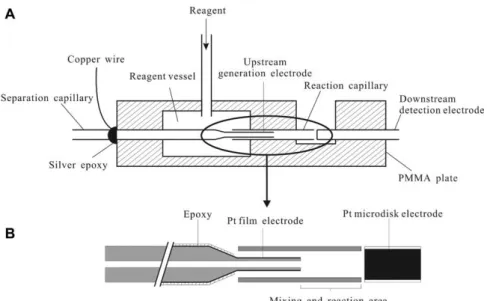
coupling complementary detectors. In CE, commonly used detectors are UV/VIS, mass spectrometry (MS), electrochemical detection, capacitively coupled contactless conductivity detection (C
4D), and laser-induced fluorescence (LIF). These detectors have to be chosen considering the structural and chemical characteristics of different classes of analytes. Using two or more of these detectors in one run means combining their power in terms of selectivity and sensitivity and compensating their weaknesses. The detectors can be connected in series or using the same detection space.
Several reviews have been published so far dealing with LC-CE and 2D CE. However, focus was put on interfacing and transfer strategies [8-12]. In the field of dual detection, two reviews have been published concentrating mainly on detection systems containing at least one electrochemical detector whereas emphasis was put on amperometric detection (AD) [13, 14]. In this work, both approaches towards selectivity enhancement, hyphenating either separation systems or complementary detectors, are reviewed.
Electrophoresis in microfluidic chips is not covered. Two-dimensional LC-CE and 2D CE
systems are described with focus on the selectivity enhancement due to the
orthogonality of the different separation mechanisms. Especially online comprehensive
approaches are described, but also some online heart-cutting techniques are
mentioned. Furthermore, dual detection systems are reviewed using the common
detectors of CE such as UV/VIS, LIF, C
4D, electrochemical detection, and MS with
particular interest on dual detection approaches containing C
4D and MS.
1.3. Selectivity enhancement by two-dimensional separation
1.3.1. Hyphenation of LC and CE
Selectivity can be enhanced by the hyphenation of two separation techniques. Favorable are separation mechanisms with high orthogonality. Therefore, combination of LC and CE is considered to be very effective. Different stationary phases of the chromatographic system and different CE modes can be chosen changing the selectivity of the system.
Possible combinations of methods are summarized in Table 1 considering their way of enhancing the selectivity.
Table 1: Hyphenation of LC and CE. Overview of possible methods and their applications considering the way of selectivity enhancement.
Method Detector
Strategy of selectivity enhancement
Sample Interface Ref
RPLCxCZE
LIF Peak capacity
Peptide standards and fluorescently labeled peptides from a tryptic digest of ovalbumin
Valve-based [3]
LIF Peak capacity
Tryptic digest of horse-heart cytochrome c
Valve-based/
optically gated [15,
16]
ESI-MS Peak capacity
Separation of a glycosylated peptide mixture
Transverse flow gated [17]
ESI-FTICR-
MS Peak capacity
Tryptic digested bovine serum albumin and human cerebrospinal fluid
Transverse flow gated [18]
LIF Immunoassay
Determination of glucagon secretion from single islets of Lagerhans and their cross-reactive species
flow gated [19]
LIF Peak capacity Determination of
neuropeptide Y flow gated [20]
LIF
Competitive affinity probe
assay
Selective
determination of Fyn Src homology 2 domain
flow gated [21]
SEC-
RPLCxCZE LIF Peak capacity (factor 5)
Separation of ovalbumin digest
Valve-based/
Transverse flow gated
[27]
ICxCZE
MS Peak capacity
Separation of nucleotides and cyclic nucleotides
Modulation
interface [4]
C4D Peak capacity
Separation of inorganic anions and haloacetic acids
Valve-based [6]
IEFxRPLC
MS Identification of proteins
Drosophila proteomics, identification of yeast soluble proteins
Valve-based, hydrodynamic
[28, 29]
MS Peak capacity
Selective detection of the test protein bovine carbonic anhydrase II in a complex protein mixture
Membrane- based
[30- 32]
MS
Identification of proteins, online
digestion
Identification of Escherichia coli proteins
Membrane-
based [33]
GFC1xIEF
Column absorption
imaging
Peak capacity
Separation of albumin and myoglobin
Membrane-
based [34]
RPLCxMEKC UV Peak capacity
Separation of a complex mixture of neutral
components in medicine
Transverse flow gated [35]
1GFC
–
gel filtration chromatographyHyphenation of LC and CZE
RPLCxCZE
A common mode in CE is CZE based on electrophoretic mobility as separation mechanism. It is most often coupled to RPLC separations based on hydrophobicity using different interfacing strategies. RPLC and CZE were first coupled comprehensively by Bushey and Jorgenson [3] in 1990. They used a computer-controlled six-port valve to transfer the effluent of the LC to the CE system by switching the valve once every minute (see Figure 1).
Figure 1: Two configurations of a six-port, computer-controlled valve transferring the effluent of the RPLC (C1) to the CZE. The effluent was filled in a sample loop (L) and then flushed by a second pump (P2) over a grounded end of the CE separation capillary (CZE) and was injected electrokinetically. PW is paper wick. W is waste [3]. Figure reprinted with permission from M.M.
Bushey, J.W. Jorgenson, Automated instrumentation for comprehensive two-dimensional high- performance liquid chromatography/capillary zone electrophoresis, Anal. Chem., 62 (1990) 978- 984. Copyright 1990 American Chemical Society.
The effluent, which was filled in a sample loop, was then flushed by a second pump over a grounded end of the CE separation capillary and was injected electrokinetically once per minute. The overflow went to waste. The system was used for the separation of peptide standards and fluorescently labeled peptides from a tryptic digest of ovalbumin.
Even if a low LC flow rate (10 µL/min) was chosen to slow down the elution of the
analytes of the first dimension, they still undersampled the LC effluent for the separation
in the second dimension as the sampling frequency was too low. To enhance the
sampling rate, the approach was further developed by Larmann et al. [15] replacing the
six-port valve by an 8-port valve. Instead of one sample loop, two sample loops were
used contra-rotating. Thus, while one sample loop was filled by the LC effluent, the
content of the second sample loop was transferred to the CZE system. Again every
overflow went to waste. The setup was used for the separation of tryptic digest of horse-
that entered the capillary. For injection, the laser beam was blocked for a short period of time. The fluorescent analytes were then separated and detected by LIF using a second laser beam (5% of the total laser power). For hyphenation to LC, the LC effluent was guided into a grounded tee connector, which split the sample. The other sides of the tee piece were connected to the waste and to a CE capillary with optical-gating, respectively [15, 16]. The use of this optical gating approach enabled fast separations due to the use of short capillaries. Separations in less than 10 minutes with CE migration times of 2.5 seconds were achieved for a sample containing fluorescein isothiocyanate- tagged tryptic digests of horse heart cytochrome c.
A major drawback of optical-gating is the limited applicability to non-fluorescent
analytes. Flow gated interfaces constitute a universal alternative enabling also the
coupling to other detection concepts such as MS. The concept of transverse flow gated
interfacing is depicted in Figure 2 [17]. The effluent of the LC was guided into the
interface by a transfer capillary, which was axially aligned to the CE separation capillary
in a distance of about 70 µm. The CZE buffer was flushed vertically through the interface
hindering an injection of the LC effluent into the second dimension. To inject sample
into the CE capillary the flow was interrupted and an injection voltage was applied to
the inlet side. Using this interface, a glycosylated peptide mixture was separated in less
than 15 min by LC×CE-electrospray ionization (ESI)-MS injecting into the CE with a
sampling rate of one injection every 15 seconds [17].
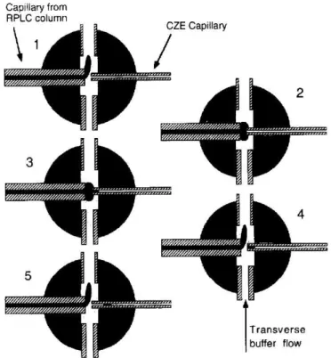
Figure 2: Scheme of the injection principle of a flow gated interface: (1) CZE mode; flush flow on and high voltage on. (2) Preinjection mode; flush flow off and CZE voltage off. Sample diffuses across the gap. (3) Injection mode; flush flow off and CZE injection voltage on. Sample is injected electrokinetically. (4) Postinjection mode; flush flow on and CZE voltage off. Not-injected sample is flushed away. (5) CZE mode; flush flow on and CZE voltage on. CE separation takes place [17].
Figure reprinted with permission from K.C. Lewis, G.J. Opiteck, J.W. Jorgenson, D.M. Sheeley, Comprehensive on-line RPLC-CZE-MS of peptides, J. Am. Soc. Mass Spectrom., 8 (1997) 495-500.
Copyright 1997 Springer Nature.
A variation of this interface was developed by Bergström et al. [18]. In this approach the LC was coupled to the CE at a right angle. The interface was fabricated in a PDMS plate having two channels crossed at slightly different height levels, which minimized the contact area in between. The CE chamber was pressurized avoiding a leaking of the LC effluent into the CE separation channel. Injection was performed by the reduction of the pressure. High voltage for CE was applied in the pressurized chamber outside the interface minimizing the risk of bubble formation. The system was applied to samples with complex biological matrices such as tryptic digested bovine serum albumin and human cerebrospinal fluid. Detection was performed by electrospray ionization Fourier transform ion cyclotron resonance MS.
Highly selective quantitative determinations of biological macromolecules were also performed implementing an immunoassay in the two-dimensional RPLCxCZE system.
After separation with RPLC the effluent was mixed on-column with the antibody reagent
applied to a competitive immunoassay for neuropeptide Y utilizing polyclonal antisera as the immunoreagent and fluorescein-labeled neuropeptide Y as tracer [20]. A similar setup was used for a competitive affinity probe assay screening mixtures for compounds inhibiting protein-ligand interactions [21]. Mixtures containing potential binding inhibitors for a specific protein-ligand interaction were separated by RPLC. The effluent was mixed online with the protein and its selective fluorescently-labeled binding partner as affinity probe. Inhibiting compounds led to a decrease in complex formation, which was detected by LIF. As model system the Fyn Src homology 2 domain and its selective phosphopeptide binding partner was used generating electropherograms in 6 seconds intervals [21].
RPLC and CZE were further coupled using an interface called “hydrodynamic interface”
[22]. The CE separation capillary was fixed on a slide bar, the inlet end immersed in a buffer vial. The LC effluent accumulated as a droplet at the LC outlet fixed in 8 cm height distance. For injection the CE capillary was immersed into the droplet moving the slide bar up leading to hydrodynamic injection due to gravity flow. Tryptic digests of proteins in D
20liver cancer tissue were analyzed by RPLCxCE - matrix-assisted laser desorption/ionization - time-of-flight - time-of-flight - mass spectrometry (MALDI-TOF- TOF-MS) identifying over 200 proteins in 1.5 hours. Transfer from the separation system to the detector was performed using a CE-MALDI interface directly depositing the effluent on a MALDI target at a 3 seconds time-interval.
Also droplet-based interfaces were described. In a microfluidic device, an oil as
immiscible carrier fluid was added online to the LC effluent leading to spontaneous
formation of nL droplets segmenting the separated analytes avoiding diffusion and
remixing of already separated peaks. These droplets were then injected into the CE after
oil filtering and droplet merging [23, 24]. The interface was applied analyzing human urine [24].
SECxCZE
Besides RPLC, also SEC was used for comprehensive hyphenation separating analytes based on their size. Interfacing was again performed using valve-based and flow gated strategies. In first approaches actuated valves with 300 to 500 nL collection loops were used [15, 25]. Orthogonality of the two techniques was shown as neither SEC nor CZE could resolve a standard protein mixture in a single dimension [15]. Lemmo and Jorgenson [26] also coupled SEC to CZE comparing a valve-based and a transverse flow- gating interface. As there was no fixed sampling volume for the flow gated interface, lower flow rates could be used improving SEC resolution. The overall sensitivity could be increased 8-fold. To enhance the selectivity further, even a 3D coupling of SEC with an RPLCxCE setup was performed using a valve-based interface [27]. Thus, orthogonal separation was performed by size, hydrophobicity, and electrophoretic mobility. The addition of the third dimension led to an increase of the peak capacity by a factor 5 compared to the 2D setup. However, high sample concentrations had to be used to overcome dilution effects caused by the interface.
ICxCZE
Hyphenating IC and CE, the two most important instrumental techniques in ion analysis are combined. While the separation mechanism of IC is based on the affinity to an ion exchange column, the mechanism of CZE is dependent on electrophoretic mobility.
Thus, their coupling is promising in terms of enhancement of peak capacity and
selectivity. The first heart-cut combination of IC and CZE was reported by Kar and
Dasgupta [37] in 1996. Almost two decades later, the first comprehensive approaches
were described [4, 6]. Two major developments were essential for this coupling. The
introduction of IC in capillary scale enabled the use of low flow rates down to
2-10 µL/min enhancing the compatibility to CE. The effluent of the IC usually contains
high salt concentrations, which disturbs the electrophoretic separation. This drawback
was overcome by the development of miniaturized ion suppressors enhancing the
detection sensitivity due to the generation of pure water as effluent. An advantage of
this (in an ideal case) matrix-free effluent is the decrease of any interferences in CE and
the effect of sample stacking due to a lower conductivity compared to the background
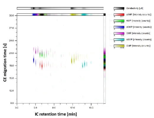
serving as CE inlet vial by a transfer capillary, which was axially aligned in close distance (<50 µm) to the CE separation capillary. The expelled effluent formed a sample cloud, which was then hydrodynamically injected into the CE capillary due to gravity flow and suction pressure of the ESI source of the MS detector. The transfer capillary was moved up and down periodically for sequential injection. Fast CE separations enabled a high sampling rate of the first dimension effluent. A model system containing nucleotides and cyclic nucleotides was chosen to show complementary separation.
Hyphenation of IEF and LC
IEFxRPLC
IEF was coupled to RPLC. The separation mechanism of IEF is based on the differences
in isoelectric points. IEF is also used as first step in 2D polyacrylamide gel
electrophoresis, which is widely used for protein separation, however, with limited
throughput and sensitivity. Thus, the combination of IEF and RPLC as two orthogonal
techniques is a promising online approach for proteomics overcoming these drawbacks
and facilitating the hyphenation to MS. Chen et al. [28, 29] described a valve-based
interface for the online integration of capillary IEF with capillary RPLC. In a first work,
after completion of the IEF, the analytes were sequentially and hydrodynamically
transferred into the RPLC until the entire sample was separated in both dimensions
using a valve with internal injection loop. The system was applied for the analysis of
Drosophila proteomics during steroid-induced programmed cell death [28]. The workwas further expanded including a stacking step of the segments before the injection into
the RPLC. Several trap columns were used to minimize peptide dilution and mixing
during the transfer between the two dimensions and to avoid interferences of the
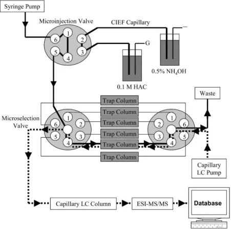
ampholyte (Figure 3) [29]. With this system, a larger number of yeast soluble proteins could be identified than with other methods already presented in literature with a protein loading of only 9.6 µg. Several further applications of the system to proteome analysis were published [39, 40].
Figure 3: Scheme of on-line integration of IEF with RPLC as a concentrating and multidimensional separation platform. Solid and dashed lines represent the flow paths for the loading of IEF fractions and the injection of fractions into a RPLC column, respectively [29]. Figure reprinted with permission from J. Chen, B.M. Balgley, D.L. DeVoe, C.S. Lee, Capillary isoelectric focusing- based multidimensional concentration/separation platform for proteome analysis, Anal. Chem., 75 (2003) 3145-3152. Copyright 2003 American Chemical Society.
A similar approach was described by Zhou et al. [30] using a microdialysis membrane
separating the IEF cathodic cell (grounded) from the separation capillary. Contrary to
the catholyte, the proteins were not able to traverse the membrane. After focusing, the
proteins were pushed hydrodynamically to a microselection valve collecting the
fractions. This setup enabled the maintenance of a linear pH gradient in the separation
capillary during the entire two-dimensional analysis as no voltage was applied on the
microselection valve. The fractions collected in the valve were further trapped on a
column, washed to remove the ampholyte, and injected into the RPLC-MS system using
a second valve [30]. In further works, storage loops were implemented between the two
two hollow fiber membrane interfaces. The first one was used to supply catholyte and electric contact similar to the approaches already described in this chapter. The second one was implemented right before the microreactor for the adjustment of buffer improving compatibility between protein separation and digestion. The setup enabled the identification of 101 proteins extracted from Escherichia coli.
Gel-filtration chromatography (GFC)xIEF
Protein analysis was also done with gel-filtration chromatography (GFC) coupled to IEF bringing the 2D polyacrylamide gel electrophoresis into a capillary format [34]. Contrary to the IEF-RPLC approaches, IEF was used as second dimension. Interfacing was performed using a microdialysis hollow fiber membrane used for desalting and carrier ampholyte mixing. After separation by GFC the effluent was split by a tee piece and injected via an 8-port injection valve into the IEF capillary passing the microdialysis interface. Detection was performed by column absorption imaging. Feasibility of the approach was shown by separation of a model mixture containing albumin and myoglobin.
Hyphenation of other LC and CE principles
RPLCxMEKC (neutral analytes)
Another CE mode is called micellar electrokinetic chromatography (MEKC). The
separation mechanism is based on the partition between the hydrophobic and the
hydrophilic compartments of a micelle. Thus, neutral analytes can be separated based
on differences in their hydrophobicity. RPLC was hyphenated to MEKC for
comprehensive two-dimensional separation of neutral species [35]. Fast MEKC
separations in the second dimension were achieved by speeding up the electroosmotic
flow (EOF). A transverse flow gating interface was used for connection of the two dimensions. Separation of a complex mixture of neutral components in traditional Chinese medicines was performed.
GFC - sub-micellar SDS array CE
Skinner et al. [41] coupled GFC with sub-micellar SDS array CE for the separation of serum. An interface was fabricated into a Plexiglas plate consisting of two BGE channels and one sample channel accommodating two separation capillaries. The two capillaries were further connected to servo actuated arms enabling the retraction from the sample channel into the BGE channel to perform separation. For injection the capillaries were moved back into the sample channel. While one capillary was in injection position, the second one was immersed in the BGE channel performing separation. Thus, sampling frequency could be enhanced using two capillaries.
1.3.2. Two-dimensional CE
Besides LC coupled to CE, also two electrophoretic methods can be coupled to enhance
selectivity. This is achieved by using electrophoretic methods based on different
separation mechanisms such as CZE, MEKC, IEF, or ITP. Further, also 2D CZE couplings
were presented using two different BGEs. There are several reviews dealing with
interfacing strategies [9, 11, 42]. The challenge is the separation and isolation of the two
electric circuits. In this chapter, two-dimensional approaches are classified by their
separation selectivity. Only online approaches are reported with emphasis on
comprehensive strategies. In Table 2, possible combinations of methods are
summarized. The way of enhancing the selectivity is considered.
LIF, MS
Online enzymatic modification
Determination of the phosphorylation status and the stoichiometry of peptides
Flow gated [47, 48]
CZExMEKC
UV Peak capacity
Separation of a tryptic digest of bovine serum albumin
Interface-
less [49]
UV Peak capacity
Determination of cationic β-blocking drugs in wastewater
Tee union
interface [50]
CZExµFFE LIF Peak capacity
Separation of a bovine serum albumin tryptic digest
Interface-
less [51]
cIEFxCZE
ESI-MS Peak capacity (Pth.= 16001)
Separation of standard proteins, proteome analysis of the
bacterium Shewanella oneidensis
Membrane-
based [52, 53]
UV Peak capacity
Separation of protein mixtures of standard proteins
Porous
junction [54, 55]
ITP-CZE
DAD
Sample preparation, preseparation
Determination of quinine and serotonin in human urine
Column
coupling [56, 57]
MS
Sample preparation, preseparation
Determination of four human angiotensin peptides, Analysis of proteinogenic amino acids
Microfluidic
interface [58, 59]
CSExMEKC LIF Peak capacity
Separation of proteins and characterization of single cells
Flow gated [60-62]
IEFxCEC
UV
Peak capacity (Pth.= 54,3201,
168 h)
Determination of human serum proteins and standard proteins
Valve-based [63]
UV Peak capacity (Pth.= 24,0001)
Separation of bovine
serum albumin digest Valve-based [64]
CZExGE LIF Peak capacity Separation of peptide mixtures, a tryptic
Interface-
less [65]
digest of trypsinogen, and <0.05% of an individual B2 neuron from Aplysia californica IEFxGE LIF Peak capacity Separation of protein
mixtures
Chip based array interface
[66]
1Pth is the maximum theoretically calculated peak capacity of the system and depends on the measurement time.
2D CE systems using CZE for both dimensions
The general separation mode of CE is CZE separating analytes based on their electromigrative behavior. Usually different mechanisms are coupled to achieve higher orthogonality. But also two-dimensional CZE-CZE systems are known using different BGEs at different pH values. Most approaches reported are heart-cutting systems. The Neusüß group [43, 44] developed an electrically isolated valve-based interface for online 2D CZE-CZE separation with ESI-MS detection. In the first dimension any ESI incompatible BGE could be used. Some regions of interest were cut by the internal sample loop and injected into the second dimension switching the valve. In the second dimension, the BGE was MS compatible enabling the detection and selective identification of the analytes [43]. The system was used for the characterization of monoclonal antibody charge variants. They were separated using a BGE, which was not compatible with ESI. The second dimension was used to separate the interfering components from the analytes of interest enabling identification by ESI-MS [44].
In another approach, a microreactor was inserted at the distal end of the first dimension
capillary [45, 46]. Proteins were separated in first dimension until the fastest compound
migrated around 75% of the capillary. Then separation was stopped applying the same
potential at the inlet and outlet vial of the protein separation capillary. Segments of the
first dimension were sequentially moved in the microreactor altering the applied high
voltages, digested and transferred to the second dimension via a flow gated interface
separating the digests [45]. A scheme of the setup is depicted in Figure 4. A replaceable
microreactor was constructed consisting of trypsin-modified magnetic beads which
were hold in place by magnets fixed at the outside of the capillary [46].
N.J. Dovichi, CE-Microreactor-CE-MS/MS for Protein Analysis, Anal. Chem., 79 (2007) 2230-2238.
Copyright 2007 American Chemical Society.
A similar setup was applied for diagonal CE using the same separation conditions in both capillaries. Thus, analytes that were not modified by an incorporated enzyme-based microreactor had the same migration times in both dimensions and were on a diagonal in a two-dimensional electropherogram. Analytes differing from the diagonal were enzymatically modified in the reactor. Thus, using immobilized alkaline phosphatase in the microreactor the phosphorylation status and stoichiometry of peptides could be monitored [47, 48].
2D CE systems using CZE in one dimension
CZExMEKC
Enhancing the orthogonality, different CE modes depending on different separation
mechanisms were combined. CZE was for example coupled to MEKC separating a tryptic
digest of bovine serum albumin using a borate buffer in the first CZE dimension and a
borate buffer containing sodium dodecyl sulfate in the second MEKC dimension. An
interface-less fused silica system was described connecting two capillaries
comprehensively (Figure 5) [49]. The two capillaries were connected by a small circular
cross-section as they were not crossing each other in the same plane. Thus, they could
be filled with separation media differing in concentration or pH. High voltage was
applied on the first capillary inlet (Figure 5, 1) for injection and separation in the first
dimension, while the outlet (Figure 5, 2) was grounded. For transferring segments into
the second dimension, voltage was applied on the first capillary inlet (Figure 5, 1), but
the outlet of the second capillary (Figure 5, 4) was grounded. Then HV was applied on
the inlet of the second capillary (Figure 5, 3) for second dimension separation. Transfer
and separation in the second dimension was repeated periodically for a comprehensive separation.
Figure 5: Interface-less two dimensional capillary electrophoresis system. The two capillaries were connected by a small circular cross-section not crossing each other in the same plane.
(A) reservoirs, (B) capillary connection, (C) fused silica capillaries (25 cm×75 µm inner diameter), (D) fused silica capillaries (5 cm×75 µm inner diameter), (E) Polyether ether ketone tubing (15 cm×0.8 mm inner diameter) and (F) detector [49]. Figure reprinted with permission from E.
Sahlin, Two-dimensional capillary electrophoresis using tangentially connected capillaries, J.
Chromatogr. A, 1154 (2007) 454-459. Copyright 2007 Elsevier.
Zhang et al. [50] coupled CZE with cyclodextrin modified MEKC for the determination of cationic β-blocking drugs in wastewater. In the first dimension also a preconcentration step by cation-selective exhaustive injection and transient ITP was performed. The capillaries were coaxially aligned and coupled by a tee union interface.
CZE x micro free flow electrophoresis (µFFE)
Continuous micro free flow electrophoresis was recently coupled to CZE inserting the CE
capillary directly into the µFFE separation channel by an edge on interface (Figure 6)
[51]. After separation in the first dimension, the analyte peak leaving the capillary
migrated directly into the µFFE separation channel without complicated fractionation
strategies. In the second dimension they were deflected laterally based on their mobility
in the perpendicular electric field. The migration time towards the detection zone is
determined by the first dimension separation, the position of an analyte peak crossing
the LIF detection zone by the second dimension μFFE separation. Peak capacities around
thousand peaks per minute (1.8 min separation window) could be achieved separating
fluorescently labeled small molecule bioamines.
Figure 6: (A) Mechanism of 2D CExμFFE separation. Analyte peaks migrate off the CE capillary directly into the μFFE separation channel where they are deflected laterally based on their mobility in the second dimension separation. (B) 2D Plot of CE separation time vs μFFE deflection distance [51]. Figure reprinted with permission from A.C. Johnson, M.T. Bowser, High-speed, comprehensive, two dimensional separations of peptides and small molecule biological amines using capillary electrophoresis coupled with micro free flow electrophoresis, Anal. Chem., 89 (2017) 1665-1673. Copyright 2017 American Chemical Society.
Capillary IEF-CZE
Capillary IEF was coupled to transient ITP-CZE separating the analytes based on orthogonal mechanisms namely their isoelectric point and their electrophoretic mobility. Further, analyte concentration was performed by transient ITP. A microdialysis junction was used as interface establishing the electrical connection and providing the anolyte and BGE for the two dimensions, respectively, reaching an estimated peak capacity around 1600. Feasibility of the system coupled to ESI-MS was shown using standard proteins [52]. The system was further applied to proteome analysis of the facultative aerobic Gram-negative bacterium Shewanella oneidensis [53]. Instead of inserting a microdialysis membrane, interfacing of capillary IEF and CZE could also be achieved by a porous junction etching a short section of the capillary wall between the two dimensions [54]. The etched wall became a porous glass membrane allowing only small ions to pass through when high voltage was applied. This interface had low dead volume as no connection of two capillaries was necessary. However, some fragileness was observed. Thus, a partially etched interface was described improving stability [55].
Further, a monolithic immobilized pH gradient was used for capillary IEF avoiding the UV
detection interferences of carrier ampholytes. The new system was used separating a protein mixture of standard proteins extracted from milk.
ITP-CZE
Coupling ITP to CZE can enhance the selectivity as ITP provides an online sample pretreatment. Interfering matrix constituents are eliminated and analytes are preseparated and concentrated before separation with CZE. As large volumes can be injected and preconcentrated in ITP, low detection limits can be achieved for direct injection of samples with complex matrices. For example, quinine in human urine was determined by ITP-CZE-UV with a limit of detection (LOD) of 8.6 ng/mL [56]. Moreover, serotonin was selectively determined in human urine by ITP-CZE using a cyclodextrin additive in the BGE [57]. Kler et al. [58] presented a glass microfluidic chip interfacing a hybrid modular system for two-dimensional ITP-CZE separations. They performed preconcentration, separation, and identification of different angiotensin peptides comparing the transfer characteristics of different microfluidic interfaces [58]. Further, they presented a method for the analysis of the 20 proteinogenic amino acids with column-coupling ITP-CE-MS using a non-aqueous ITP method [59]. For efficient two- dimensional separation, microfluidic devices used as interfaces were characterized and optimized concerning low dead volume [58, 67].
Other 2D CE systems
CSExMEKC
Two-dimensional separation using capillary sieving electrophoresis (CSE) and MEKC is
promising in terms of orthogonality for protein analysis as analytes are first separated
by size and then by hydrophobicity. Dovichi’s group developed a flow-gated interface
for CSE-MEKC coupling with sequential injection to the second dimension performed
electrokinetically. They described various applications for protein separations and single
cell characterization [60-62]. The technology was applied for the separation of a protein
homogenate from the bacteria Deinococcus radiodurans [60]. Moreover, the expression
profiles of single cells and homogenates were compared using MCF-7 breast cancer cells
[61]. The system was expanded for medium-throughput analysis using five separation
capillaries in parallel for the analysis of expression fingerprints from a homogenate lung
cancer cell line [62].
results. Wei et al. [64] used IEF coupled to pressurized CEC via a microinjection valve interface separating a bovine serum albumin digest with a theoretical peak capacity of 24,000. Again reversed phase CEC was used for the second dimension offering on- column refocusing of the effluent fractions, enhanced separation, and elution speed due to the combination of hydrodynamic flow and EOF.
Gel electrophoresis (GE) in 2D CE
Gel electrophoresis (GE) especially polyacrylamide GE is widely used in protein separation. Two-dimensional systems based on GE in capillary scale were developed. Liu et al. [65] presented a two dimensional CZE x channel GE approach. Coupling was performed without interface moving the CZE capillary outlet at a selected speed across the entrance of gel-filled channels leading to a continuous deposition of effluent.
Mixtures of peptides, a tryptic digest of trypsinogen, and <0.05% of an individual B2 neuron from the marine mollusk Aplysia californica were separated by the system achieving theoretical plate numbers between 20,000-50,000 even if the authors admitted that the system was not completely orthogonal.
Lu et al. [66] developed a chip capillary hybrid device for 2D IEFxGE. IEF occurred in the chip whereas GE as second dimension was performed in an array of capillaries (Figure 7).
The device consisted of three chips buttered together whereas the middle part was
movable. After IEF in the channel of the chip (marked in red, from A1 to C2), the middle
part was moved leading to subdivisions of the chip channel. The channels were now
connected to the capillaries in the array and gel electrophoresis was performed.
Figure 7: Chip capillary hybrid device for two-dimensional capillary IEF - capillary GE. Left: hybrid device at the first dimension separation position. After IEF in the channel of the chip (marked in red, from A1 to C2), the middle part was moved leading to subdivisions of the chip channel. Right:
hybrid device at the second dimension separation position. The channels were connected to the capillaries in the array performing gel electrophoresis [66]. Figure reprinted with permission from J.J. Lu, S. Wang, G. Li, W. Wang, Q. Pu, S. Liu, chip-capillary hybrid device for automated transfer of sample preseparated by capillary isoelectric focusing to parallel capillary gel electrophoresis for two-dimensional protein separation, Anal. Chem., 84 (2012) 7001-7007. Copyright 2012 American Chemical Society.
put mainly on amperometric detection concepts and C
4D has just been treated marginally. In this review, focus is put on concepts containing C
4D or MS detection. Dual detection with amperometric detection and dual optical detection concepts are summarized briefly. In Table 3, the concepts and their applications are shown pointing out the advantage concerning selectivity due to complementary detection.
Table 3: Overview of the dual detection concepts and their applications pointing out the advantage for selectivity enhancement due to complementary detection.
Combination Separation
method Samples for complementary determination Ref
UV/C4D
CZE Determination of a mixture of inorganic and organic ions such as vitamins
[68- 70]
CZE Quantification of incompletely separated amino
acids [71]
MEKC Determination of caffeine and taurine [72]
CZE Determination of inorganic ions and neutral UV
absorbing substances in human urine [73]
LEDIF/C4D
CZE
Simultaneous detection of inorganic cations and fluorescein isothiocyanate labeled amino acids and peptides
[74]
MEKC Monitoring of nicotine and cotinine derivatization
detecting reaction intermediates and products [75]
LEDIF/UV/
C4D CZE
Determination of inorganic ions, underivatized amino acids, fluorescent and fluorophore-labeled compounds
[76]
C4D-UV CZE Determination of a model mixture of 29 organic
acids (partly UV absorbing) occurring in urine [77]
UV-C4D CZE Determination of the dissociation constants of the
proteinogenic amino acids (partly UV absorbing) [78]
C4D-UV CZE Determination of several drugs in dietary
supplements [79]
C4D-AD MEKC
Determination of unconjugated aromatic acids in urine samples: electroactive biomarkers, non- electroactive species and matrix components
[80, 81]
C4D-C4D
CZE Detection of inorganic model ions [82]
CZE Determination of glycerol and its electrooxidation
products [83]
C4D-MS
CZE Determination of phenolic compounds [84]
CZE Determination of sugars, biogenic amines, and
carboxylic acids [85]
NACE Determination of organic biomolecules and inorganic
ions [86]
UV-IM-MS Kinetic CE
Study of the conformation and enzymatic activity of transglutaminase in presence of small molecule inhibitors
[87]
LIF-MS CZE
Determination of β-carboline alkaloids, analysis of confiscated ayahuasca samples, ethanolic plant extracts, and labeled and non-labeled N-glycans
[88- 90]
AD-AD CZE Determination of phenolic acids in a whiskey sample
using different oxidizing potentials [91]
AD-AD CZE Determination of gluthatione and gluthatione
disulfide by indirect detection [92]
LIF-DAD CZE Analysis of phenolic compounds in grape skin [93]
UV-CL CZE Determination of labeled amino acids, peptides, and proteins
[94- 97]
LIF/LS CZE Detection of fluorescent analytes and non-
fluorescent contaminations [98]
LEDIF-ECL CZE Detection of alkaloids and amino acids [99]
1.4.1. C
4D in dual detection approaches
C
4D with optical detection
C
4D as part of dual detection is mostly coupled to optical detection such as UV/VIS but
also to fluorescence detection. Optical detection methods are limited to a group of
analytes fulfilling the structural requirements e.g. to absorb light with wavelengths in
the UV/VIS range. Contrary, C
4D is a robust and universal detector complementing the
optical detector. The dual detection approaches can be divided in two groups
concerning the relative position of the two detectors. In some constructed devices the
two detectors share a detection cell. Thus, the detection occurs at the same point of the
capillary. This was achieved by placing the window for optical detection inside the
detection gap of the C
4D detection [68, 69, 71-76]. In the second group, the detectors
were placed in series leading to different effective lengths for C
4D and optical detection
[77, 78, 100-103]. In these approaches, the C
4D was located inside the capillary cassette
two silica fibers were fixed vertically onto the separation capillary in the 2 mm gap between the two electrodes. Stable signals were obtained determining a mixture of inorganic and organic ions. However, in this setup it was rather difficult and time- consuming to replace the separation capillary. Therefore, another modified setup was proposed based on a C
4D containing two semi-tubular electrodes embedded in a groove on a plexiglas box [69, 70]. The setup is depicted in Figure 8.
Figure 8: Scheme of a dual UV/C4D detector: (1) fixed plexiglas plate; (2) electrodes; (3) optical fiber bringing the radiation from the source; (4) silicone rubber ring; (5) screw pressing the silicone rubber ring to the optical fiber; (6) separation capillary; (7) movable plexiglas plate; (8) photodiode; (9) optical shielding; (10) cable connecting the photodiode with the spectrophotometric detector circuitry; (11) fixing screws [69]. Figure reprinted with permission from M. Novotný, F. Opekar, I. Jelínek, K. Štulík, Improved dual photometric-contactless conductometric detector for capillary electrophoresis, Anal. Chim. Acta, 525 (2004) 17-21.
Copyright 2004 Elsevier.
A single optical fiber was placed in the gap between the electrodes touching the
capillary. Fixation was achieved by a movable plexiglas plate containing a hole to a large
area photodiode. This design enabled easy exchange of the separation capillary. The
sensitivity of the C
4D was found twice as high as for the original detector. The dual detector was used for the determination of a food supplement containing several vitamins and inorganic cations [69]. It was further used for the quantification of the incompletely separated amino acids tyrosine and proline by comparing the signals of the two detectors as proline is not UV active and thus only tyrosine was detected by the UV detector [71].
Opekar and coworkers [72] developed a dual UV/C
4D detector for the use with very short capillaries (10.5 cm) enabling rapid separations. Therefore, the whole separation and detection apparatus including the separation capillary, the buffer vials, and the dual detection cell was implemented in a commercially available UV/VIS detector. The structural design of the C
4D was also based on the work of Tůma et al. [70] using two semi-tubular electrodes. For optical detection, an optical fiber transferred the radiation from the monochromator of the UV/VIS detector to the detection window located again between the two electrodes of the C
4D. The signal passing through the separation capillary was detected by a diode, which was also part of the commercial detector, on the opposite of the capillary. Complementarity of the two detectors was shown determining caffeine (UV) and taurine (C
4D) by MEKC. This dual detector approach was further used for the determination of ammonia, creatinine, uric acid, and hippuric acid in human urine using pressure-assisted injection [73].
Besides UV detection, a dual detector cell was also developed for fluorescence detection and C
4D [74]. Two tubular stainless steel electrodes were used for C
4D shielded by a copper plane to minimize the stray capacitance. The optical window was located between one electrode and the copper plane leading to slightly different effective cell volumes of the C
4D and the fluorescence detection. As excitation source, a high brightness blue light emitting diode (LED) was used. The emitted fluorescence was collected by an optical fiber fixed perpendicularly and passed through interference filters to a photo multiplier. Simultaneous detection of several inorganic cations (C
4D) and fluorescein isothiocyanate labeled amino acids and peptides (LED-induced fluorescence, LEDIF) was performed to show the complementary use.
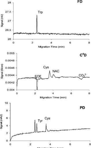
Selectivity can be further enhanced by implementing a third detector. A three-in-one
approach was described based on a standard fiber-optic adapter placed between two
Figure 9: Separation of a model mixture of amino acids showing complementarity of fluorescence detection (FD), photometric detection (PD), and C4D. Conditions: Trp 0.37 mM, Tyr 0.75 mM, Cys 0.94 mM, NAC 0.56 mM separated in 20 mM N-cyclo-hexyl-2-aminoethanesulfonic acid (pH 9);
LED 255 nm; frequency: 100 kHz; voltage: +15 kV; injection: 10 s, 1 kPa; capillary: ID=75 μm, leff
= 31.5 cm [76]. Figure reprinted with permission from M. Ryvolová, J. Preisler, F. Foret, P.C.
Hauser, P. Krásenský, B. Paull, M. Macka, Combined contactless conductometric, photometric, and fluorimetric single point detector for capillary separation methods, Anal. Chem., 82 (2010) 129-135. Copyright 2012 American Chemical Society.
The detector setup (however without UV detection) was further employed for the
monitoring of nicotine and cotinine derivatization by MEKC separating reaction
intermediates and products [75]. The C
4D enabled the determination of the EOF and the non-fluorescent products, which could be formed during the reaction.
Dual detection connected in series
Coupling of UV/VIS detection and C
4D in series is easily done implementing the C
4D into the cassette of a commercial CE instrument or placing it somewhere else along the capillary. A model mixture of 29 organic acids occurring in urine was determined by CE- C
4D-UV [77]. The dual approach enabled the detection of all analytes. C
4D was used for the small, fast migrating acids such as oxalic acid or aspartic acid whereas photometry was the detection principle of choice for the UV absorbing acids like orotic, vanillic, or hippuric acid. Moreover, also two unseparated acids could be quanitified comparing the detector signals as one was not UV absorbing and thus just visible in the C
4D [71, 77].
The complementarity of C
4D and UV detection was also used for the determination of dissociation constants of the 20 standard proteogenic amino acids [78]. The analytes lacking of chromophore such as alanine, leucine, or proline were detected by C
4D. In contrast, some amino acids were better detected using photometry as they exhibited similar conductivity to the BGE. A recent study used dual C
4D-UV detection to determine several drugs in over 100 formulations of dietary supplements sold for weight loss, fat burning, appetite reduction, and metabolite acceleration [79]. The combination of the two detectors enabled rapid screening and determination of the drugs in complex samples and gave also information on matrix interferences.
In some applications, C
4D and UV were used to compare the data or for optimization of the method instead of focusing on complementarity [100-103, 106]. A CE-C
4D-indirect UV method for the determination of γ-hydroxybutyric acid in saliva was developed and the performance of both detectors was validated [100]. Moreover, formate in blood samples could be analyzed by in-line coupling of microextractions across polymer inclusion membranes with CE-C
4D-UV [101]. In this application, the detection sensitivity of C
4D was found to be about an order of magnitude better than for UV/VIS detection.
The dual detection approach was also used with ITP [102] and with transient ITP-CE for
the analysis of paralytic shellfish toxins in mussel samples [103].
high selectivity and sensitivity of AD. Hence, AD was used for the quantification of the electroactive biomarkers whereas C
4D was chosen for the non-electroactive species and to get background information of the chemical composition and matrix of the sample.
Thus, information obtained with the two detectors complemented each other rendering the results more convincing and reliable [80, 81].
Dual C
4D was also reported [82, 83]. Stojkovic et al. [82] implemented a second detection channel into the C
4D arranged in a bridge configuration to the first one. A second capillary was inserted parallel to the separation capillary using the same inlet and outlet vial. This led to the generation of a reference signal enabling the compensation of baseline drifts due to changes in temperature or buffer composition.
The reference signal could also be generated looping the separation capillary back into the second detection channel. Feasibility of the system was demonstrated detecting inorganic model cations. Recently, dual C
4D was used for the determination of glycerol and its electrooxidation products (neutral diols and carboxylates) [83]. The two detectors were placed in 10 cm and 50 cm effective length of the separation capillary.
The fast migrating compounds were detected at the second detector while the slow migrating ions were detected at the first detector reducing the overall analysis time.
1.4.2. MS in dual detection approaches
C
4D with MS detection
The coupling of CE and MS was first introduced by Smith et al. [107] and has evolved
enormously in the last decades. Nowadays, MS can be considered as the ultimate
detector providing high selectivity and enabling the identification of analytes giving
molecular weight and structural information [108-110]. In earlier studies using dual C
4D- MS detection, the universal C
4D was not used as full detector for quantitative analysis complementary to the MS. Rather it was necessary for the optimization of the timing of a voltage switching step for a two-dimensional ITP-CE-MS separation [58] or for an interface-free CE-MS method development [111]. Recently, however, two dual C
4D-MS approaches were described demonstrating the complementarity of the two detectors enhancing the overall selectivity [84, 85]. The challenge in these approaches lied in the opposing requirements of the two detectors. For ESI-TOF-MS, volatile BGEs are essential whereas for C
4D, BGEs with rather low conductivity and high ionic strength are advantageous. Beutner et al. [84] used C
4D with ESI-TOF-MS for the determination of various phenolic compounds showing complementary response behavior for the different analytes. Moreover, the C
4D was used to monitor the EOF. Parameter selection was adjusted to the separation requirements and the compatibility of both detectors.
An NH
4Ac based BGE was taken as compromise ensuring good compatibility with the MS and an acceptable signal-to-noise ratio of the C
4D. Detector-induced band broadening could be neglected in the CE-C
4D-MS system.
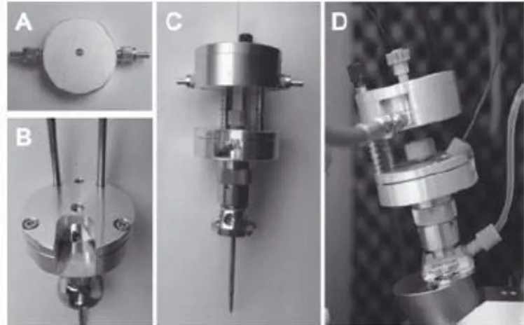
Figure 10: Photograph of a dual C4D-ESI arrangement: (a) C4D immobilized on a (b) 3D-printed support, (c) ESI interface, (d) electrical connections, (e) separation capillary, (f) sheath liquid, and (g) nebulizer gas [85]. Figure reprinted with permission from K.J.M. Francisco, C.L. do Lago, A capillary electrophoresis system with dual capacitively coupled contactless conductivity detection and electrospray ionization tandem mass spectrometry, Electrophoresis, 37 (2016) 1718-1724. Copyright 2016 John Wiley and Sons.
Figure 11: Electropherograms of an extract of a food supplement obtained by NACE with dual C4D-MS detection showing complementary information. The sample contained choline (1), acetylcholine (2, IS), Na+ (3), chloride (4), bromide (5, IS), nitrate (6), thiamine (7), and nicotinamide (8, EOF). The length of the separation capillary (ID= 50 µm) was 30 cm, which was the effective length to the MS. The effective length to the C4D was 15.2 cm. Injection time was 0.1 s. Separation was performed in a 2 M HAc/ACN BGE containing 4 mM NH4Ac applying a HV of 10 kV [86]. Figure reprinted with permission from A. Beutner, B. Scherer, F.-M. Matysik, Dual detection for non-aqueous capillary electrophoresis combining contactless conductivity detection and mass spectrometry, Talanta, 183 (2018) 33-38. Copyright 2018 Elsevier.
Another approach used non-aqueous BGEs as they are highly compatible with ESI due
to their volatility. Further, they exhibit low background conductivity being promising for
the use with C
4D [86]. A non-aqueous capillary electrophoresis (NACE) method using an
acetonitrile (ACN) based BGE was developed. Additionally, the influence of the ID of the
separation capillary was investigated considering both detectors. A capillary with ID 50
µm was found to be best suited for the dual approach. The method was applied to a food supplement quantifying the content of organic biomolecules with MS as well as inorganic ions with C
4D (Figure 11).
In summary, even if the MS is more sensitive and selective for most of the analytes, the hyphenation to a low-selectivity detector such as C
4D can enhance the gained information. The ionization efficiency of the ESI-MS is analyte-dependent and most TOF- MS systems have limitations determining analytes with low m/z. Thus, for some analytes such as inorganic ions the C
4D can be more sensitive. Furthermore, the EOF can be monitored without the requirement of an EOF marker in the sample. The C
4D can also provide information about unexpected features of the sample.
UV and fluorescence detection with MS detection
Coupling MS with UV detection is often easily performed connecting commercial equipment in series. However, the sensitivity of the MS detection is much higher compared to UV detection limited by the length of the optical pathway. Furthermore, only analytes with special structural requirements are detectable by UV. Consequently, dual detection with UV and MS was rarely used for complementary quantitative analysis.
The MS was rather used for identification of UV detected peaks [87] or the UV detector was used separately for optimization reasons [106]. Mironov et al. [87] developed a method based on kinetic CE coupled on-line with UV detection and ion mobility mass spectrometry (CE-UV-IM-MS) both detectors giving complementary information. This approach was applied to study the effect of small-molecule inhibitors on the conformational distribution and the enzymatic activity of a human tissue transglutaminase. UV detection was used to quantify the interconversion dynamics of separated protein conformers whereas conformer sizes, molecular weights, and structures were identified by IM-MS.
A problem often stated is the huge distance between the detection sites of the UV detector and the MS rendering a direct comparison of the two recordings difficult.
Overcoming this drawback, Foret et al. [113] developed a miniaturized, integrated CE-
UV-ESI-MS interface. It incorporated a fiber optic detection cell monitoring the UV-
active compounds just prior their admittance into the MS allowing also the use of short
separation capillaries.
Figure 12: Pictures of a CE-LIF-MS setup. (A) LIF detection cell; (B) the lower platform mounted on the CE-MS sprayer; (C) full setup with sprayer, lower and upper platform (with detection cell) and capillary; (D) CE-LIF-MS setup mounted on top of the MS inlet with attached optical fibers, sheath liquid tubing and nebulizer tubing [89]. Figure reprinted with permission from C. Huhn, L.R. Ruhaak, J. Mannhardt, M. Wuhrer, C. Neusüß, A.M. Deelder, H. Meyer, Alignment of laser- induced fluorescence and mass spectrometric detection traces using electrophoretic mobility scaling in CE-LIF-MS of labeled N-glycans, Electrophoresis, 33 (2012) 563-566. Copyright 2012 John Wiley and Sons.
![Figure 10: Photograph of a dual C 4 D-ESI arrangement: (a) C 4 D immobilized on a (b) 3D-printed support, (c) ESI interface, (d) electrical connections, (e) separation capillary, (f) sheath liquid, and (g) nebulizer gas [85]](https://thumb-eu.123doks.com/thumbv2/1library_info/3735718.1509011/37.892.133.348.645.981/photograph-arrangement-immobilized-interface-electrical-connections-separation-capillary.webp)