Article
Physioxia Expanded Bone Marrow Derived
Mesenchymal Stem Cells Have Improved Cartilage Repair in an Early Osteoarthritic Focal Defect Model
Girish Pattappa
1,*
,†, Jonas Krueckel
1,†, Ruth Schewior
1, Dustin Franke
1, Alexander Mench
1, Matthias Koch
1, Johannes Weber
1, Siegmund Lang
1, Christian G. Pfeifer
1,
Brian Johnstone
2, Denitsa Docheva
1, Volker Alt
1, Peter Angele
1,3and Johannes Zellner
1,41
Laboratory of Experimental Trauma Surgery, Department of Trauma Surgery,
University Hospital Regensburg, Franz Josef Strauss Allee 11, 93053 Regensburg, Germany;
jonas.kreuckel@ukr.de (J.K.); ruth.schewior@ukr.de (R.S.); dustinfranke@web.de (D.F.);
alexander.mench@ukr.de (A.M.); matthias.koch@ukr.de (M.K.); johannes1.weber@ukr.de (J.W.);
siegmund.lang@ukr.de (S.L.); christian.pfeifer@ukr.de (C.G.P.); denitsa.docheva@ukr.de (D.D.);
volker.alt@ukr.de (V.A.); angele@sporthopaedicum.de (P.A.); johannes.zellner@ukr.de (J.Z.)
2
Department of Orthopaedics and Rehabilitation, Oregon Health & Science University, 3181 SW Sam Jackson Park Rd, OP31, Portland, OR 97239, USA; johnstob@ohsu.edu
3
Sporthopaedicum Regensburg, Hildegard von Bingen Strasse 1, 93053 Regensburg, Germany
4
Department of Trauma Surgery, Caritas Hospital St. Josef, Landshuter Strasse 65, 93053 Regensburg, Germany
*
Correspondence: girish.pattappa@ukr.de; Tel.:
+49-941-943-1743†
These authors contributed equally to this work.
Received: 8 July 2020; Accepted: 14 August 2020; Published: 17 August 2020
Abstract: Focal early osteoarthritis (OA) or degenerative lesions account for 60% of treated cartilage defects each year. The current cell-based regenerative treatments have an increased failure rate for treating degenerative lesions compared to traumatic defects. Mesenchymal stem cells (MSCs) are an alternative cell source for treating early OA defects, due to their greater chondrogenic potential, compared to early OA chondrocytes. Low oxygen tension or physioxia has been shown to enhance MSC chondrogenic matrix content and could improve functional outcomes of regenerative therapies. The present investigation sought to develop a focal early OA animal model to evaluate cartilage regeneration and hypothesized that physioxic MSCs improve in vivo cartilage repair in both, post-trauma and focal early OA defects. Using a rabbit model, a focal defect was created, that developed signs of focal early OA after six weeks. MSCs cultured under physioxia had significantly enhanced in vitro MSC chondrogenic GAG content under hyperoxia with or without the presence of interleukin-1 β (IL-1 β ). In both post-traumatic and focal early OA defect models, physioxic MSC treatment demonstrated a significant improvement in cartilage repair score, compared to hyperoxic MSCs and respective control defects. Future investigations will seek to understand whether these results are replicated in large animal models and the underlying mechanisms involved in in vivo cartilage regeneration.
Keywords: mesenchymal stem cells; chondrogenesis; hypoxia; cartilage; early osteoarthritis
1. Introduction
Articular cartilage lines the surfaces of synovial joints and facilitates loading and friction-free joint movement during locomotion [1]. Following traumatic injury, newly formed tissue is fibrocartilaginous and has different structural, biochemical and mechanical properties, compared with normal articular
Biology2020,9, 230; doi:10.3390/biology9080230 www.mdpi.com/journal/biology
Biology2020,9, 230 2 of 21
cartilage. The newly formed tissue remains susceptible to progressive degeneration that can lead to a significant reduction in a patient’s physical function. This degenerative disorder is known as osteoarthritis (OA) and does not solely affect articular cartilage, but all structures within the joint [2,3].
According to the Deutsche Gesellschaft fur Orthopaedie und Unfallchirurgie (DGOU) registry between October 2013 and June 2014, 60% of treated defects were degenerative. In a multi-centre study from 400 patients, approximately 35% had chondral lesions resulting from degenerative conditions [4,5]. Current strategies for the treatment of cartilage lesions have involved the use of surgical intervention (e.g., microfracture) or cell-based therapies [6]. Two cell-based products currently used in clinical practice are autologous chondrocyte implantation (ACI) or matrix-assisted chondrocyte transplantation (MACT). However, cell-based therapies for this condition have an increased failure rate compared with traumatic cartilage defects. A recent publication examining long-term follow-up (15 years) of OA patients treated with MACT, showed that there was a worsening of patient clinical scores (e.g., Tegner, EuroQol visual analog scale) with time and a great majority of patients (60%), either underwent re-operation or experienced clinical failure [7]. A reason for the poor clinical outcome is due to the inflammatory environment created, specifically the presence of the cytokine, interleukin-1 β (IL-1 β ), correlated with poor MACT outcomes post-transplantation [5,8]. Due to the high proportion of degenerative cartilage lesions that require therapeutic intervention, regenerative options are required to overcome this challenging situation.
Recent studies have begun to focus on alternative cell sources for treating degenerative cartilage lesions. Mesenchymal stem cells (MSCs) are a potential cell source due to their multipotent differentiation (osteogenic, adipogenic and chondrogenic) and can be harvested from a variety of tissue sources (e.g. bone marrow, adipose (lipoaspirate from liposuction or Hoffa fat pad) or synovium tissue) with minimal donor site morbidity [9–11]. MSC chondrogenesis is induced via high density pellet or micromasses in the presence of pro-chondrogenic growth factors (e.g. transforming growth factor-beta (TGF-β)) to develop a cartilaginous matrix [12]. Studies have developed scaffolds/biomaterials that allow seeding or encapsulation of cells to create cartilaginous grafts for clinical translation [13,14].
A more robust cartilage matrix is formed from MSCs compared to articular chondrocytes, as the latter require extensive expansion to produce the requisite numbers of cells for clinical application.
Furthermore, chondrocytes upon redifferentiation produce fibrocartilaginous tissue after extensive expansion, in contrast to MSC chondrogenesis [15]. However, the implantation of cartilaginous MSC grafts in nude mouse models has resulted in ectopic bone formation in the long-term [16,17].
Therefore, environmental stimuli that stabilize the cartilage phenotype are required, prior to implantation. Examples include mechanical loading (e.g., hydrostatic pressure, compression) or biochemical (e.g., oxygen tension, growth factors) factors [18,19]. Studies have extensively investigated the use of low oxygen tension or physioxia in both chondrocytes and MSC chondrogenesis (reviewed in Pattappa et al., 2019 [18]). Chondrocytes reside under a low oxygen environment (2–5% oxygen) in vivo, with a low oxygen gradient developed from the superficial to deeper regions of the tissue [20,21].
These low oxygen conditions are nominally termed as hypoxia. Recent publications have used the term, physioxia, in reference to the physiological oxygen tension that is far below atmospheric oxygen tension (20% oxygen). For the purposes of this publication, physioxia refers to low oxygen culture, whilst 20%
oxygen is known as hyperoxia. Upon in vitro culture under physioxia, MSCs have been shown to significantly upregulate chondrogenic genes (SOX9, COL2A1 and ACAN) that lead to increased collagen II and GAG content within pellets or scaffolds. However, recent studies have demonstrated that the beneficial effects of physioxia on chondrogenesis is a donor dependent response [22,23].
Subsequent in vivo implantation of physioxia preconditioned chondrogenic MSCs into nude mouse and rabbit models resulted in increased cartilaginous matrix formation (GAG and collagen II) with reduced bone formation [24,25]. In contrast, there was no difference in cartilage matrix formation between hyperoxic and physioxic preconditioned chondrogenic MSCs in a sheep model [26].
Previous animal models for OA have primarily used either anterior cruciate ligament (ACL)
resection, destabilized medial meniscus (DMM) or intra-articular enzymatic injection [27,28].
These models create diffusive OA that is difficult to treat and does not reflect the clinical scenario that utilize cell-based regenerative therapies. One aim of this study was to develop a model for focal early OA that induced a focal defect with no joint instability or changes to surrounding tissues in the joint (e.g., meniscus or ACL). Using this model, we evaluated the efficacy of physioxia expanded/physioxic MSCs in cartilage regeneration. It was hypothesized that physioxic MSCs would have improved cartilage regeneration compared with hyperoxic MSCs in both post-traumatic and focal early OA cartilage defects.
2. Materials and Methods
2.1. Animals and Study Design
The animal experiments used in the study were approved by the local ethics committee (Regierung von Unterfranken, 55.2 2532-2-300, approval date: 23 December 2016). The study was performed according to the approved guidelines and regulations. Thirty-five male New Zealand White rabbits (aged 5–6 months old) were used for the animal experiments. The groups and number of animals described in each study are described in Table 1. All animals were housed in single-animal cages in an air-conditioned environment under a 12 h/12 h day/night rhythm for the whole study period with free access to food and water.
Biology 2020, 9, x FOR PEER REVIEW 5 of 22
pain, carprofen (5 mg/kg) was administered for five days post-surgery. Rabbits were sacrificed at 6 (n = 3) or 12 (n = 3) weeks post-defect creation. Histological sections were created from these defects, stained for Safranin-O/fast green (Section 2.7) and then were evaluated for OA using the OARSI score for rabbits (Section 2.8) by three blinded observers [30,31]. The time-point that was evaluated histologically to show signs of focal early OA was used for subsequent MSC treatment studies. For these studies, this model is defined as a focal early OA model, whilst defects treated immediately after creation are known as a post-trauma model.
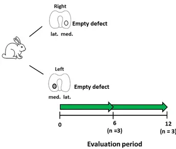
Figure 1. Schematic diagram describing the experimental setup for the development of the focal early
OA model. Number of animals (n) in each group is stated in brackets.
2.6. IL-1β Measurement in Cartilage
At the time of sacrifice for evaluating the focal early OA model, cartilage pieces surrounding the defect were removed using a scalpel and then frozen for analysis. Cartilage pieces were washed in ice cold PBS and homogenized in 200 μL 8 M urea/2% SDS solution using a PreCellys homogenizer (Bertin Instruments, Montigny le Bretonneux, France). The lysate was centrifuged for 5 min at 1000×g (4 °C) and the supernatant was transferred to a fresh tube. The protein concentration of the supernatant was determined using the BCA Protein Assay kit (Biorad, DC Protein Assay, Hercules, CA, USA) according to the manufacturer’s instructions. ELISA detection for rabbit IL-1β was performed according to manufacturer’s protocol (rabbit IL-1β; R and D systems, UK).
2.7. MSC Treatment for Focal Early OA and Post-Trauma Models
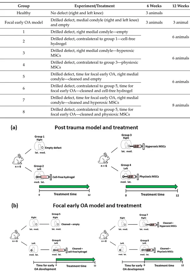
Rabbits were grouped into either post-trauma (groups 1–4; Figure 2a) or focal early OA (groups 5–8; Figure 2b) models. The post-trauma defect model is described as a focal defect that is created and immediately treated (Figure 2a), while the focal early OA defect is a focal defect that is generated, and then left to develop signs of early OA around the edges of the defect, over a designated time period (results from Section 2.5), and then treated (Figure 2b).
For the post-trauma model (Figure 2a), defects were created on both medial condyles of right and left knee of each rabbit and then either left empty (group 1; n = 6 rabbits) or injected with a commercially available hyaluronic acid hydrogel (TETEC, Tübingen, Germany) [32] on the contralateral medial femoral condyle without cells (group 2; n = 6). In the case of the focal early OA model (Figure 2b), two surgeries were performed, the first involved defect creation as described and then following development of focal early OA, a second surgery was undertaken to clean the defect using a 22G needle to remove repair tissue. This was then either left empty (Group 5; n = 6), or injected with the described cell-free hydrogel (Group 6; n = 6) on the controlateral medial femoral condyle.
In the case of MSC treated defects for both models, autologous rabbit MSCs (passage 2; n = 26, including donors from the in vitro study (Section 2.2)) isolated from bone marrow were pre-expanded
Figure 1.
Schematic diagram describing the experimental setup for the development of the focal early
OA model. Number of animals (n) in each group is stated in brackets.
Biology2020,9, 230 4 of 21
Table 1.
Summary of the groups and respective treatments for the animals used in the in vivo studies.
See Figures
1and
2for a schematic representation of the table.
Group Experiment/Treatment 6 Weeks 12 Weeks
Healthy No defect (right and left knee) 3 animals
Focal early OA model Drilled defect, medial condyle (right and left knee)
and empty 3 animals 3 animal
1 Drilled defect, right medial condyle—empty
6 animals 2 Drilled defect, contralateral to group 1—cell-free
hydrogel
3 Drilled defect, right medial condyle—hyperoxic
MSCs 6 animals
4 Drilled defect, contralateral to group 3—physioxic MSCs
5 Drilled defect, time for focal early OA, right medial condyle—cleaned and empty
6 animals 6 Drilled defect, contralateral to group 5, time for
focal early OA—cleaned and cell-free hydrogel 7 Drilled defect, time for focal early OA, right medial
condyle—cleaned and hyperoxic MSCs
8 animals 8 Drilled defect, contralateral to group 5, time for
focal early OA—cleaned and physioxic MSCs
Biology 2020, 9, x FOR PEER REVIEW 6 of 22
in parallel under either hyperoxia or physioxia (see Section 2.2). MSCs were counted using a haemacytometer and seeded into the hydrogel at a density of 50 × 10
6cells/mL according to a previously described protocol [32]. The cell seeding density was based on previous in vivo experiments using MSCs for cartilage repair in animal models [33,34]. MSC-hydrogels were injected into the defect (approx. 20 μL) with one medial femoral condyle treated with physioxic MSCs, whilst the contralateral medial femoral condyle had hyperxoic MSCs applied (Figure 2). Prior to joint closure, all hydrogel groups were allowed to gel for five minutes and joint motion was used to observe whether hydrogel remained in the defect. For post-trauma defects, hyperoxic MSCs (Group 3; n = 6) and physioxic MSCs (Group 4; n = 6) were immediately injected following defect creation (Figure 2a). In the focal early OA model, hyperoxic (Group 7; n = 8) and physioxic (Group 8; n = 8) MSCs were applied in a second surgery, following the designated period time for focal early OA development (Figure 2b). All rabbits were put back into their individual cages with no restrictions on movement. Prior to these experiments and to evaluate whether hydrogel remained within the defect, a pilot study was conducted, whereby defects were created on both knees as described and then immediately implanted with cell-free hydrogel. The rabbits were euthanized at 12 weeks post-MSC implantation for the respective model (for the pilot study, only 1 week post hydrogel application) by an intravenous overdose application of narcoren (0.5 g/kg).
Figure 2. Schematic diagram describing the defect models of (a) Post-trauma and (b) focal early OA with the respective treatment groups for these models. Number of animals (n) in each group is stated in brackets.
2.8. Decalcification and Histology/Immunohistochemistry
Figure 2.
Schematic diagram describing the defect models of (a) Post-trauma and (b) focal early OA
with the respective treatment groups for these models. Number of animals (n) in each group is stated
in brackets.
2.2. In Vitro Rabbit Bone Marrow Culture
Bone marrow derived MSCs were extracted from the iliac crest of skeletally mature New Zealand white rabbits (n = 8; 5–6 months old) following anesthesia that was applied intramuscularly using a combination of 0.6 mL/kg ketamine (10%) and xylazine (2%). The bone marrow was harvested from the iliac crest via a small incision into the bone cortex with an 18G needle and collected into a heparin-containing syringe. Bone marrow was resuspended in fresh culture media, composed of low glucose Dulbecco’s Modified Eagle Medium (DMEM; Invitrogen, Karlsruhe, Germany) with 10% fetal bovine serum (FBS; PAN Biotech, Aidenbach, Germany), 1% penicillin/streptomycin (Invitrogen), 1% HEPES (Sigma-Aldrich, Steinheim, Germany)) and 5 ng/mL basic fibroblastic growth factor (bFGF;
Peprotech, Hamburg, Germany). Bone marrow was centrifuged at 2000 rpm for five minutes and supernatant was carefully removed. The total number of nucleated cells was counted in the bone marrow aspirate and then seeded at a density of 2.5 × 10
5cells/cm
2. In parallel, flasks were cultured, either in a standard cell culture incubator (20% oxygen, 5% CO
2and 70% N
2) or a low oxygen incubator (2% oxygen, 5% CO
2, 93% N
2). This manuscript uses terminology stated in our previous manuscript, whereby 2% oxygen is known as physioxia, whilst 20% oxygen is described as hyperoxia [23]. The first media change was performed at five days post-seeding and then subsequent media changes were conducted twice a week.
Cells were trypsinized at 90% confluence and then cells counted using a hemacytometer.
MSCs were cultured under their designated oxygen conditions for a further four passages with the data used to generate population growth curves. At each passage, photomicrographs were taken and images were analysed for cell area using ImageJ software (National Institutes of Health, Bethesda, MD, USA). In each image, ten individual cells from each image per condition and donor were evaluated.
2.3. In Vitro Chondrogenic Differentiation
At passage 2, donor paired hyperoxic and physioxic MSCs (n = 8) were used to create pellet cultures, as previously described [23]. In brief, 4 × 10
5MSCs were formed in polypropylene V-bottom 96-well plates by centrifugation at 250 × g for 5 min in 300 µL chondrogenic medium that consisted of serum-free high-glucose DMEM containing 10 ng/mL TGF-β1 (R&D systems), 100 nm dexamethasone, 50 µ g/mL ascorbic acid-2-phosphate (all Sigma-Aldrich, Steinheim, Germany), 1 mM sodium pyruvate (Invitrogen) and 1% ITS (PAN Biotech GmbH, Aidenbach, Germany). A set of pellets were cultured in the presence of 0.1 ng/mL IL-1β (Peprotech), based on previous work [29]. Pellets were then cultured under their respective expansion oxygen conditions for 21 days with media changes performed every 2–3 days. In the case of physioxia pellets, media was pre-equilibrated in a physioxia incubator prior to replenishment.
2.4. Wet Weight and GAG Content Measurement
After 21 days in culture, the wet weight of pellets was measured using a balance. Macroscopic images of pellets were photographed with an optical microscope (PL2000, Optech, Germany).
Triplicate pellets from each group were digested with 150 µ g/mL papain in PBS, pH 6.0, containing 8 mM sodium EDTA, 6 mM L-cysteine (all Sigma-Aldrich). Digested pellet GAGs were quantified against a standard curve generated using bovine chondroitin sulphate A (Sigma-Aldrich) diluted in papain buffer as standard in serial dilution. DMMB dye (18 µg/mL in 0.5% ethanol, 0.2% formic acid, 30 mM sodium formate, pH 3) was added to standards and samples and absorbance measured at 575 nm (Tecan, Crailsheim, Germany).
2.5. Focal Early OA Model Development
The in vivo study was conducted on skeletally mature New Zealand white rabbits (5–6 months old)
using procedures approved by our local ethics committee (Regierung von Unterfranken, 55.2 2532-2-300,
approval date: 23 December 2016). The rabbits were anesthetized using a combination of 0.6 mL/kg
Biology2020,9, 230 6 of 21
ketamine (10%) and xylazine (2%), applied intramuscularly into the rabbit. The first animal study involved the development of a focal early OA model for the evaluation of cell-based therapies.
Following anaesthesia, the medial femoral condyle was exposed through a medial parapatellar incision with the patella dislocated to the lateral side. A 2.3 mm diameter (2 mm deep) defect was created on the medial condyle using a handheld dental drill (Surgic Pro, NSK Europe, Eschborn, Germany).
The patella was placed back into position and then skin was closed using sutures. A second defect was created on the contralateral medial femoral condyle using the same procedure (Figure 1).
The defects were left empty to observe the development of OA with time. Post-operatively, the rabbits were placed into their cages with no restrictions on movement and weight-bearing. To control pain, carprofen (5 mg/kg) was administered for five days post-surgery. Rabbits were sacrificed at 6 (n = 3) or 12 (n = 3) weeks post-defect creation. Histological sections were created from these defects, stained for Safranin-O/fast green (Section 2.7) and then were evaluated for OA using the OARSI score for rabbits (Section 2.8) by three blinded observers [30,31]. The time-point that was evaluated histologically to show signs of focal early OA was used for subsequent MSC treatment studies. For these studies, this model is defined as a focal early OA model, whilst defects treated immediately after creation are known as a post-trauma model.
2.6. IL-1β Measurement in Cartilage
At the time of sacrifice for evaluating the focal early OA model, cartilage pieces surrounding the defect were removed using a scalpel and then frozen for analysis. Cartilage pieces were washed in ice cold PBS and homogenized in 200 µ L 8 M urea/2% SDS solution using a PreCellys homogenizer (Bertin Instruments, Montigny le Bretonneux, France). The lysate was centrifuged for 5 min at 1000 × g (4
◦C) and the supernatant was transferred to a fresh tube.
The protein concentration of the supernatant was determined using the BCA Protein Assay kit (Biorad, DC Protein Assay, Hercules, CA, USA) according to the manufacturer’s instructions. ELISA detection for rabbit IL-1 β was performed according to manufacturer’s protocol (rabbit IL-1 β ; R and D systems, UK).
2.7. MSC Treatment for Focal Early OA and Post-Trauma Models
Rabbits were grouped into either post-trauma (groups 1–4; Figure 2a) or focal early OA (groups 5–8; Figure 2b) models. The post-trauma defect model is described as a focal defect that is created and immediately treated (Figure 2a), while the focal early OA defect is a focal defect that is generated, and then left to develop signs of early OA around the edges of the defect, over a designated time period (results from Section 2.5), and then treated (Figure 2b).
For the post-trauma model (Figure 2a), defects were created on both medial condyles of right and left knee of each rabbit and then either left empty (group 1; n = 6 rabbits) or injected with a commercially available hyaluronic acid hydrogel (TETEC, Tübingen, Germany) [32] on the contralateral medial femoral condyle without cells (group 2; n = 6). In the case of the focal early OA model (Figure 2b), two surgeries were performed, the first involved defect creation as described and then following development of focal early OA, a second surgery was undertaken to clean the defect using a 22G needle to remove repair tissue. This was then either left empty (Group 5; n = 6), or injected with the described cell-free hydrogel (Group 6; n = 6) on the controlateral medial femoral condyle.
In the case of MSC treated defects for both models, autologous rabbit MSCs (passage 2; n = 26,
including donors from the in vitro study (Section 2.2)) isolated from bone marrow were pre-expanded
in parallel under either hyperoxia or physioxia (see Section 2.2). MSCs were counted using a
haemacytometer and seeded into the hydrogel at a density of 50 × 10
6cells/mL according to a
previously described protocol [32]. The cell seeding density was based on previous in vivo experiments
using MSCs for cartilage repair in animal models [33,34]. MSC-hydrogels were injected into the
defect (approx. 20 µL) with one medial femoral condyle treated with physioxic MSCs, whilst the
contralateral medial femoral condyle had hyperxoic MSCs applied (Figure 2). Prior to joint closure,
all hydrogel groups were allowed to gel for five minutes and joint motion was used to observe whether hydrogel remained in the defect. For post-trauma defects, hyperoxic MSCs (Group 3; n = 6) and physioxic MSCs (Group 4; n = 6) were immediately injected following defect creation (Figure 2a).
In the focal early OA model, hyperoxic (Group 7; n = 8) and physioxic (Group 8; n = 8) MSCs were applied in a second surgery, following the designated period time for focal early OA development (Figure 2b). All rabbits were put back into their individual cages with no restrictions on movement.
Prior to these experiments and to evaluate whether hydrogel remained within the defect, a pilot study was conducted, whereby defects were created on both knees as described and then immediately implanted with cell-free hydrogel. The rabbits were euthanized at 12 weeks post-MSC implantation for the respective model (for the pilot study, only 1 week post hydrogel application) by an intravenous overdose application of narcoren (0.5 g/kg).
2.8. Decalcification and Histology/Immunohistochemistry
Femoral condyles were extracted from the rabbits and were fixed in 4% paraformaldehyde (PFA) for 48 h at 4
◦C. Condyles were rinsed in 0.1M phosphate buffer and decalcified in 10%
Ethylenediaminetetraacetic acid (EDTA; pH 8) in PBS. EDTA solution was changed three times a week and condyles placed on a manual shaker. Following decalcification, condyles were incubated with increasing sucrose concentrations (10–30%) and then embedded in Tissue-Tek (Sakura, Zoeterwoude, The Netherlands). Embedded condyles were cryosectioned at 8 µ m using a cryostat (Leica CM 1950, Leica Biosystems, Germany). MSC chondrogenic pellets were embedded using the same protocol without the requirement for decalcification.
Sections of the defect region were stained with safranin-O (0.1% w/v)/Fast green (0.05% w/v) (Sigma Aldrich, Steinheim, Germany) to stain sulphated glycosaminoglycans and collagens in the embedded condyles. For chondrogenic pellets, sulphated glycosaminoglycan content was detected by histochemical staining with DMMB (0.05% 1, 9-dimethylmethylene blue, 0.5% ethanol, 0.2% formic acid, 30 mM sodium formate, pH 3). Sections used for immunohistochemistry were rehydrated and antigen retrieval was performed at room temperature. To observe whether hyaline cartilage is formed (high collagen II staining) or fibrocartilage was formed (staining for collagen I and II), immunohistochemical staining for collagen I and II was performed. For collagen I and collagen II (both Calbiochem, Darmstadt, Germany), sections were treated with 3 mg/mL pepsin (Sigma-Aldrich) in 1xcitric/phosphate McIlvaine buffer for 15 min. Sections were blocked with a blocking buffer (10% goat serum in 1xPBS) and then incubated overnight with primary antibody to probe for rabbit collagen I (mouse monoclonal, 1:100) and collagen II (mouse monoclonal, 1:100). Following primary antibody incubation, biotinylated secondary antibody (goat anti-mouse IgG, 1:100) was applied and positive staining was visualized using nickel- and cobalt-enhanced 3, 3
0-diaminobenzidine (DAB).
2.9. Cartilage Scoring
Three blinded observers with prior knowledge of cartilage repair and histological assessment scored five safranin-O/fast green stained sections per condyle. Defect only sections were evaluated for cartilage degeneration using the OARSI histological score for rabbits (Table S1) [30,31]. For cartilage regeneration, control (defect only, cell-free hydrogel) and MSC treated defects were scored using the Sellers scoring system (Table S2) [35,36]. This is an inverse scoring system, whereby the lower the score, the better the cartilage repair.
2.10. Statistical Analysis
All statistical analysis was performed using Graphpad Prism v7 (GraphPad, La Jolla, CA, USA).
A Shapiro-Wilk normality test was used to test for normality of the data with p < 0.05, indicating use
of non-parametric statistical test. Pellet wet weight and GAG content was analyzed using two-way
ANOVA with Tukey post-hoc test. Individual Sellers score parameters were tested for significance
using a Kruskal-Wallis test with Dunn’s post hoc test. Comparison between OARSI and total Sellers
Biology2020,9, 230 8 of 21
score used a One way ANOVA with Tukey post-hoc test. A pairwise t-test was used to compare Sellers score between hyperoxia and physioxia MSC treated defects in both animal models. Correlation analysis between in vitro per pellet GAG and in vivo Sellers score outcome was conducted using Pearson correlation co-efficient test. Significance was set at * p < 0.05.
3. Results
3.1. Development of A Focal Early OA Model
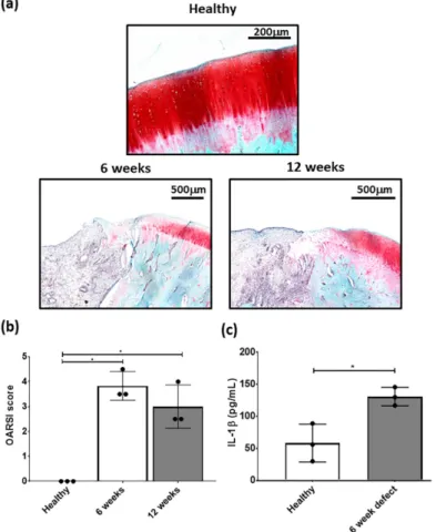
Focal osteochondal defects with a diameter of 2.3 mm and a depth of 2 mm were created on the posterior portion of the medial femoral condyle. At both, 6 and 12 weeks after defect creation, gross evaluation showed areas of degeneration with cartilage abrasion and softening in the region around the defect in the medial compartment. Histological examination showed that the structure of the cartilage layer adjacent to the defect had evidence of cell death, clustering and proliferation with fissures formed in the tissue. In the area of the defect, there were no signs of cartilaginous tissue with no safranin-o/fast green staining (Figure 3a).
Biology 2020, 9, x FOR PEER REVIEW 8 of 22
fissures formed in the tissue. In the area of the defect, there were no signs of cartilaginous tissue with no safranin-o/fast green staining (Figure 3a).
OARSI scoring of the defect had a mean score between 3 and 4 at 6 and 12 weeks post-defect creation and was significantly different from healthy cartilage (Figure 3b). Furthermore, there was a significant elevation in IL-1β concentration in the cartilage adjacent to the defect at 6 weeks post- defect (Figure 3c). Based on this data, subsequent focal early OA experiments with the respective treatments, were carried out at 6 weeks post defect creation. Although, both time points showed signs of focal early OA, the 6 week time point had the earliest signs of the condition and was used for subsequent MSC studies.
Figure 3. Representative (a) safranin-O/fast green stained healthy, 6 and 12 week defects used for the examination of focal early OA in the defect. (b) OARSI score for healthy, 6 and 12 week defects post- surgery and (c) measurement of IL-1β in cartilage from healthy and defect cartilage. Data represent mean ± S.D.; n = 3, with dots representing the mean from three blinded scorers (five sections per defect). *p < 0.05.
3.2. Rabbit MSCs Undergo Greater Population Doublings and Have Enhanced Chondrogenesis Under Physioxia
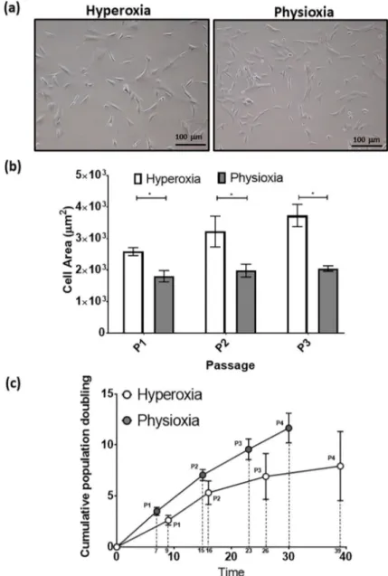
The typical MSC fibroblastic morphology was observed under both oxygen tensions, with MSCs cultured under physioxia appearing to be smaller (Figure 4a) [37]. Measurements of individual MSCs demonstrated that physioxic MSCs were significantly smaller than hyperoxic MSCs (p < 0.05; Figure 4b). Furthermore, hyperoxic MSCs increased their cell area with passage, whilst physioxic MSCs remained the same. An analysis of their cellular proliferation over four passages in culture, showed Figure 3.
Representative (a) safranin-O/fast green stained healthy, 6 and 12 week defects used for the examination of focal early OA in the defect. (b) OARSI score for healthy, 6 and 12 week defects post-surgery and (c) measurement of IL-1β in cartilage from healthy and defect cartilage. Data represent mean
±S.D.; n
=3, with dots representing the mean from three blinded scorers (five sections per defect).
* p
<0.05.
OARSI scoring of the defect had a mean score between 3 and 4 at 6 and 12 weeks post-defect
creation and was significantly different from healthy cartilage (Figure 3b). Furthermore, there was a
significant elevation in IL-1 β concentration in the cartilage adjacent to the defect at 6 weeks post-defect
(Figure 3c). Based on this data, subsequent focal early OA experiments with the respective treatments,
were carried out at 6 weeks post defect creation. Although, both time points showed signs of focal
early OA, the 6 week time point had the earliest signs of the condition and was used for subsequent MSC studies.
3.2. Rabbit MSCs Undergo Greater Population Doublings and Have Enhanced Chondrogenesis Under Physioxia
The typical MSC fibroblastic morphology was observed under both oxygen tensions, with MSCs cultured under physioxia appearing to be smaller (Figure 4a) [37]. Measurements of individual MSCs demonstrated that physioxic MSCs were significantly smaller than hyperoxic MSCs (p < 0.05;
Figure 4b). Furthermore, hyperoxic MSCs increased their cell area with passage, whilst physioxic MSCs remained the same. An analysis of their cellular proliferation over four passages in culture, showed that physioxic culture resulted in greater MSCs numbers and significantly shorter population doubling times between passages relative to hyperoxia (* p < 0.05; Figure 4c). In the case of hyperoxic MSCs, there was a cessation of cell growth after approximately 30 days in culture in the majority of donors, whereas physioxic MSCs continued to expand at the same rate for all donors (Figure 4c).
Biology 2020, 9, x FOR PEER REVIEW 9 of 22
that physioxic culture resulted in greater MSCs numbers and significantly shorter population doubling times between passages relative to hyperoxia (*p < 0.05; Figure 4c). In the case of hyperoxic MSCs, there was a cessation of cell growth after approximately 30 days in culture in the majority of donors, whereas physioxic MSCs continued to expand at the same rate for all donors (Figure 4c).
Figure 4. (a) Photomicrographs of rabbit MSCs cultured under hyperoxia and physioxia. (b) Cell area measurements for individual MSCs from photomicrographs (10 individual cells measured per image) and (c) cumulative cell growth of MSCs under physioxia and hyperoxia. Data represent mean ± S.D.;
n = 8; *p < 0.05.
Figure 4.
(a) Photomicrographs of rabbit MSCs cultured under hyperoxia and physioxia. (b) Cell area measurements for individual MSCs from photomicrographs (10 individual cells measured per image) and (c) cumulative cell growth of MSCs under physioxia and hyperoxia. Data represent mean
±S.D.;
n
=8; * p
<0.05.
Biology2020,9, 230 10 of 21
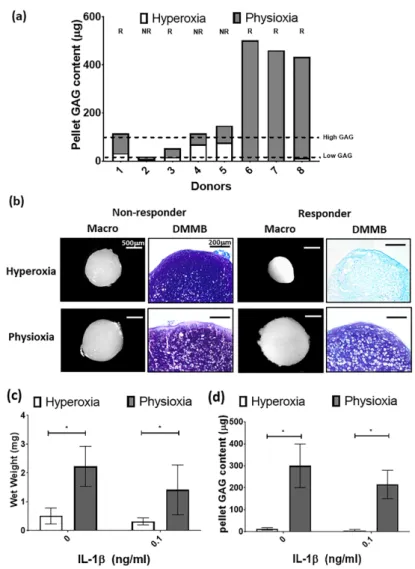
For the purpose of the subsequent in vivo studies, MSC chondrogenesis was performed for donors under their expansion conditions to evaluate their response to physioxia. There were donors either responsive (R) or non-responsive (NR) to physioxia based upon their pellet GAG content (Figure 5a).
A physioxia responsive donor is one where there was a 2-fold increase in pellet GAG under physioxa relative to hyperoxia, whereas a non-responder was one that a had a less than 2-fold increase [23].
Additionally, these responsive donors had a low intrinsic baseline chondrogenesis under hyperoxia [22].
Histological evaluation of non-responsive donors showed pellets of similar size and DMMB staining intensity. In contrast, physioxia responsive donors had larger pellets and greater DMMB staining intensity under physioxia compared to hyperoxic cultures (Figure 5b). Analysis of only physioxia responsive donors demonstrated a significant increase in pellet wet weight (* p < 0.05; Figure 5c) and GAG content (* p < 0.05; Figure 5d) under these conditions, when compared to hyperoxic culture. In the presence of IL-1 β , a decrease in pellet wet weight and GAG content was observed under hyperoxic conditions. However, the corresponding physioxic condition, demonstrated a significant increase in pellet wet weight and GAG content compared to equivalent hyperoxic cultures (* p < 0.05; Figure 5b,c).
Biology 2020, 9, x FOR PEER REVIEW 10 of 22
For the purpose of the subsequent in vivo studies, MSC chondrogenesis was performed for donors under their expansion conditions to evaluate their response to physioxia. There were donors either responsive (R) or non-responsive (NR) to physioxia based upon their pellet GAG content (Figure 5a). A physioxia responsive donor is one where there was a 2-fold increase in pellet GAG under physioxa relative to hyperoxia, whereas a non-responder was one that a had a less than 2-fold increase [23]. Additionally, these responsive donors had a low intrinsic baseline chondrogenesis under hyperoxia [22]. Histological evaluation of non-responsive donors showed pellets of similar size and DMMB staining intensity. In contrast, physioxia responsive donors had larger pellets and greater DMMB staining intensity under physioxia compared to hyperoxic cultures (Figure 5b). Analysis of only physioxia responsive donors demonstrated a significant increase in pellet wet weight (* p < 0.05;
Figure 5c) and GAG content (* p < 0.05; Figure 5d) under these conditions, when compared to hyperoxic culture. In the presence of IL-1β, a decrease in pellet wet weight and GAG content was observed under hyperoxic conditions. However, the corresponding physioxic condition, demonstrated a significant increase in pellet wet weight and GAG content compared to equivalent hyperoxic cultures (* p < 0.05; Figure 5b,c).
.
Figure 5. (a) Pellet GAG content under physioxia and hyperoxia for chondrogenic pellets. Dotted lines
represent threshold for high (physioxia non-responsive) and low (physioxia responsive) GAG donors.
R represents physioxia responders and NR are physioxia non-responders. (b) Representative macroscopic and DMMB stained chondrogenic pellets of physioxia non-responder and responder pellets. Pellet (c) wet weight; and (d) GAG content for physioxia responders with or without the presence of IL-1β. Data represent mean ± S.D.; n = 5. *
p < 0.05.Figure 5.
(a) Pellet GAG content under physioxia and hyperoxia for chondrogenic pellets. Dotted
lines represent threshold for high (physioxia non-responsive) and low (physioxia responsive) GAG
donors. R represents physioxia responders and NR are physioxia non-responders. (b) Representative
macroscopic and DMMB stained chondrogenic pellets of physioxia non-responder and responder
pellets. Pellet (c) wet weight; and (d) GAG content for physioxia responders with or without the
presence of IL-1β. Data represent mean
±S.D.; n
=5. * p
<0.05.
3.3. Physioxic Preconditioned MSCs Support An Improvement in Cartilage Repair in A Post-Trauma Cartilage Defect
Defects on the medial femoral condyle (Figure 6a) that were left empty after six weeks showed tissue repair with filling of the defect (Figure 6b). For the focal early OA model, the defect was cleaned using a 22G needle to remove the repair tissue (Figure 6c), prior to application of cell-free or MSC-hydrogel into the defect. A pilot study demonstrated that injected hydrogel stayed within the defect after one week post-implantation (Figure 6d).
Biology 2020, 9, x FOR PEER REVIEW 11 of 22
3.3. Physioxic Preconditioned MSCs Support An Improvement in Cartilage Repair in A Post-Trauma Cartilage Defect
Defects on the medial femoral condyle (Figure 6a) that were left empty after six weeks showed tissue repair with filling of the defect (Figure 6b). For the focal early OA model, the defect was cleaned using a 22G needle to remove the repair tissue (Figure 6c), prior to application of cell-free or MSC- hydrogel into the defect. A pilot study demonstrated that injected hydrogel stayed within the defect after one week post-implantation (Figure 6d).
Figure 6. Macroscopic image of (a) empty defect immediately after creation and at (b) 6 weeks (defect cricled) post-surgery. (c) Cleaned defect prior to hydrogel implantation and (d) presence of hydrogel (black circle) in defect after one week in the joint.
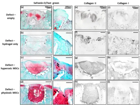
MSC treated defects had cartilaginous tissue formation with safranin-O and collagen II staining within the treated region. Application of physioxic MSCs induced greater safranin-O and collagen II staining (Figure 7d,h) with hyperoxic (Figure 7c,g). Furthermore, there was evidence of more chondrocytes and early column formation in the cartilage layer upon application of physioxic MSCs relative to hyperoxic MSCs (Figure 7c,d). In contrast, empty and cell-free hydrogel defects had fibrocartilaginous tissue with minimal safranin-o staining (Figure 7a,b) and only collagen II (Figure 7e,f) and I (Figure 7i,j) present.
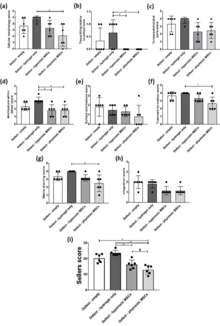
In general, scoring of cartilage regeneration showed improved scores for physioxic MSC treated defects (Figure 8). Assessment of individual parameters of the Sellers score showed a significant improvement in cellular morphology, new tissue filling, matrix staining, tissue architecture within the defect and new tidemark formation with physioxic MSCs compared to hydrogel only treated defects (*p < 0.05; Figure 8a,b,d,f,g). No significant differences in score were found for tissue integration, percentage new subchondral bone formation or surface architecture, although scores were lower for MSC treated defects (Figure 8c,e,h).
Figure 6.
Macroscopic image of (a) empty defect immediately after creation and at (b) 6 weeks (defect cricled) post-surgery. (c) Cleaned defect prior to hydrogel implantation and (d) presence of hydrogel (black circle) in defect after one week in the joint.
MSC treated defects had cartilaginous tissue formation with safranin-O and collagen II staining within the treated region. Application of physioxic MSCs induced greater safranin-O and collagen II staining (Figure 7d,h) with hyperoxic (Figure 7c,g). Furthermore, there was evidence of more chondrocytes and early column formation in the cartilage layer upon application of physioxic MSCs relative to hyperoxic MSCs (Figure 7c,d). In contrast, empty and cell-free hydrogel defects had fibrocartilaginous tissue with minimal safranin-o staining (Figure 7a,b) and only collagen II (Figure 7e,f) and I (Figure
Biology 2020, 9, x FOR PEER REVIEW7i,j) present.
12 of 22Figure 7. Representative images of (a–d) Safranin-O/Fast green, (e–h) collagen II and (i–l) collagen I staining for (a,e,i) empty defect, (b,f,j) hydrogel only, (c,g,k) hyperoxia and (d,h,l) physioxia treated post-trauma defects at 12 weeks post treatment. Box dotted line represents cartilage region evaluated for Sellers score and cross (x) signifies artefacts in the section. Arrows depict chondrocytes/chondrocytic columns in the cartilage layer.
The total Sellers score was found to have a lower score for MSC treatments compared with empty and cell-free hydrogel treated defects. Both hyperoxic and physioxic MSCs showed a significant improvement in the score compared to the controls (*p < 0.05; Figure 8i). Furthermore, physioxic MSCs resulted in a significant improvement in tissue regeneration, compared with hyperoxic MSCs (# p < 0.05; Figure 8i).
Figure 7.
Representative images of (a–d) Safranin-O/Fast green, (e–h) collagen II and (i–l) collagen I
staining for (a,e,i) empty defect, (b,f,j) hydrogel only, (c,g,k) hyperoxia and (d,h,l) physioxia treated
post-trauma defects at 12 weeks post treatment. Box dotted line represents cartilage region evaluated
for Sellers score and cross (x) signifies artefacts in the section. Arrows depict chondrocytes/chondrocytic
columns in the cartilage layer.
Biology2020,9, 230 12 of 21
In general, scoring of cartilage regeneration showed improved scores for physioxic MSC treated defects (Figure 8). Assessment of individual parameters of the Sellers score showed a significant improvement in cellular morphology, new tissue filling, matrix staining, tissue architecture within the defect and new tidemark formation with physioxic MSCs compared to hydrogel only treated defects (* p < 0.05; Figure 8a,b,d,f,g). No significant differences in score were found for tissue integration, percentage new subchondral bone formation or surface architecture, although scores were lower for MSC treated defects (Figure 8c,e,h).
Biology 2020, 9, x FOR PEER REVIEW 13 of 22
Figure 8. Individual scoring parameters of the Sellers score for (a) cellular morphology, (b) tissue filling, (c) % new subchondral bone, (d) architecture within the defect, (e) surface architecture, (f) new tidemark formation (g) new cartilage matrix staining and (h) cartilage integration with surrounding cartilage between groups in the post-trauma model. *p < 0.05 from Kruskal-Wallis test with Dunn’s multiple comparison test. (i) The overall Sellers score for cartilage repair in a post trauma model). *p
< 0.05 from one way ANOVA with Tukey post-hoc test, # p < 0.05 from pairwise t-test. Data represent the mean ± S.D. of n = 6 rabbits with dots representing the mean from three blinded scorers (five sections per defect).
Figure 8.
Individual scoring parameters of the Sellers score for (a) cellular morphology, (b) tissue filling,
(c) % new subchondral bone, (d) architecture within the defect, (e) surface architecture, (f) new tidemark
formation (g) new cartilage matrix staining and (h) cartilage integration with surrounding cartilage
between groups in the post-trauma model. * p
<0.05 from Kruskal-Wallis test with Dunn’s multiple
comparison test. (i) The overall Sellers score for cartilage repair in a post trauma model). * p
<0.05
from one way ANOVA with Tukey post-hoc test,
#p
<0.05 from pairwise t-test. Data represent the
mean
±S.D. of n
=6 rabbits with dots representing the mean from three blinded scorers (five sections
per defect).
The total Sellers score was found to have a lower score for MSC treatments compared with empty and cell-free hydrogel treated defects. Both hyperoxic and physioxic MSCs showed a significant improvement in the score compared to the controls (* p < 0.05; Figure 8i). Furthermore, physioxic MSCs resulted in a significant improvement in tissue regeneration, compared with hyperoxic MSCs (
#p < 0.05; Figure 8i).
3.4. Physioxic Preconditioned MSCs Demonstrate A Significant Enhancement in Cartilage Repair in A Focal Early Osteoarthritic Defect
Similar to the post-trauma model, MSC application had more safranin-O and collagen II staining compared to empty and hydrogel only treated defects in a focal early OA model (Figure 9).
Examination of physioxic MSCs treated defects showed chondrocytes and columns beginning to form in the cartilage layer, whilst hyperoxic MSC therapies had fewer chondrocytes or columns forming in this layer. No safranin-O (Figure 9a,b) and minimal collagen staining (Figure 9e,i,f,j) was observed in control defects. This observation corresponded to lower scores for MSC treatments independent of cultured oxygen tension compared to empty and cell-free hydrogel only defects (Figure 10). Specifically, cellular morphology, new tissue filling, percentage new subchondral bone formation, surface architecture, tissue architecture in the defect and tidemark formation were found to have a significant improvement with physioxic MSC application compared to control defects (* p < 0.05; Figure 10a–d,f). For hyperoxic MSCs, significant differences were only found for surface architecture compared with cleaned and empty defects (* p < 0.05; Figure 10e). No significant difference between groups was observed for new cartilage integration and matrix staining (Figure 10g,h).
Biology 2020, 9, x FOR PEER REVIEW 14 of 22
3.4. Physioxic Preconditioned MSCs Demonstrate A Significant Enhancement in Cartilage Repair in A Focal Early Osteoarthritic Defect
Similar to the post-trauma model, MSC application had more safranin-O and collagen II staining compared to empty and hydrogel only treated defects in a focal early OA model (Figure 9).
Examination of physioxic MSCs treated defects showed chondrocytes and columns beginning to form in the cartilage layer, whilst hyperoxic MSC therapies had fewer chondrocytes or columns forming in this layer. No safranin-O (Figure 9a,b) and minimal collagen staining (Figure 9e,i,f,j) was observed in control defects. This observation corresponded to lower scores for MSC treatments independent of cultured oxygen tension compared to empty and cell-free hydrogel only defects (Figure 10).
Specifically, cellular morphology, new tissue filling, percentage new subchondral bone formation, surface architecture, tissue architecture in the defect and tidemark formation were found to have a significant improvement with physioxic MSC application compared to control defects (*p < 0.05;
Figure 10a–d,f). For hyperoxic MSCs, significant differences were only found for surface architecture compared with cleaned and empty defects (*p < 0.05; Figure 10e). No significant difference between groups was observed for new cartilage integration and matrix staining (Figure 10g,h).
Figure 9. Representative images of (a–d) Safranin-O/Fast green, (e–h) collagen II and (i–l) collagen I staining for (a,e,i) empty defect, (b,f,j) hydrogel only, (c,g,k) hyperoxia and (d.h,l) physioxia treated focal early OA defects at 12 weeks post treatment. Box dotted line represents cartilage region evaluated for Sellers score and cross (x) signifies artefacts in the section. Arrows depict chondrocyte/
chondrocytic columns in the cartilage layer.
There was found to be a significant reduction in Sellers score with MSC presence, independent of expanded oxygen tension compared to controls (cell-free hydrogel and empty) (*p < 0.05; Figure 10i). A pairwise comparison of MSC therapies showed a significant improvement using physioxic MSCs (# p < 0.05; Figure 10i).
Figure 9.
Representative images of (a–d) Safranin-O/Fast green, (e–h) collagen II and (i–l) collagen I
staining for (a,e,i) empty defect, (b,f,j) hydrogel only, (c,g,k) hyperoxia and (d.h,l) physioxia treated
focal early OA defects at 12 weeks post treatment. Box dotted line represents cartilage region evaluated
for Sellers score and cross (x) signifies artefacts in the section. Arrows depict chondrocyte/ chondrocytic
columns in the cartilage layer.
Biology2020,9, 230 14 of 21
Biology 2020, 9, x FOR PEER REVIEW 15 of 22
Figure 10. Individual scoring parameters of the Sellers score for (a) cellular morphology, (b) tissue
filling, (c) % new subchondral bone, (d) architecture within the defect, (e) surface architecture, (f) new tidemark formation (g) new cartilage matrix staining, and (h) cartilage integration with surrounding cartilage between groups in a focal early OA model. *
p < 0.05 from Kruskal-Wallis test with Dunn’smultiple comparison test. (i) The overall Sellers score for cartilage repair in a focal early OA model. *
p < 0.05 from one way ANOVA with Tukey post-hoc test, # p < 0.05 from pairwise t-test. The datarepresent the mean ± S.D. of n = 6–8 rabbits with dots representing the mean from three blinded scorers (five sections per defect).
Figure 10.
Individual scoring parameters of the Sellers score for (a) cellular morphology, (b) tissue filling, (c) % new subchondral bone, (d) architecture within the defect, (e) surface architecture, (f) new tidemark formation (g) new cartilage matrix staining, and (h) cartilage integration with surrounding cartilage between groups in a focal early OA model. * p
<0.05 from Kruskal-Wallis test with Dunn’s multiple comparison test. (i) The overall Sellers score for cartilage repair in a focal early OA model. * p
<
0.05 from one way ANOVA with Tukey post-hoc test,
#p
<0.05 from pairwise t-test. The data
represent the mean
±S.D. of n
=6–8 rabbits with dots representing the mean from three blinded scorers
(five sections per defect).
There was found to be a significant reduction in Sellers score with MSC presence, independent of expanded oxygen tension compared to controls (cell-free hydrogel and empty) (* p < 0.05; Figure 10i).
A pairwise comparison of MSC therapies showed a significant improvement using physioxic MSCs (
#p < 0.05; Figure 10i).
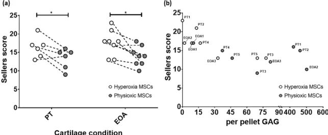
3.5. Paired Analysis Shows An Improvement in Cartilage Regeneration with Physioxic MSCs and A Correlation Between Sellers Score and Per Pellet GAG Content
Comparison of only MSC groups in either model, showed that in both post trauma (5/6) and focal early OA (7/8) models, a better Sellers score was achieved with physioxic MSCs compared with hyperoxic MSCs (* p < 0.05; Figure 11a). Plotting the in vivo Sellers score outcome against in vitro per pellet GAG content for those rabbit donors evaluated for both parameters, showed a tendency towards a lower Sellers score with increased per pellet GAG. However, this trend was not statistically significant (p > 0.05; Figure 11b).
Biology 2020, 9, x FOR PEER REVIEW 16 of 22
3.5. Paired Analysis Shows An Improvement in Cartilage Regeneration with Physioxic MSCs and A Correlation Between Sellers Score and Per Pellet GAG Content
Comparison of only MSC groups in either model, showed that in both post trauma (5/6) and focal early OA (7/8) models, a better Sellers score was achieved with physioxic MSCs compared with hyperoxic MSCs (* p < 0.05; Figure 11a). Plotting the in vivo Sellers score outcome against in vitro per pellet GAG content for those rabbit donors evaluated for both parameters, showed a tendency towards a lower Sellers score with increased per pellet GAG. However, this trend was not statistically significant (p > 0.05; Figure 11b).
Figure 11. (a) Dot plot describing paired comparison between physioxia and hyperoxic MSCs
treatment in post-trauma (PT) and focal early OA (EOA) models (*
p < 0.05). (b) A dot plot depictingthe relationship between per pellet GAG and subsequent Sellers score outcome for the treated defect.
PT signifies post trauma model and EOA represents focal early OA model with respective animal number given.
4. Discussion
The present investigation sought to understand the effect of physioxic MSCs on cartilage repair in two different clinical scenarios, post trauma and focal early OA cartilage lesions. For the latter scenario, a focal defect was created using a dental drill that induced osteoarthritic-like changes at the defect margins after six weeks and met the criteria described for focal early OA (Figure 3) [38].
Treating defects with a hyaluronic acid hydrogel seeded with physioxic MSCs resulted in a significant improvement in cartilage repair scores compared with hyperoxic MSCs in both clinical scenarios (Figures 7–10).
In vivo studies evaluating cell-based therapeutic strategies for cartilage repair have primarily focused on post-traumatic defects and late stage OA. In the latter, current animal models induce a diffusive OA throughout the joint through either intra-articular enzyme injection (e.g., collagenase or papain) or resection of joint structures (e.g., meniscus or ACL) [27,28]. However all these methods for OA induction are non-reversible, whereas the clinical target for cell-based regenerative approaches to OA are focal lesions rather than diffusive OA. Animal models that create a focal early OA lesion with no joint instability or changes in joint structures are needed to evaluate regenerative approaches. Existing focal OA models only induce a mild degeneration in the joint and they do meet the minimal criteria for early OA, e.g., impaction or groove models [39].
In the present investigation, a dental drill device was used to create a focal defect in the posterior portion of the medial femoral condyle due to the high biomechanical load applied during joint movement that would lead to a more degenerative lesion with time [40,41]. The strength of the model is that it induces a reproducible and consistent focal defect that develops into a focal early OA local to the surrounding area rather than throughout the defect. Furthermore, this model can be used to evaluate the efficacy of treating focal early OA lesions over time using methods that either stop or
Figure 11.
(a) Dot plot describing paired comparison between physioxia and hyperoxic MSCs treatment in post-trauma (PT) and focal early OA (EOA) models (* p
<0.05). (b) A dot plot depicting the relationship between per pellet GAG and subsequent Sellers score outcome for the treated defect.
PT signifies post trauma model and EOA represents focal early OA model with respective animal number given.
4. Discussion
The present investigation sought to understand the effect of physioxic MSCs on cartilage repair in two different clinical scenarios, post trauma and focal early OA cartilage lesions. For the latter scenario, a focal defect was created using a dental drill that induced osteoarthritic-like changes at the defect margins after six weeks and met the criteria described for focal early OA (Figure 3) [38].
Treating defects with a hyaluronic acid hydrogel seeded with physioxic MSCs resulted in a significant improvement in cartilage repair scores compared with hyperoxic MSCs in both clinical scenarios (Figures 7–10).
In vivo studies evaluating cell-based therapeutic strategies for cartilage repair have primarily focused on post-traumatic defects and late stage OA. In the latter, current animal models induce a diffusive OA throughout the joint through either intra-articular enzyme injection (e.g., collagenase or papain) or resection of joint structures (e.g., meniscus or ACL) [27,28]. However all these methods for OA induction are non-reversible, whereas the clinical target for cell-based regenerative approaches to OA are focal lesions rather than diffusive OA. Animal models that create a focal early OA lesion with no joint instability or changes in joint structures are needed to evaluate regenerative approaches.
Existing focal OA models only induce a mild degeneration in the joint and they do meet the minimal
criteria for early OA, e.g., impaction or groove models [39].
Biology2020,9, 230 16 of 21