Microenvironment of osteoarthritic cartilage and subchondral bone influences chondrogenic differentiation, extracellular matrix production and composition of bone
marrow-derived stem cells and articular chondrocytes
Dissertation to obtain the doctoral degree (Dr. rer. nat.) in natural science from the Faculty of Biology of the University Regensburg
By
Michaela Leyh (geb. Priller)
from Karlskron, Germany
-2014-
This work was carried out between September 2008 and May 2014 at the Department of Experimental Orthopedics of the University Hospital of Regensburg, Centre for Medical Biotechnology, Germany.
Submitted December 2014
Under the supervision of Prof. Dr. Rainer Deutzmann and Prof. Dr. Susanne Grässel
Request for examination submitted on: November 2014
Date of examination: 9 April 2015
Examination board: Chairman: Prof. Dr. Arne Dittmer First reviewer: Prof. Dr. Rainer Deutzmann Second reviewer: Prof. Dr. Susanne Grässel External reviewer: Prof. Dr. Reinhard Sterner
<Table of content
Table of content
Table of content --- 3
Acknowledgments --- 6
Abbreviations --- 7
Abstract --- 10
Zusammenfassung auf Deutsch --- 12
1 State of the Art / General introduction --- 14
1.1 Epidemiology of Osteoarthritis (OA) in human joints --- 14
1.2 Biology of articular cartilage --- 14
1.2.1 Organization of articular cartilage --- 14
1.2.2 Differences between normal and OA cartilage --- 15
1.2.3 Biomechanical properties of cartilage --- 18
1.3 Biology of subchondral bone --- 19
1.3.1 Organization of subchondral bone --- 19
1.3.2 Differences of normal and OA subchondral bone --- 21
1.4 Biology of bone marrow derived mesenchymal stem cells (BMSC) --- 22
1.4.1 BMSC and their regenerative potential --- 22
1.4.2 Chondrogenic differentiation of BMSC --- 24
1.4.3 BMSC as trophic mediators --- 26
1.5 Cartilage repair and tissue engineering --- 27
1.5.1 Cartilage repair studies --- 27
1.5.2 Biomaterials and fibrin in tissue engineering --- 28
1.5.3 Coculture models in tissue engineering --- 29
1.6 Animals in cartilage trauma repair models --- 33
1.6.1 Animal models in cartilage repair --- 33
1.6.2 Ovine cartilage repair models --- 34
2 Aim of the thesis --- 36
3 Material and methods --- 38
3.1 Culture and isolation of articular cartilage, subchondral bone and chondrocytes--- 38
3.2 Culture and isolation of mesenchymal stem cells (BMSC) and adipose-derived stem cells (ASC) --- 40
3.3 Flow cytometric characterization of BMSC and differentiation into different lineages --- 41
3.4 Cocultivation of fibrin gel embedded BMSC/chondrocytes during chondrogenic differentiation --- 43
3.5 Stimulation of fibrin gel embedded BMSC and/or chondrocytes --- 44
<Table of content
3.6 Cell vitality and proliferation in fibrin gel cocultures --- 45
3.7 Isolation and quantification of messenger RNA --- 46
3.8 Histology and immunofluorescence of fibrin gel embedded cultures --- 48
3.9 Biochemical analysis of fibrin gel cell lysates --- 48
3.9.1 DMMB Assay --- 48
3.9.2 Collagen I and II ELISA --- 49
3.9.3 Collagen III Dot-Blot --- 49
3.10 Immunoblotting of Sox9 and phospho Sox9 --- 49
3.11 Analysis of supernatants (DMMB und hydroxyproline-assay, PTHrP, Fibronectin, bFGF, IL-1β, IL-6 and IL-8 ELISA) --- 51
3.11.1 Hydroxyproline -assay --- 51
3.11.2 PTHrP, fibronectin, bFGF, IL-1ß, IL-6 and IL-8 ELISA --- 51
3.11.3 DMMB Assay --- 52
3.12 Screening for differentially expressed factors in coculture supernatant with LC-MS --- 52
3.13 Biomechanical testing--- 53
3.14 Statistical analysis --- 54
4 Results --- 55
4.1 Part I: Microenvironment of cartilage influences chondrogenic differentiation and ECM production of BMSC and chondrocytes--- 55
4.1.1 Bone marrow derived BMSC can be differentiated into different lineages --- 55
4.1.2 Cells embedded in fibrin gel co- or tricultured with OA-cartilage remain vital and proliferative for up to 28 days --- 56
4.1.3 mRNA expression of differentiation/dedifferentiation markers is altered in cartilage cocultures --- 57
4.1.4 Microenvironment of cartilage alters immunofluorescent staining pattern of collagens --- 58
4.1.5 Collagen I, II and III and GAG contents in fibrin gel cell lysates of mono-, co- and tricultures --- 60
4.1.6 Sox9 is not influenced by OA-cartilage --- 62
4.1.7 Comparison of soluble ECM fragments, PTHrP, bFGF and cytokines in supernatants of mono-, co- and tricultures --- 63
4.1.8 LC-MS analysis revealed differentially expressed factors in BMSC cartilage coculture supernatants --- 66
4.1.9 Microenvironment of OA-cartilage alters biomechanical properties of new extracellular matrix --- 67
4.2 Part II: Microenvironment of subchondral bone influences chondrogenic differentiation and ECM production of BMSC and chondrocytes --- 69
<Table of content
4.2.1 Cells embedded in fibrin gel and co- or tricultured with subchondral bone remain vital
for up to 28 days --- 69
4.2.2 mRNA expression of differentiation/dedifferentiation markers is altered in subchondral bone co- and tricultures --- 69
4.2.3 Microenvironment of subchondral bone alters immunofluorescent staining pattern of collagens --- 72
4.2.4 Fibrin gel cell lysates reveal different GAG, collagen I, II and III content in mono-, co- and tricultures --- 74
4.2.5 Sox9 in BMSC is not influenced by subchondral bone --- 75
4.2.6 Comparison of soluble ECM molecule fragments, PTHrP and cytokines in supernatants of mono-, co- and tricultures --- 76
4.2.7 Microenvironment of OA-subchondral bone alters biomechanical properties of newly regenerated cartilage --- 80
4.3 Part III: Effect of IL-1β, IL-6 or IL-8 stimulation on chondrogenic differentiation and ECM production of BMSC and chondrocytes--- 82
4.3.1 mRNA expression of (de)-differentiation markers is altered in cytokine stimulated fibrin gels --- 82
4.3.2 Stimulation with cytokines revealed different GAG, collagen I, II and III content in fibrin gel lysates --- 86
4.4 Part IV: Influence of normal ovine cartilage on mRNA expression of ovine BMSC, mixed and chondrocyte cultures --- 87
5 Discussion --- 89
5.1 General discussion --- 89
5.2 Part I: Microenvironment of cartilage coculture influences BMSC differentiation and ECM production --- 93
5.3 Part II: Microenvironment of subchondral bone coculture influences BMSC differentiation and ECM production --- 103
5.4 Part III: Stimulation with IL-1β, IL-6 and IL-8 during chondrogenesis --- 110
5.5 Part IV: Influence of coculture with normal ovine cartilage on BMSC differentiation --- 114
6 Summary and conclusion --- 115
References --- 117
Publications, Awards, Posters and Presentations --- 129
Acknowledgments
Acknowledgments
I want to thank Prof. Dr. Susanne Grässel, Department of Orthopedics, University Hospital Regensburg, for being my mentor and greatly contributing to the development of my thesis. The versatile administrative and academic skills that I have acquired during the last few years are due to her excellent support. The gained skills and the achieved qualification are a good basis for a successful career. I cannot thank her enough.
Special thanks to Prof. Dr. Rainer Deutzmann, Department of Biology and Preclinical Medicine, University of Regensburg for allowing me to work on this thesis under his patronage. He was supporting the success of my thesis by LC-MS analysis together with Dr. Astrid Bruckmann, who I also want to thank for her abundant help and guidelines.
I am also deeply grateful that Prof. Dr. Joachim Grifka, Director of the Department of Orthopedics, University Hospital Regensburg, provided the basic conditions for the accomplishment of this work.
An exceptional thank to all of my amiable colleagues, especially to Lilly Weger for her excellent technical assistance, for being a great friend and a mental support. She really made my days brighter.
I also want to convey my gratitude to Prof. Dr. Jürgen Strutz and to Prof. Dr. Holger Gassner, Department of Otorhinolaryngology, University Hospital Regensburg, who made it possible that I could finish my doctoral thesis in parallel to my duties in their lab. I am indebted to the remarkable indulgence and support of Dr. Frank Haubner and Petra Eberl.
Additionally, I want to thank Jens Schaumburger and Hans-Robert Springorum for being an abundant source of bone marrow and explanted knee joints, Johann Haas (Landgasthof Haas, Karlskron) and Albert Obermayr (Metzgerei Obermayr, Deimhausen) for provision of ovine femur from slaughterhouse waste and Drs. Uta Delling and Henriette Juelke for isolation of ovine BMSC.
Grateful thanks to my collaboration partners Dr. Lutz Dürselen and Andreas Seitz, University of Ulm, Institute of Orthopedic Research and Biomechanics, ZMFU, who supported me with generation, analyses and interpretation of the biomechanical properties.
I also want to thank R. Holmdahl and K. Rubin from the Developmental Studies Hybridoma Bank, University of Iowa, who developed the CIIC1 antibody under the auspices of the NICHD.
Finally, I want to thank my family and friends for being a great pool of hope, for their encouragement to never give up and to brighten my mood. Especially I have to thank my parents for their financial support that enabled me to finalize my thesis.
This study was supported by funds from the DFG (GR 1301/8-1) and from the German Society for Orthopedics and Orthopedic Surgery (DGOOC).
Abbreviations
Abbreviations
3D Three-dimensional ACAN Aggrecan gene ALP Alkaline phosphatase
APS Ammonium peroxodisulfate solution ASC Adipose derived stem cells
BCA Bicinchoninic acid
bFGF basic fibroblast growth factor BMSC Bone marrow derived stem cells BMP Bone morphogenetic protein BSA Bovine serum albumin cDNA Complementary DNA
Ch Chondrocytes
Col Collagen
COMP Cartilage oligomeric matrix protein Ct Threshold cycle
Da Dalton
DAPI 4´,6´-diamidino-2-phenylindole, dihydrochloride DMEM Dulbecco´s modified eagle´s medium
DMMB Dimethylmethylene blue DMSO dimethylsulfoxide DNA Deoxyribonucleic acid
dNTP mixture of deoxyribonucleotides dATP, dCTP, dGTP, dTTP E. coli Escherichia coli
ECM Extracellular matrix
EDTA Ethylene-diamine-tetra acetic acid
Abbreviations
ELISA enzyme linked immunosorbent assay et al. et alii (from Latin: and others) EtOH Ethanol
E∞ equilibrium modulus
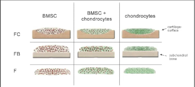
F monocultures (without subchondral bone explants) FACS Fluorescence activated cell sorting
FB co- and tricultures with subchondral bone explants FC co- and tricultures with articular cartilage explants FCS Fetal calf serum
g gravitation
GAG Glycosaminoglycan HA aggregate modulus
IBMX 3-isobutyl-1-methylxanthine IGF Insulin-like growth factor Ihh Indian hedgehog homolog IL Interleukin
k hydraulic permeability kb kilo bases
LC-MS Liquid chromatography–mass spectrometry LDH Lactate dehydrogenase
MALDI-TOF Matrix Assisted Laser Desorption/Ionization- Time Of Flight Mixed mixed cultures of BMSC and chondrocytes
MMP Matrix metalloprotease mRNA messenger RNA
MW molecular weight
n number of independent samples
N Newton
Abbreviations
NB normal bone
OA osteoarthritis
oBMSC ovine bone marrow derived stem cells OD optical density
PAGE Polyacrylamide gel electrophoresis PBS Phosphate buffered saline
PCR Polymerase chain reaction PCNA Proliferating cell nuclear antigen PDGF Platelet-derived growth factor PFA Paraformaldehyde
PTHrP Parathyroid hormone related peptide qPCR Quantitative PCR
RNA Ribonucleic acid
RT-PCR Reverse transcriptase PCR SD Standard deviation
SDS Sodium dodecyl sulphate
Sox Sex related homeobox containing transcription factors TEMED Tetramethylethylenediamine
TGF-β Transforming growth factor beta TNF-α Tumor necrosis factor-α
Tris Tris(hydroxymethyl)-aminomethan
U units
Abstract
Abstract
Objective
Osteoarthritis (OA) is characterized by an imbalance in cartilage and subchondral bone homeostasis, which could be potentially treated or improved by cell-based therapies. In the present study, a reproducible in vitro coculture model was established to evaluate the influence of normal and OA- cartilage or subchondral bone explants on chondrogenic differentiation of human bone marrow derived stem cells (BMSC), human adipose derived stem cells (ASC) and phenotype of OA- chondrocytes. Signals from the articular cartilage or the underlying subchondral bone were hypothesized to induce phenotypic shifts and an altered re- and differentiation potential in cocultured BMSC, ASC and differentiated chondrocytes from OA patients. To provide a chondrogenic environment, cells were embedded in fibrin gel and kept in chondrogenic medium for up to 28 days.
Methods
A reproducible coculture model of ASC, BMSC, mixed cultures (BMSC and chondrocytes in equal ratio) and chondrocytes embedded in fibrin gel seeded on articular cartilage or subchondral bone explants (= co- and tricultures) compared with monocultured cells in fibrin gel without explants was established. Human OA-tissues, OA-chondrocytes and BMSC were derived from patients subjected to arthroplasty. Normal (healthy) human tissues were received from knees of rare trauma affected donors (treated for sports accidents). ASC were isolated from subcutaneous fat tissue obtained from patients undergoing elective body contouring procedures. Ovine cartilage, chondrocytes and BMSC were obtained from normal (healthy) pasture sheep. Gene expression analysis, biochemical assays (ELISA, Dot-blot, DMMB, Hydroxyproline), immunofluorescence staining and biomechanical tests were used to characterize the properties of newly generated extracellular matrix (ECM) from chondrocytes and chondrogenically differentiated ASC and BMSC.
Results
In general, all cell regimens cocultured with OA-explants exhibited reduced gene expression patterns of collagens I, II, III and X in comparison with monocultures. Significant lower levels of collagen I and II protein (BMSC) and significant lower collagen I and III protein (mixed cultures) were detected in cartilage co- or tricultures, while co- and triculture with subchondral bone inhibited collagens in general. In contrast, no changes in glycosaminoglycan (GAG) synthesis were observed for cartilage co- and tricultures, while reduced GAG production was observed in subchondral bone co- and triculture lysates. Repetition of key experiments with normal ASC confirmed inhibitory effects for OA- subchondral bone. Co- and triculture with normal cartilage or subchondral bone explants showed no
Abstract
or only reduced inhibitory effects on chondrogenic differentiation or collagen gene expression. In addition, biomechanical properties of the OA-cartilage or subchondral bone co- and tricultured cell- fibrin gels were affected. All co- and triculture regimens tended to exhibit lower Young´s modulus and aggregate modulus compared with monocultures. In contrast, hydraulic permeability seemed to be higher in co- and tricultures. Supernatants of cartilage and subchondral bone co- and tricultures contained significant higher IL-1β, IL-6 and IL-8 levels, as well as significant more soluble GAGs compared with controls. In general, stimulation of monocultures with IL-1β induced matrix metalloproteinase (MMP)2, and MMP3 and reduced collagens I, II and X gene expression and led to a downregulation of aggrecan gene expression. Stimulation with IL-6 reduced aggrecan, MMP3 and MMP13 gene expression and mainly reduced collagen I, II and III gene expression of BMSC and/or chondrocytes. In contrast, IL-8 stimulation of mixed and chondrocytes monocultures had only little effects, while IL-8 stimulated BMSC showed reduced collagen I, II and III gene expression.
Conclusions
Taken together our results suggest an inhibitory effect of factors from the microenvironment of OA- cartilage and subchondral bone on production of collagens. This indicates a distinct modulatory influence, which affects the composition of the de novo produced ECM from cocultured cells and leads to impaired mechanical strength and biochemical properties of the newly formed matrix.
Experiments with ASC, normal cartilage and subchondral bone explants either might hint to disease status induced effects (OA vs. trauma) or to an effect caused by different mean age of cell and tissue donors. Soluble signal factors, i.e. pro-inflammatory cytokines (including IL-1β and IL-6), released from OA-cartilage, OA-subchondral bone, chondrocytes and osteoblasts, might partly mediate these effects on newly formed extracellular matrix properties. Thus, the microenvironment of neighbored OA-cartilage seems to provide both: promoting and inhibiting signals for BMSC differentiation and suggests that the balance of these factors determines the destiny of BMSC. This knowledge can be used to develop new strategies for cell based cartilage regeneration.
Zusammenfassung auf Deutsch
Zusammenfassung auf Deutsch
Die Mikroumgebung von arthrotischem Knorpel und subchondralem Knochengewebe beeinflusst die chondrogene Differenzierung sowie die ECM Produktion und
Zusammensetzung von BMSC und artikulären Chondrozyten
Charakteristisch für Osteoarthrose (OA) ist ein Ungleichgewicht der Knorpel- und subchondralen Knochen Homöostase. Die Behandlung und der Verlauf dieser Erkrankung könnte durch zellbasierte Therapien deutlich verbessert werden. In dieser Studie wurde ein reproduzierbares in vitro Kokulturmodell entwickelt, um den Einfluss von OA-Knorpel und -Knochen Explantaten auf die chondrogene Differenzierung von adipogenen Stammzellen (ASC), von humanen mesenchymalen Stammzellen aus dem Knochenmark (BMSC) und den Phänotyp von OA-Chondrozyten zu untersuchen. Vermutlich verursachen Signalfaktoren aus dem artikulären Knorpel und der darunterliegenden subchondralen Knochenschicht einen Shift im Phänotyp oder eine Veränderung im (Re-) Differenzierungspotential von ASC, BMSC und differenzierten Chondrozyten. Um eine chondrogene Mikroumgebung zu schaffen, wurden die Zellen in Fibringel eingebettet und bis zu 28 Tage in chondrogenem Medium kultiviert.
In einem Kokulturmodell wurden in Fibringel eingebettete ASC, BMSC, Chondrozyten oder ein Gemisch aus BMSC und Chondrozyten (zu gleichen Teilen) entweder zusammen mit osteoarthrotischem Knorpel oder subchondralem Knochen kultiviert (= Ko- und Trikultur) und mit den entsprechenden Zellen in Monokultur ohne Explantat verglichen. Alle humanen OA- Gewebestücke wurden aus Patienten entnommen, die einer Arthroplastik unterzogen wurden.
Gesundes humanes Gewebe wurde aus Knien von Unfallpatienten gewonnen, die wegen einer Sportverletzung behandelt wurden. ASC wurden aus Unterhautfettgewebe isoliert, das von Patienten einer kosmetischen Fettabsaugung stammt. Genexpressionsanalysen, biochemische Assays (ELISA, Dot-blot, DMMB, Hydroxyprolin), Immunfluoreszenzfärbungen und biomechanische Tests wurden verwendet, um die Eigenschaften der neu gebildeten Matrix von Chondrozyten und chondrogen differenzierten BMSC zu bestimmen.
Im Allgemeinen zeigten alle kokultivierten Zellbedingungen verminderte Genexpression von Kollagen I, II, III und X verglichen mit Monokulturen. Auf Proteinebene konnte signifikant weniger Kollagen I und II (BMSC) und signifikant weniger Kollagen I und III (gemischte Kulturen) in den Knorpel-Ko- und Trikulturen nachgewiesen werden. Die Ko- und Trikultur mit subchondralem Knochen zeigte eine generelle Verminderung der Expression aller Kollagene. Im Gegensatz dazu konnte keine Änderung in
Zusammenfassung auf Deutsch
der Glykosaminoglykan (GAG) Synthese der Knorpel Ko- und Trikultur nachgewiesen werden, während in der Ko- und Trikultur mit subchondralem Knochen temporär eine Reduktion der GAG Synthese beobachtet werden konnte. Die Wiederholung von Schlüsselexperimenten mit gesunden ASC konnte die inhibitorischen Effekte induziert durch Ko- und Trikultur mit OA-subchondralem Knochen bestätigen. Ko- und Trikultur mit normalem Knorpel oder subchondralem Knochen zeigte keine oder nur verminderte inhibierende Effekte auf die chondrogene Differenzierung oder Gen Expression von Kollagen. Die biomechanischen Eigenschaften der kokultivierten Fibringele waren ebenfalls beeinträchtigt. Alle Ko- und Trikulturbedingungen zeigten tendenziell ein geringeres Elastizitätsmodul und Aggregationsmodul im Vergleich zur Monokultur. Im Gegensatz dazu schien die hydraulische Permeabilität in den Ko-und Trikulturen höher zu sein. Die Überstände von Knorpel- und Knochen Ko- und Trikulturen enthielten im Vergleich mit den Kontrollen signifikant mehr IL-1β, IL-6 und IL-8, und auch mehr lösliche GAGs, die größtenteils von den Explantaten stammten. Die Stimulation der Monokulturen mit IL-1β induzierte im Allgemeinen MMP2, MMP3 und MMP13 und reduzierte die Genexpression der Kollagene I, II und X sowie von Aggrekan. Die Stimulation mit IL-6 verminderte die Aggrekan, MMP3 und MMP13 Genexpression und größtenteils auch der Kollagene I, II und III in BMSC und/oder Chondrozyten. Im Gegensatz dazu hatte die Stimulation mit IL-8 nur geringe Effekte auf gemischte und Chondrozyten Monokulturen, während in den IL-8 stimulierten BMSC eine Reduktion der Kollagene I, II und III beobachtet werden konnte.
Zusammenfassend deuten die Ergebnisse dieser Studie auf einen inhibitorischen Effekt von Faktoren aus der Mikroumgebung von OA-Knorpel oder -subchondralem Knochen auf die Kollagenproduktion hin. Diese Ergebnisse lassen auf einen modulierenden Einfluss schließen, der zu verminderten mechanischen und biochemischen Eigenschaften der neu gebildeten extrazellulären Matrix führt. Die Experimente mit ASC sowie mit normalen Knorpel und subchondralen Knochen Explantaten könnten entweder auf krankheitsbedingte Effekte (OA vs. Trauma) hinweisen oder auf einen Effekt aufgrund des unterschiedlichen Durchschnittsalters der Zell- und Gewebe Spender. Vermutlich regulieren lösliche Signalfaktoren wie z.B. proinflammatorische Zytokine, abgegeben von OA-Knorpel, OA-subchondralem Knochen, Chondrozyten oder Osteoblasten, zumindest teilweise diese negativen Effekte auf die chondrogene BMSC Differenzierung und die Qualität der neu gebildeten ECM.
Dennoch birgt die direkte Mikroumgebung von OA-Knorpel beides: fördernde und inhibierende Signale der BMSC Differenzierung. Dies lässt darauf schließen, dass die Balance dieser Faktoren das Schicksal der BMSC bestimmt. Die in dieser Studie neu gewonnenen Erkenntnisse können in Zukunft dazu beitragen, neue Strategien für die zellbasierte Knorpelregeneration zu entwickeln und vor allem die Qualität des neu gebildeten Knorpels entscheidend zu verbessern.
State of the Art / General introduction
1 State of the Art / General introduction
1.1 Epidemiology of Osteoarthritis (OA) in human joints
The human knee joint is one of the most important sources of pain in the western population, in people over 55 years old and nearly everybody over 65 years is affected by osteoarthritis (OA) (Hunter and Felson 2006). Possible causes for OA development are increasing age, mechanical stress, a high body mass index and a genetic component (Breedveld 2004). OA is a chronic joint disease and develops usually undetected many years before it is diagnosed. Notably, a correlation with pain is not mandatory and OA becomes painful in about 50% of patients (Pereira, Peleteiro et al. 2011). Still it is the leading cause of disability, which besides limitations in motility and quality of life is the high economic burden in form of direct and indirect health-related costs (Felson 2004; Hiligsmann, Cooper et al. 2013). Thus, ineffective treatments focusing only on symptoms - mainly pain reduction – should better include a change of lifestyle like weight reduction, adequate footwear and continuing exercises (Roddy and Doherty 2006). Anyhow, after diagnosis and radiographic confirmation for patients with severe end stage OA, a total replacement of the joint and implantation of a prosthesis is inevitable. Drawback in this regard is that the prosthesis has a limited lifespan and patients risk a revision surgery (Wiegant, van Roermund et al. 2013). Therefore, alternatives for successful OA- treatment are urgently required to regenerate cartilage tissue and avoid implantation of an artificial prosthesis.
1.2 Biology of articular cartilage
1.2.1 Organization of articular cartilage
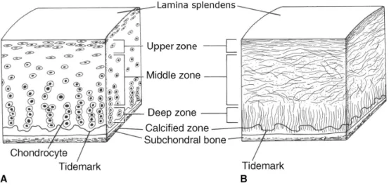
Normal articular cartilage is accurately organized and consists of different zones (Fig. 1); the outermost region is called lamina splendens, named after the light breaking reflections of parallel fibers (Hollander, Dickinson et al. 2010). The upper zone of cartilage is rich in thin collagen fibrils parallel to the articular surface, which protects the cartilage from shear forces. Characteristic is a high collagen content, with an increased collagen I content and lowest amount of aggrecan compared with deeper zones. Cells in the upper cartilage region are relatively flat, small and separated. Additionally, regulatory proteins like TGF-β1 and -3 as well as BMP1-6 (Anderson, Hodges et al. 2000; Yamane, Cheng et al. 2007)and MMPs (-1, -3, -8 and -13) are expressed but are only involved in matrix turnover, not in matrix degradation (Tetlow, Adlam et al. 2001; Tchetina 2011)
State of the Art / General introduction
In the middle zone, resting round shaped chondrocytes are residing in chondrons, surrounded by thick, low organized fibers and an ECM consisting of high collagen II and aggrecan amounts (Pfander, Swoboda et al. 2001).
The deep zone of articular cartilage has the highest aggrecan and lowest water content and the collagen fibrils with the largest diameter, which are oriented perpendicular to the joint surface (Maroudas, Bayliss et al. 1980).Chondrocytes in the deep zone are enlarged and arranged in columns right angled to the cartilage surface (Pearle, Warren et al. 2005).
The deep zone merges into the hypertrophic zone, which is separated by the so-called tidemark region from calcified cartilage that links the cartilage layer with the subchondral bone. The calcified cartilage zone is rich in hydroxylapatit crystals and hypertrophy markers like collagen X, ALP and MMP13 are expressed (Aigner, Hemmel et al. 2001; Pearle, Warren et al. 2005).
Figure 1: Schematic overview of articular cartilage
Articular cartilage is characterized by a specific arrangement of chondrocytes and ECM components. It is divided into several different zones beginning with the lamina splendens at the articular joint surface, followed by the upper, middle and deep zone, which are separated by the tidemark from the calcified zone and the subchondral bone (Drawing is adopted from (Jazrawi, Alaia et al. 2011)).
1.2.2 Differences between normal and OA cartilage
Normal articular cartilage is characterized by a special ECM composition mainly based on proteoglycan (aggrecan) and collagens. Collagen II is the most dominant collagen, which forms a fibrillar structure together with other collagens (XI and IX) as well as with non-collagenous proteins
State of the Art / General introduction
Mature normal articular cartilage has a low cell density as with the end of growth, articular chondrocytes are hardly proliferating and exhibit only a low metabolism. Nevertheless, articular cartilage shows a carefully balanced activity of matrix turnover with respect to cartilage ECM degradation and synthesis (Poole 1997; Buckwalter and Mankin 1998; Tchetina 2011). In general, intact hyaline cartilage shows only low gene expression of collagens I, II, VI, IX, X and XI but a high turnover of aggrecan (Hardingham, Fosang et al. 1994; Aigner and McKenna 2002; Poole 2003).
Importantly, hyaline cartilage has no innervations and no blood flow and thus is characterized by a low supply with nutrients and oxygen. Additionally neither pain nor overloading can be perceived (Benedek 2006). For exactly that reason, a high impact load is unfavorable, because it is able to provoke cartilage damage and bears the risk for OA development (Newberry, Mackenzie et al. 1998).
The impact level and its tenure are important, because the time period of maximal compression is more fatal if it occurs abruptly than gradually despite of the same amplitude. During this kind of strain, rupture of the collagen meshwork could happen if a critical impact is reached (Newberry, Garcia et al. 1998; Torzilli, Grigiene et al. 1999). The rupture of cartilage ECM - typically for traumatic sports injuries - represents a severe structural damage of the joint and induces the beginning of structural cartilage degradation because of constant loading alterations. Additionally it causes post- traumatic cell death, mediates acute joint inflammation and initiates repair mechanisms like an increase of pro-inflammatory cytokines, basic fibroblast growth factor (bFGF), MMPs, aggrecanases, and release of matrix fragments (Buckwalter, Anderson et al. 2013).
Figure 2 shows an overview of initiation and progression of OA including responsible factors and changes in gene expression and phenotype of chondrocytes and cartilage. More detailed, in early OA, cells residing in arthritic cartilage regions become active and restart metabolism. Especially TGF-β and collagen II production is enhanced most probably as an attempt to repair destroyed cartilage (Kouri and Lavalle 2006). Additionally, collagenases, aggrecanases, cytokines, collagen X, Indian hedgehog homolog (Ihh) and caspase 3 are upregulated in early stage cartilage lesions. During progression of OA, the balance of cartilage turnover is disturbed and MMPs – especially MMP13 – aggrecanases, collagen X and alkaline phosphatase (ALP) are further upregulated. In combination with downregulation of Sox9, TGF-β, TNF-α and aggrecan expression, a dramatic loss of aggrecan occurs in the upper zone. Additionally, the natural zonal organization of cartilage breaks down and results in large areas of destroyed inflamed cartilage tissue (Tchetina, Squires et al. 2005; Appleton, McErlain et al. 2007; Tchetina 2011). These regions represent a unique OA-microenvironment with release of high amounts of pro-inflammatory cytokines like IL-1β, IL-6, IL-8, IL-10 or TNF (Moldovan, Pelletier et al. 2000; Lu, Evans et al. 2011),and growth factors like FGF, PTHrP or TGF-β as well as
State of the Art / General introduction
ECM fragments like collagens, fibronectin and soluble GAGs (Tchetina, Kobayashi et al. 2007; Huang and Wu 2008).
Finally, destruction of cartilage is dominant in late OA and ECM production disappears because chondrocytes shift their phenotype from reparative to degradative resulting in total breakdown of cartilage (Kouri and Lavalle 2006). Besides a metabolic imbalance, activation of the whole endochondral ossification program starting with cell proliferation through articular chondrocyte hypertrophy and apoptosis has been identified as an important determinant of OA progression (Alsalameh, Amin et al. 2004; van den Berg 2011). Although the view of a generalized OA- chondrocyte hypertrophy is controversial, signaling molecules relevant for endochondral ossification may be involved in OA pathogenesis. Reactivation of embryonic differentiation pathways underscore the clinical relevance of chondrocyte differentiation associated processes for OA pathogenesis (Dreier 2010; Pitsillides and Beier 2011).
As a result, this alteration in ECM composition and turnover, mainly the loss of proteoglycan, leads to changes in biomechanical properties, like higher permeability and an increased water content causing a decrease in hydraulic pressure. Hence, the diminished stability in weight bearing regions contributes to an enhanced cartilage loss and promotes further stress, inflammation and cell death leading to more pressure on residing chondrocytes (Akizuki, Mow et al. 1987; Lai, Hou et al. 1991;
Setton, Mow et al. 1994; Kouri and Lavalle 2006). Consequently, the microenvironment of OA- cartilage is not comparable with the microenvironment provided by normal articular cartilage.
State of the Art / General introduction
Figure 2: Scheme of initiation and progression of OA
Schematic overview of events initiating early OA and leading to progression of late OA. Possible contributing factors are in the boxes above and cellular and morphological changes are specified below (scheme is adopted from (Goldring and Goldring 2007)).
1.2.3 Biomechanical properties of cartilage
With every step we take our knee joint bears a multiple of our body weight and the articular cartilage has the important duty to distribute the load equally and frictionless over the whole joint. In return, normal articular cartilage has a unique composition of natural biphasic hyaline ECM which is mainly based on water (70-85%), proteoglycans (5-7%), foremost aggrecan, and collagens (10-20%) mainly collagen II (Mow, Holmes et al. 1984; Pearle, Warren et al. 2005).In the porous and permeable solid matrix, aggrecan is responsible for properties like elasticity and compressibility (Chambers, Cox et al.
2001; Lu and Mow 2008), while collagen II is in charge of shear and tensile properties of cartilage which enables a high viscoelastic and mechanical stability (Pearle, Warren et al. 2005). At last, water contributes 90% to load transmission on account of low permeability of cartilage. Pressurization of water provides an equal weight distribution and protects the solid phase of the matrix from load burden (Maroudas 1976; Mankin 1982; Soltz and Ateshian 1998).
In case of cartilage, diseases like OA, a rupture of tendons, damage of meniscus or lesions in the cartilage due to age, the normal/optimal distribution of weight is disturbed and the mechanical stimulation of additional local pressure on cartilage and chondrocytes can lead to alterations in cell- gene expression or to further cartilage wear out (Kouri, Aguilera et al. 2000; Abramson, Attur et al.
2006). Moreover, Salter et al. even suggest that OA-cartilage has a different sensing of the mechanical environment compared with normal cartilage leading to an inappropriate response to mechanical stimulation resulting in progression of disease by changes in the ECM and MMP gene expression (Salter, Hughes et al. 1992; Millward-Sadler, Wright et al. 2000; Salter, Millward-Sadler et al. 2002).
Key roles in biomechanical properties have, among others, the correct deposition of proteoglycans and collagens. Collagen fibrils which form the fibrillar matrix and proteoglycans which form the extra- fibrillar matrix determine the level of hydration and stability of the ECM and due to that the mechanical properties of articular cartilage. Therefore, GAG and collagen contents of ECM-tissue are useful for prediction of mechanical tissue properties (Steinert, Ghivizzani et al. 2007; Yan, Zhou et al.
2009).
In general, analysis of biomechanical properties covers attributes of stiffness, elasticity, compressibility and permeability (Busby, Grant et al. 2013). These attributes are dependent on load,
State of the Art / General introduction
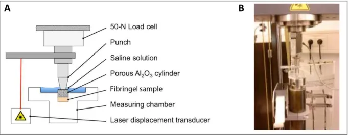
time and type of compression (Gannon, Nagel et al. 2012). Unconfined compression is a simple laboratory test method where no radial space limitations occur and specimens were compressed uniaxial (Hatami-Marbini and Etebu 2013). Confined compression is performed in a confining chamber, which simulates the in situ situation. That means the top and bottom of a cylindrical specimen is limited in space and only e.g. buffer solution is able to disappear via an effluent.
Therefore, the fibrin gel is loaded hydrostatically to the desired confining pressure on all sides. The resulting measured parameters are used for calculation of common specimen properties (Mow and Guo 2002; Busby, Grant et al. 2013).
Biomechanical properties of a tissue are defined by the following parameters: Young´s modulus, aggregate modulus and hydraulic permeability. More detailed, Young´s modulus provides information about the stiffness and is performed under unconfined compression. Aggregate modulus provides information about the compressibility and hydraulic permeability about the flow rate of water (both applied under confined compression) (Mow and Guo 2002; Busby, Grant et al. 2013).
1.3 Biology of subchondral bone 1.3.1 Organization of subchondral bone
The so-called subchondral bone of the human knee joint is located beneath the layer of articular cartilage (Sanchez, Deberg et al. 2005). In contrast to articular cartilage, which has only one resident cell type (chondrocytes), subchondral bone consists of different cell types: osteoblasts, osteocytes, osteoclasts, and due to the bone marrow also of HSC and BMSC. The main cell-type, osteoblasts, are bone forming cells with an active metabolism secreting ECM components like collagen I which accounts for about 90% of the non mineralized bone matrix. The remaining 10% of non-mineralized bone matrix consists of glycoproteins and proteoglycans. Osteoblasts are also responsible for mineralization of the matrix, by secretion of proteins that are essential for mineralization like alkaline phosphatase (ALP), osteocalcin and osteopontin. When osteoblasts are trapped in the ECM they have produced by themselves, they become osteocytes (Grabowski 2009). Recent discoveries show that osteocytes are multifunctional cells, with important regulatory functions in bone and mineral homeostasis (Dallas, Prideaux et al. 2013; Li, Song et al. 2014). They are perceptive to mechanotransduction and turn mechanical stress into endocrine signals, which regulate bone remodeling of osteoclasts and osteoblasts (Bonewald 2002; Dallas, Prideaux et al. 2013). Another important cell type in bone are osteoclasts, which arise through fusion of hematopoietic cells of the monocyte/macrophage lineage and thus are characterized by numerous cell nuclei. Osteoclasts are in charge of bone resorption and remodeling and can be found next to remodeling bone surfaces
State of the Art / General introduction
were they secrete acid and proteolytic enzymes forming a resorption lacuna. They are known to be attracted by chondrocytes, which release MMPs and generate fragments of ECM molecules that allure osteoclasts and promote vascularization (Grabowski 2009).
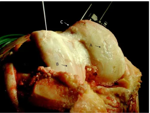
Since the adjective ‘‘subchondral’’ is vague, in the present study the subchondral bone is defined as a layer of bony lamella beneath the calcified cartilage, which are separated by the cement line (Fig. 3).
Though the structure of the subchondral bone is very inconsistent, it always consists of a thin woven layer of the subchondral bone plate and the subarticular spongiosa (trabecular bone), which expands into the marrow cavity, a supporting three-dimensional structure with impact absorbing properties (van der Harst, Brama et al. 2004). Notably, the structure and assortment of the mineral content and the organic bone matrix assigns properties of subchondral bone like absorption capacity or distribution and transfer of load (Meunier and Boivin 1997; Day, Ding et al. 2001; Donnelly, Chen et al. 2010).
Subchondral bone primarily is based on collagen I fibrils which form parallel sheets continuing to the lamellae of the bone trabeculae (Inoue 1981), which are arranged in right angles to the joint surface and are perpendicular crossed by smaller trabeculae (Meachim and Allibone 1984). In a section tangential to the articular surface, little spaces built of joined plates look like a honeycomb. Further from the surface, these spaces enlarge and progressively extend right angular to the articular surface to form the subarticular spongiosa (Singh 1978). A direct connection of the uncalcified cartilage with the spongiosa cavities is provided by little spaces that form an osteochondral connection via vascular channels (Green, Martin et al. 1970; Inoue 1981; Milz and Putz 1994).
In contrast to the tidemark, which is crossed by collagen fibrils, no collagen fibrils cross the cement line. Importantly, sympathetic (Grassel 2014) and sensory (Ashraf and Walsh 2008) nerve fibers can grow along with subchondral blood vessels through canals in the subchondral bone plate into the calcified cartilage, causing nutrients and factors to reach chondrocytes in the calcified zone. Calcified cartilage nutrition depends on diffusion, however, a high density of channel formation in mechanically stressed zones suggests a particularly good blood supply as a response to long-term stress (Berry, Thaeler-Oberdoerster et al. 1986; Madry, van Dijk et al. 2010).
Variations in natural organization concern the shape of the cement line, the thickness, density and composition of the subchondral bone plate as well as number and type of perforations going into the calcified cartilage (Madry, van Dijk et al. 2010). Additionally, variations in the trabecular structure and in mechanical properties are reported in weight bearing vs. non-weight bearing areas (Bullough, Yawitz et al. 1985; Armstrong, Read et al. 1995).
State of the Art / General introduction
In summary, the subchondral bone has two main functions: it contributes to shock absorption during mechanical stress and it maintains the articular cartilage with nutrients.
1.3.2 Differences of normal and OA subchondral bone
Research has focused on chondrocytes and cartilage as mediators of OA but there is strong evidence that also other cells and tissues of the joint like synovium or subchondral bone are influenced in OA pathogenesis (Sanchez, Deberg et al. 2005). Because of a close connection to articular cartilage via channels in the cement line, subchondral bone is not just an essential impact absorber, it additionally influences articular cartilage metabolism.
In early OA, the connecting interface between cartilage and subchondral bone is characterized by high rates of bone remodeling and turnover, mainly in the damaged regions. Proliferation of bone cells leads to thickening of the subchondral plate and diminishes the mineral density of subchondral bone, which influences its load bearing properties (Dore, Quinn et al. 2009). In this case, bone deformation under load is raised and a decrease in the elastic modulus can be observed. Under impact, these reduced biomechanical properties (Day, Ding et al. 2001) can lead to cracks or local defects in the subchondral bone plate, where a continuous pressure of water from the cartilage into the subarticular spongiosa causes bone resorption (Aspenberg and Van der Vis 1998).
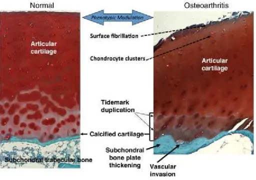
Altered load distribution is followed by changes in anatomy demonstrating that the subchondral bone plate responds sensitively to modifications in the local environment. Since the subchondral bone plate provides mechanical and metabolic functions and is a dynamic location of remodeling, mechanical distress in the interphase of cartilage and bone results in enhanced OA progression (Kouri, Aguilera et al. 2000; Goldring and Goldring 2007), namely an increased remodeling and turnover of subchondral bone, thinning trabecular structures, subchondral bone cysts at the borders of the joint, bone marrow lesions and sclerosis of subchondral plate (Burr 2004; Burr 2004; Felson and Neogi 2004; Karsdal, Leeming et al. 2008). Additionally, calcification of the tidemark region leads to a decrease in cartilage thickness and increase of subchondral plate thickness (Fig. 3) (Lane, Villacin et al. 1977; Hill, Gale et al. 2001; Burr 2004).
Importantly, an OA rat model of the osteochondral junction and tissue samples from OA patients show presence of sensory nerve fibers and expression of nerve growth factor (NGF) in the region of vascular channels. Therefore, osteochondral angiogenesis and sensory fibers probably can cause symptomatic pain (Suri, Gill et al. 2007; Walsh, Bonnet et al. 2007; Ashraf and Walsh 2008).
State of the Art / General introduction
Taken together, each of both anatomically connected tissues - articular cartilage and subchondral bone - is affected by changes in the mechanical properties of the other one (Aspenberg and Van der Vis 1998; Van der Vis, Aspenberg et al. 1998). Therefore, OA has to be seen completely as a disease of the whole joint and not separately of single tissues as it involves both: the articular cartilage and the subchondral bone as shown in figure 3.
Figure 3: Anatomy of normal and osteoarthritic articular cartilage and subchondral bone
Normal human articular cartilage (left side) is separated by the tidemark from a thin zone of calcified cartilage.
Followed by subchondral bone, wich is connected via numerous canals with the calcified cartilage. In contrast, osteoarthritic human articular cartilage (right side) shows clear signs of surface fibrillation and cartilage ECM degradation, in detail, increase of cartilage calcification, duplication of the tidemark and enhanced vascular invasion from subchondral bone (Drawing is adopted from (Goldring 2012)).
1.4 Biology of bone marrow derived mesenchymal stem cells (BMSC) 1.4.1 BMSC and their regenerative potential
There are in general two types of stem cells: embryonic stem cells and adult stem cells. The latter are a rich source for a variety of repair cells, which are able to help out in case of tissue injuries and replace damaged cells (Sylvester and Longaker 2004). They can be found in almost every tissue and can differentiate into a broad range of specialized cells. Stem cells isolated from respective tissues are predestined to differentiate into cells hosted along these lineages. In the bone marrow, even two different types of stem cells coexist, in particular hematopoietic stem cells (HSC) and mesenchymal
State of the Art / General introduction
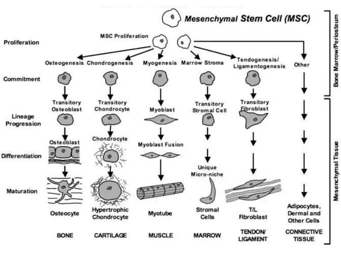
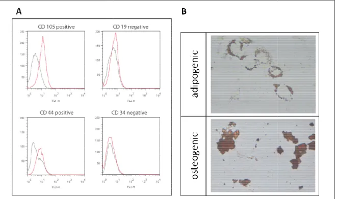
stem cells (MSC) (Sylvester and Longaker 2004). These adult bone marrow derived MSC (BMSC) are capable for differentiation in vitro after an appropriate stimulation into several different lineages and become amongst others adipocytes, osteoblasts or chondrocytes (Pittenger, Mackay et al. 1999; Kolf, Cho et al. 2007) (Fig. 4).
Figure 4: Regenerative potential of MSC
MSC have the potential to differentiate into a broad range of specialized cells and form new tissues like bone (osteocytes), cartilage (chondrocytes), muscle (myocytes), marrow stroma (marrow stromal cells), tendon/ligament (tenocytes/ligament cells), fat (adipocytes) and connective tissues (connective tissue cells).
Growth factors and cytokines control differentiation into the different lineages. Therefore, therapeutically approaches in regenerative medicine, which use adult BMSC are promising as they facilitate tissue repair and regeneration of damaged tissue. (Drawing is adopted from (Caplan and Dennis 2006)).
The differentiation potential of BMSC, implanted into focal cartilage defects and their ability to produce a proper ECM, which integrates closely with the adjacent normal tissue is highly dependent on response of the BMSC to the tissue microenvironment. The microenvironment is provided by cell- cell interactions with surface proteins of neighbor cells via integrins or CD44 and by cell-matrix interactions with insoluble matrix molecules like collagens, glycoproteins and proteoglycans (Lutolf and Hubbell 2005; Steinert, Ghivizzani et al. 2007). Additionally soluble molecules released from the
State of the Art / General introduction
neighboring tissue like growth factors (IGF I, TGF-β, FGF) (Denker, Nicoll et al. 1995), cytokines (IL-1, TNFα) (van den Berg, Joosten et al. 1999) and chemokines (IL-8) are important for proper differentiation (Garner, Stoker et al. 2011). Therefore, an important focus for successful differentiation lies on the interaction of BMSC and their microenvironment. A better understanding of these cues will facilitate regenerative therapies using adult BMSC, which have a promising future in musculoskeletal tissue repair.
1.4.2 Chondrogenic differentiation of BMSC
Regulation of chondrogenic differentiation of BMSC and production of a typical hyaline cartilage involves a variety of molecular mechanisms: stem cell intrinsic factors, paracrine factors derived from neighboring cells like undifferentiated BMSC, osteoblasts or chondrocytes and microenvironmental components independent from cells like availability of nutrients and oxygen, adhesion molecules, ECM epitopes or a 3D surrounding (Coimbra, Jimenez et al. 2004; Goldring, Tsuchimochi et al. 2006;
Birmingham, Niebur et al. 2012). Additionally, chondrogenic differentiation of BMSC and deposition of new ECM can be induced in vitro by stimulation with growth factors like BMPs or TGF-β superfamily members in a controlled culture condition with accurately defined culture medium.
Stimulated BMSC start upregulation of chondrocyte specific genes like fibronectin, N-cadherin, COMP, Collagen IX and during progressed differentiation also of typical articular proteins like collagen II, aggrecan, hyaluronan and chondroadherin (Majumdar, Wang et al. 2001; Sekiya, Larson et al. 2005; Park, Yang et al. 2009).
Molecular mechanisms during the tightly and complex regulated chondrogenic differentiation are not yet fully understood. BMSC pass through several maturation stages in which characteristic factors are activated. After initiation of chondrogenesis, BMSC start cell proliferation and differentiate to chondrocytes under the influence of growth and differentiation factors, like bFGF or bone morphogenetic protein (BMP). In early chondrogenesis, stem cells differentiate to resting chondrocytes with the largest increase in expression of genes coding for cartilage ECM macromolecules like proteoglycans, cartilage oligomeric matrix protein (COMP) and collagen II, IX and XI. In case of hyaline cartilage, this type of chondrocyte is stably maintained in the joint (Mastrogiacomo, Cancedda et al. 2001; Goldring, Tsuchimochi et al. 2006).
Further, interactions of transcription factors, such as Sox9 and Runx2 determine whether chondrocytes remain articular or undergo terminal differentiation before ossification (Mastrogiacomo, Cancedda et al. 2001; Goldring, Tsuchimochi et al. 2006). In embryonic development, chondrocyte differentiation processes during endochondral ossification resulting in
State of the Art / General introduction
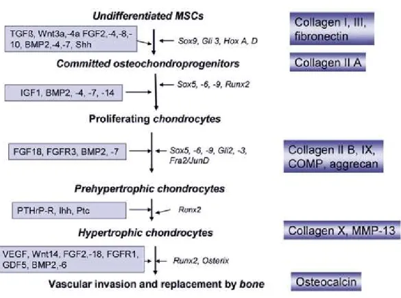
bone formation. Maturing chondrocytes proliferate, increase their metabolic activity and enlarge in size while they deposit collagen X and produce alkaline phosphatase (ALP). The late stage of terminal differentiation is marked by mineralization, vascularization and invasion of bone cells and bone marrow cells (Goldring, Tsuchimochi et al. 2006; Fischer, Dickhut et al. 2010). A common, yet unresolved problem during chondrogenesis of BMSC in vitro is that after 3-4 weeks most of the BMSC exhibit a hypertrophic phenotype.
Figure 5: Chondrogenic differentiation during endochondral bone formation.
Schematic representation of different stages of chondrogenic differentiation of mesenchymal stem cells (MSC).
Growth and differentiation factors are listed on the left side of the arrows and transcription factors on the right side. Stage specific ECM marker proteins are listed in blue boxes at the right side of the figure. (Drawing is adopted from (Grassel and Ahmed 2007))
In the early phase of chondrogenesis the most prominent transcription factors belong to the SOX family genes, mainly SOX9, SOX5 and SOX6 (Fig. 5) (Lefebvre and de Crombrugghe 1998; Sekiya, Tsuji et al. 2000). However, regulation of chondrogenesis is more complex and the progression of chondrogenesis is additionally governed by other transcription factors, DNA-binding proteins, nuclear receptors, matrix proteins, matrix modifiers as MMPs and adhesion molecules, many extracellular ligands and their receptors (Mundlos and Olsen 1997; de Crombrugghe, Lefebvre et al. 2000;
Lefebvre, Behringer et al. 2001). Later, during terminal differentiation mainly PTHrP and Ihh
State of the Art / General introduction
stimulate upregulation of collagen X, ALP and MMP13 in chondrocytes, which synthesize mineralized matrix followed by vascular invasion and cell death at the end (Fig. 5) (Barry, Boynton et al. 2001;
Goldring, Tsuchimochi et al. 2006).
A better understanding of the mechanisms of chondrogenic differentiation and hypertrophy is fundamental for further stem cell based therapies, because for a successful cartilage repair a stable articular phenotype and prevention of terminal differentiation is essential. To obtain a high quality functional regenerative tissue with superior mechanical load capacity, proper differentiated BMSC which produce a highly organized functional ECM are required, as ECM components are strongly correlated to mechanical strength of engineered cartilage-like tissue (Mauck, Yuan et al. 2006).
1.4.3 BMSC as trophic mediators
Moreover, BMSC are trophic mediators, which secrete a variety of cytokines and growth factors that reflect their functional status in their specific microenvironment (Prockop 1997; Caplan and Dennis 2006; Kolf, Cho et al. 2007). These factors are able to feed back to the cell itself and govern the functional status and physiology by inducing intracellular signaling or by instructing another cell in the neighborhood to secrete functionally active factors (Caplan and Dennis 2006). A profound clinical use of BMSC may be due to their trophic effects, when BMSC at the site of an injury produce paracrine and autocrine acting factors that are known to suppress the local immune system, stimulate angiogenesis, reduce scar formation and apoptosis and enhance mitosis and differentiation of residing stem cells into regenerative tissue (Caplan and Dennis 2006). Additionally, the therapeutical potential of BMSC is provided by paracrine secretion of cytokines and chemokines like VEGF, FGF-2 or IL-6 via stimulation of survival pathways, induction of differentiation and stemness or regulation of anti-inflammatory effects (Burdon, Paul et al. 2011).
A recent study by Pricola et al. affirmed that IL-6 is probably a “stemness” factor, which contributes to the MSC undifferentiated status and that IL-6 is necessary for proliferation of MSC, protects from apoptosis and inhibits adipogenic and chondrogenic differentiation in an ERK1/2 dependent pathway (Pricola, Kuhn et al. 2009). BMSC used for therapy of chronically inflammatory diseases as OA have two main functions: Firstly, they can either differentiate into chondrocytes or they can mediate mitosis and differentiation of residing neighbor cells and secondly, BMSC can suppress apoptosis and inflammatory processes by release of trophic factors.
State of the Art / General introduction
1.5 Cartilage repair and tissue engineering 1.5.1 Cartilage repair studies
Most tissues containing stem cells have an elaborated self-repair ambition, with exception of adult articular cartilage. Damaged articular cartilage reveals only minimal or low self-repair capacity, especially in defects of critical size and therefore it urgently requires potent tissue engineering-based therapeutic methods (Caplan, Elyaderani et al. 1997; Cheng, Hardingham et al. 2014).
Lars Peterson and Mats Brittberg were clinical pioneers, who performed the first cell based approach for treatment of full-thickness cartilage or osteochondral lesions via autologous chondrocyte implantation (ACI) in 1994 (Brittberg, Lindahl et al. 1994). Until now, there appeared to be many approaches with promising clinical results for cell based cartilage defect treatment of symptomatic osteochondral defects. Examples are autologous mosaicplasty (Reverte-Vinaixa, Joshi et al. 2013), matrix-assisted autologous chondrocyte transplantation (MACT) or microfracturing with or without biomaterial support. For autologous mosaicplasty, ACI and MACT, cartilage is harvested from a healthy non-weight bearing region of the joint and grafts or ex vivo expanded cells were re- implanted into the defect. Drawbacks of these methods are limited cell and graft availability as well as risk of donor morbidity (damage at the donor site) (Brittberg, Peterson et al. 2003; Bedi, Feeley et al. 2010). During microfracturing, little cavities were created in the subchondral bone leading to bleeding into the defect. Natural occurring stem cells and the developing fibrin-clot built the fundament of defect repair. Anyhow, recent studies showed a risk for promotion of fibrocartilage development by using this method (Harris, Siston et al. 2010; Holtzman, Theologis et al. 2010).
With increasing cell demand for regeneration of large damaged cartilage areas, chondrocytes need to be expanded in vitro, but chondrocytes de-differentiate and their chondrogenic capacities to produce stable cartilage ECM disappear (Mandelbaum, Browne et al. 2007). Hence, re-differentiation capacity of chondrocytes is desirable and studies showed that a 3D environment like it is provided by scaffolds or hydro gels is able to reactivate a chondrogenic phenotype after expansion (Endres, Neumann et al.
2012). An alternative autologous cell source to replace chondrocytes are chondrogenic differentiated BMSC, which have a high chondrogenic differentiation potential triggered by growth factors and cytokines (Caplan and Dennis 2006). Notably, regulatory obstacles in clinical use of BMSC-based treatments are impeding progress of tissue engineering strategies for cartilage repair (Grassel and Lorenz 2014).
Nevertheless, BMSC might be superior to expanded chondrocytes or cartilage grafts, as they are well expandable in vitro, retain their differentiation capacity over several passages and are capable of cartilage like matrix production in a proper environment. As a result, therapeutically approaches in
State of the Art / General introduction
regenerative medicine which use adult BMSC, would be promising as they might facilitate tissue repair and regeneration of damaged cartilage.
1.5.2 Biomaterials and fibrin in tissue engineering
Interaction between BMSC and ECM components provides an instructive microenvironment and contributes to differentiation of BMSC into chondrocytes. This suggests a beneficial effect by using BMSC in conjunction with synthetic or natural scaffolds, which provide a 3D environment (Lutolf and Hubbell 2005; Lee, Yu et al. 2008). Additionally, a conductive effect of a 3D culture is given for chondrocytes, which maintain their articular character instead of de-differentiating in monolayer (Schagemann, Mrosek et al. 2006; Francioli, Candrian et al. 2010).
Several studies proved the suitability of different 3D culture models for cartilage repair, like collagen or gelatin scaffolds (George, Kuboki et al. 2006; Brochhausen, Sanchez et al. 2013) and hydrogels including photopolymerized hyaluronic acid, agarose (Williams, Kim et al. 2003), Matrigel, PuraMatrix (Dickhut, Gottwald et al. 2008) or alginate (Ho, Cool et al. 2010).
A commonly used hydrogel scaffold is based on fibrin (fibrin gel or fibrin glue) that encapsulates BMSC or chondrocytes and mimics the structure and function of a natural ECM by maintaining a round morphology of cells and thus facilitating deposition of new ECM components (Dickhut, Gottwald et al. 2008; Ho, Cool et al. 2010). Fibrin is a natural non-cytotoxic, biocompatible and biodegradable polymer that can be smoothly polymerized and casted from its basic constituents and has been in clinical use for several years. A big advantage of fibrin gel is a close contact of the implanted BMSC/chondrocytes to the defect tissue borders, as it can directly integrate. Additionally, various in vivo and in vitro studies demonstrate the potential of fibrin gels in the field of tissue engineering together with human or animal cells. The benefit of combining cells with fibrin is well established for cartilage regeneration, since numerous studies managed to successfully repair defects in animal models. For example, Fussenegger et al. accomplished to reconstruct cartilage in a rabbit-model using stabilized autologous fibrin-chondrocyte constructs (Fussenegger, Meinhart et al.
2003), Sims et al. produced a well formed cartilaginous matrix using bovine chondrocytes suspended in fibrin glue in vivo (Sims, Butler et al. 1998) and Mesa et al. demonstrated that ovine articular chondrocytes from 8 year old sheep can be rejuvenated in vivo when encapsulated in fibrin gel (Mesa, Zaporojan et al. 2006). Moreover, fibrin constructs containing rabbit BMSC which were provided with constant levels of growth factors during culture time enable the development of articular cartilage in vitro and in vivo (Park, Yang et al. 2009). Even a human pilot study using fibrin- ACI reports good clinical improvement 2 years after surgery with a favorable clinical outcome and
State of the Art / General introduction
regenerated tissue shows evidence for similar characteristics to normal cartilage (Kim, Choi et al.
2010).
Depending on the experimental setup, some studies show that fibrin is degraded too fast accompanied with extensive shrinkage or replacement by fibrous tissue (Homminga, Buma et al.
1993; van Susante, Buma et al. 1999), while other groups were successful with cartilage reconstruction (Ting, Sims et al. 1998). Degradation of fibrin can be slowed by addition of high concentrations of antifibrinolytic substances like aprotinin and tranexamic acid or by elevating the fibrinogen/thrombin ratio causing alterations in the crosslinking during polymerization (Fussenegger, Meinhart et al. 2003).
Taken together, a 3D environment, as is provided by biodegradable porous scaffolds or hydro gels like fibrin gel, positively contributes to chondrogenic differentiation capacity and ECM production of BMSC and is beneficial for cartilage neogenesis (Williams, Kim et al. 2003; Erickson, Huang et al.
2009).
1.5.3 Coculture models in tissue engineering
1.5.3.1 Microenvironment of cartilage and chondrocytes
For long-term repair and regeneration of focal cartilage defects, chondrocytes or BMSC are implanted at the site of injury, however, not much attention has been paid to microenvironmental effects of neighboring cartilage or subchondral bone. This is specifically evident in diseases affecting diarthrodial joints as osteoarthritis (OA). The differentiation potential of BMSC, implanted into focal cartilage defects and their ability to produce a proper and stable ECM, which integrates closely with the adjacent normal tissue is highly dependent on response of the BMSC to the local tissue microenvironment provided by cell-cell and cell-matrix interactions and factors released from the neighboring tissue. Soluble and insoluble factors are working together to regulate cell commitment and tissue morphogenesis in native tissue (Lutolf and Hubbell 2005; Steinert, Ghivizzani et al. 2007;
Leyh, Seitz et al. 2014 a).
Moreover, it has been demonstrated that BMSC are able to differentiate into a specific cell type, depending on the environment they are actually residing (Djouad, Delorme et al. 2007). Crosstalk between BMSC and cartilage ECM components could be a strongly determining factor for the differentiation of BMSC into chondrocytes. These interactions provide an instructive microenvironment suggesting a beneficial effect on differentiation of BMSC into chondrocytes (Lutolf and Hubbell 2005; Lee, Yu et al. 2008). In addition, normal articular rat cartilage microenvironment
State of the Art / General introduction
enhances chondrogenic differentiation capacity of rat BMSC and leads to a higher collagen and glycosaminoglycan content in the ECM while at the same time it prevents hypertrophic differentiation (Ahmed, Dreier et al. 2007).
However, the effect of diseased cartilage and OA-chondrocytes on chondrogenic differentiation of BMSC is poorly understood. OA-chondrocytes secrete factors, such as pro-inflammatory cytokines and chemokines that are believed to have a negative effect on locally residing stem cells and inhibit cartilage repair in vivo. OA related cartilage lesions and fissures release high amounts of ECM degradation products like fragments of collagen, fibronectin or GAGs. So far, they have not been target for BMSC-based therapies, as this would imply to implant cells into the neighborhood of diseased tissue, where they are confronted with an altered microenvironment of the neighboring cartilage and subchondral bone tissue (Aung, Gupta et al. 2011).
Articular cartilage is unique because residing chondrocytes express a stable chondrogenic phenotype whereas in the growth plate or during endochondral ossification chondrocytes underwent terminal differentiation and hypertrophic cells are replaced by bone due to ossification of cartilage (Goldring, Tsuchimochi et al. 2006).
Recent studies demonstrate the ability of paracrine factors released by articular cartilage tissue or articular chondrocytes to induce chondrogenesis of BMSC and to suppress terminal differentiation and matrix calcification of growth plate chondrocytes in vitro and might stabilize articular differentiation of BMSC. Candidate factors are for example FGF-2, TGF-β or PTHrP (Bohme, Winterhalter et al. 1995; Jikko, Kato et al. 1999). Studies analyzing the effect of chondrocyte coculture on spontaneous chondrogenic differentiation of BMSC also show positive effects on collagen II gene and protein expression even without exogenous TGF-β supplementation, suggesting secreted factors that induce chondrogenic differentiation in BMSC (Vats, Bielby et al. 2006; Ahmed, Dreier et al. 2007; Lettry, Hosoya et al. 2010). However, the identity of these factors needs to be determined yet in order to develop protocols, which allow stabilization of the chondrogenic phenotype of BMSC and to reduce their tendency to undergo hypertrophy. Until now, numerous scientists have set up many different coculture studies using chondrocytes and BMSC to enlighten the accurate mechanism for chondrogenic induction and to find the best protocol for generation of a stable articular chondrocyte phenotype.
Interestingly, the ratio of cocultured BMSC and articular chondrocytes regulate whether differentiation proceeds towards a cartilaginous or osseous phenotype. For example, culturing articular chondrocytes with BMSC in a 2:1 ratio induces both phenotypes simultaneous in a 3D- alginate hydrogel construct indicating that chondrocytes provide the necessary factor(s) or cell