• Keine Ergebnisse gefunden

Functional characterization of Pol5 in the maturation of both ribosomal subunits

N/A
N/A
Protected

Academic year: 2021

Aktie "Functional characterization of Pol5 in the maturation of both ribosomal subunits"

Copied!
176
0
0

Wird geladen.... (Jetzt Volltext ansehen)

Volltext

(1)

Functional characterization of Pol5 in the maturation

of both ribosomal subunits

DISSERTATION

ZUR ERLANGUNG DES

DOKTORGRADES DER NATURWISSENSCHAFTEN (DR. RER. NAT.) DER FAKULTÄT FÜR BIOLOGIE UND VORKLINISCHE MEDIZIN

DER UNIVERSITÄT REGENSBURG

vorgelegt von

Christina Braun, geb. Schmidt

aus Regensburg

im April 2020

(2)
(3)

Das Promotionsgesuch wurde eingereicht am:

15. April 2020

Die Arbeit wurde angeleitet von:

PD Dr. Jorge Pérez-Fernández

Das Promotionskolloquium wurde abgehalten am:

15. Oktober 2020

Die vorliegende Arbeit wurde mit „summa cum laude“ benotet.

Regensburg, 16. Oktober 2020

______________________________________

Christina Braun

(4)
(5)

So, don't worry about tomorrow Take it today

AC/DC – Have a Drink on Me

Für meine Familie

(6)
(7)

Contents

Abstract ... 1

Zusammenfassung ... 2

1. Introduction ... 3

1.1 Overview of the relevance of ribosomes and their biogenesis ... 3

1.2 Functional and structural description of mature ribosomal subunits ... 4

1.2.1 The 40S or small subunit (SSU) ... 4

1.2.2 The 60S or large subunit (LSU) ... 5

1.2.3 The 80S ribosome ... 7

1.3 Transcription of the ribosomal DNA ... 8

1.4 Processing of the ribosomal RNAs ... 9

1.4.1 Co-transcriptional processing ... 9

1.4.2 Post-transcriptional processing ... 11

1.4.3 Regulation of co- and post-transcriptional processing at the A0, A1, and A2 sites…….. ... 12

1.4.4 Recycling events during pre-rRNA processing... 13

1.5 Assembly of the small subunit ... 14

1.5.1 Stepwise formation of the SSU processome ... 14

1.5.1.1 Assembly of the 5’ETS particle ... 15

1.5.1.2 Assembly of the 5’ domain, the central domain, and the 3’ major domain 17 1.5.1.3 Assembly of the 3’ minor domain and finishing of the SSU processome .... 19

1.5.2 Transition to the cytoplasmic pre-40S particle... 21

1.5.3 Final maturation of the small subunit ... 21

1.6 Assembly of the large subunit ... 23

1.6.1 Stepwise assembly of the nucleolar pre-60S particle ... 23

1.6.1.1 Assembly of the solvent-exposed side ... 23

1.6.1.2 Assembly of the polypeptide exit tunnel ... 27

1.6.1.3 Assembly of structural hallmarks... 28

1.6.2 Development of the nucleoplasmic pre-60S particle ... 29

1.6.3 Final maturation of the large subunit ... 30

1.7 Quality control mechanisms during ribosome biogenesis ... 31

1.7.1 Avoiding premature ribosomes to be engaged in translation ... 32

1.7.2 Inspecting pre-ribosomes to be functional ... 32

1.7.2.1 Probing the structure ... 32

1.7.2.2 Inspecting the composition ... 33

1.7.3 Degrading non-functional ribosomes stalled in translation ... 34

1.7.4 Sensing the balanced production of both ribosomal subunits ... 35

(8)

1.8 Classification of Pol5 in the context of ribosome assembly ... 36

1.9 Objectives of this thesis ... 37

2. Results ... 39

2.1 Pol5 interactions within pre-ribosomes ... 39

2.1.1 Characterization of the UTP-A complex ... 39

2.1.2 Analysis of pre-ribosomal particles containing tUTP or UTP-A components 44 2.2 Functional characterization of Pol5 (published in Braun et al., 2020) ... 45

2.2.1 Association of Pol5 with pre-rRNA ... 45

2.2.1.1 Analysis of pre-rRNA species predominantly associated with Pol5 ... 45

2.2.1.2 CRAC of pre-rRNA species contacting Pol5 ... 47

2.2.2 Depletion studies of Pol5 ... 48

2.2.2.1 Setup of a Pol5 depletion system and growth analysis ... 48

2.2.2.2 Analysis of pre-rRNA synthesis influenced by Pol5 depletion ... 50

2.2.2.3 Determination of the pre-rRNA processing phenotype induced by Pol5 depletion ... 51

2.2.3 Polysome profiles recorded in the presence and absence of Pol5 ... 53

2.2.4 The role of Pol5 in LSU assembly ... 54

2.2.4.1 Influence of Pol5 depletion on the protein composition of pre-60S particles ... 54

2.2.4.2 Influence of Pol5 depletion on pre-rRNA processing and composition of pre-60S particles ... 58

2.2.5 The role of Pol5 in SSU assembly ... 60

2.3 Pol5 in rDNA transcription ... 62

2.3.1 rDNA occupancy of Utp4, Utp5, and Pol5 compared to RNA Pol I ... 62

2.3.2 Influence of Pol5 on the rDNA occupancy of RNA Pol I ... 63

2.4 Domain characterization of Pol5 by mutant analyses ... 65

2.4.1 Prediction of the Pol5 tertiary structure ... 65

2.4.2 Functional analysis of NTD and CTD ... 65

2.4.2.1 Growth analysis of Pol5 mutants ... 65

a)N-terminal truncation mutants of Pol5 ... 66

b)C-terminal truncation mutants of Pol5 ... 67

c)AIM domain mutants of Pol5 ... 68

2.4.2.2 The expression of Pol5 NTD and CTD in trans ... 70

2.4.2.3 The role of NTD and CTD in release of tUtps ... 72

a)Affinity purification of Utp5 upon expression of NTD mutants ... 73

b)Affinity purification of Utp5 upon expression of CTD and AIM mutants 74 2.4.2.4 The role of the AIM domain in pre-60S particles... 76

2.4.3 Heterologous expression of NTD and CTD in E. coli ... 78

3. Discussion ... 83

3.1 Method achievement: 4tU-pulse labeling in S. cerevisiae ... 83

(9)

3.2 The role of Pol5 in ribosome biogenesis ... 84

3.2.1 Depletion analysis ... 84

3.2.2 Domain characterization ... 85

3.2.3 Looking for UTP-A ... 86

3.3 Pol5 participates in folding of 25S rRNA domain III ... 87

3.4 Release of Noc2 from pre-60S particles might depend on Pol5 ... 89

3.5 Pol5 is required for the recycling of SSU-processome components ... 89

4. Material & Methods ... 93

4.1 Material ... 93

4.1.1 Host bacteria ... 93

4.1.2 Yeast strains ... 93

4.1.3 Plasmids ... 94

4.1.4 Oligonucleotides... 97

4.1.4.1 Primer for PCR amplification and sequencing ... 97

4.1.4.2 Primer for qPCR amplification ... 100

4.1.4.3 Probes for northern blot detection and primer extension ... 101

4.1.5 Enzymes ... 102

4.1.6 Antibodies ... 102

4.1.7 Media and buffers ... 103

4.1.8 Kits……... ... 108

4.1.9 Chemicals and consumables ... 108

4.1.10 Equipment ... 109

4.1.11 Software ... 110

4.2 Methods ...111

4.2.1 Work with E. coli ... 111

4.2.1.1 Cultivation of bacterial cells ... 111

4.2.1.2 Transformation of electro-competent bacterial cells by electroporation 111 4.2.1.3 Transformation of chemo-competent bacterial “Rosetta Star” cells by heat shock ... 111

4.2.1.4 Long-term storage of plasmid DNA in E. coli ... 111

4.2.1.5 Purification of plasmid DNA from E. coli (“mini-preparation”) ... 112

4.2.1.6 Heterologous expression of HIS-tagged yeast proteins in E. coli ... 112

4.2.1.7 Affinity purification of HIS-tagged yeast proteins expressed in E. coli ... 112

4.2.2 Work with S. cerevisiae ... 113

4.2.2.1 Cultivation and harvest of yeast strains ... 113

4.2.2.2 Long-term storage of yeast strains ... 114

4.2.2.3 Preparation of competent yeast cells... 114

4.2.2.4 Transformation of competent yeast cells... 114

4.2.2.5 Generation of yeast strains for this study ... 114

4.2.2.6 Generation of yeast plasmids for this study... 115

(10)

4.2.2.7 In vivo growth test of yeast strains by drop assay ... 115

4.2.2.8 Cell culture to determine the depletion phenotype of yeast strains ... 116

4.2.2.9 Purification of genomic DNA from yeast ... 116

4.2.2.10 Denaturing protein extraction from yeast ... 116

4.2.2.11 4-thio Uracil-pulse labeling of yeast cells ... 117

4.2.2.12 [5-6-

3

H]-Uracil-pulse-chase labeling of yeast cells ... 117

4.2.2.13 Preparation of yeast cell extracts for affinity purifications ... 117

4.2.2.14 Yeast affinity purification using IgG-coupled sepharose beads or ANTI- FLAG M2 affinity gel for downstream western and northern blot analysis ... 118

4.2.2.15 Yeast affinity purification using IgG-coupled magnetic beads for downstream western blot and semi-quantitative mass spectrometry analysis ... 118

4.2.2.16 Yeast affinity purification for downstream in-gel-trypsin digest and mass spectrometry analysis (according to Krogan et al., 2004) ... 119

4.2.2.17 Yeast chromatin immunoprecipitation (ChIP) ... 120

4.2.2.18 Yeast polysome fractionation with low salt or low Mg

2+

concentrations 121 4.2.3 Work with DNA... 122

4.2.3.1 Polymerase chain reaction (PCR) ... 122

4.2.3.2 Quantitative real-time PCR (qPCR) ... 123

4.2.3.3 Ethanol precipitation of DNA ... 123

4.2.3.4 Native agarose gel electrophoresis ... 123

4.2.3.5 Quantitation of DNA ... 124

4.2.3.6 AQUA cloning ... 124

4.2.3.7 Digestion of DNA with restriction endonucleases ... 124

4.2.3.8 Dephosphorylation of vector DNA ... 124

4.2.3.9 Gel extraction of DNA fragments ... 125

4.2.3.10 DNA Ligation ... 125

4.2.3.11 Order of oligonucleotides and sequencing of DNA ... 125

4.2.4 Work with RNA ... 125

4.2.4.1 RNA extraction ... 125

4.2.4.2 Biotinylation of 4tU-labeled samples ... 126

4.2.4.3 Analysis of RNA with high molecular weight by formaldehyde agarose gel electrophoresis ... 126

4.2.4.4 Analysis of RNA with low molecular weight by urea polyacrylamide gel electrophoresis ... 127

4.2.4.5 Northern blotting of formaldehyde agarose gel via passive capillary transfer ... 127

4.2.4.6 Northern blotting of urea polyacrylamide gel via electro transfer ... 127

4.2.4.7 IR-dye conjugated Streptavidin labeling and detection of biotinylated RNA ... 127

4.2.4.8 Radioactive probe labeling and detection of RNA ... 128

4.2.4.9 Radioactive primer extension with total RNA extracts ... 128

4.2.5 Work with protein ... 129

4.2.5.1 Determination of protein concentration by Bradford Assay ... 129

(11)

4.2.5.2 SDS-polyacrylamide gel electrophoresis (SDS-PAGE)... 130

4.2.5.3 Coomassie staining of SDS-polyacrylamide gels ... 130

4.2.5.4 Western blotting of SDS-polyacrylamide gels ... 130

4.2.5.5 Ponceau S staining after western blotting ... 130

4.2.5.6 Immunodetection of transferred proteins ... 131

4.2.6 Semi quantitative mass spectrometry analysis (according to Jakob et al., 2012 and Ohmayer, PhD thesis, 2014) ... 131

4.2.6.1 iTRAQ® labeling of protein samples ... 131

4.2.6.2 HPLC run ... 132

4.2.6.3 MS plate preparation ... 132

4.2.6.4 Chosen MS and MS/MS settings ... 132

4.2.6.5 Database search and settings for iTRAQ quantification ... 133

4.2.6.6 Analysis of MS raw data ... 134

5. References ... 135

6. Supplemental figures ... 153

7. List of figures ... 157

8. List of tables ... 159

9. Publications & Presentations ... 161

10. Danksagung ... 163

(12)
(13)

Abstract

Ribosome biogenesis is a complex process. In the yeast Saccharomyces cerevisiae, it requires the concerted action of 79 ribosomal proteins (RPs), more than 150 assembly factors (AFs), four ribosomal RNAs (rRNAs), and several snoRNAs. During the past 50 years, most studies focused on the pathway how ribosomes are synthesized and how this process is regulated. Using multidisciplinary approaches, we know most of the factors involved in the synthesis of ribosomes, the steps in which they participate, and the structure of several ribosomal intermediates. In addition, for several AFs their specific catalytic activities and putative functions have been proposed. Nevertheless, there are many AFs whose function is far from being understood or even weakly related to ribosome synthesis.

Among the not well characterized factors, the protein Pol5 was initially described as a B- type like DNA polymerase with a putative role in the assembly of the small ribosomal subunit and in rDNA transcription due to its physical association with proteins participating in these processes.

In this study, Pol5 was characterized and it was described as the fourth AF involved in the maturation of both ribosomal subunits. By performing a detailed phenotypic analysis of yeast cells in dependency of Pol5 expression, the role of Pol5 in the initial folding of the polypeptide exit tunnel within the large ribosomal subunit could be defined. In contrast to the published association of Pol5 with AFs involved in the assembly of the small subunit, the data presented in this thesis do not support a stable association of these early assembling proteins under optimal growth conditions. However, Pol5 might play a role in the recycling of several AFs of the small subunit, which is required to maintain an ongoing synthesis of ribosomes. Moreover, part of the N-terminal domain of the protein, and more specifically, a motif participating in the recruitment of the nuclear exosome, is suggested to be involved in the recycling process.

Thus, Pol5 might connect the recycling of AFs of the small subunit with the correct

assembly of the large subunit. This theory, which is compatible with alternative

regulatory pathways, contributes to a rational explanation for the balanced synthesis of

both ribosomal subunits in eukaryotic organisms.

(14)

Zusammenfassung

Ribosomenbiogenese ist ein komplexer Prozess, der in der Hefe Saccharomyces cerevisiae auf der aufeinander abgestimmten Wirkungsweise 79 ribosomaler Proteine (RPs), von über 150 Assemblierungsfaktoren (AFs), vier ribosomaler RNAs (rRNAs) und verschiedener snoRNAs beruht. Während der vergangenen 50 Jahre lag der Fokus der wissenschaftlichen Arbeit darauf, wie Ribosomen synthetisiert werden und wie dieser Prozess reguliert ist. Dank multidisziplinärer Ansätze kennen wir inzwischen den Großteil der Faktoren, die in die Synthese von Ribosomen involviert sind, und können einordnen, an welchen Assemblierungsschritten diese Faktoren beteiligt sind. In diesem Zusammenhang konnten auch die Strukturen verschiedener ribosomaler Assemblierungsintermediate aufgeklärt werden. Von einigen AFs wird angenommen, dass sie katalytisch aktiv sind, während für andere nur über mögliche Funktionen spekuliert werden kann. Die Funktion vieler AFs ist bis dato nur wenig verstanden und steht häufig kaum in direktem Zusammenhang mit der Ribosomenbiogenese.

Zu den wenig charakterisierten Faktoren gehört auch Pol5. Dieses Protein wurde ursprünglich als B-Typ-ähnliche DNA-Polymerase mit einer möglichen Rolle in der Assemblierung der kleinen ribosomalen Untereinheit und in der Transkription der rDNA beschrieben. Diese Funktion von Pol5 wurde angenommen, da es mit Proteinen assoziiert gefunden wurde, die an beiden genannten Prozessen beteiligt sind.

In dieser Arbeit wird die funktionelle Charakterisierung von Pol5 vorgestellt, wobei dieses Protein als vierter AF beschrieben wird, der an der Reifung beider ribosomaler Untereinheiten mitwirkt. Mithilfe einer detaillierten Phänotyp-Analyse, in der Hefezellen verglichen wurden, die Pol5 exprimieren oder depletieren, konnte die Beteiligung von Pol5 an der Faltung des Polypeptid-Exit-Tunnels innerhalb der großen ribosomalen Untereinheit festgestellt werden. Trotz der früher beschriebenen Assemblierung von Pol5 an AFs der kleinen Untereinheit, deuten die Daten dieser Arbeit nicht auf eine stabile Assoziation von Pol5 mit den AFs der kleinen Untereinheit unter idealen Wachstumsbedingungen hin. Dennoch stützen meine Daten die Vermutung, dass Pol5 für das Recycling einiger AFs während der Reifung der kleinen Untereinheit benötigt werden könnte. Die Freisetzung der AFs ist unerlässlich, wenn die effiziente Synthese der Ribosomen aufrechterhalten werden soll. Außerdem deuten die Ergebnisse darauf hin, dass ein Teil der N-terminalen Domäne von Pol5, genauer gesagt ein Protein-Motiv, das das nukleäre Exosom rekrutieren kann, am Recycling der AFs beteiligt ist.

Das bedeutet, Pol5 könnte die Disassemblierung des Vorläufers der kleinen Untereinheit

mit der korrekten Assemblierung der großen Untereinheit verknüpfen. Diese Theorie

wird im Rahmen dieser Arbeit ausführlich diskutiert und stimmt mit alternativen

Regulationsmechanismen überein. Außerdem würde sie eine Erklärung liefern, wie die

stöchiometrische Produktion beider ribosomaler Untereinheiten in Eukaryoten möglich

ist.

(15)

1. Introduction

1.1 Overview of the relevance of ribosomes and their biogenesis

Ribosomes are molecular machineries present in the cytoplasm and they are responsible for the translation of mRNAs into proteins. For this reason, ribosomes are essential elements for every living cell and highly conserved components among all known organisms (Woolford and Baserga, 2013).

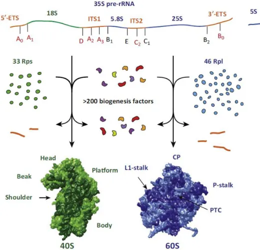
Figure 1: Schematic overview of ribosome biogenesis in Saccharomyces cerevisiae obtained from Kressler et al. (2017).

Ribosomal proteins (green and blue) and assembly factors (variegated) of the 40S (green) and the 60S (blue) subunits are synthesized in the cytoplasm from mRNAs produced by RNA polymerase II (RNA Pol II).

35S pre-rRNA and 5S rRNA (dark blue) are synthesized by RNA polymerase I (RNA Pol I) and RNA polymerase III (RNA Pol III), respectively. External and internal transcribed spacers (5’ETS, ITS1, ITS2, 3’ETS; orange) are removed from 35S pre-rRNA during ribosome biogenesis by endo- and exonucleolytic cleavages at processing sites indicated in red and black, respectively. Mature 40S subunits contain 33 ribosomal proteins (light green) and the 18S rRNA (dark green). Mature 60S subunits contain 46 ribosomal proteins (light blue) and the 5.8S, 25S, and 5S rRNAs (dark blue). Structural hallmarks of both mature subunits are indicated. CP is short for central protuberance, P-stalk for phospho-stalk, and PTC for peptidyl transferase center.

To decode the mRNA and join the single amino acids together to form a polypeptide chain,

ribosomes use two main active sites, the decoding center and the peptidyl transferase

(16)

center (PTC). The former is located within the small ribosomal subunit (SSU) and the latter is part of the large ribosomal subunit (LSU). Joining of both subunits builds a mature ribosome. Related to their sedimentation under high centrifuge forces, the small and the large subunits in eukaryotes are known as the 40S and 60S subunits, respectively, while the mature eukaryotic ribosome is known as 80S ribosome (Deley, 1964). Ribosomes are a paradigm for fundamental studies on ribonucleoprotein particles (RNPs) because they are a main component of every cell, composed of RNAs (ribosomal RNA, rRNA) and proteins (ribosomal proteins, RPs). In the yeast Saccharomyces cerevisiae, the SSU contains 33 RPs and the 18S rRNA, whereas the LSU harbors 46 RPs and the 25S, 5.8S, and 5S rRNAs (Figure 1) (Klinge and Woolford, 2019; Kressler et al., 2017).

In eukaryotic cells, ribosome assembly is a challenging process involving the action of three RNA polymerases. RNA polymerases I (RNA Pol I) and III (RNA Pol III) transcribe the ribosomal DNA (rDNA) in the nucleolus to produce the precursor transcripts of the mature rRNAs. RNA polymerases II (RNA Pol II) and III produce small nucleolar RNAs required for the synthesis of ribosomes. Furthermore, RNA Pol II synthesizes the mRNAs, which are translated in the cytoplasm to form the RPs. In addition, the synthesis of ribosomes takes place along different cellular compartments, starting in the nucleolus and ending in the cytoplasm, where the mature ribosomes are required. Synthesis of ribosomes requires processing and folding of the rRNA, the incorporation of the 80 RPs, and the transport of the ribosomal subunits to the cytoplasm. These processes need the action of about 200 assembly factors (AFs) and 77 small nucleolar RNAs (snoRNAs) (Figure 1) (de la Cruz et al., 2015).

Since exponential growing yeast cells contain about 200,000 ribosomes and they divide every 90 minutes, each cell needs to produce about 1,000 ribosomes per minute. Thus, synthesis of ribosomes is a very dynamic process requiring 60% of total cellular transcription and with 50% of RNA Pol II activity dedicated to the production of enough RPs and AFs (Warner, 1999). Moreover, the choreography of the more than 200 AFs (including snoRNAs) participating in the synthesis of both ribosomal subunits needs to be fine-tuned to avoid an excessive formation of non-functional particles.

1.2 Functional and structural description of mature ribosomal subunits

1.2.1 The 40S or small subunit (SSU)

The mature 40S subunit adopts, compared to the mature 60S subunit, a relatively simple

conformation consisting of five main parts named as head, beak, body, platform, and

central pseudoknot (Figure 2). The mRNA tunnel crosses the SSU between the beak and

body. The active site of the small subunit, the decoding center, lies in the middle of the

small subunit close to the central pseudoknot (Klinge and Woolford, 2019).

(17)

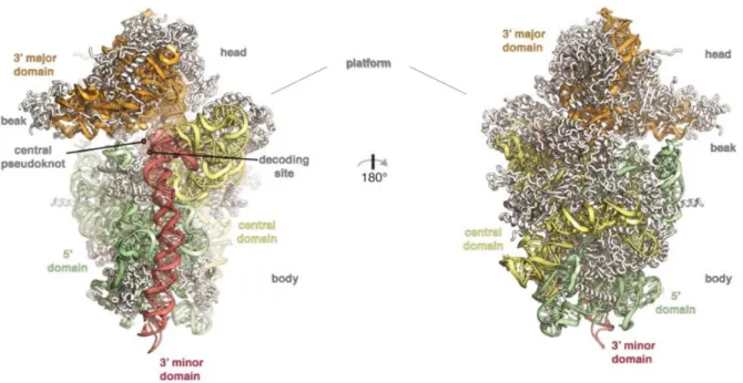
Figure 2: Tertiary structures of the mature small ribosomal subunit adapted from Klinge and Woolford (2019).

Cartoon representation of the subunit interface (left) and the solvent-exposed side (right) of the small ribosomal subunit. The subdomains of the 18S rRNA are colored and indicated. RPs are depicted in white.

Positions of head, beak, body, platform, central pseudoknot, and decoding center are annotated.

The rRNA component of the SSU is the 18S rRNA, which harbors four structural domains:

5’ domain, central domain, 3’ major domain, and 3’ minor domain. The 3’ minor domain is oriented upright within the body along the subunit interface surrounded by the 5’

domain and the central domain (on the left in Figure 2). The 3’ major domain lies exclusively in the upper part of the SSU structure and constitutes head and beak. Thus, the central domain connects head and body. The central pseudoknot forms in the middle of the subunit at the interface of the other four domains and it is required for the function of the decoding center (on the left in Figure 2) (Ben-Shem et al., 2011; Klinge and Woolford, 2019).

1.2.2 The 60S or large subunit (LSU)

The mature 60S subunit harbors at least eight main sites with specific names and

functions, which are described in detail in Figure 3. In the right part of the structure, the

phospho-stalk (P-stalk) is the binding site of translation factors and the sarcin ricin loop

(SRL) triggers GTP hydrolysis in translation factors with GTPases activity (Pérez-

Fernández et al., 2005; Santos and Ballesta, 2005; Voorhees et al., 2010). The binding sites

for tRNAs known as, A-, P-, and E-sites (Aminoacyl-, Peptidyl-, and Exit-) localize in the

middle of the LSU. The nascent polypeptide chain runs along the LSU through the

polypeptide exit tunnel (PET) (Konikkat and Woolford, 2017). On the top, the central

protuberance (CP), composed of 5S rRNA and the RPs Rpl5 and Rpl11, enhances

translational fidelity possibly by allowing the communication between the different

functional centers (Dinman, 2005; Smith et al., 2001).

(18)

Figure 3: Functional centers of the mature large ribosomal subunit depicted in the quaternary structure obtained from Konikkat and Woolford (2017).

The 60S subunit is oriented to visualize the subunit interface, containing the peptidyl transferase center (PTC) and other active sites. Above, the central protuberance (CP) containing the 5S rRNA (brown) and the RPs Rpl5 and Rpl11 (pale yellow) is depicted. On the right, the RNA-binding site of the phospho-stalk (P- stalk) is shown in blue and the sarcin ricin loop (SRL) in red. In the center, the helices H89 and H91 (yellow) belong to the tRNA entry tunnel and mark the beginning of the tRNA accommodation corridor with A- and P-sites. The ending of the corridor at the E-site can be identified by Rpl42 colored in purple. In addition, the A-site finger is depicted in green and Rpl10 in red. In the center, the position of the polypeptide exit tunnel (PET) is indicated. It proceeds toward the solvent-exposed side (on the reverse side).

The LSU contains three mature rRNA components, named 25S, 5.8S, and 5S. The primary

structure of 25S rRNA contains six conserved domains (I to VI from 5’ to 3’ end). Domains

I, II, and the 5.8S rRNA lie at the solvent-exposed surface of the LSU (on the right in Figure

4). Domains IV and V build the PTC containing the A-, P-, and E-sites (on the left in Figure

4). Domains II, III, and V participate in formation of the PET and domain III surrounds the

exit of the PET at the solvent-exposed side (on the right in Figure 4). Both domains III and

VI bridge the subunit interface and the solvent-exposed side. Domains I and III trap the

5.8S rRNA (on the right in Figure 4), while domains II and V dock the 5S rRNA and form

the PTC (on the left in Figure 4) (Ben-Shem et al., 2011; Klinge and Woolford, 2019).

(19)

Figure 4: Tertiary structures of the mature large ribosomal subunit adapted from Klinge and Woolford (2019).

Cartoon representation of the subunit interface (left) and the solvent-exposed side (right) of the large ribosomal subunit. The subdomains of the 25S rRNA are colored and indicated, as well as 5.8S and 5S rRNAs.

RPs are depicted in white. Positions of the central protuberance, L1-stalk, P-stalk, GTPase activating center (named SRL in Figure 3), peptidyl transferase center, and polypeptide exit tunnel are annotated.

1.2.3 The 80S ribosome

The 40S and 60S subunits assemble in 80S ribosomes during translation initiation. This process starts in eukaryotic cells with the scanning of the mRNA by the 43S preinitiation complex to identify the start codon. Therefore, the 40S subunit guides the mRNA coming from the mRNA entry tunnel (Figure 5) between beak and body through the ribosome (Ben-Shem et al., 2011).

Figure 5: Structure of the mature 80S ribosome (PDB ID: 4V88).

Cartoon representation of the 80S ribosome consisting of 40S subunit (light green) and 60S subunit

(orange). Positions of head, beak, and mRNA entry tunnel of the 40S subunit and central protuberance (CP)

and polypeptide exit tunnel (PET) of the 60S subunit are annotated.

(20)

Once the start codon is identified by the 43S particle carrying the initiator-tRNA, the 60S subunit joins to form the 80S ribosome. The association of the 60S subunit is allowed by initiation factor eIF2 triggered GTP hydrolysis, which leads to the dissociation of initiation factors from the 43S particle, which precluded unspecific association with 60S subunits (Kimball, 1999). The formation of the 80S creates the A-, P-, and E-sites, which are occupied during elongation with tRNAs. Therefore, an amino-acylated tRNA enters the tRNA tunnel and binds to the A-site, while the tRNA connected to the polypeptide chain sits at the P-site. The peptide bond formation between the new amino acid and the polypeptide chain is carried out by the PTC. After the formation of the peptide bond, tRNAs are translocated. The tRNA at the P-site moves toward the E-site. The tRNA at A- site is now bound to the nascent polypeptide and moves toward the P-site releasing the A-site for the association of a new amino-acylated tRNA. Once the stop codon is reached, the release factors transfer a water molecule to the polypeptide chain and trigger GTP hydrolysis, terminate translation, disassemble the 80S ribosome, and release the completed polypeptide chain.

1.3 Transcription of the ribosomal DNA

In eukaryotic cells, the synthesis of ribosomes begins with the transcription of the rDNA in the nucleolus. In S. cerevisiae, each cell contains around 150 copies of the rDNA, which are organized as head-to-tail tandem repeats on chromosome XII (Figure 6) (Petes, 1979;

Schweizer et al., 1969).

Figure 6: rDNA locus of S. cerevisiae obtained from Woolford and Baserga (2013).

Schematic representation of the rDNA locus on chromosome XII of the yeast S. cerevisiae encoding around

150 rDNA repeats. One repeat is depicted in more detail containing the sequences of 5S, 18S, 5.8S, and 25S

rRNAs. As indicated, 5S rRNA is produced by RNA Pol III in the opposite direction and separated from the

remaining rRNA sequences by NTS2 (non-transcribed spacer). RNA Pol I produces the 35S pre-rRNA

transcript reaching from 5’ETS to 3’ETS (external transcribed spacer). NTS1 (non-transcribed spacer)

separates the rDNA loci.

(21)

Each copy contains the sequence of the 35S gene encoding for three out of four rRNAs (18S, 5.8S, 25S) and the 5S rRNA gene, which are located on complementary strands and separated by non-transcribed spacers (NTS; Figure 6). RNA Pols I and III transcribe the 35S and 5S genes, respectively, in opposed senses. The 35S pre-rRNA contains the sequences of the 18S, the 5.8S, and the 25S rRNAs, which are separated by the internal transcribed spacers ITS1 and ITS2 and flanked by the external transcribed spacers 5’ETS and 3’ETS (see Figure 1 and Figure 6) (Udem and Warner, 1972).

RNA Pol I is an outstanding RNA polymerase because it exclusively transcribes the 35S gene. In contrast, besides 5S rRNA synthesis, RNA Pol III produces tRNAs and several other RNAs, which include the RNA component of the signal recognition particle and snoRNAs (Dieci et al., 2007).

In S. cerevisiae, RNA Pol I consists of 14 subunits and it needs several transcription factors to be recruited to the promoter region of the 35S gene (Engel et al., 2018; Keener et al., 1997; Keys et al., 1994, 1996; Lalo et al., 1996; Siddiqi, 2001; Steffan et al., 1996;

Yamamoto et al., 1996). Both transcription initiation and elongation by RNA Pol I are regulated steps in rRNA synthesis (French et al., 2003; Zhang et al., 2010). Moreover, the production of rRNAs can be primary regulated by the number of rDNA copies accessible for transcription (Sandmeier et al., 2002). Due to the high activity of RNA Pol I required to support the demand of ribosomes, actively transcribed rDNA can even be visualized by electron microscopy with the so-called “Miller spread”-technique, initially developed by Oscar Miller using amphibian oocytes (Miller and Beatty, 1969).

1.4 Processing of the ribosomal RNAs

35S pre-rRNA is processed by several endo- and exonucleolytic cleavages to produce the mature rRNAs. Processing of the pre-rRNA might occur as co- and post-transcriptional events.

1.4.1 Co-transcriptional processing

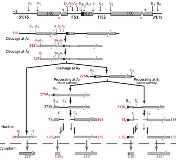
Co-transcriptional processing involves the cleavage events at sites A0, A1, and A2 (Figure 7). Co-transcriptional cleavage in A2 occurs in 50 to 70% of the nascent transcripts under optimal growth conditions (Kos and Tollervey, 2010; Osheim et al., 2004). However, it is not clear which are the molecular mechanisms behind the selection for co- or post- transcriptional processing. In any case, the efficient cleavage at these sites requires the formation of the first pre-ribosomal particle called SSU processome (Dragon et al., 2002).

Based on the accumulation of the major SSU precursor (Figure 7), the order of cleavage events has been established by convention.

Therefore, processing at sites A0 and A1 within the 5’ETS region constitutes the primary

cleavage events (Figure 7). Cleavage at the A1 site produces the mature 5’ end of the 18S

rRNA and it requires the evolutionarily conserved nucleotides upstream of the A1

(22)

sequence in the 5’ETS (Venema et al., 1995) and the stem-loop structure downstream of A1 within the sequence of the mature 18S rRNA (Sharma et al., 1999).

Cleavage at the A2 site splits the 35S transcript into the pre-rRNAs for the small and the large ribosomal subunit (20S and 27SA2 pre-rRNAs, Figure 7). As for the A1 cleavage, positioning of the cleavage site A2 is defined by a downstream stem-loop structure (Allmang et al., 1996). Although, cleavages at the A1 and A2 sites might involve the activity of Utp24 (Bleichert et al., 2006; Wells et al., 2016), some authors suggest Rcl1 as the putative endonuclease for at least the A2 site (Billy et al., 2000; Delprato et al., 2014; Horn et al., 2011). As an alternative, it is possible that the cleavage activity of Utp24 is supported by Rcl1 (Khoshnevis et al., 2019).

Figure 7: pre-rRNA processing in S. cerevisiae adapted from Braun et al. (2020).

Schematic representation of the pre-rRNA processing pathway(s) in yeast. Names of the precursors and mature rRNAs are indicated in red. Cleavage sites producing 18S rRNA are depicted in red. Processing sites for the maturation of 5.8S and 25S rRNAs are colored blue. The dashed line represents the nuclear envelope.

In contrast with this systematic cleavage pattern, some studies suggest the existence of

alternative processing pathways, which are evident when yeast cells are cultivated under

stress conditions (Kos-Braun et al., 2017). In fact, previous work of our group, using highly

sensitive methods as affinity purifications, showed the presence of all possible precursors

associated with AFs. These data suggest the stochastic cleavage at A0, A1, A2, and A3 sites

(23)

as a first-come/first-served model for the early steps of ribosome biogenesis (Boissier et al., 2017).

1.4.2 Post-transcriptional processing

At least the final maturation of SSU particles and the processing of LSU pre-rRNAs take place as post-transcriptional processing events. Moreover, at least 30% of transcripts are processed post-transcriptionally at the A0, A1, and A2 sites and this ratio increases when cells are cultivated in suboptimal growth conditions (exhausted nutrients, galactose containing medium, stationary growth phase, etc.) (Kos-Braun et al., 2017). It is supposed that the post-transcriptional cleavages at sites A0, A1, and A2 occur in the same order as in the co-transcriptional process, but as for co-transcriptional cleavage, also alternative processing pathways might be possible (Boissier et al., 2017). Afterward, the resulting 20S pre-rRNA is immediately exported from the nucleolus into the cytoplasm (Udem and Warner, 1973). In the cytoplasm, the endonuclease Nob1, which already associates with the pre-rRNA in the nucleolus, reaches the processing site D, cleaving and producing the mature 18S rRNA (see Figure 7) (Fatica et al., 2003; Lamanna and Karbstein, 2009;

Pertschy et al., 2009).

The second product of the cleavage at the A2 site is the 27SA2 pre-rRNA, precursor of 5.8S and 25S rRNAs. 27SA2 is processed at the A3 site by the RNase MRP, which requires the correct formation of the 3’ETS by release of the nascent transcript via endonucleolytic cleavage of Rnt1 at site B0 (Allmang and Tollervey, 1998; Kufel et al., 1999). Therefore, processing events at the 5’ and 3’ ends of the 27SA2 pre-rRNA are connected, most likely by the interaction of Rpl3 with both ends of the 27SA2 pre-rRNA. Moreover, Rpl3 is required for the association of the early acting AFs, which participate in the initial formation of the pre-60S particle (Gamalinda et al., 2014; Hitchen et al., 1997; Kufel et al., 1999; Rosado et al., 2007a).

After A3 cleavage, the 5’-3’ RNases Rat1, Xrn1, and Rrp17 process the 5’ end of 27SA3 pre- rRNA to the B1 S site present in the 5’ of the 27SB pre-rRNA (El Hage et al., 2008; Henry et al., 1994; Oeffinger et al., 2009). As an alternative, an unknown endonuclease might process the 27SA2 pre-rRNA directly at B1 L (see Figure 7) (Faber et al., 2006). These two pathways define the 5’ end of the 5.8S, which is seven nucleotides longer in the 5.8S L rRNA.

However, the two processing pathways are asymmetrical and the of 5.8S S rRNA accounts for up to 80% of the total 5.8S. Interestingly, inhibition of MRP leads to an exclusive synthesis of 5.8S L rRNA (Chamberlain et al., 1998; Chu et al., 1994; Lygerou et al., 1996;

Schmitt and Clayton, 1993), and accordingly, the absence of factors involved in the major pathway as Rrp5 and Dbp3 causes an imbalance in the production of 5.8S S and 5.8S L

rRNAs (Eppens et al., 1999; Lebaron et al., 2013; Venema and Tollervey, 1996; Weaver et al., 1997).

In parallel to the processing of the mature 5’ end of the 5.8S rRNA, an unknown 3’-5’

exonuclease produces the mature 3’ end of the 25S rRNA at the B2 site (see Figure 7)

(Elela et al., 1996; Fernández-Pevida et al., 2015; Kufel et al., 1999).

(24)

Independently of the pathway producing the 5’ end of the 27SB pre-rRNA, the next step includes the separation of 5.8S and 25S rRNA precursors by the endonucleolytic cleavage of the 27SB pre-rRNA at site C2 within the ITS2 region by Las1 (see Figure 7) (Gasse et al., 2015). Two secondary structures were initially predicted for the ITS2 called “ring” and

“hairpin”. Both structures are formed by alternative base-pairing within the ITS2 and the conformational change between both might trigger C2 cleavage (Côté et al., 2002; Joseph et al., 1999; Peculis and Greer, 1998; Yeh and Lee, 1990). Nevertheless, a third conformation showing a long helical organization is supposed to be the most accessible structure for the C2 endonuclease (Coleman, 2015; Pöll et al., 2017).

Cleavage at C2 produces the 7S and the 26S pre-rRNAs and the subsequent cleavage at site E leading to the mature 5.8S rRNA is a complex multi-step process (see Figure 7) (Mitchell et al., 1996). The first steps are carried out by the exosome (Lebreton et al., 2008a; Liu et al., 2006; Mitchell et al., 1997) and among several other factors, the helicase Mtr4 guides the exosome to the pre-rRNA (de la Cruz et al., 1998a; Fromm et al., 2017;

Jackson et al., 2010). The obtained 6S pre-rRNA is finally matured to 5.8S rRNA in the cytoplasm, where the last steps are performed by a concerted action of the putative non- essential 3’-5’ exonucleases Rex1, Rex2, and Rex3 and by the putative non-essential nuclease Ngl2 (Faber et al., 2002; van Hoof et al., 2000; Thomson and Tollervey, 2010).

The processing of 26S pre-rRNA at the C1 site by exonucleases Rat1, Rrp17, and Xrn1, as well as the AF Las1, forms the mature 5’ end of the 25S rRNA (see Figure 7) (Gasse et al., 2015; Geerlings et al., 2000; Oeffinger et al., 2009; Schillewaert et al., 2012).

1.4.3 Regulation of co- and post-transcriptional processing at the A0, A1, and A2 sites

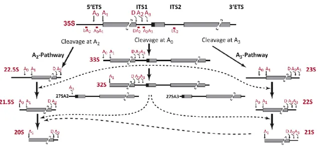
Growth of the yeast cells under stress or beyond exponential growth phase can lead to increased post-transcriptional pre-rRNA processing at the expense of co-transcriptional processing. Post-transcriptional processing takes place on the completely synthesized 35S pre-rRNA either after terminated transcription or after cleavage at site B0. As a result, higher amounts of 35S and 23S pre-rRNAs are detected.

The second cleavage step after cut at B0 might occur either at sites A0, A1 (producing 33S

or 32S pre-rRNA, respectively), or at site A3 (producing the 23S pre-rRNA reaching from

5’ETS to A3) (Figure 8) (Allmang and Tollervey, 1998). The preferential use of the A3

pathway, producing 23S and 27SA3 pre-rRNAs, may directly depend on the nutrient

availability and may be regulated by the TORC1 (Tor complex 1) pathway, which also

controls the transcription activity of all three RNA polymerases. Tor1 kinase is

constitutively active and inactivated in the absence of nutrients. Besides transcription,

TORC1 activates the CK2 kinase, which leads to preferential cleavage at the A2 site (Kos-

Braun et al., 2017). Contrary, when the CK2 kinase is not activated, the A3 pathway is

predominantly used. Upon nutrient depletion, both 27SB (derived from 27SA3) and 23S

pre-rRNAs might be non-fully productive intermediates, which are not further processed

(Kos-Braun et al., 2017), most possibly due to the absence of RPs (Philippi et al., 2010).

(25)

Figure 8: Alternative pre-rRNA processing adapted from Boissier et al. (2017).

Schematic representation of the alternative pre-rRNA processing pathways for removal of the 5’ETS sequence of the 35S pre-rRNA. Names of the pre-rRNAs and cleavage sites are depicted in red. Probes used for northern blot detection of the pre-rRNA intermediates are indicated in red below 35S precursor and named after their hybridization sites.

All intermediates depicted in Figure 8 can be observed when components of the exosome are mutated (Allmang et al., 2000). Non-productive pre-rRNA intermediates are polyadenylated by the TRAMP complex (LaCava et al., 2005). Poly(A) tail targets the exosome to aberrant intermediates, which are degraded (Allmang et al., 2000; Wery et al., 2009; Zanchin and Goldfarb, 1999). Nevertheless, the removal of aberrant intermediates by the exosome might require additional cleavage events at novel cleavage sites (Choque et al., 2018).

In contrast, affinity purification of AFs increases the sensitivity for the detection of alternative processed transcripts, which are visualized and might represent productive intermediates. It is possible that the reduced synthesis of RPs in suboptimal growth conditions slows down the processing of particles requiring the assembly of large sets of RPs and speeds up the processing of particles less dependent on RPs. This mechanism would explain an increase in the lifetime of early particles but a decrease in the lifetime of the late ones. In addition, the absence of the exosome might reduce the recycling of AFs and, therefore, increase the half-life of aberrant pre-rRNA intermediates (Figure 8), which are usually short-living intermediates due to the fast kinetics producing mature 18S rRNA.

1.4.4 Recycling events during pre-rRNA processing

As explained in the previous section, the exosome and the TRAMP complex are responsible for the degradation of dead-end products, which accumulate when pre-rRNA intermediates cannot be further processed (Dez et al., 2006; LaCava et al., 2005).

Moreover, the 3’-5’ exonuclease activity of the exosome and 5’-3’ exonucleases are

directly involved in pre-rRNA processing for removal of the ETS and ITS regions. The

5’ETS-A0 fragment is degraded by the exosome, which is recruited by the helicase Mtr4

(Allmang et al., 2000; Bernstein et al., 2008; Suzuki et al., 2001). The second 5’ETS

(26)

fragment reaching from A0 to A1 is a substrate of the exosome and the nucleolar 5’-3’

exoribonuclease Rat1 (Allmang et al., 2000; Johnson, 1997; Petfalski et al., 1998). The D- A2 fragment resulting from cleavages at A2 and D is degraded by the cytoplasmic 5’-3’

exoribonuclease Xrn1 (Johnson, 1997; Stevens et al., 1991).

After A3 cleavage, Rat1, Xrn1, and Rrp17 also process the 27SA3 pre-rRNA to the B1 S site present in the 5’ of the 27SB pre-rRNA (El Hage et al., 2008; Henry et al., 1994; Oeffinger et al., 2009). It is assumed that the correct assembly of Rpl17 at this point is required to stop the degradation process toward the B1 site. This implies that aberrant pre-60S intermediates, which lack at least Rpl17, are directly degraded instead of being further processed (Henry et al., 1994; Sahasranaman et al., 2011). Within the ITS2 region, the E- C2 fragment is degraded first by the exosome in the nucleolus and by other 3’-5’

exonucleases in the cytoplasm (see 1.4.2). Recruitment of exosome would occur most possibly by recruitment of Mtr4 through Nop53 (Cepeda et al., 2019; Falk et al., 2017;

Thoms et al., 2015). The remaining ITS2 fragment reaching from C2 to C1 is degraded by the 5’-3’ exonucleases Rat1, Rrp17, and Xrn1 and the B2-B0 fragment of the 3’ETS is degraded by an unknown 3’-5’ exonuclease (see 1.4.2).

Altogether, the mentioned events are important to recover the excised nucleotides and to recycle AFs, which are associated with the spacer regions (5’ETS, ITS1, and ITS2) or accumulated in dead-end products (Houseley and Tollervey, 2009).

1.5 Assembly of the small subunit

During assembly of the small subunit, three defined particles are identified.

In the early steps of ribosome synthesis, the first pre-SSU particle or small subunit processome (SSU processome) is formed in the nucleolus. However, post-transcriptional cleavage allows the formation of the 90S pre-ribosome, which contains the 35S pre-rRNA and for instance the SSU-processome factors but also early AFs of the large subunit. The third particle corresponds to the pre-40S particle, which is formed in the nucleus (after A0, A1, and A2 cleavages) and transported to the cytoplasm, where the last cleavage event takes place and the 40S or small subunit is produced.

1.5.1 Stepwise formation of the SSU processome

The formation of the SSU processome can be currently explained by two non-exclusive models. The hierarchical model defines a stepwise process, in which the assembly of some AFs and RPs is required for the further association of others (e. g. Dutca et al., 2011; Pérez- Fernández et al., 2007, 2011) . The second model only establishes a relevant role for rRNA domains on the consecutive recruitment of AFs (Hunziker et al., 2019). In both cases, the assembly of the SSU processome starts with the assembly of the 5’ETS particle.

Subsequently, the different domains of the 18S rRNA are folded and gradually integrated

into the precursor particle (Figure 9).

(27)

Figure 9: Schematic model for SSU-processome assembly adapted from Peña et al. (2017).

tUTP/UTP-A complex (orange) is bound first to the 5’ETS (red) and forms the base of the SSU processome.

Binding of UTP-B complex (turquois), U3 snoRNP (blue), and some other AFs (yellow, green) completes the 5’ETS particle. 5’ domain (moss green), as well as the central domain (light gray), 3’ major domain (dark gray), and 3’ minor (min) domain (purple) are transcribed, folded, and decorated with factors in a hierarchical manner.

The finally assembled SSU processome contains 36 AFs and 18 RPs out of the 33 RPs present in the mature 40S subunit. Within the available models of the SSU processome, the folding of the 5’ and central domains resembles the folding state in the mature small subunit, whereas the 3’ major and minor domains still require large structural reorganization to adopt the conformation of the mature subunit (Sun et al., 2017; Zhang et al., 2016b).

1.5.1.1 Assembly of the 5’ETS particle

The first step within the formation of the 5’ETS particle involves the association of the heptameric tUTP/UTP-A (derived from U three protein; Dragon et al., 2002) complex with the 5’ETS of the nascent transcript produced by RNA Pol I (Gallagher et al., 2004; Pérez- Fernández et al., 2007). The tUTP complex formed by the proteins Utp4, Utp5, Utp8, Utp9, Utp10, Utp15, and Utp17/Nan1, was identified as functional subcomplex of the SSU processome, which might also participate in the efficient transcription of the rDNA (Gallagher et al., 2004). With a protein composition similar to the tUTP complex, the UTP- A complex was identified containing Pol5 instead of Utp5 (Krogan et al., 2004). Since the nomenclature of tUTP and UTP-A complexes is not always clearly used in the literature, we will refer to them as two independent complexes containing either Utp5 or Pol5, respectively, or as tUTP/UTP-A complex when the protein composition is unclear. The association of the tUTP/UTP-A complex with the 5’ETS is required for the subsequent stable assembly of the hexameric complexes UTP-B (consisting of Utp1/Pwp2, Utp6, Utp12, Utp13, Utp18, Utp21) and U3 snoRNP (composed of C/D box U3 snoRNA, Nop1, Snu13, Nop56, Nop58, Rrp9) (Figure 10) (Boissier et al., 2017; Dosil and Bustelo, 2004;

Pérez-Fernández et al., 2007; Watkins et al., 2000). In addition, both complexes are

mutually required for their stable association onto the pre-rRNA (Pérez-Fernández et al.,

2007). The final assembly of the 5’ETS particle is achieved by the association of the Mpp10

complex (consisting of Imp3, Imp4, Mpp10) (Pérez-Fernández et al., 2011; Sá-Moura et

al., 2017) and some other AFs, like Utp3/Sas10, Sof1, Utp7, Utp11, and Bud21/Utp16 (see

(28)

Figure 10 and Figure 12) (Chaker-Margot et al., 2015). Finally, the proteins Fcf2, as well as the putative A1 and A2 endonuclease Fcf1/Utp24, are recruited to the 5’ETS (Zhang et al., 2016b).

Figure 10: Protein components of the 5’ETS particle adapted from Chaker-Margot et al. (2015).

Schematic representation of the protein and RNA composition of the 5’ETS particle defined by mass spectrometry. 5’ETS sequence of the pre-rRNA is depicted in gray, U3 snoRNP containing the U3 snoRNA is colored orange, AFs are showed in beige, and Utp3 as predicted exosome interactor is depicted in pink.

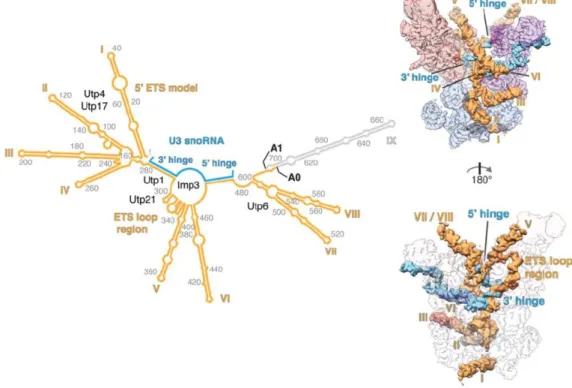
The tUTP/UTP-A complex associates with helices I to IV of the 5’ETS (Figure 11). While Utp8, Utp9, and Utp17 assemble at the most 5’ end of the 5’ETS, the remaining four proteins Utp4, Utp5, Utp10, and Utp15 are incorporated downstream (Hunziker et al., 2016). Utp17, Utp4, and Utp15 might stabilize the first three helices, while Utp5 and Utp10 connect the tUTP/UTP-A complex to the UTP-B complex by interaction with the UTP-B components Utp21 and Utp18. The UTP-B component Utp18 is a central interactor within the 5’ETS particle by binding to the tUTP/UTP-A component Utp10, the UTP-B components Utp21 and Utp6, and the U3 snoRNP components Nop58 and Snu13 (Hunziker et al., 2016). Within the UTP-B complex the tWD domains of Utp1 and Utp21 interact together and their C-terminal domains associate with the C-terminal domains of Utp12 and Utp13 (Boissier et al., 2017; Zhang et al., 2016a). In addition, Utp21 bridges the association with Utp6 and Utp18 (Boissier et al., 2017; Hunziker et al., 2016; Pöll et al., 2014; Zhang et al., 2016a). We have proposed that the presence of Utp12 and Utp13 is not required for the assembly of the UTP-B core components Utp6, Utp18, Utp21, and Utp1, but the association with the 5’ETS particle requires the complete formation of the UTP-B complex (Boissier et al., 2017).

The U3 snoRNA base pairs the middle part of the 5’ETS through its 5’ and 3’ hinge regions (Figure 11) (Beltrame and Tollervey, 1992, 1995; Beltrame et al., 1994; Dutca et al., 2011;

Marmier-Gourrier et al., 2011). This association might be stabilized by interactions with the tUTP/UTP-A and UTP-B complexes (Pérez-Fernández et al., 2007) and the U3 snoRNA may participate in folding of the helices V and VI and stabilization of pre-ribosomal particles. The initial interaction between U3 snoRNA and 5’ETS seems to be mediated by the 3’ hinge, whereas the incorporation of additional AFs, like UTP-B components, U3 snoRNP factors, and Mpp10 complex might depend on the additional binding of the 5’

hinge to the 5’ETS (Dutca et al., 2011; Marmier-Gourrier et al., 2011). Association of Imp3

and Imp4 might occur concomitantly with Mpp10 as a single protein complex (Pérez-

Fernández et al., 2011), which has no influence on the stability of the 5’ETS particle itself,

(29)

but it is essential for later assembly steps of the SSU processome (Gérczei et al., 2009;

Pérez-Fernández et al., 2011; Shah et al., 2013).

Figure 11: Secondary and tertiary structures of the 5’ETS particle adapted from Chaker-Margot et al. (2017).

On the left, the secondary structure model of the 5’ETS (orange) and the interaction with the U3 snoRNA (blue) are depicted. Helix IX is not observed in the EM density map and it is depicted in gray. Names of some of the associated proteins, the pre-rRNA domains, and the A0 and A1 processing sites are indicated. On the right, front (above) and back (below) view of the tertiary structure of the 5’ETS particle with RNA elements colored as on the left are shown. tUTP/UTP-A complex is colored in light blue, U3 snoRNP in light violet, and UTP-B complex in light red.

Distant regions of the 5’ETS are also connected by UTP-B, with helices VII and VIII bound by Utp6, the 5’ETS loop region between helices IV and V by Utp1 and Utp21, and helix V by Utp18 (Chaker-Margot et al., 2017), and therefore suggesting a chaperone activity for UTP-B (Chaker-Margot et al., 2017). In addition, Utp18 may play an additional role in recruiting the exosome, due to the presence of an arch-interaction motif (AIM) domain, which binds the “arch” domain in Mtr4 (Jackson et al., 2010; Thoms et al., 2015).

Altogether, the 5’ETS particle has a very dense structure, which is stabilized by many protein-protein interactions maintaining the folding of the RNAs and providing a base for the assembly of the remaining AFs in the SSU processome (Barandun et al., 2017; Chaker- Margot et al., 2017; Hunziker et al., 2016; Sun et al., 2017; Zhang et al., 2016b).

1.5.1.2 Assembly of the 5’ domain, the central domain, and the 3’ major domain

Domains of the 18S rRNA assemble after formation of the 5’ETS particle has taken place (see Figure 12) (Chaker-Margot et al., 2017; Sun et al., 2017; Zhang et al., 2016b).

The 5’ domain of the 18S rRNA includes the first 18 helices. The RPs Rps9, Rps23, Rps24,

and Rps30 associate with the helices 3, 4, and 15 to 18, while Rps4, Rps6, Rps8, and Rps11

(30)

associate with helices 5 to 14. Helix 6 contains the binding site of the C/D box snoRNA U14, which is required for 2’-O methylation of nucleotides 414 and 415 within the 5’

domain (Chaker-Margot, 2018; Liang and Fournier, 1995; Zhang et al., 2016b). AFs like Enp2, Bfr2, and Lcp5 are stably bound between helices 14 and 15 and they recruit AFs like the helicase Dbp4/Hca4 (Soltanieh et al., 2014). Lcp5 contains an exosome interaction domain and it may assist Utp18 to bring the exosome in the proximity of the 5’ETS domain (Barandun et al., 2017; Chaker-Margot et al., 2015; Wiederkehr et al., 1998). Although the function of most of the AFs recruited to the 5’ domain is not yet defined, assembly of the 5’ domain is required for the subsequent incorporation of the other rRNA domains within the SSU processome (Chaker-Margot et al., 2015; Sun et al., 2017; Zhang et al., 2016b).

Next, the central domain formed by helices 19 to 27 is folded into the nascent SSU processome (see Figure 12). The helices 20, 22, 23, and 26 are bound by six RPs (Rps1, Rps7, Rps13, Rps14, Rps22, and Rps27) (Sun et al., 2017). In addition, the H/ACA box snoRNAs snR30 and snR10 base-pair within the central domain. Although both snoRNPs are required for small subunit biogenesis, their specific functions remain elusive and they need to be removed to achieve the mature conformation of the 18S rRNA (Atzorn et al., 2004; Bohnsack et al., 2008; Fayet-Lebaron et al., 2009; Kiss et al., 2010; Martin et al., 2014). During the formation of the central domain, among others, the large AF Rrp5 is recruited and it is required for the stable assembly of the UTP-C complex, formed by Utp22, Rrp7, and the four CKII subunits (Krogan et al., 2004; Pérez-Fernández et al., 2007;

Torchet et al., 1998). Similar to the other UTP complexes, UTP-C promotes the folding and stabilization of the SSU precursor particle by interactions with RPs and rRNA sequences (Lin et al., 2013; Pérez-Fernández et al., 2007). Strikingly, Rrp5 interacts with the central domain and with other binding sites throughout the 35S pre-rRNA, being also relevant for the synthesis of the large subunit (Eppens et al., 1999; Lebaron et al., 2013; Zhang et al., 2016b). Finally, Utp13 and Sof1, which associate with the 5’ETS, contribute to the folding of the central domain onto the 5’ETS via interactions with Utp7 and the UTP-B complex and provide additional stabilization by connecting the central domain with the 5’ETS (Barandun et al., 2017; Chaker-Margot et al., 2017; Sun et al., 2017; Zhang et al., 2016b).

The 3’ major domain of the 18S rRNA contains helices 28 to 43 (see Figure 12). However,

helices 35 to 40 are not resolved in Cryo-EM structures, most likely because they are

initially poorly folded and only a few proteins are associated during these early steps. The

RPs Rps5, Rps16, and Rps28 associate with helices 28 to 30 and 41 to 43, while Rps12 and

Rps31 bind to helices 32 to 34. Altogether, five AFs are recruited at this step. The AFs

Nsr1, Mrd1, and Nop9 associate with helix 28 close to one of the 18S rRNA binding sites

of the U3 snoRNA (Chaker-Margot et al., 2015; Sun et al., 2017; Zhang et al., 2016b). Since

U3 snoRNA binding sites at the 18S rRNA might be later involved in the formation of the

central pseudoknot, it is suggested that these three AFs contribute to chaperoning this

RNA element (Zhang et al., 2016b). Similar to Utp13 and Sof1, the Mpp10 complex might

stabilize the SSU-processome by connecting the first and the last helices (helix 28 and

helix 43) of the 3’ major domain with the 5’ETS (Barandun et al., 2017; Chaker-Margot,

2018; Sun et al., 2017; Zhang et al., 2016b).

(31)

1.5.1.3 Assembly of the 3’ minor domain and finishing of the SSU processome

The final step of SSU processome formation is the folding of the 3’ minor domain (Jakob et al., 2012). In contrast to the 3’ major domain, a multitude of AFs are recruited at this step and many interactions, stabilizing the folding of the other domains and the structure of the complete SSU processome, are formed (Figure 12) (Chaker-Margot et al., 2017).

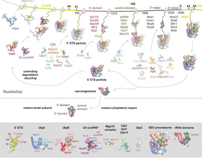
Figure 12: Schematic representation of the co-transcriptional assembly of the SSU processome obtained from Barandun et al. (2018).

Early events of small subunit assembly in the nucleolus and subsequent maturation resulting in the mature cytoplasmic small subunit are depicted. The relevant parts of the rDNA locus are depicted including 5’ETS and ITS1 (yellow), 5’ domain (red), central domain (green), 3’ major and minor domains (gray) of the 18S rRNA. Factors of the assembled SSU processome are shown as schematic outline. Transient components are listed and colored according to their associated rRNA domain. Below, the SSU processome and its major components are depicted in detail.

The 3’ minor domain contains the helices 44 and 45. Helix 44 is the longest within the 18S rRNA and it contacts the UTP-C component Utp22 present at the central domain and the UTP-B components Utp12 and Utp13 at the 5’ETS (Hunziker et al., 2016; Sun et al., 2017).

The AFs Utp30, Bms1, Rcl1, Kre33, Nop14, Noc4, Utp14, Utp20, Enp1, Pno1/Dim2, Rrp12,

and Rrt14 and the helicases Dhr1 and Dhr2 were found to be assembled at this point

(Chaker-Margot et al., 2015; Zhang et al., 2016b). In contrast during folding of the 3’ minor

(32)

domain, helicases like Dbp4, which unwind the snoRNA-rRNA interaction might release 14 earlier AFs and the snoRNAs U14, snR30, and snR10 (Soltanieh et al., 2014).

The heterodimer Rcl1-Bms1 bridges the domains 3’ major and 5’ (Chaker-Margot et al., 2017). This position might allow the GTPase activity of Bms1 to exert mechanical force and facilitate the structural rearrangement of the SSU processome required for A2 cleavage (Delprato et al., 2014). In addition, Kre33 connects the 3’ minor domain to 3’

major and 5’ domains and Utp20 the 3’ minor domain to 5’ domain and helices VII and VIII of the 5’ETS (Chaker-Margot et al., 2017). The heterodimer Nop14-Noc4 assembles once the head structure is formed and bridges the 5’ETS, the 5’ domain, and the central domain by association with tUTP/UTP-A, Mpp10-Imp5, and Bms1-Rcl1 (Barandun et al., 2017; Jakob et al., 2012). The AF Utp14 might contribute to join together helix V of the 5’ETS and 3’ domains by association with Utp6 and Utp7 to Sof1 (Barandun et al., 2017).

Finally, the endonuclease of the D site, Nob1, is recruited into the SSU processome concomitantly with the folding of the ITS1 (Chaker-Margot et al., 2015).

Figure 13: Structure of the SSU processome of S. cerevisiae adapted from Chaker-Margot (2018).

Each view of the structure is rotated by 120°. 5’ETS (yellow) is bound by tUTP/UTP-A complex (pale yellow), UTP-B complex (dark yellow), and U3 snoRNP (red). The domains of the 18S rRNA are depicted in green (5’ domain), orange (central domain), bright pink (3’ major domain), and dark pink (3’ minor domain). The associated AFs are shown in similar colors, respectively.

A special feature of the mature SSU processome is the physical separation of the four domains of the 18S rRNA, which might facilitate their individual downstream maturation (Figure 13) (Barandun et al., 2017; Chaker-Margot et al., 2017). Interestingly, several AFs (Utp20, Rrp5, Enp1, Krr1, and Pno1/Dim2) might prevent the SSU processome from adopting a premature conformation, and apparently, the formation of the central pseudoknot is inhibited by the presence of Bms1, Nop14, Imp4, Faf1, and Mpp10. Bms1, Nop14, Imp4, and Faf1 might stabilize the base pairing between the U3 snoRNA and the 5’ domain of the 18S rRNA, which needs to be destabilized to form the central pseudoknot.

In addition, Mpp10 holds helix 44 of the 3’ minor domain in a partially unfolded

conformation (Barandun et al., 2017; Chaker-Margot, 2018; Sun et al., 2017; Zhang et al.,

2016b; Chaker-Margot et al., 2015, 2017).

(33)

1.5.2 Transition to the cytoplasmic pre-40S particle

Subsequent maturation of the SSU processome in the cytoplasmic pre-40S particle requires several extensive structural rearrangements (Figure 14).

First, the cleavages at A1 and A2 sites must take place and 18S rRNA must adopt the central pseudoknot structure. Both events seem to be coordinated by the action of the helicase Dhr1, which unwinds at least one of the rRNA-U3 snoRNA duplexes and the resulting open structure would be the substrate for the endonuclease Utp24 (Bleichert et al., 2006; Sardana et al., 2015; Wells et al., 2016).

Second, the 5’ETS particle must be removed in order to recycle the AFs (see Figure 12), possibly achieved by the exosome (Allmang et al., 2000). As previously explained, either the UTP-B component Utp18, through its AIM domain, or the AFs Utp3 and Lcp5, because of their resemblance to the exosome cofactor Rrp47, might mobilize the exosome to the 5’ETS particle (Chaker-Margot, 2018; Costello et al., 2011; Klinge and Woolford, 2019).

Figure 14: Structural rearrangement of the SSU processome to obtain the mature small ribosomal subunit adapted from Chaker-Margot (2018).

Comparative view of the 18S rRNA domains in the SSU processome (A) and the mature small ribosomal subunit (C). The required rotations of the domains to achieve the mature conformation are demonstrated in B, showing the domains in their SSU-processome conformation in pale colors. The rearrangements begin in the nucleus and are finished in the cytoplasm.

Third, most AFs, with the exception of Enp1, Pno1/Dim2, and Rrp12, must be removed, the AFs Rio2, Dim1, Tsr1, and Ltv1 must associate, and the RPs Rps15, Rps18, and Rps28 join the pre-rRNA to allow the export of pre-40S particles. Ltv1 interacts with Enp1 and replaces Nop14, while Tsr1, a catalytically inactive GTPase, substitutes Bms1 (Chaker- Margot, 2018; Ferreira-Cerca et al., 2005; Heuer et al., 2017; Klinge and Woolford, 2019;

McCaughan et al., 2016; Scaiola et al., 2018; Schäfer et al., 2003). These assembly steps assure the export competence of pre-40S particles (Peña et al., 2017).

1.5.3 Final maturation of the small subunit

The pre-40S complex transported into the cytoplasm contains only a few AFs (Figure 15),

which in general hold the complex in a premature conformation that is translationally

(34)

inactive (Scaiola et al., 2018). It is very likely that the stepwise release of the factors and the association of RPs directly lead to the final maturation of the small subunit (Heuer et al., 2017; Scaiola et al., 2018).

The factors Enp1 and Ltv1 lie between head and body on the solvent-exposed side and they maintain the pre-head structure by preventing the formation of one of the three-way- junctions required for the mature head structure (Collins et al., 2018; Strunk et al., 2011, 2012). The missing connection between helices 34, 35, and 38 requires the association of Rps3, Rps10, and Rps14, which is blocked by Enp1 and Ltv1 (Heuer et al., 2017; Scaiola et al., 2018).

Figure 15: Structure of the cytoplasmic pre-40S subunit adapted from Scaiola et al. (2018).

Overview of the front and back of the pre-40S structure. 20S pre-rRNA is depicted in gray, RPs in light blue, Tsr1 in yellow, Rio1 in blue, Pno1 in green, Enp1 in red, and Ltv1 in purple. Structural hallmarks are annotated.

The core structure of A- and P-sites at the decoding center is composed of the central pseudoknot, already built by helices 1, 2, and 28 of the 5’ domain and by helices 44 and 45 of the 3’ minor domain. Factors Tsr1 and Rio2 are positioned at the subunit interface and they block the final formation of the decoding center by association with helices 28 and 44 (Heuer et al., 2017; Strunk et al., 2011). Dim1 (not depicted in Figure 15), Tsr1, and Rio2 prevent the premature association of initiation factors (Heuer et al., 2017;

Scaiola et al., 2018; Strunk et al., 2011). In addition, the conformation of the premature head structure allows the association of Pno1, which protects the D site of the 20S pre- rRNA from premature cleavage (Scaiola et al., 2018).

Phosphorylation of Enp1 and Ltv1 by the kinase Hrr25 triggers their release and induces

the gradual conformational change of the described particle toward the mature small

subunit (Figure 16) (Schäfer et al., 2006). The conformational change allows the

association of Rps3 (Mitterer et al., 2016) and the formation of the beak causes the

rotation of the head and induces the ATPase activity of Rio2, which is required for its

release (Ferreira-Cerca et al., 2012). Disassembly of Rio2 induces the subsequent release

of Tsr1 and enables helix 44 to adopt its mature conformation in proximity to helix 28,

allowing the association of Rps26. Incorporation of Rps26 changes the position of Pno1

and exposes the D site to the nuclease Nob1 (Heuer et al., 2017). This last step may

represent a checkpoint, assuring that 20S pre-rRNA cleavage is not carried out until the

Abbildung

Figure 1: Schematic  overview  of ribosome biogenesis in  Saccharomyces cerevisiae  obtained  from  Kressler et al
Figure  2:  Tertiary  structures  of  the  mature  small  ribosomal  subunit  adapted  from  Klinge  and  Woolford (2019)
Figure  3:  Functional  centers  of  the  mature  large  ribosomal  subunit  depicted  in  the  quaternary  structure obtained from Konikkat and Woolford (2017)
Figure  4:  Tertiary  structures  of  the  mature  large  ribosomal  subunit  adapted  from  Klinge  and  Woolford (2019)
+7

Referenzen

ÄHNLICHE DOKUMENTE

"Community Medicine" aufgebaut. Ein Eckpfeiler dieses Schwerpunktes ist die Integration der Problemstellungen der Lehre, Forschung und medizinischen Versorgung.

Based on these data and on structures of isolated L12, it was envisioned that the stalk is organized into three structural and functional elements, that are connected by

YbeY is a putative ribosomal endoribonuclease which has been implicated, among other things, to be involved in quality control of 70S ribosomes, in 17S pre-rRNA maturation

Dynamic  instability  results  from  GTP  hydrolysis  within  the  beta  tubulin  subunit  that  occurs  upon  assembly  and  destabilizes  the  lattice  by 

However, one has to keep in mind that Creld mutant flies have a higher mitochondrial mass, which is reflected in a higher ratio of protein content of the mitochondrial fraction

Additionally, pbl27 plants were also impaired in resistance against the bacterial pathogen Pst DC3000 hrcC suggesting that PBL27 plays a role in signal transduction of a

The observed CRAC sites for Has1 were consistent with its previously reported functions in the release the U14 snoRNA from pre-40S particles and regulating the release of a

To verify the indel within uL5 and its processed pseudogene, 1323 (uL5) and 346 (pseudo- gene) random cattle samples were genotyped using FRET 8 (primer sequences are listed in