Noc3p for Large Ribosomal Subunit Maturation in Saccharomyces cerevisiae
DISSERTATION
ZUR ERLANGUNG DES
DOKTORGRADES DER NATURWISSENSCHAFTEN (DR. RER. NAT.) DER FAKULTÄT FÜR BIOLOGIE UND VORKLINISCHE MEDIZIN
DER UNIVERSITÄT REGENSBURG
vorgelegt von
Fabian Teubl
aus Wörth an der Donau
Im August 2020
Das Promotionsgesuch wurde eingereicht am:
06. August 2020
Die Arbeit wurde angeleitet von:
Prof. Dr. Joachim Griesenbeck
Unterschrift:
___________________________
Fabian Teubl
Contents
1 Summary ... 1
2 Zusammenfassung ... 3
3 Introduction ... 5
3.1 The eukaryotic ribosome – Structure and function ... 5
3.1.1 Overview ... 5
3.1.2 Cryo-EM as state of the art for addressing ribosomal structure ... 6
3.1.3 Structure and components of the small ribosomal subunit ... 7
3.1.4 Structure and components of the large ribosomal subunit ... 8
3.1.5 Structure and function of the 80S or mature ribosome ...10
3.2 Transcription of the genes encoding for the ribosomal RNAs in the nucleolus ...13
3.3 Processing and modification of ribosomal RNAs ...16
3.3.1 Co-transcriptional processing ...16
3.3.2 Post-transcriptional processing ...19
3.4 Modification of ribosomal RNAs ...22
3.5 Assembly of the ribosomal subunits ...23
3.5.1 Assembly of the SSU ...25
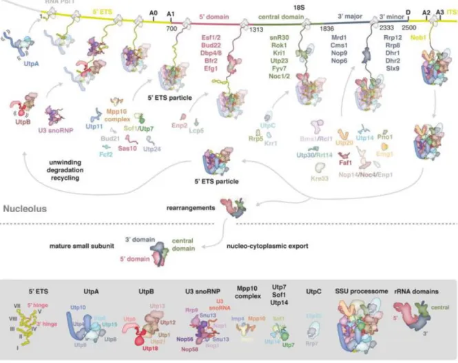
3.5.1.1 Formation and assembly of the small subunit processome - the initial precursor for both ribosomal subunits ...25
3.5.1.2 Final assembly of the small subunit ...32
3.5.2 Assembly of the LSU ...34
3.5.2.1 Nucleolar stages of large subunit assembly ...34
3.5.2.2 Nuclear stages of large subunit assembly ...45
3.5.2.3 Final cytoplasmic maturation of the large subunit ...49
3.6 Structural dynamics accompanying large subunit biogenesis ...51
3.7 RNA structure probing ...53
3.8 Classification of Noc3p in the context of large subunit biogenesis ...56
3.9 Objectives of this work ...62
4 Results ... 63
4.1 Single molecule cryo-electron microscopy reveals different populations of large ribosomal subunit precursors associated with Noc3p and delivers first structural hints on Noc2p ...63
4.2 Functional characterization of the Noc2p/Noc3p interaction and their association with pre-ribosomes ...72
4.2.1 Noc2p interacts with Noc3p via domain 6 close to its C-terminus ...72
4.2.2 Structure based mutagenesis of Noc3p ...76
4.2.4 Different domains of Noc3p have different roles on the assembly with Noc2p
and with pre-LSUs ...81
4.3 Noc3p associates with pre-ribosomes of an intermediate maturate state and its recruitment and release is dependent on the pre-ribosomal assembly state ...86
4.4 The 25S rRNA domain III undergoes significant conformational rearrangements upon depletion of certain LSU r-proteins ...91
4.5 Depletion of certain LSU domain III r-proteins does not affect the structure and orientation of the ITS2 ... 101
4.6 N-terminal truncations of rpL34 are mostly lethal and only conditionally assembled in ribosomes ... 103
4.7 Structure probing of Noc3p associated pre-ribosomes reveals a misfolded 25S rRNA DIII independent of the rpL34 truncation variant ... 108
5 Discussion ... 115
5.1 The correct pre-ribosomal assembly state accounts for the timely controlled recruitment and release of Noc3p ... 115
5.1.1 Prerequisites for the recruitment of Noc3p to pre-LSU particles ... 115
5.1.2 Prerequisites for the release of Noc3p from pre-LSUs ... 116
5.1.2.1 Assembly defects upon depletion of LSU r-proteins affect the release of Noc3p ... 116
5.1.2.2 Conformational rearrangements produced during folding of domain III impede the release of Noc3p ... 117
5.1.2.3 The ITS2 remains unaffected in Noc3p release mutants and shapes with the proposed ring-pin model ... 119
5.1.2.4 Absence of late acting LSU r-proteins L2/L43 impede Noc3p release and are diminished from pre-LSUs upon depletion of Noc3p ... 120
5.2 N-terminal truncations of rpL34 have an impact on pre-rRNA processing, Noc3p release and the structural integrity of LSU domain III ... 123
5.3 Structural and functional characterization of Noc2p/Noc3p ... 125
5.3.1 Single molecule cryo-EM provides structural information on the C-terminus of Noc3p and suggests volume density for Noc2p ... 125
5.3.2 Structural important sites for the formation of the Noc2p/Noc3p heterodimer and the association with pre-ribosomes ... 126
6 Material and Methods ... 129
6.1 Material ... 129
6.1.1 Yeast Strains ... 129
6.1.2 Plasmids ... 134
6.1.3 Oligonucleotides ... 140
6.1.3.1 Primer for PCR amplification and sequencing ... 140
6.1.3.2 Primer for primer extension analyses ... 141
6.1.3.3 Probes for northern Blotting ... 141
6.1.4 Antibodies (used for western blotting) ... 142
6.1.5 Chemicals ... 142
6.1.6 Media and buffers ... 142
6.1.7 Kits ... 148
6.1.8 Enzymes ... 148
6.1.9 Consumables ... 148
6.1.10 Equipment... 150
6.1.11 Software ... 151
6.2 Methods ... 152
6.2.1 Work with Saccharomyces cerevisiae ... 152
6.2.1.1 Cultivation and harvest of yeast strains ... 152
6.2.1.2 Preparation of competent yeast cells... 152
6.2.1.3 Transformation of competent yeast cells ... 152
6.2.1.4 Generation of yeast strains expressing epitope tag fusion proteins ... 153
6.2.1.5 Purification of genomic DNA from yeast ... 153
6.2.1.6 Growth kinetic analysis of yeast strains ... 153
6.2.1.7 Spot test analysis of yeast strains ... 154
6.2.1.8 Long term storage of yeast strains ... 154
6.2.2 Work with E. coli ... 154
6.2.2.1 Cultivation of bacterial strains ... 154
6.2.2.2 Preparation of chemical competent bacterial cells ... 154
6.2.2.3 Transformation of chemical competent bacterial cells with DNA by heat shock ... 155
6.2.2.4 Preparation of electric competent bacterial cells ... 155
6.2.2.5 Transformation of electro-competent bacterial cells with DNA by electroporation ... 155
6.2.2.6 Purification of plasmid-DNA from E.coli ... 156
6.2.3 Work with DNA ... 156
6.2.3.1 Native agarose gel electrophoresis of DNA ... 156
6.2.3.2 Purification of DNA fragments from agarose gel ... 156
6.2.3.3 Polymerase chain reaction ... 156
6.2.3.4 Ethanol precipitation of DNA ... 156
6.2.3.5 DNA quantification using UV spectroscopy ... 157
6.2.3.6 Digestion of DNA with restriction endonucleases ... 157
6.2.3.7 Dephosphorylation of DNA fragments ... 157
6.2.4 Work with RNA ... 158
6.2.4.1 RNA extraction ... 158
6.2.4.2 Denaturing agarose gel electrophoresis for high molecular weight RNA . 158 6.2.4.3 Denaturing acryl amide gel electrophoresis for low weight molecular RNA ... 158
6.2.4.4 Northern blotting (passive capillary transfer) ... 158
6.2.4.5 Northern blot (electro transfer) ... 159
6.2.4.6 Radioactive probe labelling and detection ... 159
6.2.4.7 Quantification of northern blots with MultiGauge ... 160
6.2.5 Work with Proteins ... 160
6.2.5.1 Determination of protein concentration ... 160
6.2.5.2 Denaturing protein extraction with TCA ... 161
6.2.5.3 SDS-polyacrylamide gel electrophoresis (SDS-PAGE) ... 161
6.2.5.4 Western blot ... 161
6.2.5.5 Ponceau S staining after western blotting ... 161
6.2.5.6 Detection of proteins by chemiluminescence ... 162
6.2.6 Affinity Purification of RNPs via Epitope Tagged Fusion Proteins ... 162
6.2.6.1 Preparation of yeast cell extracts ... 162
6.2.6.2 Affinity purification of (pre)-rRNPs using IgG coupled magnetic beads .... 163
6.2.6.3 Affinity purification of pre-ribosomes using IgG coupled sepharose beads ... 163
6.2.6.4 Affinity purification of pre-RNPs using anti-FLAG antibody coupled sepharose beads... 164
6.2.7 Local tertiary structure probing by MNase fusion proteins ... 164
6.2.7.1 Yeast strains / generation ... 164
6.2.7.2 Affinity purification and subsequent MNase digest of pre-RNPs ... 164
6.2.7.3 Primer extension ... 165
6.2.7.4 Sequencing reaction ... 166
6.2.7.5 Gel electrophoresis for low molecular weight DNA ... 166
7 References ... 167
8 List of Figures ... 203
9 List of Tables ... 205
10 Publications and Presentations ... 207
11 Acknowledgements / Danksagung ... 209
1
1 Summary
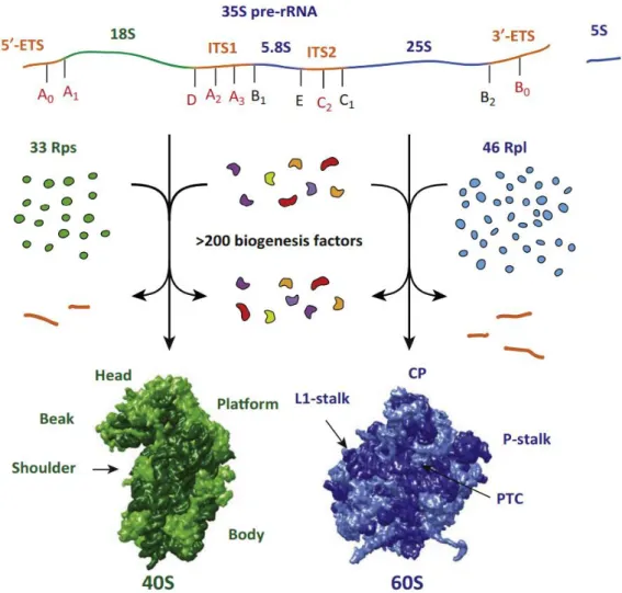
Ribosomes are macromolecular ribonucleoprotein (RNP) particles that catalyze the translation of mRNA into proteins in all living cells in all kingdoms of life. The ongoing production of ribosomes ensures the availability of a sufficient amount of proteins, which are required for various cellular processes. Ribosome biogenesis represents a highly energy consuming and complex process. Starting in the nucleolus, RNA polymerase I and III transcribe the ribosomal RNA (rRNA) encoding genes to generate precursor rRNAs, which undergo a series of processing, folding, modifying and assembly events in the course of maturation. In the yeast Saccharomyces cerevisiae, 79 ribosomal proteins (RPs), four rRNAs, several snoRNAs and more than 150 transiently acting biogenesis factors operate in a concerted and highly dynamic network to generate the mature ribosomal subunits.
During the past decades, a lot of effort has been made to elucidate and further understand the synthesis of ribosomes as paradigm of RNP biogenesis. Extensive research on this field has produced detailed characterization of ribosome biogenesis factors like the maturation step for which they are crucial for or the molecular function they exert. Nevertheless, the role of many factors remains further and requires extensive further investigation.
The “Noc-proteins” belong to the broad group of biogenesis factors, which have been identified essential for large ribosomal subunit (LSU) maturation (Noc1p/2p/3p). Accompanying studies have indicated the existence of the dimeric complexes Noc1p/Noc2p and Noc2p/Noc3p, which possibly associate with early and intermediate LSU precursor particles, respectively.
In this study, focus was laid on the role of Noc3p. Single molecule cryo-EM revealed the association of Noc3p with subsets of different pre-ribosomal populations of intermediate maturation states.
Thereby, a time interval for the association of Noc3p with pre-LSU particles could be deduced within a series of nuclear maturation steps. Simultaneously, first structural characterization could be obtained for the position of Noc2p, the Noc3p binding partner.
Furthermore, the basis of the time window, in which association of Noc3p with pre-ribosomes has been observed and the prerequisites for its recruitment and release to and off pre-LSUs has been explored. The results revealed that both recruitment and release of Noc3p depends on the pre- ribosomal assembly state. The varying impact of the single ribosomal proteins (r-proteins) has been discussed in regard to their known binding sites in LSU particles.
Applying a versatile tethered tertiary structure probing approach allowed investigation of possible structural alterations that impact or postpone the timely related release of Noc3p. Indeed, an impeded release could be backtracked to an altered conformational state of pre-LSU domain III upon knock- down of particular r-proteins.
2
Finally, structure-based mutagenesis of Noc3p helped to reveal sites within the protein that are essential for the formation of the heterodimer with Noc2p as well as for the association with the pre- LSU.
Further studies will be necessary to figure out the binding mode of Noc3p to pre-ribosomes and to which extent it might as well rely on the presence of Noc2p. For this, structural information on Noc2p as whole would greatly help to further address this issue. Altogether, the results presented in this work allow to hypothesize towards a role of Noc3p as sensor for LSU biogenesis and provide a solid base for further investigation.
3
2 Zusammenfassung
Ribosomen sind makromolekulare Ribonukleoprotein- (RNP) Partikel, die in allen lebenden Zellen und in allen Bereichen des Lebens vorkommen. Sie katalysieren die Translation von mRNA zu Proteinen.
Die kontinuierliche Produktion von Ribosomen stellt letztlich die Verfügbarkeit von Proteinen in ausreichend hohen Mengen für verschiedene zelluläre Prozesse sicher. Bei der Ribosomenbiogenese handelt es sich um einen komplexen Prozess, der mit hohem Energiebedarf verbunden ist. Die Synthese von Ribosomen beginnt im Nukleolus, in dem die RNA Polymerasen I und III die ribosomalen RNA (rRNA) kodierenden Gene transkribieren und so Vorläufer-rRNAs generieren. Diese durchlaufen während ihres Reifungsverfahrens eine Vielzahl an Prozessierungs-, Faltungs-, Modifizierungs- und Assemblierungsschritten. In der Hefe Saccharomyces cerevisiae wirken 79 ribosomale Proteine (RPs), vier rRNAs, einige snoRNAs und mehr als 150 Biogenesefaktoren in einem abgestimmten und hochdynamischen Netzwerk zusammen, um die reifen ribosomal Untereinheiten zu erzeugen. Die Biogenesefaktoren interagieren dabei transient in einem definierten Zeitfenster mit den prä- ribosomalen Partikeln. Im Laufe der vergangenen Jahrzehnte wurde viel Aufwand betrieben, um dieses Paradigma der RNP-Biogenese weiter aufzuklären und zu verstehen. Umfangreiche Forschung auf diesem Feld hat dazu beigetragen, das Detailwissen zu ribosomalen Biogenesefaktoren zu erweitern.
Dies schließt beispielsweise den Reifungsschritt ein, für welchen sie benötigt werden oder ihre molekulare Funktion. Nichtsdestotrotz bleibt die Rolle vieler dieser Faktoren bis dato schwer definierbar, was weitere intensive Untersuchungen erfordert.
Die „Noc-Proteine“ gehören als solche zu dieser großen Gruppe der Biogenesefaktoren. Sie wurden ursprünglich als essentielle Faktoren für die Reifung der großen ribosomalen Untereinheit (LSU) identifiziert (Noc1p/2p/3p). Begleitende Studien haben die Existenz eines Noc1p/Noc2p- und eines Noc2p/Noc3p- Dimers aufgedeckt, welche möglicherweise jeweils mit frühen und intermediären LSU- Partikeln assoziiert sind.
In der vorliegenden Arbeit wurde der Fokus auf die Rolle von Noc3p gelegt. Ergebnisse aus Kryo- Elektronenmikroskopie auf Einzelmolekülebene deuten auf die Assoziation von Noc3p mit einer Teilmenge an verschiedenen prä-ribosomalen Populationen eines intermediären Reifungszustandes hin. Dadurch konnte ein Zeitintervall über den Verlauf einiger nukleären Reifungsschritte definiert werden, für diese eine Assoziation mit Noc3p beobachtet werden konnte. Gleichzeitig gab es erste strukturelle Hinweise auf die Position von Noc2p, dem Bindungspartner von Noc3p.
Um die Basis, die dieses Zeitfenster definiert, weiter zu verstehen, wurden die Voraussetzungen für die Rekrutierung und die Freisetzung von Noc3p im Kontext von prä-LSU Partikel genauer untersucht.
Die Ergebnisse deuten darauf hin, dass sowohl die Rekrutierung als auch die Freisetzung von Noc3p vom prä-ribosomalen Assemblierungszustand abhängig sind. Die unterschiedlichen Einflüsse der
4
einzelnen ribosomalen Proteine (r-Proteine) wurden in Bezug auf deren bekannte Bindungsstellen in LSU-Partikeln diskutiert.
Die Anwendung eines vielseitig einsetzbaren Ansatzes zur Strukturanalyse ermöglichte die Untersuchung etwaiger struktureller Veränderungen, die die zeitlich gesteuerte Freisetzung von Noc3p beeinflussen oder gar verzögern. Tatsächlich konnte dabei eine beeinträchtigte Freisetzung von Noc3p beobachtet werden, welche sich auf konformationelle Änderungen in der prä-LSU Domäne III als Konsequenz der Abwesenheit bestimmter r-Proteine zurückverfolgen ließ.
Letztlich konnten durch strukturbasierte Mutagenese von Noc3p Bereiche innerhalb dieses Proteins identifiziert werden, die unabdingbar für die Bildung des Heterodimers mit Noc2p und für die Assoziation mit prä-Ribosomen sind.
Weitere Studien werden nötig sein, um die Bindung von Noc3p an prä-Ribosomen genauer aufzuklären, insbesondere zu welchem Ausmaß hierfür Noc2p erforderlich ist. Hierfür wären strukturelle Daten zu Noc2p als Ganzes sehr hilfreich. Insgesamt erlauben die in der vorliegenden Arbeit präsentierten Ergebnisse eine Hypothese über die Rolle von Noc3p als Sensor für die LSU Biogenese. Dies bietet eine solide Basis für zukünftige Untersuchungen.
5
3 Introduction
3.1 The eukaryotic ribosome – Structure and function
3.1.1 Overview
Ribosomes are macromolecular ribonucleoproteins complexes that fulfill the same function in all growing cells in all domains of life – the translation of mRNA into proteins. They constitute a complex and essential element within the cell that has been highly conserved throughout evolution (Woolford and Baserga 2013).
The term “ribosome” was initially proposed by Richard B. Roberts in 1958 in the course of the 1st symposium of the Biophysical Society on “Microsomal Particles and Protein Synthesis” at the Massachusetts Institute of Technology (MIT) (Richard B. Roberts 1958). This expression helped to designate the RNPs of the microsome fraction that have been already described by several scientists at that time. Initially, Siekevitz and colleagues showed, that microsomal fractions contain indispensable elements that help to incorporate amino acids into proteins (Siekevitz 1952).
Ribosomes can generally be classified in prokaryotic 70S and eukaryotic 80S particles, whereas the unit
“S” (short for “Svedberg”) stands for the sedimentation coefficient. Since it considers the point, at which sedimentation and diffusion reach a state of equilibrium during centrifugation, the sedimentation coefficient does not only consider the pure molecular weight of proteins but as well their size and spatial structure in a native solution (Svedberg and Fåhraeus 1926).
All ribosomes are composed of a large and a small ribosomal subunit (LSU and SSU, respectively), whereas the eukaryotic 80S ribosome is larger in size compared to its prokaryotic 70S counterpart.
Nevertheless, the general functions of the ribosomal subunits are identical across different organisms, respectively. In this work, all following statements refer to the situation encountered in the eukaryotic unicellular organism Saccharomyces cerevisiae unless stated otherwise. The large, 60S ribosomal subunit incorporates three RNA molecules (rRNAs), the 25S, 5.8S and 18S RNA (3396, 158 and 121 nucleotides (nt), respectively) and 46 ribosomal proteins (r-proteins). The small 40S subunit harbors solely one 18S rRNA (1800 nt) and 33 r-proteins (Klinge and Woolford 2019; Kressler et al. 2017). In brief, an important function of the SSU is to decode the sequence information on the mRNA template, which encodes for a certain protein. Binding sites for the tRNAs are contiguously located, thus facilitating codon-anticodon recognition between mRNAs and tRNAs loaded with amino acids. The LSU contains the peptidyl transferase center (PTC) that catalyzes the formation of a peptide bond between two amino acids (Schmeing and Ramakrishnan 2009). Notably, due to the fact that the PTC is nearly completely deprived of proteins, this reaction is predominantly mediated by the RNA components (Nissen et al. 2000), thus classifying the ribosome as ribozyme (Cech 2000). Together, both SSU and LSU build up the mature 80S ribosome (DELEY 1964) through interactions at their subunit interfaces.
6
Notably, the emergence of high resolution structures has strengthen the knowledge that not only the interaction between the subunits, but as well mechanistic processes for instance the binding of mRNA and tRNA and especially the formation of peptide bonds rely on the RNA components of the ribosome (Ban et al. 2000; Nissen et al. 2000; Carter et al. 2000; Harms et al. 2001; Yusupov et al. 2001; Ben- Shem et al. 2010; Ben-Shem et al. 2011).
3.1.2 Cryo-EM as state of the art for addressing ribosomal structure
To understand the processes underlying ribosome biogenesis has been an ambitious goal since their discovery. Throughout the last decades a lot of effort has been made to develop and refine new technologies to further enlighten ribosome assembly. In order to fully understand the mechanisms and processes that underlie the formation of ribosomes, it is necessary to know the exact structure of the respective particles.
Therefore, imaging techniques gained enormous importance for direct visualization of (pre-) ribosomes. X-ray crystallography has been used by structural biologists for decades. Especially around the turn of the millennium, a series of high-resolution crystal structures of bacterial and archaeal ribosomes were described (see 3.1.1). However, the main challenge in crystallographic studies is to figure out the right conditions for well-diffracting crystals. It is therefore of great importance to ensure that the isolated samples of ribosomes are highly homogeneous and can form well-ordered crystals.
Unfortunately, for some proteins it can take months or years to crystallize them, whereas others never crystallize at all. In addition, several further problems have complicated the work with crystallography, for instance the stability of the resulting crystals or phasing problems.
Nowadays, single molecule cryogenic electron microscopy (cryo-EM) has largely superseded X-ray crystallography, especially for structure determination of macromolecular complexes (Carroni and Saibil 2016). Importantly, cryo-EM does not require crystals, but ribosomes can be directly observed after freezing, ex vivo. Additionally, cryo-EM requires a much smaller amount and a less concentration of samples for analysis and can tolerate impurities as well as structural heterogeneity of the sample.
In fact, different sub-states of ribosomes or of their precursors can be deduced which allows an insight into the dynamic behavior of these particles (see for instance Kater et al. 2017; Sanghai et al. 2018b).
An important step in the improvement of cryo-EM was the invention of direct-electron detectors enabling to achieve near atomic resolutions (Kühlbrandt 2014). Due to the constant further development of both soft- and hardware related with cryo-EM, this technique provides a powerful tool for receiving high resolution structure of mature ribosomes and ribosomal intermediates. Even though the evaluation of cryo-EM data is more complex and requires higher computing power it represents the method of choice for studying the structure and function of (pre-) ribosomes that is routinely used in laboratories over the whole globe.
7
3.1.3 Structure and components of the small ribosomal subunit
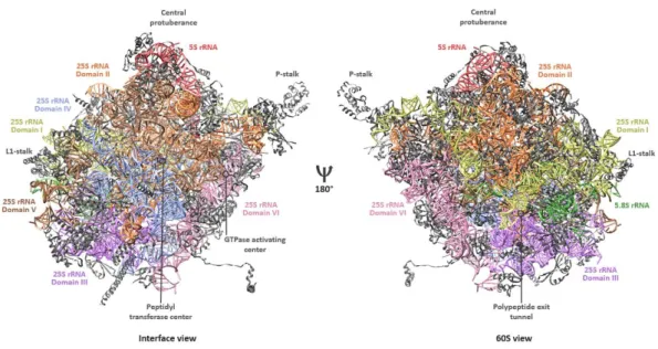
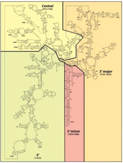
The 40S or small ribosomal subunit adopts, compared to the large subunit, a relatively simple structural conformation. The RNA component of the small subunit, the 18S rRNA, is subdivided in four phylogenetically conserved secondary structure domains: the 5´-, central-, 3´-minor and the 3´-major domain. These fold into tertiary structures, interacting with 33 r-proteins, thereby forming the head, beak, platform and the body (Figure 1, left side). These constitute characteristic architectural landmarks of the mature small ribosomal subunit (Klinge and Woolford 2019).
The 3´ major domain is almost exclusively located in the upper part of the SSU which forms the head and the beak, a protrusion of the 18S rRNA surrounded by proteins. The 3´minor domain is located at the subunit interface, vertically oriented to the head and surrounded by the 5´- and the central domain.
The latter one connects the head and the body (Figure 1, right side).
The central pseudoknot represents a key feature of the small ribosomal subunit that brings together all of the four secondary structure subdomains of the 18S rRNA. This interface regions is important for the overall architecture of the 18S rRNA and the decoding center, which is located in close vicinity in the middle of the small subunit (Ben-Shem et al. 2011).
Figure 1: Overall structure of the mature small ribosomal subunit from Saccharomyces cerevisiae.
Cartoon representation of the small ribosomal subunit shown from an interface view (left side) and from an 40S- or solvent side view (right side) (PDB 4V88). The 18S rRNA subdomains are color-coded and indicated. Ribosomal proteins are shown in grey. Positions of structural landmarks (head, beak, platform, body, central pseudoknot) and of functional sites (decoding center) are annotated.
The mRNA passes the small subunit through a channel between the beak and body and exists between the platform and head. A series of high resolving crystal structures of different prokaryotic ribosomes have allowed to track the path of the mRNA through the small subunit (Yusupova et al. 2001). As
8
bacterial and eukaryotic ribosomes share an evolutionarily conserved core which attributes their intrinsic functions, the course of mRNA trough the SSU in eukaryotic organisms is the same. Structural data obtained by X-ray crystallography and cryo-EM have completed these findings (Melnikov et al.
2012). The initial entry of mRNA to the small subunit however shows substantial differences between pro- and eukaryotes. In prokaryotic, but not archaeal organisms, the shine Dalgarno sequence initializes binding of the SSU onto mRNA (Gualerzi and Pon 2015), whereas in eukaryotes the mechanism of mRNA recognition and recruitment appears to be more sophisticated involving the 5´- cap structure of mRNA and additional factors, ultimately forming the pre-initiation complex (Jackson et al. 2010; Fraser 2015).
3.1.4 Structure and components of the large ribosomal subunit
The 60S or large ribosomal subunit adopts a more condensed, crown-like conformation including several characteristic landmarks and functional centers. The large subunit harbors three RNA molecules – the 25S, the 5.8S and the 5S rRNAs. The former one is subdivided in six phylogenetically conserved secondary structures – domains I-VI – which form its defined tertiary structure, together with the interaction of 46 r-proteins. In general, the subdomains of the LSU are more intertwined with each other than are the 18S rRNA subdomains of the SSU.
Figure 2: Overall structure of the mature large ribosomal subunit from Saccharomyces cerevisiae.
Cartoon representation of the large ribosomal subunit shown from an interface view (left side) and from an 60S- or solvent side view (right side) (PDB 4V88). The 25S rRNA subdomains as well as the 5.8S and the 5S rRNAs are color-coded and indicated. Ribosomal proteins are shown in grey. Positions of structural landmarks (P-stalk, L1-stalk, central protuberance) and of functional sites (peptidyl transferase center, polypeptide exit tunnel, GTPase activating center (“SRL” in Figure 3) are annotated.
The solvent side is largely structured both by the 25S rRNA domains I, II and by the 5.8S rRNA (Figure 2, right side), whereas domains IV and V form the subunit interface. The 5.8S rRNA base-pairs with
9
domain I and is sandwiched between domains I and III. The latter one is located at the bottom of the subunit, linking domains localized there. Domains III and VI bridge the subunit interface with the solvent exposed side. The 5S rRNA is assembled on top of the LSU, contacting domains II and V (Ben- Shem et al. 2011; Klinge and Woolford 2019).
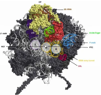
Functional sites are mostly localized at or near the subunit interface (Figure 3) (Konikkat and Woolford 2017). The P-stalk is shown on the right side of the LSU (Figure 3, blue). At its basis lies the sarcin ricin loop (SRL) which has obtained its name by the cytotoxins α-sarcin and ricin that target this structural motif and completely abolish translation (Endo and Wool 1982; Endo et al. 1987). Both the P-stalk and the SRL form the GTPase-associated center (GAC). The P-stalk recruits and activates translation factors and the SRL triggers GTP hydrolysis to GDP of these factors (Santos and Ballesta 2005; Diaconu et al.
2005; Voorhees et al. 2010).
The tRNA accommodation corridor begins with helices of 25S rRNA domain V that form the tRNA entry tunnel (Figure 3, yellow) and continues with the A-, P- and E- (Aminoacyl-, Peptidyl- and Exit-) sites for tRNA binding. In the middle of this corridor, at the P-site, localizes the polypeptide transfer center (PTC) formed by domains IV and V and nearly completely deprived of proteins. At this site, the peptide bond formation between two amino acids and the hydrolysis of peptidyl-tRNA bonds are catalyzed.
The PTC is adjacent to the entrance of a tunnel, the polypeptide exit tunnel (PET). Nascent proteins pass through the tunnel before they are released from the ribosome at the opposite, solvent exposed site. The exit of the PET is surrounded by the 25S rRNA domain III.
The central protuberance (CP) of the mature LSU is located atop of the particle comprising the 5S rRNA and the LSU r-proteins rpL5 and rpL11 (Figure 3, “CP”). It is involved in inter-subunit interactions with domains II and V and assembled r-proteins, respectively (Ben-Shem et al. 2011). A plethora of studies on the 5S rRNA has been performed throughout the decades and several functions have been attributed (for review see Gongadze 2011). Importantly, it has been shown to enhance translational fidelity. However, translation involves all functional centers of the ribosome since this process constitutes its dedicated function. Therefore, the central protuberance is supposed to operate as signal transducer between the functional centers of the large subunit through physical connections, thus helping to coordinate translation events (Smith et al. 2001; Dinman 2005; Kiparisov et al. 2005).
The L1 stalk (Figure 3, left side) is suggested to facilitate opening of the E-site by moving to a different conformation, thereby contributing to the efficient release of the tRNA (Andersen et al. 2006).
The A-site finger (ASF), which is as well highlighted in the structure protrudes to the inter-subunit space, located in close proximity to the A-site. It has been shown to attenuate the motion of the A-site tRNA, acting as translocation attenuator that helps to maintain the reading frame (Komoda et al. 2006).
10
Figure 3: Functional centers of the large ribosomal subunit of Saccharomyces cerevisiae.
Cartoon representation of the 60S ribosomal subunit from an interface side view in order to primarily visualize its functional centers. At the top, the 5S rRNA and associated LSU r-proteins L5/L11 are shown. Two further characteristic landmarks, the P-stalk (on the right, blue) and the L1-stalk (on the left) are indicated. The SRL-loop is shown in red and the tRNA entry tunnel in yellow which depicts the beginning of the tRNA accommodation corridor that continues with the A- and P-sites for tRNAs and ends with rpL42 (purple) at the tRNA E-site. The PTC is indicated in the middle of the tRNA accommodation corridor at the P-site, the PET emerges to the solvent side and is indicated as well. The A-site finger (green) and the adjacent rpL10 (red) are shown as well. Adopted from (Konikkat and Woolford 2017).
3.1.5 Structure and function of the 80S or mature ribosome
Both the small as well as the large ribosomal subunit contain an evolutionarily conserved core that assigns the main functions of the ribosome. However, eukaryotic ribosomes are at least 40 % larger in size compared to their prokaryotic counterparts, which is due to additional elements present in the subunits. Eukaryote specific additives are extra ribosomal proteins and insertions/extensions into existing r-proteins, which could be visualized by the availability of the first high resolution crystal structures of Saccharomyces cerevisiae and Tetrahymena thermophila (Ben-Shem et al. 2010; Ben- Shem et al. 2011; Klinge et al. 2011; Rabl et al. 2011). A further eukaryote specific feature are blocks of additional rRNA, termed “expansion segments” (ES) (Gerbi 1996), which emerge from the conserved secondary structure core of the mature rRNAs and are mainly located on the solvent exposed peripheral surface of the subunits (Hsiao et al. 2009; Melnikov et al. 2012). Notably, these ES are not fully eukaryote specific elements, as ES were found in several archaeal organisms (see for instance Armache et al. 2013). Some archaea contain even supersized ES that are longer than eukaryotic ones (Penev et al. 2019).
11
The two subunits of Saccharomyces cerevisiae differ significantly from each other regarding the distribution of expansion segments and of r-proteins. In the small subunit, most rRNA is concentrated at the bottom, whereas the r-proteins are scattered all over its surface, mostly on the solvent exposed site. Conversely, the expansion segments of the large subunit form a nearly continuous ring around the PET, spanning from the P-stalk to the L1-stalk (for detailed description of the subunits see 3.1.3 and 3.1.4). The LSU r-proteins are mostly associated with this ring-structure, which results in the same characteristic distribution. Moreover, it has been shown that eukaryote specific r-proteins are often contacted by eukaryote specific ESs (Ben-Shem et al. 2011).
Information concerning the detailed roles of most expansion segments remains sparse although several properties and functions could already be attributed. For instance, deletion of ES27L of tetrahymena was lethal and lead to an accumulation of pre-rRNA. This indicated, that this ES might play a role in either pre-rRNA processing or the stability of mature rRNAs (Sweeney et al. 1994).
Furthermore, replacement of the ES with unrelated sequences of similar size or a homologous sequence from another organism in incorrect orientation was lethal as well, suggesting that the ES are not simply spacers that can be replaced by any sequences of corresponding size (Sweeney et al. 1994).
Similar observations in yeast corroborated these findings (Jeeninga et al. 1997). Recent studies revealed the association of the methionine amino peptidase (MetAP) with ES27L, which indicates a function in translation fidelity. Deletions at specific sites within the ES27 lead to increased amino acid misincorporation, read-through- and frameshifting-errors (Fujii et al. 2018).
In general, the deletion of ES mostly interferes with the correct production of mature rRNAs an hence impedes the proper biogenesis of ribosomes (Ramesh and Woolford 2016). Moreover, increased rRNAs of more complex organisms than prokaryotic ones could confer increased functionality to the ribosome. For instance, characterization of the binding partners of ES7 revealed, amongst others, the association of several aminoacyl tRNA synthetases with the ES. This connection is supposed to maintain a close interaction of the synthetases with each other and with the translation machinery (Gómez Ramos et al. 2016). Therefore, the increased complexity of eukaryotic translation, especially regarding initiation and elongation might also explain their presence (for selected reviews describing the current knowledge of the translation process see Wilson and Doudna Cate 2012; Voorhees and Ramakrishnan 2013; Hinnebusch 2014; Rodnina 2016; Dever et al. 2016; Hinnebusch 2017; Schuller and Green 2018;
Shirokikh and Preiss 2018).
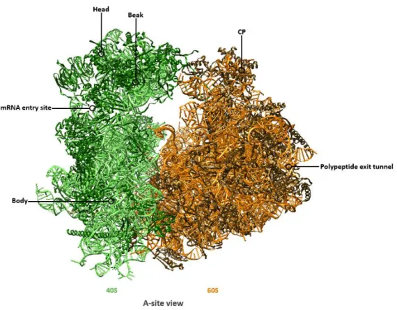
During translation initiation, the small ribosomal subunit joins the large ribosomal subunit to form an asymmetric assembly. Thereby, the head of the SSU interacts with the CP of the LSU (Figure 4).
Importantly, the integrity of the two subunits to each other within the mature ribosome is strengthened by the formation of inter-subunit bridges.
12
Figure 4: Overall structure of the mature 80S ribosome from Saccharomyces cerevisiae.
Cartoon representation of the mature 80S ribosome from an A-site view, consisting of the small 40S subunit (rRNA chain in light green, r-proteins in dark green) and of the large 60S ribosomal subunit (rRNA in orange, r-proteins in brown) (PDB 4V88).
Positions of the head, beak, body and the mRNA entry site of the SSU as well as the central protuberance (CP) and the polypeptide exit tunnel (PET) of the LSU are annotated.
In fact, the translation process is not static but highly dynamic. During elongation, when seen form the solvent side of the small subunit, the SSU rotates a few degrees counterclockwise in a ratcheting motion relatively to the large subunit (Frank and Agrawal 2000). Additionally, the head domain of the SSU swivels relatively to the body (Zhang et al. 2009a; Spahn et al. 2004). These movements are important for mRNA- and tRNA- translocation, termination and other processes. The inter-subunit interactions change with each rearrangement and are dynamic in composition. Therefore, inter- subunit bridges are indispensable to maintain the communication between the subunits during protein synthesis. The crystal structure of the yeast ribosome reveals 17 inter-subunit bridges which are, in contrast to the prokaryotic counterpart, mainly formed by the protein components of the subunits (Spahn et al. 2001; Yusupov et al. 2001; Ben-Shem et al. 2011).
13
3.2 Transcription of the genes encoding for the ribosomal RNAs in the nucleolus
The nucleolus defines a substructure within the nucleus that can be observed as dark spot by light microscopy. In 1781 Felice Fontana was the first to figure the nucleolus in the nucleus which he described as an egg-formed body occupied by a spot in the middle. Further, more explicit descriptions of the nucleolus emerged during the 18th century (for a historical review see Montgomery 1898).
According to the current state of knowledge, the nucleolus represents a functional intra-nuclear, membrane less organelle, which is present in all eukaryotic cells analyzed so far. It constitutes the ribosome factory of the cell where rDNA transcription, initial pre-rRNA processing and first assembly events of the earliest pre-ribosomal particles take place (Raška et al. 2006; Hernandez-Verdun et al.
2010). The structure of the nucleolus is not homogenous. In fact, three morphological compartments defined by their texture and contrast were observed in electron microscopy analyses of eukaryotes – fibrillar centers (FC), a dense fibrillar component (DFC) and a granular component (GC), which are functionally assigned to processes in ribosome biogenesis (Léger-Silvestre et al. 1999; Trumtel et al.
2000). However, the differentiation and organization of the nucleolar sub-components among different organism is challenging (discussed in the review of Thiry and Lafontaine 2005).
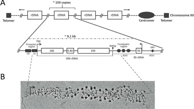
The nucleolus forms around the nucleolar organizer region (NOR), sites where multiple rDNA copies cluster in arrays. In the yeast Saccharomyces cerevisiae ~150 copies of the genes encoding the ribosomal RNAs, referred to as “rDNA repeats” are localized on chromosome XII as tandem array (Petes and Botstein 1977; Petes 1979; Long and Dawid 1980). Each rDNA repeat has a size of ~9.1 kb and is arranged in two transcriptional units (Figure 5, A). The first unit encodes an operon-like gene that produces a 35S pre-rRNA transcript containing the sequences for mature 18S, 5.8S and 25S rRNAs with a total size of ~6.6 kb. As indicated in Figure 5 (A), the sequences for the mature rRNAs are flanked by spacers (ETS/ITS), which have to be removed during pre-rRNA processing (see 3.3). The 35S precursor rRNA molecule is transcribed by RNA polymerase I (Pol I). The second unit encodes for the 5S rRNA, arranged in reverse direction, which is flanked on both sides by non-coding intergenic spacers (IGS), thereby separated from the first unit (Philippsen et al. 1978). The 5S rRNA is transcribed by RNA polymerase III (Pol III) (Weinmann and Roeder 1974).
14
Figure 5: Organization of the rDNA locus in Saccharomyces cerevisiae and visualization of “Miller spreads”.
(A) Organization of the rDNA encoding genes, arranged in a cluster of arrays (~ 150 copies) located on chromosome XII. The composition of one rDNA repeat which has a length of approximately 9,1 kb is illustrated in more detail. The 25S, 5.8S and the 18S rDNA sequences which make up the 35S rDNA are arranged in an operon-like structure, separated by internal transcribed sequences and flanked by external sequences (not annotated here, for illustration see Figure 6). The 5S rDNA is separated by the intergenic spacer 1, the region separating the 35S rDNA repeats from neighboring by the intergenic spacer 2 (IGS1/IGS2). The transcription directions of the 35S rDNA by Pol I and of the 5S rDNA by Pol III in opposite directions are indicated by arrows. The positions of relevant DNA elements are marked: The upstream element (UE) and the core element (CE) which are part of the rDNA promoter, the cleavage site of Rnt1p (triangle), the IGS1 including the T-rich element (circle), the Reb1p-binding site (trapeze) and the replication fork barrier (cross), and the IGS2 which contains an autonomous replicating sequence (ARS, diamond). (B) Electron micrograph of a Miller chromatin spread which shows actively a transcribed rDNA repeat. Pol I molecules are marked by arrows. Adapted from (Osheim et al. 2004).
The 14 subunit Pol I is an outstanding polymerase, as it is solely dedicated to transcribe the 35S pre- rRNA. In contrast, Pol III transcribes, besides the 5S rRNA, the tRNA genes and several other non-coding RNAs (ncRNAs) including the RNA component of the signal recognition particle and different small nucleolar RNAs (snoRNAs) (for review see Dieci et al. 2007). The requirements for an efficient initiation of Pol I transcription are well studied. It requires the action of the four proteins or protein modules, the core factor (CF), TATA box binding protein (TBP), upstream activating factor (UAF) and Rrn3p.
Moreover, two regulatory cis elements in the Pol I promotor region, the core element (CE) ranging from -28 to +8 relative to the transcription start site (TSS) and the upstream element (UE) located around bases -146 to -51, are necessary (Musters et al. 1989; Kulkens et al. 1991; Choe et al. 1992).
The Pol I specific initiation factor Rrn3p associates directly with the A43 subunit of Pol I and is strictly required to render Pol I competent for initiation (Yamamoto et al. 1996; Milkereit and Tschochner 1998; Peyroche et al. 2000). The heterotrimeric CF consisting of Rrn6p, Rrn7p and Rrn11p (Keys et al.
1994; Lin et al. 1996; Lalo et al. 1996) binds to the CE in the promoter region and can recruit the
15
initiation competent Rrn3p-Pol I to the rDNA, possibly via interaction between Rrn6p and Rrn3p (Peyroche et al. 2000). Notably, Pol I, Rrn3p and CF bound to the CE are sufficient for a very basal transcription in vitro. However, the transcription rate tremendously enhances upon binding and interaction with UAF and TBP (Keener et al. 1998). UAF consists of Rrn5p, Rrn9p, Rrn10p, Uaf30p and the two histones H3 and H4, which are suggested to mediate efficient binding to the UAE (Keys et al.
1996; Keener et al. 1997; Siddiqi et al. 2001). TBP binds, accordingly to UAF, to the UE and interacts both with components of the UAF and the CF namely Rrn9p and Rrn6p, respectively. The interaction with Rrn9p was shown to be more strong than with Rrn6p, which indicates a CF dependent recruitment by UAF, bridged through TBP (Steffan et al. 1996; Steffan et al. 1998; Keener et al. 1998). Therefore, the current model of Pol I initiation proposes UAF and TBP bound to the rDNA promoter region.
Thereby, UAF and TBP recruiting CF, which provides a platform for binding of initiation competent Rrn3p-Pol I through CF recruitment to form the pre-initiation complex (PIC) that ultimately results in a high-level transcription of 35S pre-rRNA.
Once transcription is initiated, Pol I maintains a high elongation rate, transcribing at a speed of ~ 40- 60 nts per second (Kos and Tollervey 2010). Regarding Pol I transcription termination, the exact mechanism is not clarified in detail. It depends on cis-elements downstream of the 35S rDNA region (T-rich sequence, Reb1-binding site, replication fork barrier) and on trans-acting factors that bind to these elements (Reb1p, Nsi1p, Fob1p). Moreover, endonucleolytic cleavage by Rnt1p followed by exonucleolytic trimming of the nascent rRNA could also contribute to the release of Pol I from the rDNA (for review see Németh et al. 2013). Previously, the availability of high-resolution structures of Pol I at different states, in association with Rrn3p, as well as initiation complexes have contributed to the understanding on how the transcription process and its regulation might occur (e.g. see Engel et al. 2013; Fernández-Tornero et al. 2013; Neyer et al. 2016; Tafur et al. 2016; Sadian et al. 2017; Sadian et al. 2019; Pilsl and Engel 2020).
Both transcription initiation and elongation of Pol I are regulated steps in rRNA synthesis (French et al.
2003; Zhang et al. 2010). During elongation, several protein factors have been found to influence Pol I activity, for instance Spt4p, Spt5p or the Paf1 complex (Paf1C). Importantly, these trans-acting factors exert their function upstream of pre-rRNA processing at the level of rDNA transcription. Due to their manipulation of the Pol I elongation properties, they influence the temporarily abundance of pre-rRNA transcripts and may therefore exert an impact on the organization of pre-rRNA processing (Schneider et al. 2006; Schneider et al. 2007; Zhang et al. 2009b; Zhang et al. 2010; Anderson et al. 2011). Another level of regulation is represented by the HMG protein Hmo1p which was found to bind on nucleosome free rDNA, thereby maintaining accessible rDNA structure (Gadal et al. 2002; Hall et al. 2006; Merz et al. 2008).
16
Finally, the synthesis of rRNA can be regulated at a very initial level through the abundance of rDNA copies that are accessible for transcription. Even in exponentially growing yeast cells, only ~ 50% of the 35S rDNA repeats are actively transcribed. In contrast, the other half is packaged into nucleosomes which seems to be important for the integrity of the rDNA locus (Ide et al. 2010).
One possibility to observe and quantitate actively transcribing Pol I complexes is the direct visualization of chromatin spreads by electron microscopy, a method that has been developed by Oscar L. Miller at the end of the 60s (Miller and Beatty 1969). Thereby, the numerous Pol I complexes, occupying a single rDNA repeat, can be observed at different positions within a single rDNA gene, having transcribed the 35S rDNA to a different degree. The emerging nascent rRNAs form the branches of the chromatin fiber, which are occupied with terminal knobs giving the “Miller-spreads” its characteristic “Christmas tree”
like appearance (Figure 5, B). Notably, the terminal knobs represent the first ribosomal precursor particles, as initial steps of pre-RNA processing and ribosome assembly occur co-transcriptionally (see 3.3.1 and 3.5.1.1).
3.3 Processing and modification of ribosomal RNAs
As mentioned above, RNA polymerase I is dedicated to the transcription of the rDNA locus, except for the 5S rRNA. Thereby, the initial 35S pre-rRNA, which contains the mature sequences for the 18S, 5.8S and 25S rRNAs is generated. Embedded in this precursor transcript, the mature rRNAs are separated by two internal transcribed spacers (ITS1 and ITS2) and flanked by external transcribed spacers (5´-ETS and 3´-ETS). These additional blocks of RNA are not present in the mature ribosomal subunits and have therefore to be removed. This involves successive series of both endonucleolytic and exonucleolytic processing events (Venema and Tollervey 1995).
3.3.1 Co-transcriptional processing
Originally, it was assumed that pre-rRNA processing in eukaryotes occurs exclusively after transcription of the rDNA by RNA polymerase I. In this situation, the 35S pre-rRNA transcript is cleaved at processing site B0 in the 3´-ETS (Figure 6) at an imperfect stem-loop by the endonuclease Rnt1p, a bacterial homologue to RNase III (Elela et al. 1996; Kufel et al. 1999). Subsequent processing steps, starting with successive cleavages within the 5´-ETS would occur after Rnt1p has performed its actions.
First evidence for co-transcriptional processing events comes from observations made by pulse-chase labelling experiments of synthesized rRNAs. The outcome of these analyses lead to the suggestion, that the introduction of base modifications followed by separating cleavage events on 35S pre-rRNA were not completely post-transcriptional (Udem and Warner 1972). In the late 80´s, further evidence for co-transcriptional processing of the 5´-ETS in Saccharomyces cerevisiae has been received. At that time, Veinot-Drebot and colleagues estimated half-lives of pre-rRNA molecules that contain ETS versus
17
the time period that is necessary to fully transcribe the 35S pre-rRNA by quantitative hybridization techniques. The results suggested that ETS removal takes place before the transcription is completed (Veinot-Drebot et al. 1988). Later, electron microscopy analysis of yeast cells in log phase helped to visualize the terminal knobs at the end of branches that appear in miller spreads, which represent the nascent RNPs. Transcripts of different lengths have been observed, dependent on the current position of Pol I within the rDNA gene. It was concluded that processing events of approximately 40-80 % occur on the nascent transcripts in the ITS1 (Osheim et al. 2004). More recently, these findings were confirmed by metabolic labelling experiments of exponentially growing yeast cells and mathematical modelling. The respective analyses demonstrated a co-transcriptional cleavage activity of pre-rRNA at a level of 70 % (Kos and Tollervey 2010).
The terminal knobs observed in miller spreads were supposed to form prior to possible processing events. In fact, compositional analysis revealed the integrity of components that are required for pre- 18S processing. Thus, the knobs have been termed small subunit processome (SSU processome) (Dragon et al. 2002). This indicates that co-transcriptional processing involves endonucleolytic cleavage events at the processing sites A0, A1, A2 and B0. However, the molecular mechanisms underlying the decision for co- or post-transcriptional processing have not been clarified yet. 5´-ETS processing starts with cleavages at site A0 and A1, resulting in 33S and 32S pre-rRNAs, respectively. The exact position of processing at site A1 is defined by evolutionarily conserved nucleotides upstream of this site in the 5´-ETS and by a stem-loop structure downstream of A1 within the 5´-region of the 18S rRNA (Venema et al. 1995; Sharma et al. 1999). Cleavage at site A2 splits the two maturing small and large subunits incorporating 20S and 27S pre-rRNA, respectively, from each other which leads to independent maturation routes. The A2 processing site is as well defined by 3´- evolutionarily conserved nucleotides that form a stable hairpin structure, similar as observed for the 3´-region of 18S rRNA (Allmang et al. 1996). However, cleavage at site A2 cannot be undoubtedly designated to one putative endonuclease. The involvement of Utp24 in cleavage at both positions A1 and A2 has been reported by some authors (Bleichert et al. 2006; Wells et al. 2016), whereas others attribute processing at site A2 to Rcl1p (Billy et al. 2000; Horn et al. 2011; Delprato et al. 2014). Finally, processing at position B0 in the 3´ETS terminates co-transcriptional processing, as this cleavage step releases the emerging pre-rRNA transcript. Interestingly, this has been found to impede processing at site A3 within the ITS1, probably depicting a quality control checkpoint (Allmang and Tollervey 1998).
Growth of the yeast cells in an environment facing stress conditions, for instance nutrient limitation may, however, lead to increased post-transcriptional processing at the expense of co-transcriptional processing. In 2017 Kos-Braun and co-workers revealed the TOR1 and CK2 kinases as responsible key- players in regulating pre-rRNA processing at a post-transcriptional level. The Tor 1 complex (TORC1) is sensitive to growth conditions as nutrient availability and environmental stress. TORC1 depicts a
18
central signaling hub in response to environmental cues mediating a variety of intracellular signaling branches (González and Hall 2017) and citations therein). Thereby, TORC1 controls the transcriptional activity of all three RNA polymerases and therefore the availability of rRNA and r-proteins for ribosome biogenesis. Among them is the downstream activation of the CK2 kinase, a prerequisite for pre-rRNA processing at site A2 along the A2-pathway (Kos-Braun et al. 2017). Inhibition of either TOR1 or CK2 leads to identical pre-rRNA processing abnormalities. As consequence, large amounts of 23S pre-rRNA resulting from a non-productive processing pathway through direct cleavage at A3 in the ITS1 prior to 5´-ETS processing were observed. As processing at site A3 is coupled to B0 processing (Allmang and Tollervey 1998), the course of these events is per definition derived to post-transcriptional processing.
Notably, experimental evidence that 23S pre-RNA is aberrant and ribosomes incorporating this pre- rRNA species are degraded is missing. Further evidence for diminishment of co-transcriptional processing activity could be obtained regarding the terminal knobs of the EM-analysis described above.
Upon heat-shock at 37 °C the resulting transcripts failed to show transcript-cleavage in contrast to the untreated control (Osheim et al. 2004). In addition further mechanisms exist that serve for fine-tuning the balance between co- and post-transcriptional processing (for review see La Cruz et al. 2018).
Therefore, co-transcriptional processing is preferred in favorable growth conditions controlled by a variety of intra- and extracellular signals.
19 Figure 6: Pre-rRNA processing in the yeast Saccharomyces cerevisiae.
(A) Schematic representation of one 25S pre-rRNA molecule. The sequences of mature 18S, 5.8SS/L and 25S rRNA are separated by two internal transcribed spacers (ITS1/ITS2) and flanked by two external transcribed spacer regions (5´-ETS/3´- ETS). The positions of the individual processing which correspond to 5´- or 3´-end of rRNA (precursors) are indicated. The positions, numbers and designation of the DNA oligo probes used for detection of the different rRNA (precursors) by northern blotting are illustrated. Note that the lengths of rRNAs and spacer sequences are not in proportion to their real lengths.
Processing site for generation of 18S rRNA are depicted in red, those for generation of 5.8SS/L and 25S rRNA are depicted in blue. (B) Alternative pathways of 27SA2 pre-rRNA processing. See text for more detailed explanation. Adopted and modified (Braun et al. 2020).
3.3.2 Post-transcriptional processing
At least the further maturation steps of both the small and the large subunit occur as post- transcriptional processing events. The 20S pre-rRNA incorporated in SSU-precursor particles are exported to the cytoplasm where the final maturation to 18S rRNA occurs (Udem and Warner 1973).
Therefore, the endonuclease Nob1p performs cleavage at the single stranded processing site D in the ITS1 (Fatica et al. 2003a; Lamanna and Karbstein 2009; Pertschy et al. 2009).
The precursor of the large subunit incorporating 27SA2 pre-rRNA represents the second product from cleavage at site A2. Further processing splits in two possible pathways both ultimately resulting in the same 25S rRNA sequence but yielding alternative forms of 5.8S rRNA that differ in length of 7 nucleotides at their 5´-ends (5.8SS/5.8SL). In the vast majority, site A3 within the ITS1 is cleaved by the endonuclease MRP (Lindahl et al. 1992; Chu et al. 1994a). Notably, in 1991 the group of Jonathan R.
Warner already isolated a temperature sensitive strain carrying a mutation in the gene designated as RRP2. This mutant strain accumulated this, at that time novel, pre-rRNA species containing sequences of the ITS1 (Shuai and Warner 1991). One year later, this phenotype was referred to a mutation in the endonuclease MRP (Lindahl et al. 1992).
In any case, cleavage at site A3 provides the entry site for 5´-3´ exonucleolytic ITS1 trimming to the B1S
site, the mature 5´-end of 27SBs pre-rRNA involving the exonucleases Rat1p, Xrn1p and Rrp17 (Henry et al. 1994; Oeffinger et al. 2009). Alternatively, in approximately 10 % of the cases, cleavage at B1L by an so far unknown endonucleolytic processing event, generates the mature 5´-end of 5.8SL (Faber et al. 2006). This alternative processing pathway results in the production of 27SA3 pre-rRNA which