Identification of novel therapeutic targets in mastocytosis:
Inhibition of survival of neoplastic mast cells by targeting IAPs and TRAIL receptors
Inaugural-Dissertation zur
Erlangung des Doktorgrades
der Mathematisch-Naturwissenschaftlichen Fakultät der Universität zu Köln
vorgelegt von Stefan Philipp Grotha
aus Hamburg
Köln, 2011
Berichterstatter: Prof. Dr. Manolis Pasparakis (Gutachter) PD Dr. Roswitha Nischt Tag der mündliche Prüfung: 04. Juli 2011
Mastocytosis is a rare disease characterized by neoplastic accumulation of mast cells in various organs, particularly in skin and bone marrow. Sporadic mastocytosis is usually caused by somatic point mutations of the receptor tyrosine kinase KIT that occur in exon 17, codon 816, and activates KIT by affecting the intracellular enzymatic site of the molecule.
Homeostasis of so altered mast cells is influenced on different levels. These cells show an elevated rate of proliferation and changes in cellular apoptosis. In this thesis the expression of the inhibitor of apoptosis protein (IAP) Survivin in neoplastic mast cells and its importance as a diagnostic marker or therapeutic target has been evaluated. For this purpose the Survivin content of normal and neoplastic mast cells was analyzed. Furthermore Survivin expression in mast cells of bone marrow samples from patients with mastocytosis and healthy donors was compared. The expression of Survivin protein in the neoplastic human mast cell line HMC-1 was downregulated by siRNA and apoptosis was subsequently induced with tyrosine kinase inhibitors or the death receptor ligand TRAIL. It is known from other hematopoietic neoplasia that Smac mimetics, inhibitors of the IAPs XIAP, cIAP1 and cIAP2, are potent therapeutics able to amplify the effect of classical cytotoxic therapies. Here the Smac mimetic LCL161 was used in combination with the tyrosine kinase inhibitor PKC412 and TRAIL to inhibit growth of the neoplastic mast cell line HMC-1.
The expression of the IAP Survivin is enhanced in neoplastic altered mast cells and can be detected in significantly higher amounts in mast cell infiltrates of the bone marrow from patients with mastocytosis compared to healthy donors underlining its value as a diagnostic marker. Downregulation of Survivin protein as well as the use of the Smac mimetic LCL161 inhibited the growth of the neoplastic mast cell line HMC-1 synergistically with tyrosine kinase inhibitors and the death receptor ligand TRAIL. These results emphasize the relevance of cellular regulation of apoptosis as a future target for the therapy of mastocytosis.
Zusammenfassung
Mastozytose ist eine seltene Erkrankung die durch eine neoplastische Akkumulation von Mastzellen in unterschiedlichen Organen, insbesondere der Haut und dem Knochenmark charakterisiert ist. Auslöser für die Mastozytose sind oftmals aktivierende somatische Punktmutationen in Exon 17, Codon 816 des Gens der Rezeptortyrosinkinase KIT, welche die intrazelluläre enzymatische Domäne des Rezeptors beeinflussen. Die Homöostase derartig veränderte Mastzellen ist auf mehreren Ebenen gestört. So konnte nicht nur eine erhöhte Proliferationsrate, sondern auch ein verändertes Apoptoseverhalten nachgewiesen werden. In dieser Arbeit wurde die Expression des inhibitor of apoptosis Proteins (IAP) Survivin in neoplastisch veränderten Mastzellen und seine Bedeutung als diagnostischer Marker sowie als therapeutisches Target untersucht. Hierzu wurden gesunde und neoplastisch veränderte Mastzellen auf ihren Survivingehalt hin überprüft sowie die Survivinexpression der Mastzellen in Knochenmarksstanzen von Mastozytosepatienten mit denen gesunder Probanden verglichen. Des weiteren wurde die Expression von Survivin in der neoplastischen Mastzelllinie HMC-1 mit Hilfe von siRNA herunterreguliert und Apoptose mithilfe von Tyrosikinaseinhibitoren und dem Todesrezeptorliganden TRAIL induziert. Aus anderen hämatopoetischen Neoplasien ist bekannt, dass sogenannte Smac mimetics, Moleküle, die die IAPs XIAP, cIAP1 und cIAP2 binden und inaktivieren können, potente Therapeutika sind, die in der Lage sind, die Wirkung klassischer zytotoxischer Therapien zu verstärken. Hier wurde das Smac mimetic LCL161 in Kombination mit dem Tyrosinkinaseinhibitoren PKC412 sowie TRAIL verwendet, um das Wachstum von HMC-1 Zellen zu hemmen.
Das IAP Survivin wird verstärkt in neoplastisch veränderten Mastzellen exprimiert und ist in Mastzellinfiltraten im Knochenmark von Mastozytosepatienten in signifikant höheren Mengen nachzuweisen als in denen gesunder Probanden, was den Wert des Proteins als diagnostischen Marker herausstellt. Die Herunterregulierung des Survivin Proteins, ebenso wie der Einsatz des Smac mimetic LCL161 hemmte das Wachstum der neoplastischen Mastzelllinie HMC-1 im Synergismus mit Tyrosinkinaseinhibitoren oder dem Todesliganden TRAIL. Dies unterstreicht die Bedeutung der zellulären Apoptoseregulation als zukünftiges Ziel der medikamentösen Therapie der Mastozytose.
1 Introduction ... 4
1.1 Mast cells ... 4
1.1.1 Development of mast cells ... 4
1.1.2 Mast cell functions ... 5
1.2 Mastocytosis ... 6
1.2.1 Clinical presentation ... 7
1.2.2 Pathogenesis ... 8
1.2.3 Therapy ... 9
1.2.4 Tyrosine kinase inhibitors ...10
1.3 Targeting of the inhibitor of apoptosis protein (IAP) family and TRAIL death receptors in cancer ...10
1.3.1 Apoptosis...10
1.3.2 The extrinsic pathway of apoptosis ...11
1.3.3 The intrinsic pathway of apoptosis ...11
1.3.4 TRAIL receptors ...13
1.3.5 Inhibitor of apoptosis protein (IAP) family...14
1.3.6 The IAP Survivin ...16
1.3.7 Smac/DIABLO ...16
1.3.8 Smac mimetics ...17
1.3.9 Preclinical and clinical studies ...17
1.4 Objectives ...19
2 Results ...20
2.1 Expression of pro- and anti-apoptotic molecules in mast cells ...20
2.1.1 Expression of TRAIL receptors in mast cells ...21
2.1.1.1 Expression of TRAIL and its receptors in human neoplastic mast cells ...21
2.1.1.2 Expression of TRAIL-R in murine mast cells ...23
2.1.2 Human neoplastic mast cells express inhibitor of apoptosis proteins (IAPs) ...25
2.1.3 Expression of Survivin in human mast cells ...26
2.1.3.1 Expression of Survivin mRNA and protein in human mast cells ...26
Table of contents 2
2.1.3.2 Expression of Survivin protein in mast cells infiltrating the bone marrow of patients
with Mastocytosis ...27
2.2 Control of mast cell survival by pro- and anti-apoptotic molecules ...29
2.2.1 TRAIL reduces viability of human neoplastic mast cells and induces apoptosis ...30
2.2.2 TRAIL induced loss of viability of BMMC is increased by SCF ...31
2.2.3 TRAIL-induced apoptosis of BMMC is increased by SCF ...32
2.2.4 Downregulation of Survivin increases mast cells sensitivity towards apoptotic stimuli..33
2.2.5 The Smac mimetic LCL161 inhibits proliferation of neoplastic human mast cells ...35
2.2.6 LCL161 does sensitize neoplastic human mast cells to TRAIL and PKC412 ...36
3 Discussion ...38
3.1 Identification of novel therapeutic targets in mastocytosis: Inhibition of survival of neoplastic mast cells by targeting of IAPs and TRAIL ...39
3.1.1 Choice of models for mast cell investigation ...39
3.1.2 Detection of apoptosis ...40
3.1.3 The IAP Survivin is highly expressed in neoplastic mast cells...41
3.1.4 TRAIL receptors in HMC-1 cells are differently expressed and murine bone marrow- derived mast cells TRAIL-R expression and function is influenced by c-KIT activity ...42
3.1.5 Downregulation of Survivin increases neoplastic human mast cells sensitivity towards TRAIL and tyrosine kinase inhibitors ...43
3.1.6 LCL161 does sensitize neoplastic human mast cells to TRAIL and PKC412 ...44
3.1.7 Conclusion and Outlook ...45
4 Materials and Methods ...47
4.1 Chemicals and enzymes ...47
4.2 Mice...47
4.2.1 Mouse strains ...47
4.3 Cells and cell culture ...47
4.3.1 Cell culture media and reagents ...47
4.3.2 HMC-1 ...48
4.3.3 KU812 ...49
4.3.4 C57.1 ...49
4.3.5 Cord Blood Derived Mast Cells (CBMC) ...49
4.3.6 Bone Marrow- Derived Mast Cells (BMMC) ...50
4.3.7 Skin Mast Cells ...50
4.3.8 JURKAT cells ...50
4.3.9 LAD-2 cells ...50
4.4 Stimulation of cells ...51
4.4.1 Materials ...51
4.4.2 Methods ...51
4.5 Analysis of cells ...52
4.5.1 Materials ...52
4.5.2 Reverse Transcription – PCR ...52
4.5.2.1 Isolation of genomic tail DNA ...53
4.5.2.2 Genotyping protocols ...53
4.5.3 Immunohistochemistry ...54
4.5.4 Western blot analysis ...54
4.5.5 Flow cytometry ...54
4.5.6 Cell proliferation...55
4.6 Oligonucleotides ...55
4.6.1 Plasmid based siRNA ...56
4.7 Antibodies for Immunohistochemistry ...57
4.8 Antibodies for Western blot analysis ...57
4.9 Antibodies for flow cytometric analysis ...58
4.10 Statistical analysis ...58
5 Abbreviations ...59
6 References ...61
7 Acknowledgements ...74
8 Erklärung ...75
Introduction 4
1 Introduction 1.1 Mast cells
Mast cells were first described by Paul Ehrlich in 1878 as cells containing granules which can be stained red with aniline blue (Metachromasy) (Ehrlich 1878). He speculated that the cells were “fattened” (german: “gemästet”) by phagocytosis and so termed them mast cells. Today it is known that these granules contain preformed mediators that can be released after stimulation (Bradding 1996; Metcalfe, Baram et al. 1997) and that mast cells are important effector cells of the immune system. They can be found in tissue forming the inner and outer barrier of an organism like the skin or the mucosa of the lung and the gut. During a type-I allergic reaction, immunoglobine E (IgE) produced by B lymphocytes binds to the membrane bound IgE receptor I (FcεRI) on the mast cell. When antigen is crosslinking the receptors, mainly preformed mediators from the cytoplasmic granules like histamine, heparin, lipid mediators or cytokines are released.
1.1.1 Development of mast cells
Mast cell progenitors (MCP) arise from CD34+ hematopoietic stem cells in the bone marrow.
These MCP emigrate via the blood circuit into different tissues where they maturate under the influence of stem cell factor (SCF) produced by different cells (i.e. fibroblasts). SCF binds to the mast cell receptor tyrosine kinase c-KIT (CD117). Mast cells are the only hematopoietic cells showing a high c-KIT expression during all stages of their development.
MCP stimulation by SCF leads to formation of granules and maturation of mast cells (Kirshenbaum, Goff et al. 1992; Valent, Spanblochl et al. 1992). While human mast cells mainly depend on SCF for maturation and proliferation in the murine system other substances like interleukins (IL-3, IL-4 and IL-9) and nerve growth factor (NGF) have been described as regulators (Matsuda, Kannan et al. 1991; Rennick, Hunte et al. 1995). Different subtypes of mature mast cells can be distinguished by their localization and their histochemical properties. Three mast cell subtypes can be classified in humans by the protease content in their granules. Mast cells only containing tryptase (MCT), tryptase and chymase (MCTC) or chymase and cathepsin G (MCC) can be distinguished (Metcalfe 2008).
While mast cells of the MCT subtype can be found mainly in the lung, the mucosa of the gut and the nose, the MCTC subtype resides in the skin, tonsils and the submucosa of the gut.
The microenvironment of the tissue in which mast cells reside determines their sensitivity towards activating stimuli independent of their subtype. While skin mast cells can be activated by poly-L-lysine, the neuropeptide substance P, compound 48/80 or morphine, lung mast cells are insensitive towards these stimuli (Lowman, Rees et al. 1988).
1.1.2 Mast cell functions
Mast cell degranulation, the exocytotic release of preformed or de-novo synthesized mediators can be triggered by various stimuli. The pattern of released substances is determined by the subtype and localization of the mast cell and the type and intensity of the stimulus. Preformed mediators can be substances like mast cell proteases, peptidoglycans, tumor necrosis factor (TNFα), vascular endothelial growth factor (VEGF) and fibroblast growth factor 2 (FGF2) while interferons, leukotrienes, chemokines can be synthesized upon activation (Marshall 2004). Besides activation via crosslinking of the FcεRI mast cell degranulation can be triggered by activation of complement receptors (C3a, C5a), Fcγ receptors, toll-like receptors and chemokine receptors. At the end the combination of triggers and their intensity determines the pattern of synthesized and released mediators from the mast cell (Gilfillan and Tkaczyk 2006). The physiological role of mast cells is their function in the innate as well as the acquired immunity in the control of infections with bacteria and parasites. It has been shown that mast cell released TNFα can help to control bacterial infection in a model of murine peritonitis (Echtenacher, Mannel et al. 1996) and nematode infections in mice lead to accumulation of mucosal mast cells in the gut with release of mast cell protease 1 (Knight, Wright et al. 2000).Toll-like receptors on mast cells are capable of binding microbial particles leading to release of cytokines like TNF, IL-6 and IL-8 stimulation immigration of effectors cells into infected tissues (Marshall 2004). Finally they are possibly linked to allograft tolerance (Lu, Lind et al. 2006) and tumor invasion (Gounaris, Erdman et al. 2007). In the context of allergy mast cells react to otherwise harmless antigen by release of several preformed mediators which directly influence the surrounding tissue. During the immediate hypersensitivity reaction, the enzyme tryptase, chymase, cathepsine G and carboxypeptidase activate a rebuilding of the connective tissue matrix by metalloproteases.
Histamine and heparin are released, increase vessel permeability and trigger smooth muscle contractions. These effects are amplified by release of lipid mediators like leukotrienes C4, D4 and E4. These lipid mediators are also responsible for intensified mucoid secretion and maintenance of inflammatory reactions. The late onset reaction again is initiated by preformed and newly synthesized mast cell mediators. By secreting cytokines like TNFα, leukotriene B4 or histamine endothelial cells are activated which upregulate expression of adhesion molecules like P-selectine, E-selectine and ICAM-1 on their surface. To these molecules leucocytes like neutrophils, monocytes and CD4+ T cells can bind and migrate into the tissue by transmigration (Koh, Dupuis et al. 1993; Robinson, Hamid et al. 1993;
Montefort, Gratziou et al. 1994).
Introduction 6
1.2 Mastocytosis
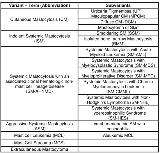
The term Mastocytosis describes disorders caused by the pathogenic increase of mast cell numbers in different tissues. The consequences that arise from mast cell burden and accumulation of their associated mediators can be both locally and systemically. Local mast cell accumulation in organs like skin, gastrointestinal tract, bone marrow, spleen and lymph nodes can be associated with different hematologic disorders (Metcalfe 2008). In Cutaneous Mastocytosis (CM) mast cell infiltrates are restricted to the skin and variants of CM are defined by their clinical presentation. In addition in Systemic Mastocytosis (SM) one or more internal organs predominantly the bone marrow are infiltrated by mast cells (Valent, Horny et al. 2001) often associated with hematologic disorders. The pathological proliferation of mast cells in Mastocytosis and the release of their mediators can cause symptoms including pruritus, flushing, nausea, diarrhea and vascular instability and in systemic Mastocytosis severe symptoms like Osteoporosis and organ failure occur. The complex pattern of symptoms and their severity leads to a classification listed by the World Health Organization (WHO) and subsequent prognosis for each case of Mastocytosis (Horny, Sotlar et al. 2007):
Variant – Term (Abbreviation) Subvariants
Cutaneous Mastocytosis (CM)
Urticaria Pigmentosa (UP) = Maculopapular CM (MPCM)
Diffuse CM (DCM) Mastocytoma of Skin Indolent Systemic Mastocytosis
(ISM)
Smoldering SM (SSM) Isolated bone marrow Mastocytosis
(BMM)
Systemic Mastocytosis with an associated clonal hematologic non-
mast cell lineage disease (SM-AHNMD)
Systemic Mastocytosis with Acute Myeloid Leukemia (SM-AML)
Systemic Mastocytosis with Myelodysplastic Syndrome (SM-MDS)
Systemic Mastocytosis with Myeloproliferative Disorder (SM-MPD)
Systemic Mastocytosis with Chronic Myelomonocytic Leukemia
(SM-CMML)
Systemic Mastocytosis with Non- Hodgkin’s Lymphoma (SM-NHL)
Systemic Mastocytosis with Hypereosinophilic Syndrome
(SM-HES) Aggressive Systemic Mastocytosis
(ASM)
Lymphadenopathic SM with eosinophilia
Mast cell Leukemia (MCL) Aleukemic MCL Mast Cell Sarcoma (MCS)
Extracutaneous Mastocytoma
Table 1.1 WHO Classification of Mastocytosis
1.2.1 Clinical presentation
Cutaneous Mastocytosis (CM)
CM shows a wide variety of skin alterations (Rueff, Dugas-Breit et al. 2006). Even though all lesions in CM are due to an abnormal elevated mast cell number in the dermis the number and morphology of efflorescences varies (Longley, Duffy et al. 1995). The most common subvariant of CM presents as disseminated macular or maculopapular rash, and is (descriptively) termed urticaria pigmentosa (UP) (Wolff, Komar et al. 2001) (Hartmann and Henz 2002). Diffuse CM is less frequently diagnosed. The solitary localized mastocytoma (of the skin) is also rare, and has a benign clinical course. Thus, most mast cell tumors of the skin appear to be benign in most cases.
Indolent systemic mastocytosis (ISM)
The most common systemic variant of mastocytosis is the indolent systemic mastocytosis (ISM) wich involves the skin with limited increase of mast cell number in other organs, in most cases the bone marrow (Valent, Horny et al. 2001). With a common onset in adult age ISM shows a prolonged clinical course in almost all patients with survival times of two decades and more. Besides bone marrow infiltrates, more than 90% of patients show maculopapular cutaneous mastocytosis / urticaria pigmentosa. Level of serum tryptase is raised in comparison to cutaneous mastocytosis with levels of 20 to 200 µg/l. A rare variant of ISM is the so called smoldering systemic mastocytosis (SSM) with pronounced mast cell infiltration (>30% in bone marrow histology), organomegaly and tryptase levels above 200 µg/l.
Systemic mastocytosis with an associated clonal hematologic non mast cell lineage disease (SM-AHNMD)
A small subgroup of patients with systemic mastocytosis develops an additional clonal disease of hematologic non-mast cell lineage cells like the myelodysplastic / myeloproliferative syndrome or myeloid leukemia. Generally these are myeloid neoplasia while systematic lymphatic diseases like lymphoma or myeloma are extremely rare in these patients.
Introduction 8
Aggressive systemic mastocytosis (ASM)
In rare cases an ISM can develop into an aggressive systemic mastocytosis (ASM) with progressive infiltration of immature mast cells and subsequent organ destruction and failure (Valent 1996). ASM is much less common than ISM comprising only about 5% of all SM patients. Possible symptoms are cytoplenias, malapsorption, bone fractures and signs of hepatopathy with loss of liver function (Horny, Sotlar et al. 2007). A rare subvariant of ASM with prominent eosinophilia of blood and tissues and generalized lymphadenopathy (clinically mimicking malignant lymphoma) has been described as lymphadenopathic mastocytosis with eosinophilia (Hauswirth, Sperr et al. 2002).
Mast cell leukemia (MCL)
In patient with mast cell leukemia smear preparations of the bone marrow show more than 20% mast cells (Horny and Valent 2001). In case of a typical mast cell leukemia numbers of circulating mast cell in the blood are generally above 10% while in cases of aleukemic mast cell leukemia the numbers are below 10%. Prognosis of MCL is poor, mostly with survival of less than two years.
Mast cell sarcoma (MCS)
Localized mast cell proliferations are also extremely rare and include both the extracutaneous mastocytoma (of the lung) and the “true” mast cell sarcoma. It is noteworthy that the mast cell sarcomas reported occurred in tissues not commonly involved by SM (larynx, colon, meningeal site). All cases showed rapid progression and generalization with the terminal phase resembling MC leukemia. (Horny, Sotlar et al. 2007).
1.2.2 Pathogenesis
The most important growth factor for human mast cells is the cytokine stem cell factor (SCF).
SCF binds to the tyrosine kinase receptor KIT (CD117) encoded by the proto-oncogene KIT.
By KIT activation SCF induces various cell functions in mast cells like proliferation, differentiation, mediator release, migration and survival (Tsai, Takeishi et al. 1991; Mekori, Oh et al. 1993). The majority of all adult patients with mastocytosis carry a somatic KIT mutation in exon 17 codon 816 with substitution of asparagine by valine (D816V) leading to an autonomous ligand independent activation of KIT (Nagata, Worobec et al. 1995) (Longley,
Tyrrell et al. 1996) (Longley, Metcalfe et al. 1999). In seldom cases substitutions by other amino acids in codon 816 i.e. D816Y, D816H and D816F (Feger, Ribadeau Dumas et al.
2002) or mutations in neighboring codons (D820G) (Liegl, Kepten et al. 2008) have been described. In contrast to adult patient only a subgroup of children with mastocytosis carry activating KIT mutations (Sotlar, Escribano et al. 2003) (Yanagihori, Oyama et al. 2005). To some extent pediatric patients show untypical (i.e. D816F, D816Y, R816K) or silent non activating (D816D) mutations. Despite these findings revealing the molecular cause of mastocytosis many questions concerning its pathogenesis still remain unanswered. In particular factors besides the activating KITD816V mutation affecting the heterogeneity of mastocytosis and factors which are active in cases without activating KIT mutations i.e. in pediatric patients still have to be discovered.
1.2.3 Therapy
Despite growing scientific knowledge no curative therapy for the treatment of mastocytosis is available (Escribano, Akin et al. 2002; Brockow 2004). Currently applied treatments base on reduction or antagonism of mediator release and reduction of mast cell numbers (Longley, Duffy et al. 1995; Golkar and Bernhard 1997; Marone, Spadaro et al. 2001). Since mastocytosis is a rare disease and treatment is not always necessary clinical studies with large patient numbers are currently not available. Thus recommended therapies base on isolated cases or case series.
In cases of cutaneous mastocytosis education of patients about diagnosis, prognosis and possible trigger factors and their avoidance is of capital importance (Allison and Schmidt 1997; Golkar and Bernhard 1997; Brockow 2004). Medication in most cases is symptomatically and in accordance with the patient’s subjective condition. Commonly corticosteroids, antihistamines or a psoralen + UVA (PUVA) treatment have to be considered (Escribano, Akin et al. 2002; Worobec and Metcalfe 2002). In case of periodical condition i.e.
weekly swelling of skin lesions or flushes, regular medication with H1 antihistamines should be prescribed. Gastrointestinal conditions can be treated by H2 antihistamines, cromoglycic acid, proton pump inhibitors and antacids. Systemic administered corticosteroids can be effective in patients with frequent anaphylactic reactions or severe systemic conditions like ascites, diarrhea or malapsorption (Metcalfe 1991). Calcium, vitamin D or bisphosphonate come into considerations in cases with osteopenia or osteoporosis (Laroche, Bret et al.
2007). In patients with progressed categories of systemic mastocytosis especially aggressive mastocytosis or mast cell leukemia cytoreductive therapies have to be considered. Interferon alpha for example can reduce mast cell numbers in patients with aggressive systemic or smoldering systemic mastocytosis (Kluin-Nelemans, Jansen et al. 1992; Lippert and Henz
Introduction 10
1996; Worobec, Kirshenbaum et al. 1996; Hauswirth, Simonitsch-Klupp et al. 2004).
Cladribin (2-chlorodeoxyadenosine, 2-CDA) according to first studies is reducing mast cell numbers in patients with aggressive mastocytosis (Tefferi, Li et al. 2001; Kluin-Nelemans, Oldhoff et al. 2003).
1.2.4 Tyrosine kinase inhibitors
During the last years the use of drugs targeting the tyrosine kinase (TK) KIT has been proposed and inhibitors have been developed. The TK inhibitor imatinib (= STI571) is of none or low effectiveness in patients with KITD816V mutation most common in patients with mastocytosis since the mutation is leading to a steric alteration in the receptor preventing imatinib from binding to the ATP binding domain of KIT (Ma, Zeng et al. 2002; Vendome, Letard et al. 2005). However in patients carrying uncommon KIT mutations in exons 9 or 10 imatinib leads to a reduction in mast cell numbers, lowered tryptase levels and improvement of clinical symptoms (Akin, Fumo et al. 2004; Zhang, Smith et al. 2006). Novel multi kinase inhibitors like midostaurin (PKC412), nilotinib (AMN107) or dasatinib (BMS354825) have been developed and undergo clinical testing (Gotlib, Berube et al. 2005; von Bubnoff, Gorantla et al. 2005; Gleixner, Mayerhofer et al. 2006). Reportedly midostaurin has remarkable effects on neoplastic mast cells in vitro as well as to induce remission in a patient with MCL carrying the activating D816V KIT mutation (Gotlib, Berube et al. 2005).
1.3 Targeting of the inhibitor of apoptosis protein (IAP) family and TRAIL death receptors in cancer
1.3.1 Apoptosis
The ability of multicellular organisms to control cell number is the prerequisite for many physiological processes. The targeted destruction of cells is executed by an active molecular suicide program so called programmed cell death. So acting as a counterpart to cell division, apoptosis enables the organism to regulate cell number and to specifically kill harmful cells.
Therefore the programmed cell death is of central importance in developmental processes and tissue homeostasis. A dysregulation with reduced or excessive cell death may lead to severe illnesses like cancer, autoimmune diseases or neurological disorders like Alzheimer or Parkinson disease (Thompson 1995).
Described early under varying names in 1972 the programmed cell death was finally named apoptosis by Kerr et al describing an uniformly cell death characterized by defined morphological changes of dying cells throughout various cell types and tissues (Kerr, Wyllie et al. 1972). Early characteristics of apoptosis are shrinking of the cell, condensation of chromatin and bubble formation of the cell wall. Later DNA is fragmented by endonucleases and the whole cell collapses. Cell debris is packed into apoptotic vesicles and taken up by phagocytotic cells. Since the cell membrane remains intact during the whole process no cellular content is released into the cells surrounding. Therefore the apoptotic cell death does not trigger inflammation in contrast to cells dying by necrosis. Necrosis is a type of cell death induced by mechanical stress or other environmental factors. Here the plasma membrane is disrupted and cell content is released. This can trigger an immune response and often is accompanied by inflammations which can harm surrounding tissues (Edinger and Thompson 2004).
1.3.2 The extrinsic pathway of apoptosis
The extrinsic or receptor mediated pathway of apoptosis for is used for example in cells of the immune system to eliminate infected or tumorigenic cells (Igney and Krammer 2002). It is triggered by binding of ligands to specific death receptors (CD95, TNFR1, TRAIL-R1 and TRAIL-R2) on the target cells surface. The binding of the ligand leads to the recruitment of adaptor molecules on the cytosolic domain of the activated receptor. These bind to initiator caspase 8 and 10 leading to dimerisation of caspases leading to autocatalysis and activation of initiator caspases. This signaling complex is called “death-inducing signaling complex”
(DISC). The initiator caspases subsequently cleave and activate effector caspases 3, 6 and 7 (Ashkenazi 2002; Lavrik, Golks et al. 2005).
1.3.3 The intrinsic pathway of apoptosis
The intrinsic or mitochondrial pathway of apoptosis is triggered by DNA damage, oncogene activation or other intracellular apoptotic signals. The outer mitochondrial membrane is permeabilized and cytochrome C as well as other apoptotic factors is released into the cytosol. The permeabilization of the mitochondrial membrane is regulated by an interaction of anti- and pro-apoptotic molecules of the Bcl-2 family of proteins. After the release of mitochondrial proteins into the cytosol a multi-protein complex of Apaf-1 and cytochrome C molecules is formed. The so called apoptosome binds and activates the initiator caspase 9.
In turn caspase 9 cleaves and activates effector caspases 3 and 7 (Riedl and Shi 2004;
Gogvadze and Orrenius 2006). The intrinsic pathway as well can be activated by death
Introduction 12
receptors. The Bcl-2 family protein BID can be cleaved by caspase 8. Cleaved BID activates the pro-apoptotic proteins Bak and Bax leading to permeabilization of the mitochondria (Korsmeyer, Wei et al. 2000).
The effector caspase activated by both apoptotic pathways cleave a variety of cellular substrates leading to the characteristic biochemical and morphological changes of the apoptotic cell like chromatin condensation, DNA fragmentation or shrinking and fragmentation of the cell (Fischer, Janicke et al. 2003).
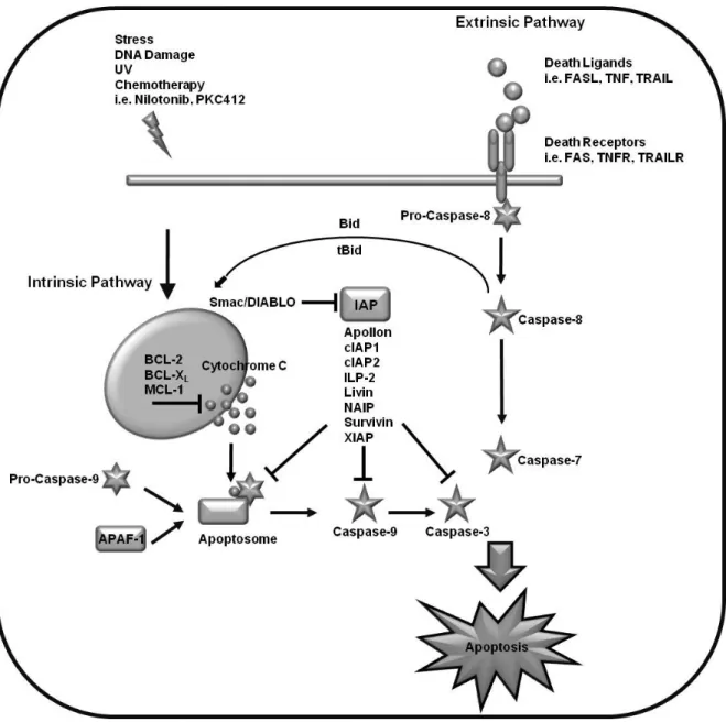
Fig. 1.1 Apoptotic pathways:
Apoptosis is initiated via two main pathways. The extrinsic pathway is triggered by binding of ligands to their respective death inducing receptor and following activation of caspase 8. Release of pro- apoptotic proteins and cytochrome C from the mitochondria following different stress signals initiates the intrinsic pathway of apoptosis leading to activation of pro-caspase 9. The activation of both initiator caspases leads to processing of effector caspases with subsequent cleavage of target proteins and cell death. The extrinsic and intrinsic pathways are connected via the cleavage of Bid to tBid by caspase 8. Depending on the cell type activation of death receptors directly leads to activation of effector caspases (type I cells) or indirectly via the mitochondria (type II cells).
1.3.4 TRAIL receptors
The tumor necrosis factor-related apoptosis-inducing ligand (TRAIL), a member of the tumor necrosis factor (TNF) superfamily of proteins, that was discovered in 1995 (Wiley, Schooley et al. 1995) and since then is under investigation as a biologically targeted anti-tumor protein since it induces apoptosis in a variety of human cancer cell lines while leaving normal cells unaffected (Walczak, Miller et al. 1999). In humans TRAIL can bind to four known membrane bound receptors, death receptor 4 (DR4)/TRAIL receptor-1 (Pan, O'Rourke et al. 1997), death receptor 5 (DR5)/TRAIL receptor-2 (Pan, Ni et al. 1997; Walczak, Degli-Esposti et al.
1997), TRAIL receptor without an intracellular domain (TRID)/decoy receptor 1 (DCR1)/TRAIL receptor-3 (Degli-Esposti, Smolak et al. 1997; Pan, Ni et al. 1997; Sheridan, Marsters et al. 1997), and TRAIL receptor-4/decoy receptor 2 (DCR2)/TRAIL receptor with a truncated death domain (TRUNDD) (Degli-Esposti, Smolak et al. 1997; Marsters, Sheridan et al. 1997; Pan, Ni et al. 1998). TRAIL receptor-3 and TRAIL receptor-4 do not possess an intracellular domain and have been proposed as competitive inhibitors of TRAIL induced apoptosis (Sheridan, Marsters et al. 1997). TRAIL receptor 1 and TRAIL receptor 2 on the other hand contain a cytoplasmic death domain capable of activating the extrinsic pathway of apoptosis by recruiting the death-inducing signaling complex (DISC) upon ligand binding and trimerization of the receptor (Hymowitz,
Christinger et al. 1999). DISC includes FAS- associated death domain protein (FADD), pro- caspase 8, and possibly cellular FADD-like IL-1β- converting enzyme inhibitory protein (cFLIP) (Kischkel, Lawrence et al. 2000; Sprick, Weigand et al. 2000). After autocatalytic cleavage and activation of pro-caspase 8 at the DISC downstream effector caspases are activated in type I cells (Kischkel, Lawrence et al. 2000). In type II cells intrinsic apoptosis can be triggered by cleavage of Bid (Barnhart, Lee et al. 2003).
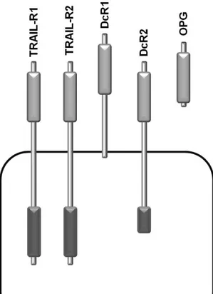
Finally TRAIL can also bind to osteoprotegerin (Emery, McDonnell et al. 1998), a soluble receptor for Receptor Activator of NF-κB Ligand (RANKL) (Fig. 1.2).
Fig. 1.2 The human TRAIL receptors:
Human TRAIL can bind to five different receptors. TRAIL receptor 1 and TRAIL receptor 2 can signal apoptosis via their cytoplasmatic death domain. Decoy receptor 1 is lacking a death domain while decoy receptor 2 has a truncated death domain unable to signal apoptosis. Osteoprotegerin is a soluble receptor for TRAIL.
Introduction 14
The different sensitivity of normal and neoplastic cells towards TRAIL induced apoptosis may be due to several reasons. Messenger RNA of all TRAIL receptors is expressed throughout a wide variety of cells and tissues (Wiley, Schooley et al. 1995). Therefore the expression pattern of apoptosis signalling versus non signalling TRAIL receptors has been proposed as a way of regulation of sensitivity to TRAIL-induces apoptosis (Marsters, Sheridan et al. 1997;
Pan, Ni et al. 1997; Pan, Ni et al. 1998) and has been confirmed for primary tumors of the gastrointestinal tract (Sheikh, Huang et al. 1999) and human breast cancer cell lines (Sanlioglu, Dirice et al. 2005). Another finding suggests that the cell cycle phase determines cell sensitivity towards TRAIL as was shown in human colon and lung cancer cell lines arrested in the G0/G1phase of the cell cycle (Jin, Dicker et al. 2002) may be due to differential expression of pro- and anti-apoptotic proteins during different cell cycle phases as has been shown in activated T cells (Algeciras-Schimnich, Griffith et al. 1999). The most prominent theories concerning regulation of TRAIL-induces apoptosis involve the differential expression of pro- and anti-apoptotic proteins within the targeted cells such as cFLIP, Bcl-2 family members, the ERK and Akt signaling pathways and the inhibitor of apoptosis proteins (IAPs).
(Griffith, Chin et al. 1998; Shiiki, Yoshikawa et al. 2000; Nesterov, Lu et al. 2001; Fulda, Meyer et al. 2002; Vaculova, Hofmanova et al. 2006; Kim, Ricci et al. 2008). Since cFLIP binds competively to FADD its suppression can sensitize some cancer cells to TRAIL- induced apoptosis (Geserick, Drewniok et al. 2008). In many tumor cell lines XIAP has been observed to be highly expressed and it may lead to TRAIL resistance by directly inhibiting caspases 3, 7 and 9, even in the presence of Smac/DIABLO (Deveraux, Takahashi et al.
1997; Schimmer, Welsh et al. 2004). Anti-apoptotic members of the Bcl-2 family regulate sensitivity towards TRAIL as has been shown for Bcl-2 and Bcl-xL (Hinz, Trauzold et al.
2000; Sinicrope, Penington et al. 2004) as well as for myeloid cell leukemia-1 protein (Mcl-1) (Clohessy, Zhuang et al. 2006).
1.3.5 Inhibitor of apoptosis protein (IAP) family
Originally IAP proteins were identified in baculoviruses. These insect viruses express IAP proteins to inhibit the apoptosis initiated by the infected host cell (Crook, Clem et al. 1993).
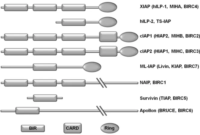
Primarily IAPs were thought to function by regulating caspases, which are cysteine proteases that are involved in apoptosis. However IAPs also influence a multitude of other cellular processes, such as ubiquitin (Ub) –dependent signaling events that regulate activation of nuclear facor-κB (NF-κB) transcription. Until now eight human IAPs have been identified:
XIAP, ILP-2, cIAP1, cIAP2, ML-IAP (Livin), NAIP, Survivin and Apollon. The defining feature of an IAP protein is the presence of the baculovirus IAP repeat (BIR) domain, a zinc-binding fold of approximately 70 amino acid residues that mediates protein-protein interactions
(Birnbaum, Clem et al. 1994; Hinds, Norton et al. 1999; Sun, Cai et al. 2000). IAPs of which there are eight in humans, carry between one and three copies of this domain (Fig. 1.3).
Fig. 1.3 Inhibitor of apoptosis proteins (IAPs):
The eight human inhibitor of apoptosis proteins are characterized by at least one BIR domain. BIR:
Baculovirus IAP repeat, CARD: caspase recruitment domain, RING: really interesting new gene, ILP:
IAP-like protein, cIAP cellular IAP. ML-IAP: melanoma IAP, NIAP: neuronal IAP, XIAP: X-linked IAP.
Additionally XIAP, cIAP1, cIAP2 and Livin have a RING domain that provides them with Ub ligase (E3) activity (Yang, Fang et al. 2000) that can link target proteins to one or more ubiquitin residues. Moreover they carry an Ub-associated (UBA) domain through which they interact with ubiquitylated proteins (Gyrd-Hansen, Darding et al. 2008; Blankenship, Varfolomeev et al. 2009). Monoubiquitination of target proteins leads to functional changes and polyubiquitination in most cases leads to proteolytic degradation. Polyubiquitinated proteins are degraded in the proteasome, a multi protein complex (Hicke 2001; Vaux and Silke 2005). Only cIAP1 and cIAP2 carry a caspase recruitment domain (CARD) located between the BIR and the RING domain. Its function is currently unknown.
Introduction 16
1.3.6 The IAP Survivin
With a molecular size of 16.5 kDa Survivin is the smallest known member of the IAP family of anti-apoptotic proteins. Survivin contains a single BIR domain but no RING finger or other identifiable domain typically found in IAPs. In healthy tissues its expression is found during embryogenesis and in adults in vascular endothelial cells (Mesri, Morales-Ruiz et al. 2001) (Blanc-Brude, Mesri et al. 2003), polymorphonuclear cells (Altznauer, Martinelli et al. 2004), T cells (Okada, Bakal et al. 2004; Xing, Conway et al. 2004) and haematopoietic progenitor cells (Xing, Conway et al. 2004; Gurbuxani, Xu et al. 2005; Leung, Xu et al. 2007). Survivin has the ability to counteract with extrinsic and intrinsic mediators of apoptosis with effecting mechanisms such as withdrawal of IL-3 (Fukuda and Pelus 2004), stimulation of death receptors of the TNFR family like FAS (CD95) (Tamm, Wang et al. 1998), TNF-related apoptosis-inducing ligand (TRAIL) (Chawla-Sarkar, Bae et al. 2004). By binding to Smac/DIABLO (Sun, Nettesheim et al. 2005) it may regulating apoptosis directly by sequestering SMAC away from XIAP (Song, Yao et al. 2003) or preventing altogether its release from mitochondria (Ceballos-Cancino, Espinosa et al. 2007). Besides its anti- apoptotic functions survivin plays a key role in modulating cell cycle dependent mechanisms such as cell division, cell stress response and cell cycle checkpoints with physical localisation to the mitotic spindle apparatus (Pennati, Folini et al. 2008). Therefore its properties tend to increase cell proliferation of tumor cells and also mutant cancer cells making survivin an attractive therapeutic target and diagnostic marker in a wide spectrum of malignancies.
1.3.7 Smac/DIABLO
The second mitochondria-derived activator of caspase (Smac) or direct IAP binding protein with low isoelectric point (pI) DIABLO was identified simultaneously by two groups in 2000 (Du, Fang et al. 2000; Verhagen, Ekert et al. 2000). It resides within the mitochondrial intermembrane space and is subsequently released into the cytosol through semiselective permeability together with other mitochondrial proteins such as Omi, adenylate kinase-2, cytochrome c and apoptosis-inducing factor upon the induction of apoptosis. Bcl-family proteins are pivotal in this process as they may facilitate (i.e. Bid (Du, Fang et al. 2000; Li, Zhao et al. 2002), Bax and/or Bak (Du, Fang et al. 2000; Kandasamy, Srinivasula et al. 2003) or inhibit (such as Bcl-2 or Bcl-xL) (Du, Fang et al. 2000; Sun, Bratton et al. 2002) the release of apoptotic mediators. There are different ways in which Smac/DIABLO contributes to the apoptotic process. Smac can bind many different IAPs (i.e. XIAP, cIAP1, cIAP2 and Survivin) (Du, Fang et al. 2000). It is able to interact with IAPs by binding to Survivins BIR1 (Gao, Zhang et al.; Sun, Nettesheim et al. 2005) as well as BIR2 and BIR3 domains of XIAP,
cIAP1 and cIAP2 (Chai, Du et al. 2000) and so preventing their interaction with caspases 3 and 7 and in case of XIAP to caspase 9 (Srinivasula, Hegde et al. 2001). Smac/DIABLO can alter levels of certain IAPs. For example it can enhance autoubiquitination of cIAP1 and cIAP2 resulting in their proteasomal degradation (Varfolomeev, Blankenship et al. 2007;
Vince, Wong et al. 2007). Furthermore, Smac/DIABLO increases tumor necrosis factor alpha (TNFα) mRNA expression leading to autocrine stimulation of the extrinsic apoptotic pathway (Varfolomeev, Blankenship et al. 2007; Vince, Wong et al. 2007).
1.3.8 Smac mimetics
Determination of the specific biochemical structures involved in the IAP-Smac interaction has facilitated the development of Smac-like molecules. The crucial N-terminus of Smac has four residues (Ala-Val-Pro-Ile) that contact the BIR3 domain of XIAP, and this structure is stabilized by electrostatic and hydrophobic forces (Liu, Sun et al. 2000; Wu, Chai et al.
2000). Once this structure was elucidated, the development of Smac mimetics, including effective nonpeptic mimetics that have advanced bioavailability and stability (Liu, Sun et al.
2000; Wist, Gu et al. 2007), provided a variety of molecules that could serve as IAP antagonists. Several different groups of Smac mimetics can be distinguished: fusion peptides of the last four to eight N-terminal residues of Smac with various modifications, plasmid based polynucleotides coding for full-length Smac and peptic or nonpeptic small molecules mimicking Smac structure.
1.3.9 Preclinical and clinical studies
TRAIL
The tumoricidal activity of TRAIL has been described in multiple preclinical trials carried out in vitro and in vivo using recombinant human TRAIL (rhTRAIL) on cell lines derived from both solid and hematologic malignancies, either alone or in combination with various chemotherapy agents or irradiation (Ashkenazi, Pai et al. 1999; Gazitt 1999; Kelley, Harris et al. 2001; Marini, Schmid et al. 2005). A way of directing TRAIL towards tumor cells is the creation of cell specific fusion proteins (Stieglmaier, Bremer et al. 2008). Combination therapy with conventional chemotherapy or radiotherapy where successful to overcome TRAIL resistance of tumors (Keane, Ettenberg et al. 1999; Mizutani, Yoshida et al. 1999;
Nimmanapalli, Perkins et al. 2001). Bortezomib, a proteasome inhibitor approved for the treatment of multiple myeloma has been shown to have a pro-apoptotic effect in combination
Introduction 18
with TRAIL possibly by its ability to increase p53 and TRAIL receptor-2 expression while decreasing cFLIP expression (Johnson, Stone et al. 2003; Sayers, Brooks et al. 2003;
Williams and McConkey 2003). Bcl-xL, Mcl-1 and cFLIP have been downregulated by the kinase inhibitor sorafenib in several human leukemia sensitizing them to TRAIL-induced apoptosis while normal CD34+ bone marrow cells stay unaffected (Rosato, Almenara et al.
2007). In human prostate cancer cells BH3I-2’ a Bcl-2 inhibitor induces apoptosis synergistically in the presence of TRAIL (Ray, Bucur et al. 2005).
To date clinical phase II trials for monoclonal TRAIL-R1 antibody TRM-1 (Mapatumumab) for treatment of non-small cell lung cancer and Non-Hodgekin’s lymphoma have been completed and the clinical phase II trial of recombinant human TRAIL AMG 951 for treatment of small cell lung cancer is still ongoing (http://clinicaltrials.gov/).
Survivin
Strong survivin expression is observed in the vast majority of cancers (Altieri 2003) and in patients with haematologic malignancies (Cong and Han 2004). In cancer elevated survivin is commonly associated with enhanced proliferative index (Sui, Dong et al. 2002; Takai, Miyazaki et al. 2002; Fields, Cotsonis et al. 2004), resistance to chemotherapy (Tran, Master et al. 2002; Zaffaroni, Pennati et al. 2002) and rate of tumor recurrence (Swana, Grossman et al. 1999).
Besides classical diagnostic methods like reverse transcription polymerase chain reaction (RT-PCR), and flow cytometric analysis several new tools have been developed to establish survivin as a diagnostic biomarker like molecular beacon technology (Yang, Cao et al. 2005), recombinant nucleic acid-reporter gene-based assays (Caldas H, Altura RA WO2006066451;2006) and LabMAP technology (Lokshin A. US20070042405; 2007).
Prominent strategies in targeting survivin in disease involve adenoviral expression of mutant survivin (Mesri, Wall et al. 2001), small hammerhead RNA (shRNA) (Caldas, Holloway et al.
2006) or small inhibitory RNA (siRNA) (Uchida, Tanaka et al. 2004). Combinational treatment in association with chemotherapeutics like gemcitabine, peclitaxel or doxorubicin results in marked downregulation of survivin expression and tumor regression (Patents reviewed in (Kanwar, Kamalapuram et al. 2010)).
A Phase II study with LY2181308 a modified antisense oligonucleotide for treatment of acute myeloid leukemia and an evaluation study for survivin mRNA as a marker in bladder cancer patients has been completed. The transcriptional survivin repressors YM155 finished phase II studies for treatment of solid tumors, melanoma and prostate cancer and EM-1421 finished phase I trials for several tumor types (http://clinicaltrials.gov/).
Smac mimetics
Over the past few years evidence for the use of Smac mimetics in the treatment of neoplastic malignancies continues to accumulate. At this stage only a few small molecule Smac mimetics have entered Phase I clinical trial like AT-406, TL32711, HGS1029 and LCL161 for treatment of advanced solid tumors or GDC-0152 for non-Hodgkin’s lymphoma (http://clinicaltrials.gov/) but the effectiveness of a multitude of substances has been proven in vitro and in vivo. For example peptides successfully induce apoptosis in human breast cancer cells in combination with paclitaxel, etoposide or doxorubicin (Arnt, Chiorean et al.
2002) and augmented TRAIL induced apoptosis in JURKAT cells (Guo, Nimmanapalli et al.
2002). Polynucleotides increased the leukemia cell lines K562 and CEM sensitivity to both UV light-induced and TRAIL-induced apoptosis (Jia, Patwari et al. 2003) and sensitized JURKAT cells to TRAIL- and epothilone-induced apoptosis (Guo, Nimmanapalli et al. 2002).
Small molecules sensitized human glioblastoma T98G cells to TRAIL induced apoptosis and HeLa cells to TNFα-induced apoptosis (Li, Thomas et al. 2004). Finally in female C.B-17- Prkdcscid mice Smac mimetic treatment was able to significantly reduce tumor growth rate (Eiseman, Lan et al. 2005).
1.4 Objectives
Aim of the work is the identification and characterization of possible therapeutic targets and diagnostic markers in mastocytosis with special attention to pro- and anti-apoptotic proteins.
Therefore the human neoplastic mast cell lines HMC-1.1 and HMC-1.2 carrying one respectively two activating mutations in the tyrosine kinase receptor KIT (CD117) were analyzed for the expression of members of the inhibitor of apoptosis protein (IAP) family and the expression of the pro-apoptotic TNF-related apoptosis-inducing ligand receptors (TRAIL-R1 and TRAIL-R2) which could be of future use as diagnostic markers or therapeutic targets in mastocytosis. With respect to ongoing research with recently generated mice carrying a mutant KIT receptor and so resembling the situation in human mastocytosis patients (Gerbaulet, Wickenhauser et al. 2010) the biological function of the murine TRAIL-R on murine bone marrow derived mast cells was characterized in this work. In two different approaches the combinational targeting of members of the IAP family of proteins and the use of either TRAIL or tyrosine kinase inhibitors as stimuli for the extrinsic respectively intrinsic pathway of apoptosis in neoplastic human mast cells were analyzed to make way for future investigation of treatment options of neoplastic mast cells diseases.
Results 20
2 Results
2.1 Expression of pro- and anti-apoptotic molecules in mast cells
Identification of pro- or anti-apoptotic molecules differentially expressed in neoplastic mast cells compared to healthy tissue could be a step in enhancing existing therapies for mastocytosis patients. Since the constitutive activation of the tyrosine kinase KIT is a hallmark of neoplasia in mastocytosis the two neoplastic mast cell lines HMC-1.1 and HMC-1.2 carrying one respectively two mutations in the KIT gene were analyzed for expression of pro- and anti-apoptotic molecules.
The IAP Survivin was found to be stronger expressed in HMC-1.2 cells indicating a link between KIT activation and Survivin expression. Further primary human mast cells generated from umbilical cord blood or isolated from skin were compared to different mast cell and non-mast cell lines for Survivin expression on the mRNA and protein level showing very low expression in the primary healthy mast cells. Comparison of bone marrow samples of mastocytosis patients and healthy donors revealed a significantly higher Survivin expression in mast cell infiltrates of the mastocytosis patients while expression in infiltrates of healthy donors was almost undetectable. This pattern highlights Survivin expression as to be more disease-related than for example the recently analyzed anti-apoptotic bcl-2 family protein Mcl1 (Aichberger, Mayerhofer et al. 2007) which is expressed in normal and neoplastic transformed mast cells.
In a second approach human neoplastic mast cells were analyzed for the expression of the TNF-related apoptosis-inducing ligand receptors (TRAIL-R1 and TRAIL-R2). Both cell lines show expression of both receptors on mRNA and protein level, and their expression on the cell surface was confirmed by flow cytometric analysis. With regard to a recently generated mouse model of mastocytosis murine mast cells were analyzed for expression of the murine TRAIL receptor and its regulation after KIT- and FcεR-activation. The murine TRAIL receptor is expressed on murine bone marrow derived mast cells and in accordance with findings in human mast cells (Berent-Maoz, Piliponsky et al. 2006; Berent-Maoz, Salemi et al. 2008) its expression is upregulated after IgE-dependent activation. Here a dose dependent regulation of the murine TRAIL receptor expression after activation of KIT with stem cell factor has been shown.
2.1.1 Expression of TRAIL receptors in mast cells
2.1.1.1 Expression of TRAIL and its receptors in human neoplastic mast cells
The expression of the TRAIL receptors TRAIL-R1 and TRAIL-R2 was analyzed in both clones of the human mast cell line HMC-1. To identify a potential differential expression both cell lines were analyzed for mRNA- and protein-expression of the receptors. The surface expression of both TRAIL receptors on HMC-1 cells was determined by flow cytometry.
Furthermore the expression of TRAIL mRNA and protein was measured since recent findings showed TRAIL expression in human neoplastic mast cells with still unclear mechanisms of regulation, release or function (Berent-Maoz, Salemi et al.). Both cell lines showed comparable expression levels with no significant differences of TRAIL, TRAIL-R1 and TRAIL-R2 mRNA, protein and surface expression of the receptors.
The human neoplastic mast cell lines HMC-1.1 and HMC-1.2 were analyzed for expression of mRNA of TRAIL and its receptors TRAIL-R1 and TRAIL-R2. Both cell lines were seeded in complete medium and harvested during logarithmic growth. The mRNA was extracted and subjected to RT-PCR. Both cell lines express mRNA of TRAIL and the receptors TRAIL-R1 and TRAIL-R2. The densitometric analysis of several PCR gels showed no significant difference between PCR products of both cell lines (Fig. 2.1).
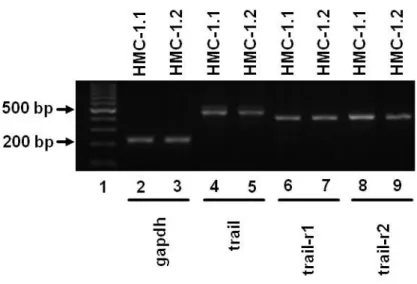
Fig. 2.1 Human neoplastic mast cells show RNA expression of trail and its receptors:
RT-PCR products of RNA isolated from HMC-1.1 and HMC-1.2 cells separated on a 2% agarose gel.
(1, 100 bp Ladder; 2+3, gapdh 226 bp; 4+5, trail 510 bp; 6+7, trail-r1 415 bp; 8+9, trail-r2 437 bp).
Data are representative of three independent experiments, all of which had similar results.
Results 22
Both neoplastic mast cell lines were subjected to Western blot analysis with specific antibodies against TRAIL and its receptors TRAIL-R1 and TRAIL-R2. Expression of all three proteins can be detected in cell lysates of both cell lines harvested during logarithmic growth (Fig. 2.2).
Fig. 2.2 Expression of TRAIL and its receptors can be detected in cell lysates of human neoplastic mast cells:
Western blot analysis of HMC-1.1 and HMC-1.2 cell lysates was performed with specific antibodies against TRAIL, TRAIL-R1 and TRAIL-R2. β-Actin was used as a loading control.
Data are representative of three independent experiments with similar results.
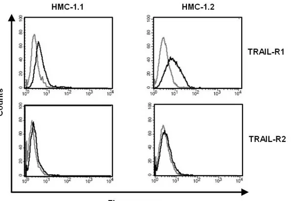
Surface expression of TRAIL-R1 and TRAIL-R2 was shown on HMC-1.1 and HMC-1.2 cells by staining both cell lines with fluorescent antibodies specific for the receptors (Fig. 2.3).
Fig. 2.3 Human neoplastic mast cells express TRAIL receptors on the cell surface:
HMC-1.1 and HMC-1.2 cells where labeled with PE-conjugated antibodies against TRAIL-R1, TRAIL-R2 (black line) or PE-conjugated isotype controls (grey line). Cells were analyzed by flow cytometry. Data are representative of three independent experiments with similar results.
2.1.1.2 Expression of TRAIL-R in murine mast cells
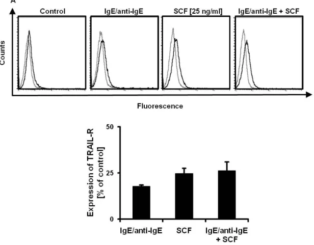
TRAIL-R is expressed on murine mast cells and its expression is upregulated by stem cell factor and IgE dependent activation
To analyze the effect of c-KIT activation and IgE-dependent activation on TRAIL-R- expression of murine mast cells, bone marrow-derived mast cells (BMMC) were either incubated with SCF or stimulated with IgE/anti-IgE or both for 24 h. Cells were harvested and stained with TRAIL-R specific or isotype antibodies and analyzed by flow cytometry (Fig. 2.4 A). Mean fluorescence was calculated and plotted as a percentage of the control (Fig. 2.4 B), showing that incubation with SCF of IgE-dependent stimulation increases TRAIL-R expression on BMMC but a combination of both has no additive effect.
Fig. 2.4 TRAIL-R expression on BMMC is upregulated by SCF and IgE dependent activation:
(A) BMMC were incubated with SCF, activated by 1 µg/ml IgE/anti-IgE or both for 24 h. TRAIL-R expression was measured by flow cytometry after staining of cells with PE conjugated TRAIL-R specific antibodies (black graphs) or PE conjugated isotype control (grey line). Data are representative of three independent experiments, all of which had similar results. (B) Mean fluorescence of stimulated and unstimulated cells was calculated. Results are presented as percent expression of TRAIL-R on stimulated cells compared to unstimulated cells (defined as 0%) and expressed as mean +/- SEM of three independent experiments.
Results 24
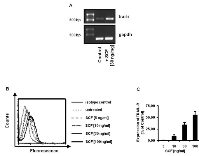
SCF dependent upregulation of TRAIL-R is dose dependent
To confirm the first flow cytometric results, RT-PCR from mRNA isolated from SCF treated BMMC after 24 h was performed and a clear increase of PCR product was observed (Fig.2.5 A). Further, BMMC were incubated with SCF for 24 h and analyzed by flow cytometry. When BMMC were incubated with increasing concentrations of SCF for 24 h an increase of TRAIL-R expression was detected showing a correlation between receptor expression and level of c-KIT activation (Fig. 2.5 B, C).
Fig. 2.5 Upregulation of TRAIL-R on BMMC by SCF is dose dependent:
RT-PCR was performed of RNA isolated from BMMC incubated with SCF (30 ng/ml) for 24 h compared to untreated control cells (A). PCR products where analyzed on a 1% agarose gel (TRAIL-R 579 bp, GAPDH 226 bp).
BMMC where incubated with different concentrations of SCF (0, 5, 10, 30 and 100 ng/ml) for 24 h and surface expression of TRAIL-R was analyzed by flow cytometry after staining of cells with PE- conjugated antibody against TRAIL-R (black lines) or PE-conjugated isotype control antibody (grey line) (B).
Mean fluorescence of stimulated and unstimulated cells was calculated (C). Results are presented as percent expression of TRAIL-R on stimulated cells compared to unstimulated cells (defined as 0%) and expressed as mean +/- SEM of three independent experiments.
2.1.2 Human neoplastic mast cells express inhibitor of apoptosis proteins (IAPs)
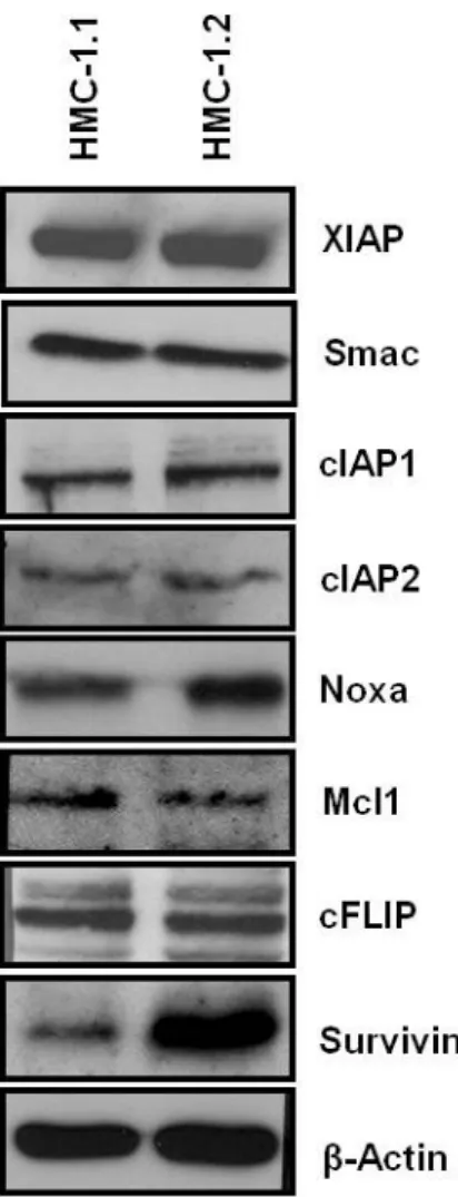
To determine proteins involved in apoptosis which are differently expressed under the control of the active tyrosine kinase KIT, the two human neoplastic mast cell lines HMC-1.1 and HMC-1.2 were analyzed. Both cell lines where seeded in complete medium, harvested during logarithmic growth and subjected to Western blot analysis with antibodies specific for X-linked inhibitor of apoptosis (XIAP), cellular inhibitor of apoptosis 1 and 2 (cIAP1, cIAP2) and Survivin, the second mitochondria-derived activator of caspase (Smac), the bcl-2 family members Mcl1 and Noxa, the cellular FLICE-like inhibitory protein (cFLIP) and β-Actin as loading control. Resulting films were analyzed densitometrically.
The expression of almost all proteins analyzed except Survivin was found to be equal in both HMC-1 subclones. Slight differences in single blots like seen below for Noxa (Fig. 2.6) could not be confirmed over the whole range of experiments. A pronounced expression of the IAP Survivin in HMC-1.2 cells was observed in all repeated experiments and was highly significant after densitometric analysis.
Fig. 2.6 Expression of IAPs in neoplastic human mast cells:
Cell lysates of HMC-1.1 and HMC-1.2 cells were analyzed with specific antibodies against XIAP, Smac, cIAP1, cIAP2, Noxa, Mcl1, cFLIP and Survivin. β-Actin was used as a loading control.
Data are representative of at least four independent experiments with similar results.
Results 26
2.1.3 Expression of Survivin in human mast cells
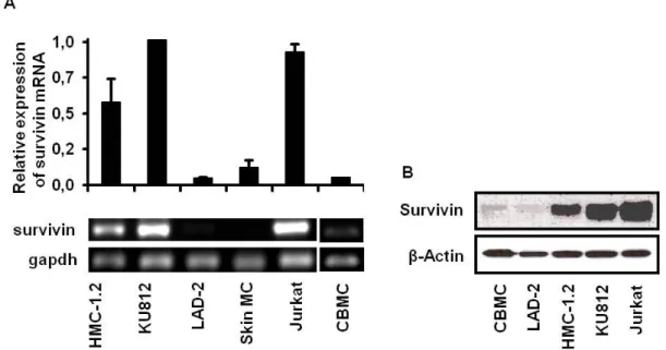
2.1.3.1 Expression of Survivin mRNA and protein in human mast cells
Different human mast cells were analyzed for expression of survivin mRNA (Fig. 2.7 A) and protein (Fig. 2.7 B) to study a possible link between Survivin expression and neoplasia.
Human skin mast cells, human umbilical cord blood-derived mast cells and the mast cell line LAD-2 which is stem cell factor dependent and carries no activating KIT mutations showed low expression of survivin mRNA expression compared to HMC-1.2 cells, the basophilic KU812 cell line and JURKAT T cells. Western blot analysis performed with antibodies specific for Survivin showed low expression of Survivin protein in human umbilical cord blood-derived mast cells and LAD-2 cells compared to HMC-1.2, KU812 and JURKAT T cells.
Survivin is expressed in all cells analysed. The amount of mRNA and protein correlates with the rate of proliferation.
Fig. 2.7 Increased expression of survivin mRNA (A) and protein (B) in human neoplastic mast cells compared to primary mast cells:
RT-PCR was performed of mRNA isolated from different mast cell lines (HMC-1.2, KU812, LAD-2), primary human skin mast cells (skin MC) and the control cell line JURKAT with primers specific for survivin and gapdh (A). Fluorescence of ethidium bromide-stained RT-PCR products was measured by densitometry (upper graph). Results are presented as relative expression of survivin mRNA compared to the most pronounced expression KU812 cells (defined as 1.0) of at least three independent experiments with similar results.
Western blot analysis was performed of cell lysates of umbilical cord blood-derived mast cells (CBMC), different mast cell lines (LAD-2, HMC-1.2, KU812) and the control fell line JURKAT with a Survivin-specific antibody. β-Actin served as a loading control (B). Data are representative of three independent experiments with similar results.
2.1.3.2 Expression of Survivin protein in mast cells infiltrating the bone marrow of patients with Mastocytosis
To verify the relevance of upregulated survivin expression in vivo, bone marrow sections with mast cell infiltrates from Mastocytosis patients were screened for co-expression of mast cell tryptase and Survivin. In all samples from Mastocytosis patients Survivin expression was strongly pronounced compared to samples from healthy donors with reactive bone marrow infiltrates (Fig. 2.8).
Mast cells in bone marrow-infiltrates of patients with mastocytosis do not show elevated staining of the interphase marker Ki67 but significant increase of the marker of G1 arrest p21 compared to mast cells in reactive bone marrow infiltrates (Baldus, Zirbes et al. 2004).
Further p21 expression in mast cells is linked to c-KIT activity (Panzenbock, Bartunek et al.
1998), so elevated Survivin expression could be due to the neoplastic state of the cells not their higher rate of proliferation. Serial sections of bone marrow specimen where stained with antibodies specific for tryptase and survivin to analyze a possible co-expression. The double staining revealed no co-localization of tryptase and Survivin in bone marrow sections of healthy patients while most mastocytosis patients showed Survivin expression in at least 30% of infiltrates (Fig. 2.8 A-D).
Fig. 2.8 Expression of Survivin protein in mast cells infiltrating the bone marrow of patients with mastocytosis:
Paraffin embedded bone marrow sections of mast cell infiltrates from patients with Mastocytosis were stained for mast cell tryptase (A, C) and Survivin (B, D).
Results 28
To determine a statistical significance of these findings the bone marrow sections were scored for their level of Survivin antibody reactivity (Table 2.1): 0, mastocytosis: no reactivity of dense focal infiltrates of mast cells, reactive bone marrow (RBM): no reactivity of mast cells; 1, mastocytosis: reactivity in <30% of infiltrates, RBM: reactivity in <30% of mast cells;
2, reactivity in 30% to 70% of infiltrates or mast cells; 3, reactivity in >70% of infiltrates or mast cells. While a high Survivin expression (score 1-3) was only observed in patients with mastocytosis, Survivin expression score was 0 in healthy (RBM) donors. The serum tryptase level of the patients has a tendency to correlate with the degree of survivin expression but this was not statistical significant.
Table 2.1 Expression of Survivin in human Mastocytosis:
Paraffin-embedded bone marrow sections of 16 patients with mastocytosis were analyzed by immunohistochemistry and compared with 3 control patients with reactive bone marrow (RBM). The diagnosis of mastocytosis was made on the basis of published criteria. Out of the 16 patients, 15 patients had indolent systemic mastocytosis and one patient had aggressive systemic mastocytosis.
Informed consent was obtained from all before bone marrow biopsies were performed.
2.2 Control of mast cell survival by pro- and anti-apoptotic molecules
After identifying Survivin as an anti-apoptotic protein being differentially expressed in human neoplastic mast cells depending on the level of KIT activation and TRAIL receptors being expressed in both subclones and murine mast cells, the susceptibility of these cells to TRAIL induced apoptosis was evaluated. HMC-1.1 and HMC-1.2 cells were incubated with recombinant human TRAIL and exhibited comparable low but significant loss of viability and an increasing number of apoptotic cells was detectable after at least 24 h of incubation.
In murine bone marrow-derived mast cells TRAIL alone failed to induce apoptosis. Only co- incubation with stem cell factor raised their susceptibility and significant apoptosis was measurable at 72 h of incubation.
In two approaches anti-apoptotic proteins were targeted in neoplastic human mast cells before apoptosis was induced by either tyrosine kinase inhibitors or TRAIL.
First the Survivin expression of HMC-1.2 cells was downregulated by Survivin-specific siRNA. This alone abolished proliferation and reduced their viability. Furthermore downregulation of Survivin combined with either tyrosine kinase inhibitor treatment or TRAIL induced apoptosis resulted in a synergistic loss of viability.
In a second approach the Smac mimetic LCL161 was used to target the IAPs XIAP, cIAP1 and cIAP2 in HMC-1.1 and HMC-1.2 cells. LCL161 alone led to accumulation of HMC-1 cells in the G1 phase of the cell cycle and to inhibited proliferation. Combination of LCL161, the tyrosine kinase inhibitor PKC412 or TRAIL reduced viability and increased the rate of apoptotic cells in a synergistical manner after 24 h.
Results 30
2.2.1 TRAIL reduces viability of human neoplastic mast cells and induces apoptosis
To investigate a dose dependent sensitivity of HMC-1 cells towards TRAIL induced apoptosis human neoplastic mast cells were incubated with human recombinant FLAG-tagged TRAIL at concentrations of 250 and 500 ng/ml. Furthermore a difference in sensitivity between both subclones should be examined. The percentage of viable cells was determined by propidium iodide uptake (Fig 2.9 A) and the rate of apoptosis by binding of Annexin-V (Fig. 2.9 B). After 48 h of incubation cells were analyzed by flow cytometry. Both the loss of viability and the increase of apoptotic cells were significant and dose dependent in HMC-1.1 and HMC-1.2 cells but there was no significant difference in loss of viability or rate of apoptosis between both cell lines.
Fig. 2.9 TRAIL induces apoptosis in neoplastic human mast cells and reduces their viability:
HMC-1.1 and HMC-1.2 cells were incubated with TRAIL-FLAG for 48 h. Viability and apoptosis was measured by FACS analysis after staining with APC-labeled Annexin-V and propidium iodide.
Data are representative of at least three independent experiments.
P values ≤ 0.05 where considered as significant (*).