Varicella-zoster virus infections – antiviral therapy and diagnosis
Abstract
Varicella-zoster virus is an important human pathogen that causes varicella after primary infection and zoster after recurrence. Following
Andreas Sauerbrei
1primary infection, the virus remains latently for life in dorsal root and
1 Institute of Virology and Antiviral Therapy, German cranial nerve ganglia. Varicella and zoster are worldwide widespread
diseases and may be associated with significant complications. This
Consulting Laboratory for manuscript presents a short overview about the fundamental knowledge HSV and VZV, Jena University including the most important clinical signs, the capabilities for antiviral
treatment and the spectrum of methods for laboratory diagnosis.
Hospital, Friedrich-Schiller University, Jena, Germany Keywords:varicella-zoster virus, varicella, zoster, antiviral therapy,
laboratory diagnosis
1 Fundamental knowledge
1.1 Virus, epidemiology and pathogenesis
Varicella-zoster virus (VZV, human herpesvirus 3 – HHV-3) is a member of the genusVaricelloviruswithin the sub- familyAlphaherpesvirinaeand the familyHerpesviridae.
It is an enveloped DNA virus with low environmental resis- tance and has a size of 150–200 nm [1]. The viral gen- ome consists of double-stranded DNA with a length of 125 kb und comprises 73 open reading frames. The ico- sahedral capsid has 162 capsomers and is surrounded by a lipid envelope comprising of host cell components and virus-encoded glycoproteins. The virus binds via gly- coproteins to cellular receptors like the mannose-6- phosphate and penetrates the cellular membrane there- after. As with all herpesviruses, the viral replication is a complex cascade-adjusted process with sequential ex- pression of α, β and γ genes. Viral replication takes place mainly in the nucleus of infected cells. Varicella-zoster virus has only one serotype. Despite a pronounced genetic homogeneity, there are nucleotide polymorphisms within the VZV genome leading to the classification of 5 major clades after whole genome sequencing, showing different geographical distributions [2]. The VZV DNA has numerous sequence homologies with the herpes simplex virus (HSV) genome.
Varicella-zoster virus is distributed worldwide in humans.
The virus is highly contagious and is transmitted predom- inantly by airborne droplet infection. Infected individuals excrete the virus via saliva or conjunctival fluid from two days before the onset of varicella exanthema. The fluid of skin vesicles is also highly infectious before the lesions are completely encrusted. In case of zoster, the risk to spread the infection is significantly lower since in most cases only the vesicle fluid is infectious. Exclusively in zoster patients with particularly pronounced immuno-
deficiency, VZV can be shed via the pharynx. While in countries with temperate climate the majority of children develop varicella before the age of 10 years, a relatively small portion of children in tropical and subtropical areas have been demonstrated to be VZV-seropositive, and varicella affects mainly adolescents and adults [3]. Before the implementation of universal varicella vaccination in Germany in 2004, VZV seroprevalence showed a rapid increase during the first decade of life and reached between 80% and more than 90% [4]. Among adults who are more than 40 years old, only isolated individuals were susceptible to VZV. In women of child-bearing age, VZV seroprevalence has been calculated as approximately 95–97%. Risk groups for life-threatening primary VZV in- fections are seronegative adults, young infants from seronegative mothers, patients with immunodeficiency, unborn children in case of maternal varicella during the first 20 (24) weeks of gestation and newborns from mothers with varicella infections shortly before or after delivery. The risk of zoster is increased in elderly people, immunodeficient patients and children after varicella during pregnancy or the first year of life.
Varicella-zoster virus is cytopathogenic during productive infection. However, after primary infection, it can establish latency in ganglion cells. Following centripedal axonal transport, circular viral DNA persists in neurons of dorsal root and cranial nerve ganglia, where it can remain quies- cent for years or even decades, respectively. From there, viruses may be reactivated and may cause recurrent in- fections called zoster after centrifugal transport via nerve axons. The cumulative incidence of VZV reactivations leading to zoster increases significantly in older people [5] in accordance with the waning VZV-specific cell-medi- ated immunity in the elderly [6]. During latency, immedi- ate early as well as early viral proteins from several open reading frames can be detected in human neurons [7].
Currently, it is assumed that there is a continuous but
low level viral replication under immunologic control during VZV latency [8].
During primary infection (incubation period 10–21 days), VZV invades the body through the mucous membranes of the upper respiratory tract and undergoes the first replication in the regional lymph nodes followed by a primary lymphocyte-associated viremia 4–6 days post infectionem(p.i.) and a peripheral blood mononuclear cell-associated secondary viremia 10–14 days p.i. dissem- inating the virus to the skin [9].
1.2 Clinical signs
Nearly all cases of the primary VZV infection result in varicella [10]. In temperate climates, the disease peaks during winter and early spring. Before the universal vari- cella vaccination was introduced, approximately 750,000 varicella cases per year were observed in Germany [11].
The clinical pictures range from harmless varicella during childhood to severe courses in immunodeficient patients of all age groups. The disease begins suddenly with an itchy rash and partially with moderate fever. Varicella exanthema is characterized initially by pinhead to pea sized erythematous macules developing consecutively to papules, water-clear vesicles, yellowish pustules and crusts. There are always different stages of exanthema simultaneously resulting in the picture of a “starry sky”.
As a rule, the contagiosity of varicella ends approximately 5–7 days after onset of exanthema with complete crusting of skin vesicles. After about 2 weeks, the exanthema is completely healed. Varicella complications have rarely been observed in immunocompetent pre-school children [12]. However, the disease is a special risk for patients with impaired cellular immune function, e.g. patients with oncological diseases, organ or bone marrow transplant- ation, autoimmunopathies, congenital immune defects or individuals infected with the human immunodeficiency virus [13], [14]. The most common complications are re- lated to secondary bacterial infections, neurological and hematological manifestations. In addition, varicella during pregnancy is associated with high risk of maternal pneu- monia and congenital transmission of the virus leading to severe fetal sequelae [15]. After varicella infection between 5 and 20 (24) gestational weeks, a congenital varicella syndrome with 30% mortality can be expected in 1–2% of the cases with the main clinical symptoms of segmental cicatricial skin lesions, neurological diseases, eye diseases and limb hypoplasia. In case of maternal varicella between 5 days before and 2 days after delivery, there is the high risk of neonatal varicella with fatal out- come in up to 20% of the cases without antiviral thera- peutic intervention. The repeated occurrence of varicella, so-called secondary varicella, is almost exclusively ob- served in patients with impaired cellular immune re- sponse. Breakthrough diseases can be considered as a new manifestation of varicella caused by the wild-type virus and occurring at the earliest at 42 days after single varicella vaccination with a prevalence of 4(–9)% in per-
sons vaccinated annually [12]. Most breakthrough dis- eases are very mild and the infectivity is low [16].
Herpes zoster, also referred shortly as zoster, always re- flects a recurrent VZV infection after endogenous virus reactivation. In Germany, zoster prevalent with more than 400,000 cases per year is one of the most common viral skin infections [17]. The study group for varicella at the Robert Koch-Institute (RKI), Berlin (Germany), has report- ed an increasing incidence of zoster especially in people aged over 50 years during the last several years, but this trend began before the universal varicella vaccination has been recommended [18]. Zoster is preceded by a prodromal phase with burning pains and/or sensory dis- turbances in the area of one to three adjoining derma- tomes. The disease begins with skin erythema followed by characteristic grouped papules developing to vesicles arising during an interval of 1–5 days. Afterwards, the vesicles dry out over 7–12 days. In immunodeficient pa- tients, the disease can follow a chronic course accompan- ied by skin lesions persisting for months and occurring repeatedly. Zoster is predominantly localized in thoracic skin regions, but with increasing age the innervation areas of trigeminal nerve are affected. Zoster disease is more severe and more frequently associated with complications in immunocompromised patients. Important complications are neurological manifestations, hemorrhagic and necrotic skin changes, bacterial super-infections, disseminations of infection, and inclusion of eyes or ears [19]. Pains lasting longer than 4 weeks and occurring again after a pain-free interval are designated as postzosteric neuralgia caused by an irreversible necrosis of ganglion cells. A peripheral sensitization of nociceptive c fibers followed by central sensitization of spinal nociceptive neurons and a degeneration of nociceptive c fibers as result of inflam- mation are discussed as pathomechanisms [11]. Zoster during pregnancy is generally not hazardous for the infant.
2 Antiviral therapy
2.1 Antiviral agents in clinical use
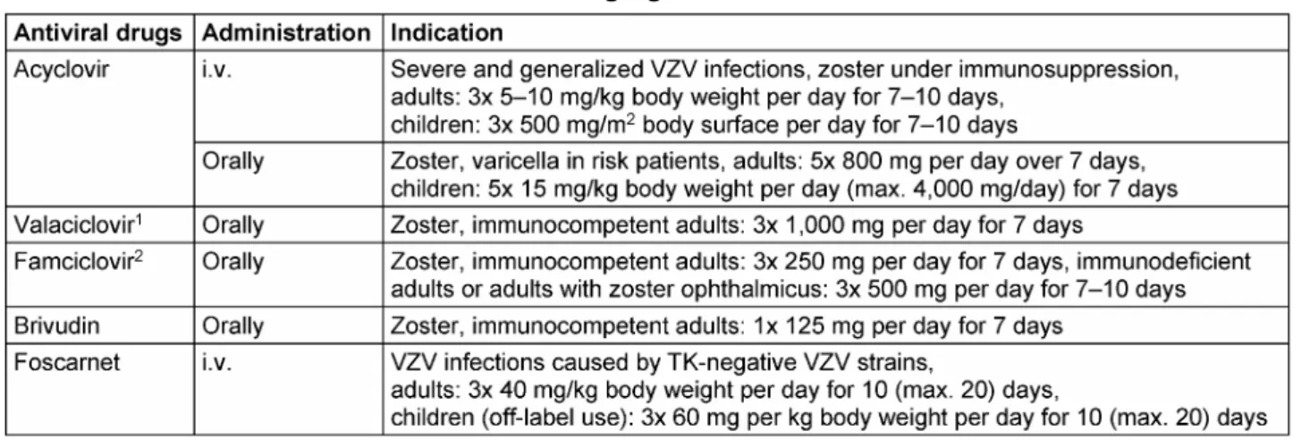
Replication of VZV in infected cells can be blocked effect- ively by the administration of antiviral agents. An early administration especially in zoster may reduce the dam- age of tissue, and thus the destruction of affected gangli- on cells can be diminished or even prevented. Primarily, the acyclic nucleoside analogs acyclovir including its prodrug valaciclovir, famciclovir (prodrug of penciclovir) and the cyclic nucleoside analog brivudin ((E)-5-(2-bro- movinyl)-2'-deoxyuridine, BVDU) are available for antiviral therapy of VZV infections (Table 1). The specificity of antiviral activity is based on the phosphorylation of these inhibitors by the viral thymidine kinase (TK) to their mono- (acyclovir, penciclovir) or diphosphates (brivudin), while the further phosphorylation steps to the triphosphate are catalyzed by cellular enzymes. Thus, the spectrum of activity is defined by the presence of the key enzyme, the viral TK. The triphosphates of the nucleoside analogs in-
Table 1: Antiviral drugs against VZV in clinical use
hibit and fix the viral DNA polymerases (pol) and are in- corporated as “false” substrate into the growing DNA chain. In case of acyclovir/valaciclovir, this results in chain termination due to the absence of the hydroxy group in 3’ position essential for further linking. In other nucleoside analogous compounds, their incorporation into DNA is possible.
Acyclovir
It is the standard therapeutic agent for antiviral treatment of VZV infections, but the oral bioavailability is only 15–30%. Oral treatment is recommended for varicella in risk patients and zoster disease in immunocompetent patients. In severe VZV infections, especially in immuno- deficient patients, acyclovir has to be administered intra- venously (i.v.). After i.v. administration, side effects on the central nervous system (CNS) have been observed occasionally, whereas the oral medication can be associ- ated with gastrointestinal symptoms. Substances with kidney toxicity should not be combined simultaneously with acyclovir. Laboratory kidney and liver parameters have to be monitored.
Valaciclovir
The prodrug (l-valyl ester) of acyclovir is administered or- ally and is converted into acyclovir by the hepatic valaciclovir hydrolase. Valaciclovir has an oral bioavailabil- ity of 54% resulting in three to four times higher drug concentrations than oral acyclovir. The consequences are longer dosing intervals and a better compliance. The administration of valaciclovir is approved for antiviral treatment of zoster in immunocompetent adults, but the drug is not approved for the use during childhood and adolescence. Possible side effects are similar to those after medication of acyclovir.
Famciclovir
This inactive diacethyl ester prodrug of penciclovir arises by separation of two ester groups in small intestine and
liver. Penciclovir is an acyclic nucleoside analog (exchange of the ether oxygen atom in the acyclic side chain by a methyl bridge) derived from gancyclovir. Since the oral absorption is very low, penciclovir is only used for topic antiviral treatment of local HSV infections. In contrast, famciclovir has a bioavailability of 77% after oral admin- istration. It is used for antiviral therapy of zoster in im- munocompetent adults and immunosuppressed patients from the age of 25 years. Similar to valaciclovir, fam- ciclovir is not approved in childhood and adolescence. In rare cases, taking of famciclovir can lead to headaches, mental confusion and nausea.
Brivudin ((E)-5-(2-bromovinyl)-2'-deoxyuridine)
This cyclic nucleoside analog, converted to its mono- and diphosphate by the viral TK, is administered orally and has a bioavailability of approximately 40%. It is used for the antiviral treatment of zoster in immunocompetent adults. Since the safety profile is unknown, brivudin is not approved for antiviral therapy in children and adoles- cents. Therefore, the risk-benefit ratio should be examined carefully before the agent can be used in children and adolescents, and the parents have to be informed (off- label use). Brivudin is generally well tolerated, but gastrointestinal disturbances, impairment of renal func- tion, increasing of liver enzymes and reversible changes of blood cell count may occur. A simultaneous adminis- tration of 5-fluorouracil or other 5-fluoropyrimidines re- sults in an enhanced and possibly dangerous toxicity.
Foscarnet (trisodium phosphonoformate)
The pyrophosphate analog foscarnet inhibits the viral DNA pol of numerous DNA and RNA viruses by the preven- tion of the pyrophosphate exchange. Since this drug is not required to be metabolized for its antiviral activity, it has also an effectiveness against TK-negative VZV strains resistant to nucleoside analogs. That is why foscarnet is recommended for alternative antiviral treatment in sus- pected clinical resistance to acyclovir expecting rarely in immunosuppressed patients with severe zoster courses.
Disturbances of renal function and toxic caused ulcers of urogenital mucosa have to be considered as important side effects.
2.2 Development of resistance
Resistance of VZV against antiviral drugs as acyclovir or foscarnet has been observed only in immunodeficient patients suffering from zoster e.g. in the acquired immune deficiency syndrome or under immunosuppression due to cancer diseases or transplantation [20], [21], [22]. For the development of resistance, the impaired immune re- sponse and long-term administration of drugs in the context of antiviral treatment or chemoprophylaxis are of crucial significance. The weakened immune response leads to a longer virus replication with the spontaneous development of an increasing number of resistant virus mutants followed by selection of resistant viruses that cannot be eliminated by the impaired immune response [23]. Generally, resistances are associated with non- synonymous mutations localized within the gene of the respective target molecule or within the gene of proteins responsible for the metabolisation or the effectiveness of antiviral agents. For acyclovir and the related nucleo- side analogs, resistance is based almost exclusively on non-synonymous mutations of TK gene (UL36) and rarely, mostly in connection with resistance against foscarnet, on amino acid (aa) changes in the DNA pol gene (UL28) [24]. In contrast to the TK that is not required for the replication of VZV, the DNA pol is essential for the viral replication cycle. According to previous findings, VZV strains resistant to acyclovir due to mutations in the TK gene are always cross-resistant to brivudin [22]. Until now, there are only a few studies in the literature in which the significance of TK and DNA pol mutations was verified by phenotypic findings. The main reason is that VZV can only be isolated rarely in cell culture from patient samples, at best, from vesicle fluid.
Thymidine kinase gene
The thymidine kinase gene of VZV has a size of 1,026 bp and is coding for 342 aa. This gene contains two con- served regions, one nucleotide (ATP)-binding site (aa 12–29) and one nucleoside (substrate)-binding site (aa 129–145). Contrary to TK of HSV-1 and HSV-2, only a low number of natural polymorphisms have been iden- tified in the VZV TK, but it is important to differentiate each of them from resistance mutations [22]. Compared to the VZV strain Dumas (GenBank accession No. X04370.1, clade 1) frequently used as reference, all European wild-type strains comprise the aa polymorphism S288L. Comparable with HSV [25], the resistance muta- tions of VZV TK gene are assigned to three phenotypes (i) TK negative (TK-, no TK activity detectable, occur most frequently), (ii) TK reduced (TKr, diminished TK activity, 1–15% of normal activity) and (iii) TK altered (TKa, altered TK substrate specificity, no phosphorylation of acyclovir and other nucleoside analogs). Resistance to acyclovir
may be caused by stop codons, frameshifts (deletions or insertions) or aa substitutions inside and outside of con- served gene regions. Since only a few validated resistance mutations have been reported in the literature to now, novel or unknown mutations must always be expected when clinically or phenotypically resistant VZV strains are analyzed genotypically.
DNA polymerase gene
This gene with a size of 3,585 bp is coding for 1,195 aa.
There are eight conserved regions designed to I to VII and A. Similar to the TK gene, only a small number of natural polymorphisms have been reported for DNA pol [22].
Resistance-related aa substitutions are localized mostly in conserved gene centers. So far, little research has been done on natural polymorphisms and resistance mutations of the VZV DNA pol gene.
3 Laboratory diagnosis
3.1 Detection of virus
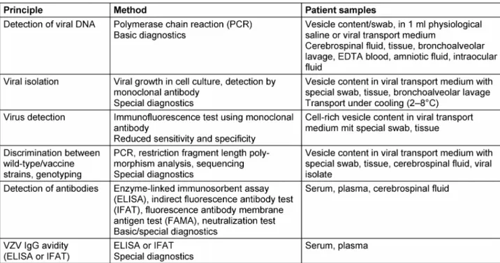
Varicella-zoster virus-positive samples have to be con- sidered as dangerous goods of the category B, risk group 2 to be shipped according to UN 3373 regulations.
Shipment is possible at room temperature, and cooling is only recommended if samples are intended for virus isolation in cell culture [26]. Acute VZV infection is diag- nosed (Table 2) by detection of the viral DNA using poly- merase chain reaction in vesicle fluids, cerebrospinal fluids, tissues, bronchoalveolar lavages, EDTA blood, amniotic fluids, or intraocular fluids (retinal necrosis) [27].
Isolation of VZV, only possible in a few cell types such as human embryonic fibroblasts, is time-consuming, requires a high degree of experience and has no clinically relevant sensitivity. In most cases, only vesicle fluids containing high virus load are suitable for viral isolation. For success- ful viral isolation, an early and careful taking as well as an optimal transport of samples are essential. Identifica- tion of viral isolates is carried out properly by immuno- fluorescence using monoclonal fluorescein-labelled anti- body. Direct qualitative detection of VZV antigens by use of commercial detection systems may provide results within a few hours, but this method has a reduced sensi- tivity and specificity. Procedures for direct detection of VZV including nucleic acids or antigens do principally not allow any differentiation between primary and recurrent infection. Discrimination between VZV wild-type and vac- cine strains can be performed by genotyping using restric- tion fragment length polymorphism analysis or sequencing [28], [29].
3.2 Detection of antibodies
Serological VZV diagnosis is especially indicated if sus- ceptible persons have to be identified. Because of the high rates of seroconversion, the determination of anti-
Table 2: Methods for laboratory diagnosis of VZV infections
body status is not necessary after varicella vaccination in healthy children, adolescents, and adults in contrast to immunodeficient vaccinees and healthcare workers [30]. In the daily laboratory practice, ligand assays or immunofluorescence tests are common for the determin- ation of VZV-specific IgG antibodies (Table 2). Regardless of the test used, each result interpreted as anti-VZV IgG positive by the respective laboratory can be used as cri- terion of immunity against varicella and individuals with borderline findings should be classified as “not immune“.
Commercially available test kits differ with respect to sensitivity. Therefore, high sensitive tests such as special glycoprotein enzyme-linked immunoassays (ELISA) or the fluorescence antibody membrane antigen test (FAMA) should be used to control immune status after varicella vaccination and for vaccine studies [31], [32]. Primary VZV infection can be diagnosed by the determination of VZV IgG seroconversion after sequential blood samples were obtained. Anti-VZV IgM will be detectable, usually in combination with anti-VZV IgG, at the earliest from the fourth day after onset of disease. Even though anti-VZV IgM is commonly used in practice to confirm active VZV infection, it has to bear in mind that IgM antibodies will be detectable with significant delay after onset of varicella exanthema and only in maximally 50–60% of patients with zoster [27]. In addition, numerous commercial VZV IgM immunoassays have a reduced sensitivity and may show false-positive results caused by cross-reactions with other herpesviruses, in particular with HSV [15]. Anti-VZV IgA may be determined frequently in persons infected latently with VZV, but high titer values exclusively correlate with zoster disease. Intrathecal VZV-specific IgG antibod- ies may be of significance for retrospective diagnosis of VZV-associated CNS infections [33]. Determination of VZV IgG avidity allows the differentiation between primary
(varicella) and recurrent infections (zoster), but there is only limited experience to this [34].
3.3 Resistance testing
In case of VZV infections, an antiviral treatment failure due to resistance is assumed if there are no clinical im- provements detectable during the administration of the antiviral medication, mostly acyclovir within 10 (–21) days [21], [24], [35]. In these cases, an alternative treatment with foscarnet is indicated. In parallel, genotypic resistant testing and, if a viral isolate can be established in cell culture, phenotypic resistant testing should be carried out. Because of the high expenditure of time (at least 3 weeks), the phenotypic resistance testing has mostly no clinical relevance, but the procedure can help to characterize novel mutations or gene variations which are so far unclear with respect to their significance for any resistance.
Phenotyping
Phenotyping has been considered as gold standard for resistance testing of VZV, but the VZV isolation in cell culture has low sensitivity. The plaque reduction assay has been established as the method of choice [22]. After adding the antiviral compound to be tested in descending dilution series, the 50% effective concentration (EC50) is estimated inducing a 50% inhibition of viral replication.
For the evaluation of possible resistance, a susceptible VZV reference strain has to be tested in each experiment as control. It is a crucial advantage that phenotyping al- lows an unambiguous interpretation of the results. How- ever, the procedures used are time- and material-consum- ing as well as non-standardized. In practice, phenotypic resistance testing can only be realized if swabs can be
obtained from vesicle fluids from which the virus can be isolated in cell culture. For interpretation of the results, the most common and reliable procedure for nucleoside analogs is to classify VZV strains as resistant if the mean EC50measured is three to five times higher than the cor- responding value of sensitive control strain [36]. For resistance to foscarnet, EC50values >300 µM have been proven to be sound [37].
Genotyping
By analogy with HSV, genotyping resistance testing of VZV is performed by means of amplification and sequencing TK and DNA pol genes [25], [38]. For the identification of non-synonymous mutations, sequence data must be compared with the published sequences of a susceptible reference strain available in the Genbank (e.g. VZV strain Dumas, accession No. X04370.1). The advantage of genotyping is a considerably shorter delay (approximately 2 days) in comparison to phenotyping and the direct testing of patient samples without virus isolation in cell culture. The restricted quantity of viral DNA may have a limiting effect. A great disadvantage is the fact that there is only little information available about assured resist- ance-associated aa substitutions. That is why only stop codons or frameshift mutations can be interpreted without doubt as related to resistance. In addition, the analysis of genotypic resistance may be difficult when a mixture of viral strains with different genotypes is present.
Therefore, it is a problem in clinical practice to define a questionable resistance of VZV strains on the basis of genotyping results alone. Recently, it has become appar- ent that phenotypic testing of recombinant VZV isolates is the best method to validate the significance of muta- tions for any resistance [39].
Notes
Competing interests
The author declares that he has no competing interests.
Acknowledgement
This review was supported by the scientific advisory board for Antiviral Therapy of the German Association for the Control of Virus Diseases and the Society of Virology.
References
1. Davison A, Eberle R, Hayward GS, McGeoch DJ, Minson AC, Pellett PE, Roizman B, Studdert MJ, Thiry E. Family Herpesviridae. In:
Fauquet C, Mayo M, Maniloff J, Desselberger E, Ball L, editors.
Virus taxonomy – Eighth Report of the International Committee on Taxonomy of Viruses. San Diego, CA: Academic Press; 2005.
p. 193–212.
2. Breuer J, Grose C, Norberg P, Tipples G, Schmid DS. A proposal for a common nomenclature for viral clades that form the species varicella-zoster virus: summary of VZV Nomenclature Meeting 2008, Barts and the London School of Medicine and Dentistry, 24-25 July 2008. J Gen Virol. 2010 Apr;91(Pt 4):821-8. DOI:
10.1099/vir.0.017814-0
3. Lokeshwar MR, Agrawal A, Subbarao SD, Chakraborty MS, Ram Prasad AV, Weil J, Bock HL, Kanwal S, Shah RC, Shah N. Age related seroprevalence of antibodies to varicella in India. Indian Pediatr. 2000 Jul;37(7):714-9.
4. Wutzler P, Färber I, Wagenpfeil S, Bisanz H, Tischer A.
Seroprevalence of varicella-zoster virus in the German population.
Vaccine. 2001 Oct;20(1-2):121-4. DOI: 10.1016/S0264- 410X(01)00276-6
5. Hillebrand K, Bricout H, Schulze-Rath R, Schink T, Garbe E.
Incidence of herpes zoster and its complications in Germany, 2005-2009. J Infect. 2015 Feb;70(2):178-86. DOI:
10.1016/j.jinf.2014.08.018
6. Gershon AA, Mervish N, LaRussa P, Steinberg S, Lo SH, Hodes D, Fikrig S, Bonagura V, Bakshi S. Varicella-zoster virus infection in children with underlying human immunodeficiency virus infection. J Infect Dis. 1997 Dec;176(6):1496-500. DOI:
10.1086/514147
7. Mahalingam R, Wellish M, Cohrs R, Debrus S, Piette J, Rentier B, Gilden DH. Expression of protein encoded by varicella-zoster virus open reading frame 63 in latently infected human ganglionic neurons. Proc Natl Acad Sci USA. 1996 Mar;93(5):2122-4. DOI:
10.1073/pnas.93.5.2122
8. Kennedy PG, Rovnak J, Badani H, Cohrs RJ. A comparison of herpes simplex virus type 1 and varicella-zoster virus latency and reactivation. J Gen Virol. 2015 Jul;96(Pt 7):1581-602. DOI:
10.1099/vir.0.000128
9. Arvin AM, Moffat JF, Redman R. Varicella-zoster virus: aspects of pathogenesis and host response to natural infection and varicella vaccine. Adv Virus Res. 1996;46:263-309. DOI:
10.1016/S0065-3527(08)60074-3
10. Schneweis KE, Krentler C, Wolff MH. Durchseuchung mit dem Varicella-Zoster-Virus und serologische Feststellung der Erstinfektionsimmunität [Varicella-zoster virus infection and the serologic determination of first-infection immunity]. Dtsch Med Wochenschr. 1985 Mar;110(12):453-7. DOI: 10.1055/s-2008- 1068845
11. Sauerbrei A, Wutzler P. Varicella-Zoster-Virus-Infektionen: Aktuelle Prophylaxe und Therapie. 1st ed. Bremen, London, Boston: Uni- Med; 2004. p. 44-53.
12. Wutzler P, Knuf M, Liese J. Varicella: efficacy of two-dose vaccination in childhood. Dtsch Arztebl Int. 2008 Aug;105(33):567-72. DOI: 10.3238/arztebl.2008.0567 13. Arvin AM. Management of varicella-zoster virus infections in
children. Adv Exp Med Biol. 1999;458:167-74. DOI:
10.1007/978-1-4615-4743-3_16
14. Hirose I, Ymamaguchi H, Inaguma D, Ono K, Shimada S, Kawada J, Shiraki K, Kimura H. Fatal varicella infection in a girl with systemic lupus erythematosus after oral acyclovir prophylaxis.
Eur J Pediatr. 2006 Apr;165(4):280-1. DOI: 10.1007/s00431- 005-0066-z
15. Sauerbrei A. Varicella-zoster virus infections during pregnancy.
In: Mushahwar K, editor. Congenital and other related infectious diseases of the newborn. Perspect Med Virol. 2007;13:51-73.
16. Vázquez M, Shapiro ED. Varicella vaccine and infection with varicella-zoster virus. N Engl J Med. 2005 Feb;352(5):439-40.
DOI: 10.1056/NEJMp048320
17. Ultsch B, Köster I, Reinhold T, Siedler A, Krause G, Icks A, Schubert I, Wichmann O. Epidemiology and cost of herpes zoster and postherpetic neuralgia in Germany. Eur J Health Econ. 2013 Dec;14(6):1015-26. DOI: 10.1007/s10198-012-0452-1 18. Siedler A, Hecht J, Rieck T, Tolksdorf K, Hengel H. Die
Varizellenimpfung in Deutschland. Eine Zwischenbilanz mit Blick auf die Masern-Mumps-Röteln- (MMR-)Impfung [Varicella vaccination in Germany. A provisional appraisal in the context of MMR vaccination]. Bundesgesundheitsblatt
Gesundheitsforschung Gesundheitsschutz. 2013 Sep;56(9):1313-20. DOI: 10.1007/s00103-013-1789-z 19. Gross G. Zoster. Manifestationsformen an der Haut,
Komplikationen und Therapie [Zoster. The manifestation forms in the skin, complications and therapy]. Dtsch Med Wochenschr.
1997 Jan;122(5):132-9. DOI: 10.1055/s-2008-1047587 20. Fillet AM, Visse B, Caumes E, Dumont B, Gentilini M, Huraux JM.
Foscarnet-resistant multidermatomal zoster in a patient with AIDS. Clin Infect Dis. 1995 Nov;21(5):1348-9. DOI:
10.1093/clinids/21.5.1348
21. Saint-Léger E, Caumes E, Breton G, Douard D, Saiag P, Huraux JM, Bricaire F, Agut H, Fillet AM. Clinical and virologic characterization of acyclovir-resistant varicella-zoster viruses isolated from 11 patients with acquired immunodeficiency syndrome. Clin Infect Dis. 2001 Dec;33(12):2061-7. DOI:
10.1086/324503
22. Sauerbrei A, Taut J, Zell R, Wutzler P. Resistance testing of clinical varicella-zoster virus strains. Antiviral Res. 2011 Jun;90(3):242- 7. DOI: 10.1016/j.antiviral.2011.04.005
23. Piret J, Boivin G. Resistance of herpes simplex viruses to nucleoside analogues: mechanisms, prevalence, and management. Antimicrob Agents Chemother. 2011 Feb;55(2):459-72. DOI: 10.1128/AAC.00615-10
24. Balfour HH Jr,Benson C, Braun J, Cassens B, Erice A, Friedman- Kien A, Klein T, Polsky B, Safrin S. Management of acyclovir- resistant herpes simplex and varicella-zoster virus infections. J Acquir Immune Defic Syndr. 1994 Mar;7(3):254-60.
25. Sauerbrei A. Diagnostik und antivirale Therapie von Herpes- simplex-Virus-Infektionen. Der Mikrobiologe. 2014;24:151-8.
26. Sauerbrei A. Windpocken (Varizellen). In: S2k-Leitlinie Labordiagnostik schwangerschaftsrelevanter Virusinfektionen.
Berlin, Heidelberg: Springer; 2014. p. 95-110. DOI:
10.1007/978-3-662-43481-9_10
27. Sauerbrei A, Eichhorn U, Schacke M, Wutzler P. Laboratory diagnosis of herpes zoster. J Clin Virol. 1999 Sep;14(1):31-6.
DOI: 10.1016/S1386-6532(99)00042-6
28. Sauerbrei A, Uebe B, Wutzler P. Molecular diagnosis of zoster post varicella vaccination. J Clin Virol. 2003 Jul;27(2):190-9.
DOI: 10.1016/S1386-6532(03)00071-4
29. Sauerbrei A, Stefanski J, Philipps A, Krumbholz A, Zell R, Wutzler P. Monitoring prevalence of varicella-zoster virus clades in Germany. Med Microbiol Immunol. 2011 May;200(2):99-107.
DOI: 10.1007/s00430-010-0178-6
30. Sauerbrei A, Wutzler P. Labordiagnostik der Varizellen. Kinderärztl Prax. 2004;Sonderheft Impfen:18-21.
31. Sauerbrei A, Färber I, Brandstädt A, Schacke M, Wutzler P.
Immunofluorescence test for sensitive detection of varicella- zoster virus-specific IgG: an alternative to fluorescent antibody to membrane antigen test. J Virol Methods. 2004 Jul;119(1):25- 30. DOI: 10.1016/j.jviromet.2004.02.012
32. Sauerbrei A, Wutzler P. Serological detection of varicella-zoster virus-specific immunoglobulin G by an enzyme-linked
immunosorbent assay using glycoprotein antigen. J Clin Microbiol.
2006 Sep;44(9):3094-7. DOI: 10.1128/JCM.00719-06 33. Sauerbrei A, Wutzler P. Laboratory diagnosis of central nervous
system infections caused by herpesviruses. J Clin Virol. 2002 Jul;25 Suppl 1:S45-51. DOI: 10.1016/S1386-6532(02)00033- 1
34. Kneitz RH, Schubert J, Tollmann F, Zens W, Hedman K, Weissbrich B. A new method for determination of varicella-zoster virus immunoglobulin G avidity in serum and cerebrospinal fluid.
BMC Infect Dis. 2004 Sep;4:33. DOI: 10.1186/1471-2334-4- 33
35. Safrin S, Berger TG, Gilson I, Wolfe PR, Wofsy CB, Mills J, Biron KK. Foscarnet therapy in five patients with AIDS and acyclovir- resistant varicella-zoster virus infection. Ann Intern Med. 1991 Jul;115(1):19-21. DOI: 10.7326/0003-4819-115-1-19 36. Morfin F, Thouvenot D. Herpes simplex virus resistance to
antiviral drugs. J Clin Virol. 2003;26:29-37. DOI: 10.1016/S1386- 6532(02)00263-9
37. Safrin S, Crumpacker C, Chatis P, Davis R, Hafner R, Rush J, Kessler HA, Landry B, Mills J. A controlled trial comparing foscarnet with vidarabine for acyclovir-resistant mucocutaneous herpes simplex in the acquired immunodeficiency syndrome.
The AIDS Clinical Trials Group. N Engl J Med. 1991 Aug;325(8):551-5. DOI: 10.1056/NEJM199108223250805 38. Schubert A, Gentner E, Bohn K, Schwarz M, Mertens T, Sauerbrei
A. Single nucleotide polymorphisms of thymidine kinase and DNA polymerase genes in clinical herpes simplex virus type 1 isolates associated with different resistance phenotypes. Antiviral Res.
2014 Jul;107:16-22. DOI: 10.1016/j.antiviral.2014.03.015 39. Brunnemann AK, Bohn-Wippert K, Zell R, Henke A, Walther M,
Braum O, Maschkowitz G, Fickenscher H, Sauerbrei A, Krumbholz A. Drug resistance of clinical varicella-zoster virus strains confirmed by recombinant thymidine kinase expression and by targeted resistance mutagenesis of a cloned wild-type isolate.
Antimicrob Agents Chemother. 2015 May;59(5):2726-34. DOI:
10.1128/AAC.05115-14
Corresponding author:
Prof. Dr. Andreas Sauerbrei
Institute of Virology and Antiviral Therapy, Jena University Hospital, Hans-Knoell-Strasse 2, 07745 Jena, Germany, Phone: +49-3641-9395700, Fax: +49-3641-9395702 Andreas.Sauerbrei@med.uni-jena.de
Please cite as
Sauerbrei A. Varicella-zoster virus infections – antiviral therapy and diagnosis. GMS Infect Dis. 2016;4:Doc01.
DOI: 10.3205/id000019, URN: urn:nbn:de:0183-id0000196
This article is freely available from
http://www.egms.de/en/journals/id/2016-4/id000019.shtml Published:2016-02-17
Copyright
©2016 Sauerbrei. This is an Open Access article distributed under the terms of the Creative Commons Attribution 4.0 License. See license information at http://creativecommons.org/licenses/by/4.0/.