TECHNISCHE UNIVERSITÄT MÜNCHEN
Lehrstuhl für Mikrobielle Ökologie
Investigation of propanediol and fucose degradation by Salmonella Typhimurium
Lena Staib
Vollständiger Abdruck der von der Fakultät Wissenschaftszentrum Weihenstephan für Ernährung, Landnutzung und Umwelt der Technischen Universität München zur Erlangung des akademischen Grades eines
Doktors der Naturwissenschaften
genehmigten Dissertation.
Vorsitzender: Univ.- Prof. Dr. S. Scherer Prüfer der Dissertation:
1. apl. Prof. Dr. T. M. Fuchs 2. Univ.-Prof. Dr. H. Daniel
Die Dissertation wurde am 08.08.2016 bei der Technischen Universität München eingereicht und durch die Fakultät Wissenschaftszentrum Weihenstephan für Ernährung, Landnutzung und Umwelt am 09.11.2016 angenommen.
Contents
List of Figures IV
List of Tables VI
Nomenclature VII
Summary XI
Zusammenfassung XIII
1 Introduction 1
1.1 Food-borne illnesses . . . 1
1.2 The genusSalmonella. . . 1
1.2.1 Salmonellainfections . . . 2
1.2.2 S.Typhimurium . . . 2
1.2.2.1 The genome . . . 2
1.2.2.2 The infection cycle . . . 3
1.3 Gut niche occupation byS.Typhimurium . . . 4
1.3.1 Host and microbiota . . . 4
1.3.2 Metabolic adaptation of enteropathogens . . . 7
1.3.3 Subversion of defence mechanisms byS.Typhimurium . . . 9
1.3.4 Exploitation of exceptional nutrient sources . . . 11
1.3.4.1 Tetrathionate respiration inS.Typhimurium . . . 15
1.4 Research objective . . . 16
2 Material and Methods 17 2.1 Material . . . 17
2.2 Microbiology . . . 17
2.2.1 Culture conditions . . . 17
2.2.1.1 Cultivation in rich medium . . . 17
2.2.1.2 Cultivation in poor media . . . 17
2.2.1.3 Turbidity measurements . . . 19
2.2.1.4 Motility test . . . 19
2.3 Molecular biology . . . 20
2.3.1 Preparation of nucleic acids . . . 20
2.3.1.1 Plasmid isolation . . . 20
2.3.1.2 Isolation of genomic DNA . . . 20
2.3.1.3 Isolation of RNA . . . 20
2.3.1.4 DNase digestion of RNA . . . 21
2.3.1.5 cDNA synthesis . . . 22
2.3.1.6 Determination of nucleic acid concentrations . . . 22
2.3.2 Agarose gel electrophoresis . . . 22
2.3.3 Polymerase chain reaction . . . 23
2.3.3.1 Standard PCR . . . 23
2.3.3.2 Purification of PCR-products . . . 24
2.3.4 Enzymatic modification of DNA/ Cloning . . . 24
2.3.5 Genetic modification of bacteria . . . 24
2.3.5.1 Preparation of electrocompetent cells . . . 24
2.3.5.2 Transformation/ electroporation . . . 25
2.3.5.3 Conjugation . . . 25
2.3.5.4 Phage transduction . . . 26
2.3.5.5 Construction ofS.Typhimurium reporter strains . . . 27
2.3.5.6 Construction ofS.Typhimurium insertion and deletion mutants 27 2.3.5.7 Complementation of gene deletions . . . 28
2.3.6 Determination of bioluminescence and fluorescence . . . 28
2.3.6.1 Quantification of gene expression . . . 28
2.3.6.2 Visualisation of gene expression . . . 29
2.3.6.3 Fluorescence microscopy . . . 29
2.3.6.4 Xenogen In Vivo Imaging System (IVIS) . . . 29
2.4 Cell culture . . . 29
2.4.1 Cell lines . . . 29
2.4.2 Culture conditions and media . . . 30
2.4.2.1 Subcultivation of human cell lines . . . 30
2.4.2.2 Storing of human cell lines . . . 30
2.4.3 Invasion assay . . . 30
2.4.4 Immunofluorescence staining . . . 31
2.5 C. elegansassay . . . 32
2.6 Software and statistical analysis . . . 33
3 Results 35 3.1 Utilisation of alternative carbon and energy sources by enteropathogens . . . 35
3.2 Distribution of the genes relevant for 1,2-propanediol and L-fucose metabolism . 39 3.2.1 Thepduandfucgenes are highly conserved amongS. enterica . . . 39
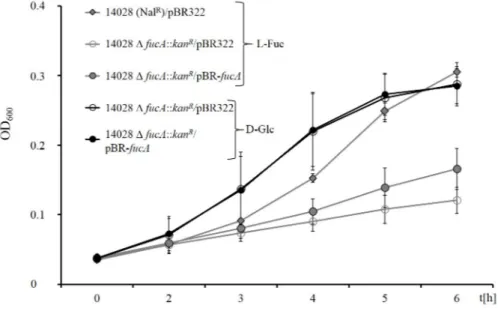
3.3 Genetic determinants for the growth ofS.Typhimurium with 1,2-PD and L-Fuc . 41 3.3.1 fucAis essential for growth with L-fucose . . . 41
3.3.2 L-fucose and 1,2-propanediol are used simultaneously . . . 43
3.3.3 Deletion ofpduCeliminates growth with 1,2-propanediol . . . 44
3.3.4 PpduApromoter activity represents growth with 1,2-propanediol . . . 46
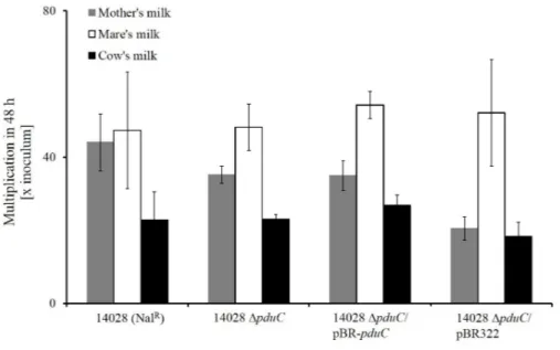
3.3.5 Role ofpduduring cultivation in milks . . . 48
3.3.6 Impact ofpduCdeletion on growth in gut content of pigs . . . 51
3.4 Thepdugenes are transcribed as one polycistronic mRNA . . . 52
3.5 Estimation of L-fucose contents in the gut . . . 54
3.6 PpduAis induced by low 1,2-PD levels . . . 55
3.6.1 Expression of thepdugenes does not seem to be completely repressed by D-Glc . . . 58
3.7 Cell culture assays . . . 60
3.7.1 S.Typhimurium invasiveness is influenced byinvAand mucus . . . 60
3.7.2 Invasion of LS174T cells is preferred over HT29-MTX . . . 61
3.7.3 Motility is crucial forS.Typhimurium to overcome the mucus layer . . . 62
3.8 Evidence of L-Fuc in the gut ofC. elegans . . . 63
3.9 Summary of the results . . . 64
4 Discussion 65 4.1 L-Fuc and 1,2-PD metabolism . . . 65
4.1.1 Origin of free L-Fuc in the gut . . . 65
4.2 Distribution of thefucandpdugenes among enteropathogens . . . 66
4.3 L-Fuc and 1,2-PD degradation byS.Typhimurium . . . 68
4.3.1 Induction ofpdugene expression . . . 71
4.3.2 Transcription of thepdugenes . . . 74
4.4 FucA and PduC are essential for L-Fuc and 1,2-PD degradation . . . 75
4.5 Invasiveness is influenced by motility, theinvlocus, and mucus secretion . . . 76
4.6 L-Fuc availability in theC. elegansglycocalyx . . . 78
4.7 Conclusion and perspectives . . . 79
Bibliography 83 5 Appendix 107 5.1 Supplier information . . . 107
5.2 List of bacterial strains . . . 111
5.3 List of primers . . . 113
5.4 Plasmids and vectors . . . 117
5.5 Media composition . . . 131
5.6 Supplementary information . . . 133
5.6.1 Gene clusters . . . 133
5.7 Acknowledgements . . . 144
5.8 List of publications . . . 145
5.9 Eidesstattliche Erklärung . . . 146
List of Figures
1.1 Subversion of host defence . . . 11
1.2 Genetic organisation ofeut, pduandcob/cbigenes . . . 13
1.3 Ethanolamine metabolism . . . 14
3.1 Nutrient availability in the GIT . . . 37
3.2 Anaerobic growth ofS.Typhimurium 14028 with L-Fuc . . . 42
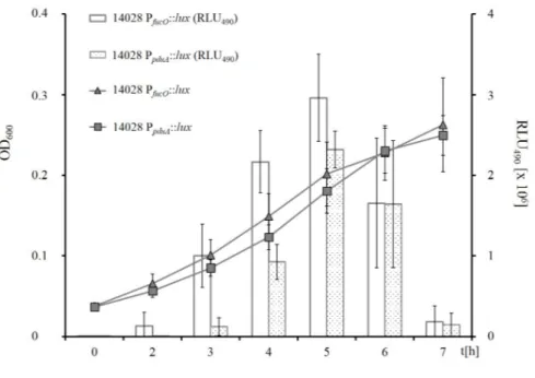
3.3 Expression offucOand thepdugenes during growth with L-Fuc . . . 44
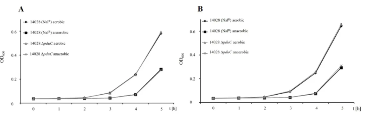
3.4 Aerobic growth of∆pduCwith 1,2-PD . . . 45
3.5 Anaerobic growth assays of strain 14028 with 1,2-PD . . . 46
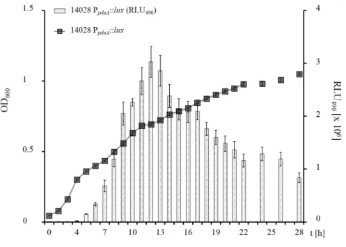
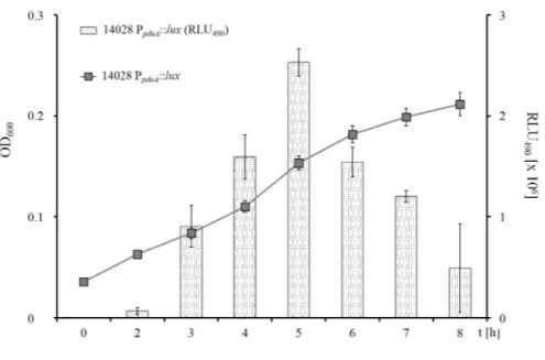
3.6 Gene expression ofpduduring aerobic growth with 1,2-PD . . . 47
3.7 Gene expression ofpduduring anaerobic growth with 1,2-PD . . . 48
3.8 S.Typhimurium 14028 growth in spent-milk media . . . 50
3.9 Growth of 14028 in gut content of pigs . . . 52
3.10 Transcriptionpdugene cluster . . . 53
3.11 Microscopic investigation of 14028 PpduA::gfpat varying 1,2-PD concentrations. . 57
3.12 Microscopic investigation for catabolite repression of PpduAby D-Glc . . . 59
3.13 Invasion assay in cell culture . . . 60
3.14 Invasion of HT29-MTX and LS174T cells . . . 61
3.15 LS174T invasion of motile and non-motileS.Typhimurium . . . 62
3.16 C. elegansinfection assay with 14028 PfucO::lux . . . 63
4.1 fucandrhagene clusters . . . 70
4.2 1,2-PD metabolism . . . 71
4.3 Regulation by PocR . . . 74
5.1 pGreen-TIR . . . 119
5.2 pBR322 . . . 120
5.3 pBR322-pduC . . . 121
5.4 pBR322-fucA . . . 122
5.5 pUTs-lux . . . 123
5.6 pUTs-PpduA-lux . . . 124
5.7 pUTs-PfucO-lux . . . 125
5.8 pUTs-invJ-lux . . . 126
5.9 pUTs-gfp . . . 127
5.10 pUTs-PpduA-gfp . . . 128
5.11 pUTs-PfucO-mCherry . . . 129
5.12 pUTs-PfucO-mCherry . . . 130
5.13 Growth 14028 PpduA::gfp . . . 137
5.14 Growth 14028 PpduA::gfpwith 1,2-PD and D-Glc . . . 138
5.15 Luminescence assay Caco-2 . . . 140
5.16 Luminescence assay LS174T . . . 142
5.17 Multiplication in LS174T cell culture . . . 143
List of Tables
2.1 LB (Bertani, 1951). . . 17
2.2 Vogel-Bonner mineral medium (Vogel and Bonner, 1956). . . 18
2.3 Lysis solution for DNA isolation. . . 20
2.4 Preparation of AqDEP C. . . 21
2.5 Reaction mixture for PCR. . . 23
2.6 Standard PCR programme. . . 24
2.7 Green plates (Maloy, 1990). . . 26
2.8 Antibody dilution buffer. . . 31
2.9 Nematode growth medium and M9-buffer (Stiernagle, 2006). . . 33
3.1 Metabolic properties of enteropathogens . . . 38
3.2 Expression ofpduandfucgenes inS. entericasubsp. entericaserovars. . . 39
3.3 Proteins encoded by thepduandfucgenes inS. entericasubspecies . . . 41
3.4 Quantitave analysis of GFP signals of 14028 PpduA::gfp . . . 55
3.5 Insufficient catabolite repression of PpduAby D-Glc . . . 59
5.1 Supplier information of chemicals, buffers and solutions. . . 107
5.2 Supplier information for cell culture material. . . 109
5.3 Supplier information about enzymes and antibodies used in the study. . . 110
5.4 Bacterial strains used in the study. . . 111
5.5 List of primers. . . 113
5.6 List of plasmids and vectors. . . 117
5.7 SOC-medium (Hanahan, 1983). . . 131
5.8 Trace element solution . . . 131
5.9 Antibiotics used in growth media. . . 132
5.10 M9 mineral medium (Sambrook and Russell, 2001). . . 132
5.11 eut, pdu, andcob/cbigene functions. . . 133
5.12 Presence of thepdugenes inS. entericaserovars . . . 135
5.13 Presence of thefucgenes inS. entericaserovars . . . 136
Nomenclature
v
v Volume per volume
w
v Weight per volume
µ Micro
◦C Degree Celsius
1,2-PD 1,2-propanediol
% Per cent
$ US-Dollar
lux Luciferase
pdu Genes for 1,2-propanediol utilisation
A Ampere
aw Activity of water
AA Amino acids
ad. Fill up to
Amp Ampicillin
Aq Water
Aqbidest Double distilled water
Arc Aerobic respiration control protein
AT Austria
BLAST Basic local alignment search tool
bp base pairs
c Centi
cf inal Final concentration
cAMP Cyclic adenosine monophosphate
CCD Charged-coupled device
cDNA Complementary DNA
cfu Colony forming unit
cfu Colony forming units
Cm Chloramphenicol
CoA Coenzyme A
CRP cAMP receptor protein
d Day
D-Glc D-glucose
DAPI 4’,6-diamidino-2-phenylindole
DE Germany
DEPC Diethylpyrocarbonate
DHAP Dihydroxy-acetone-phosphate
DMSO Dimethyl sulphoxide
DNA Deoxyribonucleic acid
E0 Standard redox potential EDTA Ethylenediaminetetraacetic acid
FCS Foetal calf serum
Fnr Fumatare-nitare reduction regulatory protein FUT2 α-(1,2)-fucosyltransferase
g Gramme
G/C-content Guanine-cytosine content in DNA
GC Gas chromatography
GFP Green fluorescent protein GIT Gastrointestinal tract
h Hour
HIP/PAP Hepatocarcinoma-intestine-pancreas/ prancreatitis associated protein
HMO Human milk oligosaccharide
Ig Immunoglobulin
iNOS Inducible nitric oxide synthase IVIS In vivoimaging system
k Kilo
Kan Kanamycin
kb Kilo base pairs
l Litre
L-Fuc L-fucose
L-Rha L-rhamnose
LB Lysogeny broth
LPS Lipopolysaccharides
M Mega
m Meter
M cell Microfold cell
Mb Mega base pairs
min Minute
mol Mole
mRNA Messenger RNA
MS Mass spectrometry
n Nano
Nal Nalidixic acid
NCBI National center for biotechnology information NeAA Non-essential amino acids
NGM Nematode growth medium
NO Nitric oxide
OD600 Optical density at 600 nm
p Pico
PBS Phosphate buffered saline
PCR Polymerase chain reaction
pH Potentium hydrogenii
RegIIIβ Regenerating islet-derived III beta RegIIIγ Regenerating islet-derived III gamma
RLU Relative light units
RNA Ribonucleic acid
ROS Reactive oxygen species
rpm Rotations per minute
RT Room temperature
RT-PCR Reverse transcriptase polymerase chain reaction
s Second
SCFA Short chain fatty acids
SCV Salmonellacontaining vacuole
SDS Sodium dodecyl sulphate
SE Sweden
Spec Spectinomycin
SPI Salmonellapathogenicity island
Strep Streptomycin
T Temperature
t Time
TA Annealing temperature
Tm Melting temperature
T3SS Type three secretion system
TCA Tricarboxylic acid
Tet Tetracycline
TLR Toll-like receptor
Tris Tris(hydroxymethyl)aminomethane
U Unit
UK United Kingdom
US United States
USA United States of America
UV Ultra-violet
V Volt
VB-NCE Vogel-Bonner no carbon-E
VB-NCE-YE Vogel-Bonner no carbon-E with yeast extract
x g Multiple of gravity
Summary
The human organism is composed of about 1013 eukaryotic (animal) cells. The gastrointestinal tract (GIT) has a surface area of about 300 m2 and is inhabited by a community of microorgan- isms that is equal to or even exceed the total number of human cells in the body (Senderet al., 2016). The number of microorganisms increases from the proximal to the distal gut and reaches its highest density in the colon. This community of resident microorganisms in the gut is called microbiota, which is of great importance to the human body. Besides other effects, it helps in di- gesting otherwise indigestible carbohydrates, affects the development of the immune system, and creates a highly competitive environment exacerbating colonisation by enteric pathogens such as SalmonellaTyphimurium. Thus,S.Typhimurium has developed strategies to overcome colonisa- tion resistance, since each year millions of cases of salmonellosis are reported worldwide.
In addition to the expression of specific virulence factors, enteropathogenic bacteria have to ad- apt their metabolism to the nutritional and physiological environment encountered outside and inside their host. The present study focusses on the utilisation of the alternative carbon source 1,2-propanediol (1,2-PD) inS.Typhimurium. 1,2-PD is the fermentation end product of bacterial growth with L-fucose (L-Fuc) or L-rhamnose (L-Rha) respectively. Both sugars are available in the GIT as they are present in food of herbal origin and constituents of mucins.
BesidesS. entericaonlyListeria monocytogenes, Clostridium perfringens, Shigella sonnei, Yersinia enterocolitica, and some enteropathogenic and enterotoxigenicEscherichia colihave been found to harbour the genes for 1,2-PD utilisation. Comprehensive database analyses conducted within the scope of this thesis revealed that the gene clusters required for 1,2-PD (pdu) and L-Fuc (fuc) degradation are conserved amongS. entericaserovars sequenced thus far. Further, the genes re- quired for tetrathionate respiration (ttr) were only found in the genomes of S. enterica and Y.
enterocolitica. Tetrathionate serves as electron acceptor for the anaerobic respiration of 1,2-PD inS.Typhimurium. It was demonstrated that thepdugene cluster, consisting of 21 genes, is ex- pressed as a single polycistronic mRNA and controlled by the promoter PpduA. The gene products ofpduCandfucAare essential for the degradation of the respective substrate as shown by growth assays. The formation and immediate utilisation of 1,2-PD was demonstrated during anaerobic growth ofS.Typhimurium 14028 with L-Fuc by monitoring the expression of genes required for L-Fuc and 1,2-PD degradation during all growth phases usinglux-promoter fusion strains. Already minor concentrations of 1,2-PD were found to be sufficient to inducepdugene expression, which is not or only weakly repressed by the simultaneous availability of 1,2-PD and alternative carbon sources such as L-Fuc or yeast extract, for example. Until the late exponential phase the presence of equimolar D-glucose (D-Glc) concentrations relative to 1,2-PD was found to repress the tran- scription of the pdugenes, because D-Glc was metabolised prior to 1,2-PD as demonstrated by monitoring the light emission of PpduA::gfp-reporter strains. The relevance of 1,2-PD degradation
was also investigated in more complex substrates such as in the gut contents of pigs, fucosylated milk, mucus-secreting cell culture and the invertebrate model. Cell culture assays using mucus- secreting cell lines confirmed the necessity of mobility for host cell invasion.
Zusammenfassung
Der menschliche Körper besteht aus circa 1013eukaryotischen (tierischen) Zellen. Der Gastorin- testinaltrakt hat eine Oberfläche von etwa 300 m2, welche von Mikroorganismen besiedelt ist. Die Anzahl der Mikroorganismen gleicht oder übersteigt die der Körperzellen (Senderet al., 2016).
Die mikrobielle Populationsdichte nimmt im proximal-distalen Verlauf des Gastrointestinaltraks zu und erreicht im Dickdarm ihr Maximum. Die im Darm ansässige Mikroflora wird auch als Mikrobiota bezeichnet und ist von höchster Wichtigkeit für den menschlichen Körper, da sie, un- ter Anderem, bei der Verdauung von, anderenfalls unverdaulichen, Kohlenhydraten behilflich ist, die Entwichklung des Immunsystems unterstützt und, durch die Erschaffung einer höchst kompe- titiven Umgebung, die Ansiedelung von pathogenen Mikroorganismen, wie besispielsweise Sal- monella Typhimurium erschwert. Daher hat S. Typhimurium Strategien entwickelt, um diesem Wettbewerb, der sogenannten Kolonisationsresistenz, zu entgehen, denn jährlich erkranken welt- weit Millionen von Menschen an Salmonellose.
Neben der Expression spezifischer Virulenzfaktoren, müssen Enteropathogene ihren Stoffwechsel an die Nährstoffverfügbarkeit und physiologischen Bedingungen, die innerhalb sowie außerhalb ihres Wirtsorganismus angetroffen werden, anpassen. Die vorliegende Arbeit befasst sich mit der Verwendung der alternativen Kolenstoffquelle 1,2-Propandiol (1,2-PD) durch S. Typhimurium.
1,2-PD ist das Produkt der bakteriellen Fermentation, der im Darm vorliegenden Zucker L-Fucose (L-Fuc) und beziehungsweise oder L-Rhamnose (L-Rha).
Die Gene, die für die Nutzung von 1,2-PD als Kohlenstoffquelle relevant sind, liegen lediglich in den Genomen der EnterobakterienS. enterica, Listeria monocytogenes, Clostridium perfringens, Shigella sonnei, Yersinia enterocoliticaund einiger enteropathogener und enterotixigenerEscheri- chia colivor. Die hier präsentierten Daten zeigen, dass nahezu alleS. entericaSerovare, die bisher sequenziert wurden, die Gene zur Nutzung von 1,2-PD (pdu) und L-Fuc (fuc) besitzen. Lediglich in den Genomen derS. entericaSerovare undY. enterocoliticakonnten die Gene, die den Bakterien die Tetrathionatatmung (ttr) ermöglichen, gefunden werden. Tetrathionat dient inS.Typhimurium als Elektronenakzeptor, um 1,2-PD anaerob oxidieren zu können. Ausgehend vom Promotor PpduA
werden diepduGene imS.Typhimurium Stamm 14028 als polycistronische mRNA exprimiert, welche die genetische Information von 21 Genen beinhaltet. Die Genprodukte der GenepduAund fucAsind essentiell für den Abbau des jeweiligen Substrats, wie in Wachstumsversuchen gezeigt werden konnte. Darüberhinaus konnte die Bildung von 1,2-PD, als Produkt der Fermentation von L-Fuc und dessen unmittelbarer Verwertung unter anaeroben Bedingungen, während der Kulti- vierung vonlux-Promotorfusionsstämmen in fucosehaltigem Medium und der Aufzeichnung der Promotoraktivitäten von Genen, die für die Verwendung von L-Fucose und 1,2-PD als Kohlen- stoffquellen benötigt werden, durch die emittierte Biolumineszenz der lux-Reporterstämme do- kumentiert werden. Der Promotor PpduA wird bereits durch geringste Konzentrationen an 1,2-PD
induziert und scheint nicht oder nur in geringem Maße durch die gleichzeitige Verfügbarkeit an- derer Kohlenstoffquellen, wie beispielsweise L-Fuc oder Hefeextrakt, gehemmt zu werden. Bis zum Erreichen der späten exponentiellen Wachstumsphase führte ie Bereitstellung äquimolarer Konzentrationen von 1,2-PD und D-Glukose (D-Glc) hingegen zu einer Repression derpduGen- expression, da D-Glc zuerst verstoffwechselt wurde, wie in Experimenten durch die Beobachtung der Lichtemission von PpduA::gfp-Reporterstämmen gezeigt werden konnte. Des Weiteren wurde die Relevanz des 1,2-PD Abbaus in komplexeren Substraten, wie dem Darminhalt von Schweinen, fucosylierter Milch, Mukus sekretierenden Zelllinen und dem Invertebratenmodell untersucht.
In Zellkulturversuchen konnte die Notwendigkeit der Bewegungsfähigkeit für die Invasion von Wirtszellen, unter der Verwendung von Mukus sekretierenden Zelllinien, bestätigt werden.
1 Introduction
1.1 Food-borne illnesses
Food-borne illnesses are caused by the consumption of contaminated beverages or foods. Symp- toms are nausea, acute gastroenteritis accompanied by watery diarrhoea, abdominal pain, vomit- ing or cramps. The causative agents are bacteria, viruses and parasites. According to the Centers for Disease Control and Prevention (CDC), about 48 million cases of food-borne illnesses oc- cur annually in the USA. This leads to at least 128,000 hospitalisations and 3,000 deaths (CDC, 2015). The European Food Safety Authority (EFSA) reported a total of 55,453 food-borne ill- nesses in Europe for the year 2012, resulting in 5,118 hospitalisations and 41 deaths (EFSA and ECDC, 2014). Non-typhoidal Salmonellafollowed by bacterial toxins, viruses and Campylob- acter, were the most common causative agents for food-borne illnesses in Europe, while in the USA Noroviruses are reported to be the major cause (EFSA and ECDC, 2014; CDC, 2015). The aforementioned incidences resulted in annual costs of about $77.7 billion in the USA (Scharff, 2012), which underlines the impact of food safety on the healthcare system. Food-borne bacterial pathogens carry many genes that enable them to adapt to various environmental conditions and pressures (Humphrey, 2004). Examples of such genes includerpoS,fur,ompR/envZ, andphoPQ, which are required for the survival of low pH conditions in the human pathogenSalmonellaTyph- imurium. The gene products of ompR/envZ are further necessary for the adaptation to osmotic pressure. In order to react to changes in oxygen availability, the pathogen harbours the genesfnr andarcAB, decreasing respiration under microaerophilic or anaerobic conditions. Further adapta- tions are, for example, heat and cold shock proteins, resistance against oxidative stress and cationic antimicrobial peptides (Rychlik and Barrow, 2005). Hence,Salmonellais regularly isolated from raw meat and meat products (Andrews-Polymeniset al., 2010; RKI, 2014), eggs and egg-products (Coxet al., 2000; RKI, 2014), but also from fruit and vegetables (Wells and Butterfield, 1997).
1.2 The genus Salmonella
Salmonellaare Gram-negative, rod-shaped, motile (peritrichously flagellated) bacteria, belonging to the family ofEnterobacteriaceae. Salmonellais positive for catalase, methyl red, Simmons’
citrate and the production of H2S, but negative for oxidase, indole and acetoin (Voges Proskauer) production, and the hydrolysis of urea (Farmeret al., 1985).SalmonellaandEscherichia coliare highly related, sharing about 71 % DNA sequence homology and may thus originate from a com- mon ancestor (McClellandet al., 2001; de Jonget al., 2012).
The nameSalmonellais derived from the American veterinary surgeon Daniel Elmer Salmon who,
together with his colleague Theobald Smith, described the bacterium for the first time in 1886 (Sal- mon and Smith, 1886).
The genus consists of Salmonella enterica andSalmonella bongori. S. enterica is further sub- divided into six subspecies on the basis of the serologic identification of O (somatic)- and H (flagellar)-antigens (Brenneret al., 2000). To date, some 2,500 serovars have been described, of which approximately 60 % belong toS. entericasubsp. enterica, which is responsible for infec- tions of warm-blooded animals including humans (Farmeret al., 1985).
1.2.1 Salmonellainfections
S. entericacan cause diverse clinical manifestations in humans. The main sources of infection are the consumption of contaminated food or water, however transmission via the faecal-oral route is also possible (Santoset al., 2001; WHO, 2013).
S.Typhi andS.Paratyphi cause systemic infections with varying clinical features (de Jonget al., 2012). Worldwide annual occurrences of typhoid fever are estimated at 22 million cases, 200,000 related deaths, and an additional six million cases of paratyphoid fever. The regions most affected are East and Southeast Asia, Africa, the Caribbean, and Central and South America (Newton and Mintz, 2014). Typhoid and paratyphoid fever are treated by administration of antimicrobials, leading to the emergence of multi-drug resistant strains (Suet al., 2004; Majowiczet al., 2010;
Newton and Mintz, 2014).
A usually less life-threatening disease is caused by non-typhoidal S. enterica serovars such as Typhimurium and Enteritidis. Symptoms like diarrhoea, nausea, vomiting and intestinal cramping occur six to 72 hours after ingestion of contaminated foods (Santoset al., 2001; WHO, 2013). The resulting gastroenteritis, also called salmonellosis, is generally self-limiting in otherwise healthy subjects and does not require special treatment. For elderly, immunocompromised and very young patients salmonellosis can also be very serious due to dehydration or bacteraemia, and subsequent sepsis (WHO, 2013; de Jonget al., 2012). With about 93.8 million estimated cases worldwide, salmonellosis is one of the main causes of acute gastroenteritis. Of these, 80.3 million cases are considered as food-borne, and 155,000 are fatal (Majowiczet al., 2010). Salmonellosis not only occurs in developing countries characterised by the lack of a clean water supply and sanitation but also in industrialised counties. Indeed in the USA one million and in Europe 91,034 illnesses are estimated to be caused by non-typhoidalSalmonellainfections (CDC, 2013; EFSA and ECDC, 2014). Although salmonellosis is a notifiable disease, about 60-90 % of the cases remain unknown (BfR, 2014; WHO, 2013).
1.2.2 S.Typhimurium
1.2.2.1 The genome
The genome of S. Typhimurium LT2 consists of the chromosome (4.857 Mb) and a virulence plasmid (pSLT). Both chromosome and virulence plasmid have a G/C-content of 53 %. The 94 kb pSLT harbours 108 protein-coding genes and is absent in S. Typhi and S. Paratyphi. The copy number of the plasmid is dependent on the conditions of growth and varies between one
to three copies (McClelland et al., 2001). The S.Typhimurium LT2 chromosome harbours four functional prophages (Gifsy-1/ -2 and Fels-1/ -2) besides 62 genomic islands and encodes 4,489 proteins (McClellandet al., 2001). Of the 62 genomic islands, ten have been said to contribute toSalmonella’s virulence, lending them the nameSalmonellapathogenicity islands (SPI) (Marcus et al., 2000; Hensel, 2004). Probably the best characterised SPIs are SPI-1 and SPI-2, responsible for the invasion of epithelial cells (SPI-1) and intracellular survival (SPI-2) (Hensel, 2004).
1.2.2.2 The infection cycle
Humans, cattle, pigs, horses, poultry, rodents, sheep and reptiles belong to the host range of S.
Typhimurium (Tsolis et al., 1999; RKI, 2014). The majority of data concerning the infection cycle ofS.Typhimurium are derived from animal studies in mice, but it is uncertain whether these findings can be extrapolated to the disease caused in humans (Tsoliset al., 1999).S.Typhimurium causes gastroenteritis in humans and calves but a systemic, typhoid-like disease in susceptible mice (Tsoliset al., 1999). The latter do not develop diarrhoea, which is the most prominent symp- tom in humans and calves. The mice die due to organ failure or bacteraemia, a complication that finds only rare occurrences in humans and cattle (Tsoliset al., 1999). The development of the streptomycin mouse model offers the possibility of studying enteric salmonellosis in mice (Barthel et al., 2003).
HoweverSalmonellavirulence and pathogenicity are mediated and influenced by the interplay of different sets of well-orchestrated virulence factors, which are dependent on the host, the host cell type, and the state of infection (Tsoliset al., 1999; Bumann, 2002).
After ingestion but prior to causing infection of the terminal ileum,S.Typhimurium is confronted with the acidic pH of the stomach, decreasing oxygen availability, increasing osmotic pressure and a mounting number of other microorganisms (Carter and Collins, 1974; Foster, 1991; Rychlik and Barrow, 2005). In mice, the primary sites of infection are microfold cells (M cells) of Peyer’s Patches (Carter and Collins, 1974). The mechanism of uptake into non-phagocytic cells is distinct from receptor-mediated phagocytosis applied by neutrophils or macrophages, and actively induced by the pathogen (Brumell et al., 1999; Galán, 1999). The capability ofSalmonellato trigger its internalisation is encoded by SPI-1, which was most likely acquired horizontally (Collazo and Galán, 1997; Galán, 1999). In total 12 SPIs have been described so far of which ten are present inS. Typhimurium (Hensel, 2004). SPI-1 and SPI-4 were described to interact in terms of host cell penetration (Gerlach et al., 2007, 2008). SPI-4 encodes a giant adhesin and the associated secretion system, but the genetic information for a type three secretion system (T3SS) and the effector molecules, which are delivered to the host cell, are located on SPI-1 (Gerlachet al., 2007;
Collazo and Galán, 1997; Marcuset al., 2000). Thus, active invasion of M cells and other non- phagocytic host cells is enabled by the translocation of effector proteins by SPI-1 T3SS, leading to cytoskeletal changes of the host cell. These cytoskeletal changes are visible as membrane ruff- ling and subsequent internalisation of the pathogen by enhanced macropinocytosis (Joneset al., 1994; Brumellet al., 1999). Shortly afterS.Typhimurium invasion the M cells die, allowing the pathogen to access the underlying lymphoid tissue, where the bacteria encounter dendritic cells, macrophages, naive B cells, CD4+ and CD8+ T-cells (Jones et al., 1994; Miller et al., 2007).
Secreted SPI-1 effector proteins not only contribute to cell invasion, but also induce responses of the host cell, such as the stimulation of cytokine production. These lead to the development of diarrhoea and the induction of programmed cell death in macrophages (Galán, 1999; Hershet al., 1999).
For the systemic spread, which requires intracellular survival and replication ofSalmonellain pha- gocytic host cells, gene products of SPI-2 are required (Marcuset al., 2000; Hensel, 2000). The pathogen resides in the host cell’s cytoplasm within a membrane compartment called theSalmo- nellacontaining vacuole (SCV), which is formed during phagocytosis of epithelial cells, macro- phages, or dendritic cells (Hensel, 2000). In a minor portion of epithelial cells, ‘hyperreplicating’
bacteria were observed by Knodleret al.(2010). The authors reported fast replication of flagel- latedS.Typhimurium in the cytoplasm of epithelial cells with consequent pyroptotic (caspase-1 dependent) cell death, and extrusion of the cells from the monolayer. It is suggested that this
‘hyperreplication’ mechanism is used by the pathogen to provide a sufficient number of invasion- primed bacteria for secondary infections or shedding propagation to other hosts, as well as to induce inflammation (Knodleret al., 2010).
In macrophages, the bacteria are able to replicate (Richter-Dahlforset al., 1997) within the SCV, but only persist in dendritic cells (Jantschet al., 2003). Via the SPI-2 encoded T3SS and secreted effector moleculesSalmonellais able to influence the host cell. It can inhibit the fusion of the SCV with endo- and lysosomes, and induce or delay cell death in order to gain time for replication or release of accumulated bacteria (Marcuset al., 2000; Kimet al., 1998; Fink and Cookson, 2007;
Klumpp and Fuchs, 2007).
1.3 Gut niche occupation by S. Typhimurium
Upon ingestion, S.Typhimurium is exposed to a series of hostile environments including the at- tainment of the densely populated gut, and the subsequent immune responses triggered by the pathogen itself (Rychlik and Barrow, 2005; Stecheret al., 2007; Knodleret al., 2010).
The bacteria need to be able to respond to a change in temperature, and the low pH in the stomach.
As soon as the bacteria reach the gut they encounter changes in osmotic pressure, the impact of bile salts on the outer membrane, decreasing oxygen availability, the presence of bacteriocins or antimicrobial peptides and the increasing density of other microorganisms along the gastrointes- tinal tract (GIT) (Rychlik and Barrow, 2005). Aside from the acquisition of nutrients, replication, and the avoidance or circumvention of the host’s immune system are essential prerequisites to infection (Falkow, 1997; Lawley and Walker, 2013).
1.3.1 Host and microbiota
Epithelial surfaces of the GIT are covered with a viscoelastic mucus gel protecting the underlying tissue from mechanical damage. This serves as a nutrient source for bacteria, but also forms a barrier to prevent the direct contact of bacteria and the epithelium (Corfieldet al., 1992). Besides being an obstacle for microorganisms, the mucus layer also prevents free diffusion of antimicrobi- als and oxygen, which consequently accumulate close to the epithelium (Donaldsonet al., 2016).
Intestinal mucus is secreted by goblet cells and consists of mucins comprised of oligosacchar- ides (glycans), attached to a protein backbone. The protein backbone is made of variable counts of repetitive proline, serine and threonine units (PTS-domain) (Corazziari, 2009; Hansson, 2012;
McGuckinet al., 2011; Moranet al., 2011). The glycosylation pattern of mucins is tissue-specific, and not conserved among species (Hansson, 2012; Moranet al., 2011). This depends on the num- ber of PTS domains as well as on the steric hindrance of adjacent glycans. Furthermore, the availability of glycosyltransferases and their substrates in the goblet cells determine the glyco- sylation pattern (Sheng et al., 2012; McGuckinet al., 2011). Here it is distinguished between secreted gel-forming, secreted non-gel-forming, and cell surface mucins. Gel-forming mucins expand after secretion, forming polymeric net-like structures. The single mucins are linked via disulphide bonds of cysteine-rich domains on N- or C-termini (Ambortet al., 2012). Cell surface mucins are of a stiff and elongated structure, contributing to the viscosity of the mucus if released from the cell’s surface. They are important for cell signalling from the surface to the cytoplasm via transmembrane linkages to cytoskeletal proteins (Moranet al., 2011). Due to its high capacity to bind water, mucus consists of about 98 % water (Hansson, 2012; Johanssonet al., 2011). In rats the mucus layer is thinnest in the small intestine (∼123µm) and thickest in the colon (∼830 µm). The mucus of the small intestine is one-layered and tightly attached to the epithelium, while two mucus layers cover the stomach and large intestine. The inner mucus layer of the small intest- ine is very thin (∼20µm) or absent, whereas it amounts to∼116µm in the colon (Atumaet al., 2001). The outer mucus layer of the colon is loose, and allows bacterial colonisation. Glycans exposed on the surface of cells or mucins are important adhesion-sites for bacterial flagella, pili or fimbriae (Chessaet al., 2009; Juge, 2012). In contrast to the outer mucus layer, the inner mu- cus layer is dense and firmly attached to the epithelium. As recently demonstrated, it is usually free from bacteria (Atumaet al., 2001; Johanssonet al., 2011), confirming the dogma that mi- croorganisms contact the epithelium only in disease states (Donaldsonet al., 2016). Roundet al.
(2011) describe an exception of the latter assumption. The authors foundBacteroides fragilis, a common gut symbiont of humans, to reside within the colonic crypts of mice (Roundet al., 2011).
B. fragilisis able to suppress antimicrobial immune responses and to utilise host-derived mucin glycans (Roundet al., 2011; Leeet al., 2013). Otherwise, upon infection or bacterial invasion of the mucus gel, the secretion of mucus is enhanced, and bacteria entrapped within the mucus are cleared from the GIT (Govindarajanet al., 2012; Moranet al., 2011).
Besides this mechanical barrier, the human GIT is inhabited by up to 1014bacterial cells per gram of contents. The density of bacterial cells increases from the small (101-107 bacterial cells per gram) to the large bowel (1012-1014 bacterial cells per gram) (Qinet al., 2010; Sekirovet al., 2010). These bacteria are called gut microbiota or microbiome. 1,000 bacterial species have been found to inhabit the human intestine, and each individual harbours at least 160 bacterial species (Qinet al., 2010). Thus, the bacterial composition of the microbiota varies between sub- jects. Nonetheless, anaerobic bacteria of the two divisionsBacteroidetesandFirmicutes(Eckburg et al., 2005) generally dominate the microbiota. BesidesBacteroidetesandFirmicutes,Actinobac- teria, Fusobacteria, Lentisphaerae, Proteobacteria, VerrucomicrobiaandDeinococcus-Thermus are present in minor proportions (Qinet al., 2010; Maccaferriet al., 2011).
In the small intestine proteins, carbohydrates and fat are digested, and monosaccharides and amino
acids are absorbed. The main functions of the large intestine are the absorption of water, electro- lytes and microbiota-derived short-chain fatty acids (SCFA) (Martins dos Santos et al., 2010).
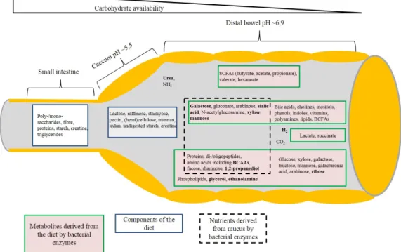
Since nutrient sources change along the GIT, the composition of the microbiota is similarly sub- ject to change. Besides amino acids, mono- and disaccharides are available in the small intestine where they support the growth of Proteobacteria (especiallyEnterobacteriaceae) andLactoba- cillales. In the caecum and large bowel complex carbohydrates are predominant, which are diet- or host-derived. These nutrients are indigestible for the host and bacteria such asE. coli. Thus, the microbiota changes in favour of bacteria, likeBacteroidesandClostridialeswhich are able to break-down more complex polysaccharides. These more complex polysaccharides also include the glycan structures of mucins (Macfarlaneet al., 2005; Johanssonet al., 2011; Kamadaet al., 2013).
In the proximal to distal course of the GIT, the availability of easy fermentable carbohydrates be- comes scarce. Thus, in the large bowel dietary oligo- and polysaccharides, sugar alcohols such as sorbitol and xylol, proteins and peptides, mucins and extruded epithelial cells all constitute avail- able nutrient sources for microorganisms (Cummings and Macfarlane, 1991; Macfarlaneet al., 1998). Microbial breakdown and fermentation of the nutrient sources referred to above lead to the production of the SCFA propionate, acetate and butyrate, H2gas, NH3, amines and phenols, for example. In turn, the SCFA serve as an energy source for the host, and are almost exclusively absorbed by the host epithelium (Cummings and Macfarlane, 1991; Macfarlaneet al., 2005; Mac- farlane and Macfarlane, 2006).
In sum, the microbiota and its enormous enzymatic capability to access and metabolise otherwise indigestible nutrient sources is beneficial to the host, as it is provided with, for instance, vitamins and SCFA (Cummings and Macfarlane, 1991; Nicholsonet al., 2012; Hooperet al., 2002). How- ever, microbiota-released monomers support the growth of many intestinal bacteria (Changet al., 2004), including that of pathogenic intruders.
The microbiota not only provides nutrients, but is also known to train the mucosal immune sys- tem, which in turn shapes the composition of the microbiota (Sekirovet al., 2010; Nicholsonet al., 2012). Further, it can actively contribute to protecting against colonisation of the gut by patho- genic bacteria (Stecher and Hardt, 2011). Stecher and Hardt (2011) describe three strategies, by which the microbiota can avoid niche occupation by enteric pathogens. 1) The direct inhibition of the pathogens by consuming available oxygen and releasing inhibitory metabolites such as SCFA or bacteriocins. 2) The stimulation of the host’s immune defence. In this situation the microbi- ota releases lipopolysaccharides (LPS) or peptidoglycan, which serve as microbial patterns. Host cells recognise these microbial patterns, which results in enhanced mucus secretion and the re- lease of antimicrobial peptides such as defensins and immunoglobulin A (IgA). 3) The depletion of favoured nutrients (Stecher and Hardt, 2011). ‘Freter’s nutrient-niche hypothesis’ claims that a great variety of growth-supporting (microbiota derived) substrates are present in the gut. However, it is suggested that the concentration of each substrate is very low and thus growth limiting. The ability of a certain bacterial species to colonise and persist within the GIT is determined by its ability to use one or a small number of nutrients more efficiently than its competitors (Freteret al., 1983; Stecher and Hardt, 2011).
1.3.2 Metabolic adaptation of enteropathogens
Pathogenic bacteria reach specific regions or tissues within the human or animal body, where they replicate and subsequently cause infection. Thus, enteric pathogens are able to adapt to the changing environments they encounter during their life cycle in order to survive, and occupy a niche within the GIT. Extracellular pathogens are frequently exposed to changing environmental conditions, whereas intracellular pathogens inhabit more stable environments (Eisenreich et al., 2010). In mammalian cells catabolic reactions such as the glycolysis and the pentose-phosphate pathway (PPP), take place in the cytosol, and the tricarboxylic-acid (TCA) cycle, theβ-oxidation of lipids, and the glutaminolysis in the mitochondria. The inner membrane of the mitochondria is also home to the cell’s energy metabolism, as the enzyme complexes of the respiratory chain are situated there. Biosynthesis of nucleosides, amino acids, fatty acids or lipids, as well as gluco- neogenesis primarily occurs in the cytosol. Hence, there is a frequent exchange of metabolites between the two compartments. Metabolism is further supported by vesicles such as peroxisomes, lysosomes, and secretory vesicles (Eisenreichet al., 2015).
Pathogenic bacteria, likeListeria monocytogenes, are adapted to replicate within the host-cytosol.
Thus, they gain direct access to the metabolites present there. Other pathogens such asS.Typh- imurium proliferate within a membrane compartment, the SCV. The membrane compartment sep- arates the pathogen from the host-cytosol. Transport systems are required to facilitate the trans- location of nutrients across the membrane barrier (Abu Kwaik and Bumann, 2015; Fuchset al., 2012; Eisenreich et al., 2015). Successful intracellular pathogens have adapted their metabol- ism to that of the host cell. Parallel utilisation of different carbon sources, like glycerol, vari- ous carbohydrates, amino acids, fatty acids and nucleosides seems to be a common strategy of intracellular pathogens, albeit each pathogen has developed individual strategies to survive and proliferate within its host cell (Steebet al., 2013; Eisenreichet al., 2015). L. monocytogenesis capable of the uptake and metabolism of amino acids, C3-, C4-, and C5-carbon substrates, and phosphorylated hexoses. In mineral media growth only occurs if arginine, the branched-chain amino acids (BCAA) leucine, valine, isoleucine, and the sulphur-containing amino acids cysteine and methionine are provided (Premaratne et al., 1991), although all genes required for the syn- thesis of the amino acids are present in theL. monocytogenesgenome (Glaseret al., 2001). Also the vitamins riboflavin, biotin, thiamine and thiotic acid have to be added to the mineral medium, as these cannot be synthesized by the pathogen (Glaseret al., 2001). During intracellular prolifer- ation, glycerol and glucose-6-P seem to be preferred carbon sources forL. monocytogenes(Eylert et al., 2008; Chatterjeeet al., 2006; Eisenreichet al., 2015).
S.Typhimurium is able to use more than 80 different carbon sources, as well as multiple electron acceptors for aerobic and anaerobic respiration, allowing the pathogen to adapt to varying envir- onmental conditions (Bumann, 2009; Gutnicket al., 1969). In infection studies in mice, Becker et al.(2006) found that metabolism is one of the most important activities ofSalmonelladuring infection. The authors state that about half of the proteins detected in proteome studies during infection were involved in metabolism (Beckeret al., 2006; Bumann, 2009, 2010). This result was surprising, as so far, metabolic enzymes have not been considered as virulence factors (Bumann, 2009). The development of vaccines or antimicrobial drugs, targeting metabolic enzymes ofSal-
monellais hampered by the robustness of theSalmonellametabolism (Beckeret al., 2006). Owing to metabolic redundancy, the pathogen can compensate the loss of a certain metabolic pathway by another, which yields the same metabolites or intermediate products (Beckeret al., 2006; Bumann, 2009, 2010).
S.Typhimurium harbours all genes required for the central metabolic pathways such as glycolysis, the Entner-Doudoroff pathway (ED), the pentose phosphate pathway (PPP), the TCA-cycle, the biosynthesis of all amino acids, vitamins and co-factors, nucleotides, fatty acids and lipids, and the main anaplerotic reactions such as the glyoxylate shunt, malic enzyme, phosphoenolpyruvate (PEP) carboxy kinase and PEP carboxylase (Götzet al., 2010; Eisenreichet al., 2015). The patho- gen is able to utilise C2, C3-, C4-, and C5-substrates as carbon sources, just as glycerol, fatty acids, pyruvate, lactate and, for example, fumarate (a C4-dicarboxylate) can be metabolised under certain growth conditions within mammalian host cells (Götzet al., 2010; Steebet al., 2013).
WhenS. Typhimurium replicates in HeLa cells or murine macrophage cell lines, glycolysis and ED pathway are upregulated, suggesting glucose (D-Glc) or C6-sugars as the main carbon source (Götzet al., 2010; Bowdenet al., 2009; Erikssonet al., 2003). While the TCA-cycle was found to be repressed in murine macrophages and HeLa cells (Götz et al., 2010), intracellular prolif- eration in systemically infected mice depends on a functional TCA-cycle (Becker et al., 2006;
Tchawa Yimga et al., 2006). In the SCV of Caco-2 cells, S. Typhimurium primarily feeds on D-Glc, but not on glucose-6-P. Mutant strains impaired in D-Glc uptake (∆ptsGand∆manXYZ) were found to incorporate less labelled (13C) C-atoms into amino acids than the wild type. Bac- teria disabled in glucose-6-P uptake (∆uhpT) did not show differential 13C-incorporation into amino acids compared to the wild type (Götzet al., 2010). Where D-Glc cannot be taken up by theSalmonellamutant strain, it uses host-derived13C3-substrates such as glycerol, pyruvate, or lactate forde novosynthesis of amino acids. Additionally, the uptake of host-derived amino acids is enhanced in the mutant strains, leading to direct incorporation of the latter into proteins (Götz et al., 2010). Thus, it can be concluded that amino acids are synthesisedde novo if rich carbon sources such as D-Glc are available while amino acid uptake from the host is increased where the bacteria feed on less energy-rich substrates (Götzet al., 2010).
During systemic infection in mice,S.Typhimurium uses at least 31 different organic and 13 in- organic host-derived carbon and energy sources in parallel (Steeb et al., 2013). As the single compounds are only available in small amounts, none could support growth as a single source of carbon and energy. Among the simultaneously utilised nutrient sources are glycerol, various hexoses and pentoses, sialic acid, fatty acids, nucleosides, and aerobic and anaerobic electron acceptors (Steebet al., 2013; Eisenreich et al., 2015; Becker et al., 2006). Glycerol and fatty acids might be derived from lipid oxidation (Steebet al., 2013; Abu Kwaik and Bumann, 2015).
Mutants compromised in aromatic amino acid, methionine or pyrimidine synthesis show virulence defects in infected animals (Leung and Finlay, 1991; McFarland and Stocker, 1987; Fieldset al., 1986). For S. Typhimurium, residing within the SCV in murine macrophages, genes encoding transport proteins for gluconate, glucuronate or galacturonate, and galactonate were found to be upregulated. Hence, the authors suggest that these carbohydrates serve as the main carbon sources for the intracellular pathogen (Erikssonet al., 2003).
After the successful invasion of a pathogen, it affects host cell metabolism in three ways: 1) The
pathogen depletes metabolites from the cytoplasm or incoming vesicles after endocytosis (Steeb et al., 2013; Eisenreichet al., 2015). 2) The pathogen secretes effector molecules that enhance the carbon metabolism of the host cell while simultaneously preventing apoptosis of the latter (Knodleret al., 2005; Yinet al., 2012). 3) Other effector molecules secreted by the pathogen trig- ger inducible nitric oxide synthase (iNOS) synthesis by the host cell, which leads to the conversion of nitric oxide (NO) to nitrate (Speeset al., 2013). Nitrate can serve as an electron acceptor for anaerobic respiration ofS.Typhimurium. Anaerobic respiration enables the pathogen to respire non-fermentable metabolites or fermentation end products. As anaerobic bacteria of the microbi- ota rely on fermentation, the pathogen gains access to a nutrient niche, which is not exploited by the microbiota (Lopezet al., 2012; Thiennimitret al., 2011; Winteret al., 2013).
It is assumed that some of the gene clusters enabling the utilisation of alternative carbon and en- ergy sources, like for example ethanolamine (eutgenes), 1,2-propanediol (1,2-PD) (pdugenes) or myo-inositol (iolgenes), have been acquired by horizontal gene transfer since the G/C-content of these ‘metabolic islands’ differs from that of the genome (Dobrindtet al., 2004; Rohmeret al., 2011). The importance of the ability to use alternative substrates not consumed by members of the microbiota remains obscure. In general, information about available and utilised substrates during proliferation ofS.Typhimurium in the lumen of the GIT is scarce (Fuchset al., 2012; Abu Kwaik and Bumann, 2013; Staib and Fuchs, 2014). Although most metabolic enzymes have been found to be dispensable for virulence, the expression of genes involved in cofactor biosynthesis, anaero- bic energy metabolism and the degradation of diverse nutrients is upregulated during infection.
Thus, metabolism seems to be directly linked to virulence (Beckeret al., 2006; Bumann, 2009;
Fuchset al., 2012).
1.3.3 Subversion of defence mechanisms byS.Typhimurium
As mentioned above, bacteria of the microbiota stimulate the host’s immune defence, secrete inhi- bitory metabolites, and deplete oxygen and nutrients (Stecher and Hardt, 2011). S.Typhimurium would not be a successful pathogen, if it were susceptible to all of the defence mechanisms en- countered in the GIT. In contrast to the microbiota, the pathogen is well equipped with genes whose products counteract some of the defence mechanisms. Additionally, the pathogen is adap- ted to the conditions in the inflamed intestine (Stecheret al., 2007; Santoset al., 2009) and even triggers inflammation (Knodler et al., 2010) (Fig. 1.1). The chemokine-like activity of antimi- crobial peptides recruits leukocytes (e.g. neutrophils). Thereby, colonisation resistance and host defence are linked (Peschel, 2002). Direct host-pathogen interaction, for example upon successful penetration of the mucus layer, and host cell invasion byS. Typhimurium, leads to the secretion of cytokines (Santoset al., 2009). Two lines of innate immune responses are activated by the se- cretion of cytokines. 1) The recruitment of neutrophils, which eliminate or kill bacteria, can also cause tissue damage. This contributes to the development of diarrhoea and inflammation. These lead to clearance of the gut, a reduction of bacteria (members of the microbiota and pathogens), and a decrease in inhibitory metabolites (Santoset al., 2009; Stecheret al., 2007, 2008). Using a mouse enteritis model, a recent study demonstrated that the bacteria of the microbiota are severely affected by the immune responses triggered duringSalmonellainfection (Deatherage Kaiseret al.,
2013). Ten days after infection,Bacteroideteswere nearly eliminated from the gut. The alteration of the microbiota, subsequently caused a change of the metabolite composition in the gut lumen.
During inflammation higher concentrations of sugar moieties, like lactose, raffinose, melibiose and galactitol accumulated, which are usually consumed by members of the microbiota. The content of high mannose and fucosylated glycans in the gut was also found to have increased upon infec- tion. Strikingly, the genes required for L-fucose (L-Fuc) degradation were found to be expressed inS.Typhimurium, residing in the inflamed intestine of mice. This suggests thatS.Typhimurium metabolises L-Fuc, alongside D-Glc, galactose, maltose and mannitol in the microbiota-depleted gut (Deatherage Kaiseret al., 2013). Furthermore, the recruitment of neutrophils and the expres- sion of iNOS results in NO-radical and reactive oxygen species (ROS) formation. The reaction between NO radicals and ROS yields peroxynitrite, which in consequence makes the generation of nitrate or S- and N-oxides possible (Winteret al., 2013; Speeset al., 2013).Enterobacteriaceae can use nitrate as well as S- and N-oxides as electron acceptors for anaerobic respiration (Winter et al., 2013). Hence, commensalE. colistrains gain a growth advantage over other members of the microbiota during inflammation because most of the latter rely on the fermentation of com- plex carbohydrates and amino acids rather than anaerobic respiration (Winteret al., 2013; Bliska and van der Velden, 2012). Only during intestinal inflammation does nitrate respiration become possible forS.Typhimurium, upon which it provides the pathogen with a growth benefit in the lu- men of the gut (Rivera-Chavezet al., 2013; Lopezet al., 2012; Bliska and van der Velden, 2012).
Nitrate reductases are also present inVibrio cholerae, Campylobacter jejuni, Bacillus cereus, En- terococcus faecalis, Clostridium perfringens and Shigella spp. (Lopez et al., 2015; Staib and Fuchs, 2014), which might confer a possible growth advantage during infection to those bacteria as well. 2) The secretion of antimicrobial proteins and peptides leads to enhanced mucus secretion, the expression of iNOS, and the secretion of lipocalin-2. Enhanced mucus secretion increases the availability of high-energy nutrients derived from mucins (Stecheret al., 2008; Corazziari, 2009).
Lipocalin-2 prevents bacterial iron-acquisition by binding the bacterial iron-chelator enterobactin (Nairzet al., 2007; Cairo et al., 2011; Santoset al., 2009; Raffatellu and Bäumler, 2010). En- terobactin is commonly found inE. coliandS. enterica(Raffatellu and Bäumler, 2010). In contrast toE. coli,S.Typhimurium harbours the alternative iron-chelator salmochelin (a glycosylated form of enterobactin), which is not bound by lipocalin-2 (Raffatellu and Bäumler, 2010; Stecher and Hardt, 2011). Further,S.Typhimurium was found to be resistant to many antimicrobial peptides and defensins secreted by members of the microbiota and the host (Peschel, 2002; Baderet al., 2005; Shiet al., 2004). Upon infection of the intestinal epithelium, enhanced secretion of C-type lectins, such as RegIIIγ (regenerating islet-derived) or RegIIIβ in mice or HIP/PAP (hepatocarci- noma-intestine-pancreas/ pancreatitis associated protein) in humans , are observed (Cash et al., 2006; Stelteret al., 2011). Peptidoglycan carbohydrate seems to be the target of the lectins (Cash et al., 2006; Stelteret al., 2011). While RegIIIγ is only bactericidal for Gram+ bacteria (Cash et al., 2006), RegIIIβkilled both Gram+and Gram−bacteria. However, RegIIIβdoes not seem to be fatal forS.Typhimurium, rather it interferes with the intestinal translocation ofS.Enteritidis in mice (Stelteret al., 2011; van Amptinget al., 2012). In comparison to mock-infected mice, oral RegIIIβtreatment resulted in increased luminal colonisation byS.Typhimurium. Survival of the pathogen could be a result of the stiff lipopolysaccharide (LPS) layer expressed on the bacterial
surface. Thus, the access of RegIIIβ to the peptidoglycan is limited, which might attenuate its antimicrobial effect onS.Typhimurium (Stelteret al., 2011).
To reach epithelial cells, chemotaxis, which enables the pathogen to move towards attractants or away from repellents, plays a crucial role inSalmonellainfections.S.Typhimurium follows a D- galactose gradient towards the epithelium (Stecheret al., 2008). Flagella and fimbriae expressed by the pathogen, are used to swim in the direction of the D-galactose gradient and thus the epi- thelium, to propel the pathogen into the mucus layers, and to attach to terminalα-(1,2)-fucose moieties of mucins and the surface of epithelial cells (Chessaet al., 2009; Stecher et al., 2008).
Furthermore, flagellin is recognised by host epithelial cells as a pathogen associated molecular pat- tern (PAMP). Flagellin binds to Toll-like receptor (TLR) 5, inducing proinflammatory responses (Stecheret al., 2004).
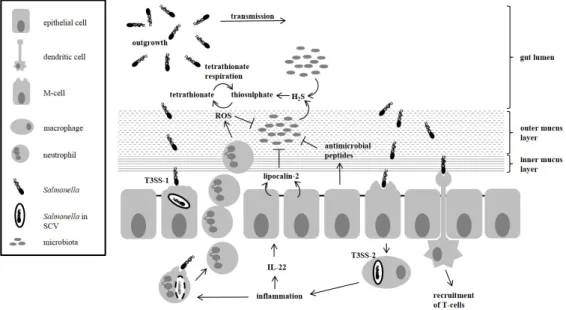
Figure 1.1:Subversion of host defence byS.Typhimurium.
The figure shows how S. Typhimurium manages to outgrow the microbiota. As de- scribed above, the pathogen is resistant to a number of antimicrobial peptides, ROS and the host-released lipocalin-2. By inducing inflammation, the pathogen creates en- vironmental conditions, which are unfavourable for bacteria of the microbiota, but be- neficial for itself. Transmission of the pathogen follows due to inflammation-induced diarrhoea. [Figure based on Stecher and Hardt (2011); Thiennimitret al.(2012)].
1.3.4 Exploitation of exceptional nutrient sources
S.Typhimurium harbours the gene clusters responsible for ethanolamine,myo-inositol, sialic acid, and 1,2-PD degradation, which either differ in G/C-content to the overall genome or are absent in closely related species. Thus, it is assumed that these metabolic traits have been acquired by horizontal gene transfer (Almagro-Moreno and Boyd, 2009; Dobrindtet al., 2004; Rohmeret al., 2011).
Sialic acids are commonly found as part of mucin- and cellular glycoconjugates, and are involved in cell-cell recognition (Varki, 1993; Almagro-Moreno and Boyd, 2009). Proliferation ofS.Typhi-
murium was impaired in gnotobiotic mice with a lack of free sialic acid in their gut, a result of the co-colonisation with a sialidase-deficientBacteroides thetaiotaomicronstrain (Nget al., 2013).
Myo-inositol, a polyol, is a component of the lipid phosphatidylinositol found in eukaryotic cell membranes. Its phosphorylated form phytate (inositol hexakisphosphate) occurs in plants and soil (Reddy, 2001; Rodrıguez and Fraga, 1999). The iol genes are thought to contribute to the vir- ulence ofS.Typhimurium in mice, pigs, chicken and calves (Carnellet al., 2007; Chaudhuriet al., 2009, 2013; Lawleyet al., 2006).
Ethanolamine is also a building block of a phospholipid, namely phosphatidylethanolamine, which is abundantly present in mammalian and bacterial membranes (Randleet al., 1969).
A study by Tsoyet al.(2009) revealed the presence of theeutgenes in the genomes of 84 bacteria belonging to different phyla (Acidobacteria, Actinobacteria, Bacteroidetes, Chlorophlexi, Firmi- cutes, Fusobacteria, andProteobacteria). Theeutgenes are required for ethanolamine utilisation.
The eutoperon was found to exist in an extended (17 genes) version and various truncated ver- sions. The long version, which is found in Enterobacteriaceae, contains the geneseutKLMNS (Tsoy et al., 2009) that encode proteins, forming a carboxysome-like microcompartment. The microcompartment is thought to entrap enzymes and substrates optimising enzymatic reactions by spatial limitation of the reaction partners, and toxic, volatile intermediates to protect the cell from damage (Kofoidet al., 1999; Penrod and Roth, 2006; Garsin, 2010).
The genes of theeutlocus are organised consecutively, in counter-clockwise orientation on theS.
Typhimurium genome. In L. monocytogenestheeut genes are still found in the same region of the genome, but are interrupted by genes required for 1,2-PD degradation (pdu) and vitamin B12 synthesis (cob/cbi) (Buchrieseret al., 2003), whereas inC. perfringenstheeut, cob/cbi andpdu genes are scattered over the chromosome (Fig. 1.2).
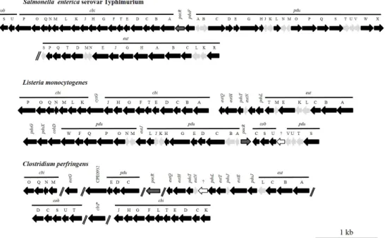
Figure 1.2:Genetic organisation ofeut, pduandcob/cbigenes in enteropathogens
Genes required for the degradation of ethanolamine, 1,2-PD and the synthesis of vit- amin B12inS. enterica, L. monocytogenesandC. perfringensare shown. Black arrows indicate genes encoding proteins of enzymatic function. Proteins forming the shell of the microcompartment are depicted in grey and unknown genes in white. The gene encoding the regulator PocR is indicated in grey surrounded by a solid black line.
Gene functions can be obtained fromTab. 5.11in the Appendix. This graph has been published in Staib and Fuchs (2014).
The transcriptional regulator EutR is expressed constitutively in small amounts (Sheppard and Roth, 1994; Roof and Roth, 1992). Only in the presence of vitamin B12and ethanolamine does EutR induce the expression of theeutoperon. The main promoter of theeutoperon is located in front ofeutS. Induction of the gene expression from this promoter results in a polycistronic mRNA transcript, which harbours the genetic information of the geneseutS-K. The global regulator CRP (cAMP receptor protein), which senses a shortage in carbon and energy sources, was found to be required foreut-gene expression (Blackwell and Turner, 1978; Roof and Roth, 1992; Kofoidet al., 1999).
Vitamin B12 is crucial for ethanolamine degradation, since it is necessary as a cofactor for eth- anolamine ammonia lyase (EutBC), and to induce eut-gene expression by EutR (Scarlett and Turner, 1976; Roof and Roth, 1992). Only four enzymes that use vitamin B12 as a cofactor are known. These are tetrahydropteroylglutamate-homocysteine methyltransferase (metH), epoxy- queuosine reductase (yjeS), ethanolamine ammonia-lyase (eutABC) and the propanediol dehyd- ratase (pduCDE) (Jeter, 1990).
Ethanolamine ammonia lyase is the first enzyme of the ethanolamine degradation pathway. The catalysed reaction yields acetaldehyde and ammonia (Fig. 1.3). Acetaldehyde can be reduced to ethanol by EutG, and ethanol is either secreted or can be metabolised to acetyl-CoA by EutE. Un- der aerobic growth conditions, acetyl-CoA can enter the central metabolism of the cell, whereas it
is converted to acetate via acetyl-phosphate under anaerobic conditions. Acetate is consequently secreted. Thus, under anaerobic conditions and the absence of a suitable electron acceptor al- lowing anaerobic respiration, ethanolamine can only serve as a nitrogen, not carbon source (Roth et al., 1996).
De novosynthesis of the cofactor vitamin B12only occurs under anaerobic growth conditions in S. Typhimurium. If the bacteria are grown aerobically with ethanolamine as the sole source of carbon and nitrogen, vitamin B12 or a precursor such as cyanocobalamin has to be provided in the medium (Jeter et al., 1984; Escalante-Semerena and Roth, 1987). To use ethanolamine as a carbon and energy source under anaerobic conditions more efficiently, the compound needs to be oxidised by anaerobic respiration. Only tetrathionate, not fumarate or nitrate, supports anaerobic respiration of ethanolamine (Rothet al., 1996).
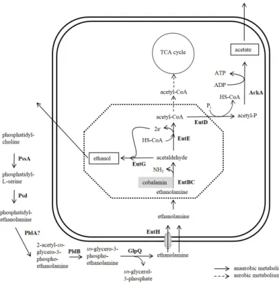
Figure 1.3:Ethanolamine metabolism ofS.Typhimurium.
The figure shows the metabolism of ethanolamine inS.Typhimurium. Enzymes are printed bold. Dashed arrows indicate aerobic degradation and solid lines show the an- aerobic metabolism of ethanolamine in the absence of tetrathionate as a terminal elec- tron acceptor. The microcompartment encoded by theeutoperon is indicated as dashed octagon. PssA: phosphatidyl serine synthase; Psd: phosphatidyl serine decarboxylase;
PldA: phospholipase A1; PldB: lysophospholipase; GlpQ: glycerophosphoryl diester phosphodiesterase; EutH: ethanolamine permease; EutBC: ethanolamine ammonia ly- ase; EutG: alcohol dehydrogenase; EutE: aldehyde oxidoreductase; EutD: phospho- transacylase; AckA: acetate kinase; . [Figure modified Garsin (2010); Staib and Fuchs (2014)]
1.3.4.1 Tetrathionate respiration inS.Typhimurium
The ttr genes, which are located on SPI-2 (Henselet al., 1999), enableS. Typhimurium to per- form tetrathionate respiration under anaerobic or microaerobic conditions (Henselet al., 1999).
Analyses of the G/C-content of SPI-2 revealed that the genomic region shows a mosaic structure and is composed of three regions, which might be of different origin (Henselet al., 1999). The first region embraces the SPI-2 virulence genes (ssaU-ssrB), the second includes the genes re- quired for tetrathionate respiration (ttrRSandttrBCA), and the third harbours genes with a thus far uncharacterised function (Henselet al., 1999; Hensel, 2000). Studies in mice have led to the assumption that only genes of the first region are relevant for systemic pathogenesis ofS.Typhi- murium (Henselet al., 1999; Hensel, 2000). Hence, tetrathionate respiration does not contribute to S. Typhimurium virulence, but is thought to be important when the bacteria are exposed to tetrathionate-containing environments (Henselet al., 1999).
The genesttrBCAencode the tetrathionate reductase, which is thought to be anchored in the cyto- plasmic membrane via TtrC (Hensel et al., 1999). Transcription of the tetrathionate reductase depends on TtrR and TtrS, forming a two component regulatory system encoded byttrRSwithin thettrgene cluster. During tetrathionate respiration, electrons are transferred from NADH + H+ through the cellular quinone pool to tetrathionate as the final electron acceptor (Rivera-Chavez et al., 2013), which is then reduced to two molecules of thiosulphate (Henselet al., 1999). The global regulator Fnr (fumarate-nitrate reduction regulatory protein) (Henselet al., 1999), but not ArcA or CRP, affectsttrexpression since anaerobic conditions are required forttrgene expression and tetrathionate respiration. Nitrate was found to slightly inhibitttrexpression, via a thus far un- known mechanism (Price-Carteret al., 2001), because nitrate is an energetically more favourable electron acceptor (nitrate-nitrite E0 = 433 mV) than tetrathionate (tetrathionate-thiosulphate E0 = 170 mV) (Lopezet al., 2012; Bliska and van der Velden, 2012).
Growth defects ofS.Typhimurium mutants unable to utilise ethanolamine were observed in bovine raw milk and egg yolk (Srikumar and Fuchs, 2011). AsS.Typhimurium is a food-borne pathogen, a contribution of ethanolamine degradation to the replication in food or the environment is also plausible. In the same study, significantly reduced proliferation rates were documented for theS.
Typhimurium mutants 14028∆eutRand 14028∆pocRin theCaenorhabditis elegansmodel. The 14028∆eutRmutant cannot use ethanolamine, and the 14028∆pocRis compromised in synthes- ising vitamin B12and 1,2-PD degradation. The degradation pathways of ethanolamine and 1,2-PD inS.Typhimurium share the following features: 1) A polyhedral microcompartment is formed to retain volatile intermediates. 2) Vitamin B12 is required as a cofactor for the central enzyme to convert the amine or diol to an aldehyde. 3) Tetrathionate is the only electron acceptor allowing anaerobic respiration of the respective substrate (Scarlett and Turner, 1976; Rothet al., 1996; Ko- foidet al., 1999; Bobiket al., 1999). Furthermore, thepdugenes were shown to be relevant for S.Typhimurium replication in macrophages (Klumpp and Fuchs, 2007). A further indication of thein vivorelevance of ethanolamine and 1,2-PD degradation inS.Typhimurium was published earlier (Stojiljkovic et al., 1995; Conneret al., 1998). Virulence defects were identified when S. Typhimurium strains deficient for ethanolamine (Stojiljkovicet al., 1995) or 1,2-PD (Conner et al., 1998) utilisation were orally administered to mice.
1.4 Research objective
Can the human gut be seen as a land of milk and honey for bacteria, or rather as a nutritional wasteland? At least for pathogens, considering the high density of bacteria in the human gut competing for food, the ability to use a variety of carbon and energy sources clearly cannot be disadvantageous. Host-derived sugars are released from complex structures such as mucins, due to the activity of enzymes secreted by members of the microbiota. These sugars not only serve as nutrients for bacteria of the microbiota, indeed they are also available for pathogens (Cummings and Macfarlane, 1991; Keeney and Finlay, 2013).
The genes for 1,2-PD degradation were previously found to be induced in murine and human cell cultures, to contribute to the replication ofS.Typhimurium in macrophages, and to be relevant for infection in nematodes and mice (Heithoffet al., 1999; Klumpp and Fuchs, 2007; Conneret al., 1998; Srikumar and Fuchs, 2011). Some details about the regulation of thepdugene cluster have been published previously (Rondon and Escalante-Semerena, 1992; Rondonet al., 1995; Bobik et al., 1992; Chenet al., 1994, 1995), but the concentration of 1,2-PD required for the induction of thepdugene expression remains unknown thus far. Whether the consecutive, clockwise oriented pdugenes are expressed as a polycistronic mRNA from a single promoter, as it is the case for the eutgenes, has not yet been published. In the last publication concerning the promoter region of thepdugene cluster, the authors suggest a single promoter for the clockwise-orientated genes of thepdugene cluster, although at that time onlypduA,pduBandpduC had been identified (Chen et al., 1995). Thus, whether additional downstream promoters exist was not known at the time.
It is assumed that 1,2-PD is available to enteropathogens in the gut, because 1,2-PD is the ferment- ation end product of bacterial growth with L-Fuc or L-Rha (Danielet al., 1998). At least L-Fuc is constantly available in the gut, as it is a constituent of mucin glycoconjugates (Muraoka and Zhang, 2011). The ability ofS. Typhimurium to break down L-Fuc and yield 1,2-PD was demonstrated (Badiaet al., 1985), and the interconnection between tetrathionate respiration, ethanolamine and 1,2-PD metabolism in the inflamed intestine has since become more concrete (Winteret al., 2010;
Bobiket al., 1997).
At first, the thesis focused on the collection of data concerning metabolites, which are available to enteropathogens in the human gut. In the following, metabolic properties of selected entero- pathogens were compared, with a special focus on metabolic pathways, required for 1,2-PD and L-Fuc degradation. Afterwards, the distribution of thepduandfucgene clusters amongsalmonel- laewas investigated in order to ascertain, whether 1,2-PD degradation is a general or an exclusive property. InS.Typhimurium strain 14028 genetic determinants of thepdugene expression were considered, which included the search for possible promoter structures within thepdugene cluster, and whether the pdugenes are transcribed as a single polycistronic mRNA or as multiple tran- scripts. Further, the minimal concentration of 1,2-PD, sufficient to inducepdugene expression, was elucidated, and whether thepdugene expression is repressed by the simultaneous presence of other carbon sources. The importance of central enzymes within the degradation pathways of 1,2- PD and L-Fuc was investigated, and whetherS.Typhimurium is able to compensate for the loss of these genes. Further examinations were conducted, which focused on the relevance of 1,2-PD de- gradation in more complex substrates, and the interaction ofS.Typhimurium and mucus-secreting cell lines.