Investigation of their Biological Properties
Zur Erlangung des akademischen Grades eines Doktors der Naturwissenschaften
vom Fachbereich Chemie der Universität Dortmund
angenommen
DISSERTATION
von
Diplom-Chemikerin
Catherine P. Katzka
aus Mountain View (Kalifornien)
1. Gutachter: Prof. Dr. Herbert Waldmann 2. Gutachter: Prof. Dr. Roger Goody
Tag der mündlichen Prüfung: 29.06.06
Die vorliegende Arbeit wurde unter der Betreuung von Prof. Dr. H. Waldmann in der
Zeit vom Februar 2002 am Institut für Organische Chemie der Universität Dortmund
sowie am Max-Planck-Institut für molekulare Physiologie bis Mai 2006 angefertigt.
Eidesstattliche Erklärung
Hiermit erkläre ich an Eides Statt, dass ich diese Arbeit selbständig und nur mit den angegebenen Hilfsmitteln angefertigt habe.
Dortmund, Mai 2006
______________________________________________________________
To my family
______________________________________________________________
Table of Contents
TABLE OF CONTENTS V
1 PREFACE 1
2 INTRODUCTION 2
2.1 L
INKINGG
ENES TOF
UNCTION2
2.1.1 Chemical Genetics 2
2.1.2 Application Areas for Chemical Genetics 5
2.1.3 Library Design for Chemical Genetics 13
2.2 T
ETRAMICA
CIDS17
2.2.1 Tetramic Acid-Containing Natural Products 17
2.2.2 Biosynthesis of Tetramic Acids 19
2.2.3 Synthetic Routes to Tetramic Acids 20
3 AIM OF THE DISSERTATION 23
4 RESULTS AND DISCUSSION 25
4.1 S
YNTHESIS OF TETRAMIC ACID LIBRARIES25
4.1.1 Synthetic plan 25
4.1.2 Implementation and Optimization of the Synthesis 27
4.2 L
ACCARINA
NALOGS
YNTHESIS39
4.2.1 Introduction 39
4.2.2 Synthetic Plan 39
4.2.3 Towards the Synthesis of a Laccarin Analog 40 4.3 S
YNTHESIS OFB
UILDINGB
LOCKS FORI
NDOLE-B
ASEDT
ETRAMICA
CIDS42
4.3.1 Introduction 42
4.3.2 Synthetic Strategy 42
4.3.3 Indole Synthesis 43
4.3.4 Tryptophan Synthesis 47
4.4 I
NVESTIGATION OF THEB
IOLOGICALP
ROPERTIES OF THET
ETRAMICA
CIDL
IBRARY50 4.4.1 Targeting the Ras Signaling Pathway 50
4.4.2 Targeting Phosphatases 64
4.4.3 Targeting Bacteria 69
5 CONCLUSION 72
6 MATERIALS AND METHODS 77
6.1 S
YNTHETICM
ATERIALS ANDM
ETHODS77
6.1.1 General 77
6.1.2 Compounds from Chapter 4.1 79
______________________________________________________________
6.1.3 Compounds from Chapter 4.2 94
6.1.4 Compounds from Chapter 4.3 95
6.2 B
IOCHEMICAL ANDB
IOLOGICALM
ATERIALS ANDM
ETHODS106
6.2.1 Materials 106
6.2.2 Methods 109
7 REFERENCES 114
APPENDIX A: ABBREVIATIONS 122
APPENDIX B: BIOLOGICAL PROPERTIES OF TETRAMIC ACIDS 124
APPENDIX C: TETRAMIC ACID
1H NMR SPECTRA 128
______________________________________________________________
1 Preface
When Paul Ehrlich discovered that methylene blue selectively stains neuronal cells, he revealed that small molecules have specific biological targets.
[1]Since then, small molecules have become an increasingly important tool for studying biological systems. The use of such chemical probes instead of genetic alterations to establish a link between gene-products and their functions is called chemical genetics. Chemical genetics relies on a collection of compounds that are able to modulate different proteins. Natural products are known to bind to proteins and may therefore serve as a fruitful source of inspiration for the synthesis of compound collections.
The tetramic acid natural product family is based on the 2,4-pyrrolidinedione core 1 (Figure 1) and is documented to affect a wide variety of biological targets.
[2]The tetramic acid core is therefore an interesting scaffold for the design of a library of chemical probes to be used in chemical genetics.
HN O
O R
1R
21 2 4 3 5
1
Figure 1: Structure of the 2,4-pyrrolidinedione (tetramic acid) 1 scaffold.
The aim of this dissertation was to synthesize a library of compounds based on the tetramic acid scaffold and use them to probe cellular and biochemical systems.
In the first part of this dissertation, the concepts of chemical genetics will be
introduced and background will be provided to the tetramic acids as a natural product
family. The goals of the thesis will then be laid out. Finally, the results of the
synthetic and biological work will be presented and discussed.
______________________________________________________________
2 Introduction
2.1 Linking Genes to Function
In 2004, the sequencing of the human genome was completed.
[3]There were high hopes that this would accelerate the understanding of complex biological systems, thereby aiding in developing novel therapies for diseases. Alone though, the genetic information from the genome sequence is not enough to comprehend intricate biological mechanisms: the link between genes and their functions needs to be established. Modern genetic methods have made it possible to rapidly identify genes and their mutant alleles by simple database operations. Gene cloning and knockout techniques allow the overexpression or silencing of proteins in lower organisms such as fruit flies (Drosophila melanogaster), zebra fish (Danio rerio) and mice (Mus musculus) to produce observable phenotypical effects. Complementarily, human cell- based models can also be engineered using molecular biological methods. Although developments in genetics have advanced the understanding of biological processes, there are still some limitations: the study of essential genes is prevented because organisms with mutations in such genes are not viable. Furthermore, genetic approaches are not well suited to studying dynamical cell biological processes that occur on a time scale of minutes or seconds. A small molecule approach can complement gene-based methods.
2.1.1 Chemical Genetics
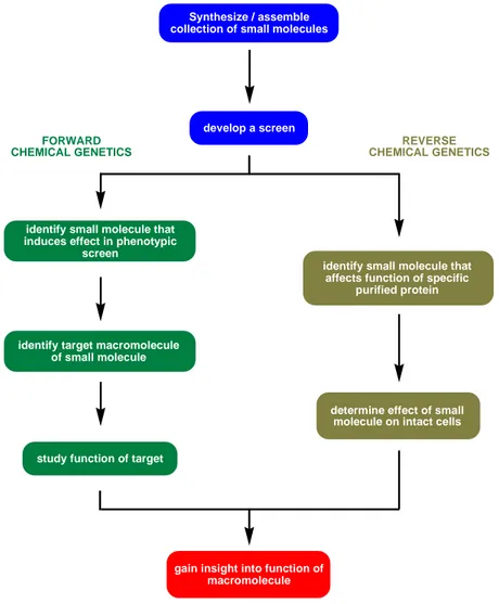
An alternative method to linking genes and proteins to their function and phenotypes
is termed chemical genetics, and uses small molecules to perturb protein networks of
biological systems.
[4]It is a multiple step approach that generally begins with the
assembly of a collection of small molecules, then with the screening of the
compounds in a developed assay and finally the identification of the modulated target
molecule. Like in classical genetics, this approach attempts to uncover the specific
macromolecules (usual proteins) that act as regulators of cellular processes. Their
functions are subsequently defined using protein biochemistry, molecular cell biology
and synthetic chemistry.
______________________________________________________________
Chemical genetics can be divided into two differing approaches: forward and reverse chemical genetics (Figure 2). These approaches will be briefly described in the following sections and will be illustrated with notable examples.
Synthesize / assemble collection of small molecules
develop a screen
identify small molecule that induces effect in phenotypic
screen
identify target macromolecule of small molecule
study function of target
identify small molecule that affects function of specific
purified protein
determine effect of small molecule on intact cells
gain insight into function of macromolecule FORWARD
CHEMICAL GENETICS REVERSE
CHEMICAL GENETICS
Figure 2: Summary of forward and reverse chemical genetics approach to understanding biological systems.
Forward chemical genetics involves the use of small molecules to screen for a desired phenotypical effect on the biological system under investigation. Once a small molecule has been identified, its molecular target needs to be revealed. This can be achieved by using molecular cell biology and protein biochemistry.
Mayer et al. used a combination of two phenotypical assays to screen a 16,320
member compound library for compounds that affect mitosis.
[5]One synthetic small
______________________________________________________________
molecule, monastrol (2), provoked the reorganization of the mitotic spindle (Figure 3).
Figure 3: (A) cell with normal mitotic bipolar spindle; (B) chemical structure of monastrol (2); (C) reorganization of mitotic spindle visualized by microscopy.
[5]The phenotype induced by monastrol had been observed before on inhibition of the mitotic kinesin protein Eg5 using anti-Eg5 antibodies.
[6;7]Biochemical investigations revealed that monastrol inhibits microtubule motility by binding Eg5.
[5]The effect of monastrol is reversible: washing the compound out allows cells to move out of mitotic arrest and complete mitosis normally. This property was used to study the function of Eg5. This protein is now an established cancer target.
[8]In reverse chemical genetics, a small molecule is used against a purified protein. This molecule is then used to “knockout” the protein in question on a cellular level.
This approach was used to screen a library of compounds to find a small molecule that binds and inactivates the protein MEK1, an enzyme whose activity is needed for cell division.
[9]A potent and selective MEK inhibitor PD184352 (3, Figure 4) was identified (50% inhibitory concentration, IC
5017 nM).
HN H
N O
O
F
F I
Cl
3
Figure 4: Chemical Structure of PD184352 (3).
Subsequent studies in vivo on mice with colon carcinomas of mouse and human origin demonstrated that tumor growth was inhibited by up to 80% upon treatment
HN NH
OH
O S
O
B
2
______________________________________________________________
with PD184352. The low toxicity, high potency and selectivity made this compound promising for the treatment of colon cancer.
Some advantages of chemical genetics over classical genetics are that temporal control is possible as small molecules can be added to the studied systems at any time point of the experiment. The effects are also reversible as the compounds can be removed metabolically or by washing. To achieve reversibility in a genetic system, conditional alleles need to be introduced; these are difficult to generate and control.
Another advantage is that small molecules have rapid effects as they are mostly diffusion limited. They can therefore be used to observe immediate effects.
Furthermore, they can be used to study critical genes in developmental stages:
whereas a cell knockout may not be viable, it may still possible to study the effects of a knockout gene product. However, the main disadvantage is that chemical genetics cannot be applied generally. Any gene, in principle, can be specifically manipulated by genetics; chemical genetics still needs selective small molecules. Moreover, the protein-targets still need to be uncovered, which at present is still a challenge.
The use of small molecules to study biological systems has had an impact on fields such as signaling,
[10;11]cell morphogenesis,
[12;13]and developmental biology.
[14]In this dissertation, the focus will lie on using small molecules to study the Ras signaling pathway, phosphatases and bacterial cell growth.
2.1.2 Application Areas for Chemical Genetics Ras Signaling Pathway
Normal cellular behavior is tightly regulated by complex signaling networks. The study of these signaling networks is important because dysfunction often leads to diseases such as cancer. One interesting example of signaling is that of the Ras/MAPK signaling pathway.
The Ras proteins belong to a family of small GTPases and play a key role in the signal
transduction from receptor tyrosine kinases to the cell nucleus for the regulation of
cell-cycle progression, cell division, survival and apoptosis.
[15;16]There are three
isoforms of Ras in mammals: N-Ras, K-Ras and H-Ras. Ras acts as a molecular
______________________________________________________________
switch by alternating from the inactive GDP-bound form to the active GTP-bound form (Figure 5).
Ras
Raf PI3K RalGDS PLCε
MEK AKT Ral Ca2+
cell-cycle progression transcription
survival transcription cytoskeletal signals
translation
transcription vesicle transport cell-cycle progression
calcium signaling
Ras
ON OFF
GTP
GEF GAP
Pi
Figure 5: Cycling of the Ras molecular switch and downstream effectors of Ras and their activities.
The intrinsic GTPase activity in vitro is slow and nucleotide exchange from the
mediums is slow as well due to the extremely high affinity of Ras to GDP. In the cell
though, these processes are aided by GTPase activating proteins (GAPs) and guanine
nucleotide exchange factors (GEFs), respectively. One of the best characterized GEFs
is Sos (Son of Sevenless). Sos’s GEF activity is mediated by its recruitment to the
plasma membrane: this occurs via the activation and binding of receptor tyrosine
kinases such as epidermal-growth-factor receptor (EGFR) to adaptor protein growth-
factor-receptor-bound protein 2 (GRB2) that in turn binds to Sos. In its active, GTP-
bound form, Ras is able to bind to and activate downstream effector proteins (Figure
5). Through these downstream effectors Ras is able to control cell proliferation,
survival and other cellular processes. One of these effectors, serine-threonine kinase
Raf, is the starting point of the much-studied mitogen-activated-protein kinase
(MAPK) pathway.
______________________________________________________________
The phosphoinositide 3-kinases (PI3Ks), another family of Ras effectors, generate second messenger lipids such as phosphatidylinositol 3,4,5-triphosphate, that then activate numerous signaling proteins like the cell surving kinase AKT/PKB. Other Ras effectors include RalGDS, the GEF of Ral, that affects transcription factors, and phospholipase Cε (PLCε) that catalyzes the hydrolysis of phosphatidylinositol 4,5- diphosphate to diacylglycerol and inositol trisphosphate resulting in the activation of protein kinase C (PKC) and the mobilization of intracellular calcium stores.
A large percentage of human tumors occur as a result of aberrant signaling in the Ras signaling cascades. Mutations in the Ras genes that compromise GTPase activity, causing Ras to accumulate in the active form, occur frequently. GAP deletion,
[17]overactivation of growth-factor receptors,
[18]or mutations or amplification of Ras effectors
[19]are also frequent causes of aberrant signaling. Particularly interesting is the role of the Ras signaling pathway in epithelial-mesenchymal transition (EMT, Figure 6).
Figure 6: Multiple signaling networks affecting EMT.
[20]EMT is a process in which cells undergo a developmental switch from a polarized,
epithelial phenotype to a highly motile fibroblastoid phenotype (Figure 7). It occurs
as a key step during embryonic morphogenesis,
[21]and has now been implicated in the
progression of primary tumors towards metastasis.
[22]It might therefore be attractive
______________________________________________________________
to target pathways involved in EMT in order to regulate tumor cell proliferation, dedifferentiation and survival.
The transforming growth factor β (TGF-β) and Ras signaling pathways have been identified as key players in EMT and metastasis.
[23]In fact, EMT requires a hyperactive Ras pathway along with TGF-β signaling.
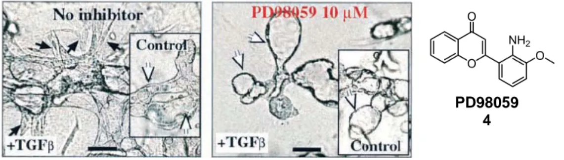
[24]The impact of these signaling pathways on epithelial plasticity was demonstrated using EpH4 cells transformed with Ha-Ras (EpRas cells). The EpRas cells underwent EMT after treatment with TGF-β. Reversal of EMT was achieved by inhibiting the Ras-Raf- MEK pathway with MEK-1 specific inhibitor PD98059 (4, Figure 7).
Figure 7: Reversal of EMT by MEK-1 inhibitor PD98059 (4). EpRas cells seeded in collagen gels in the presence or absence (inset) of TGF- β and left untreated (left image) or treated with 10 µM PD98059 (right image).
[24]These pathways are not alone in inducing EMT: Wnt/β-catenin signaling along with downstream effectors of Ras and TGF-β also promote EMT. For example, if E- cadherin is degraded or transcriptionally repressed, or if glycogen synthase kinase 3β (GSK-3β) activity is suppressed by activated PI3K (activated by upstream receptor tyrosine kinases [RTKs] or Ras), then the cytoplasmic amount of β-catenin increases and enhances transcription of T cell factor/lymphocyte enhancer factor (TCF/LEF) genes leading to cell proliferation and EMT (orange arrows in Figure 6, p.7).
[25]EMT-like cell behavior can be induced in epithelial cells by transforming them with oncogenic proteins such as Ras or Wnt.
[26]Due to the resulting phenotypical change, these cells can be used in a forward chemical genetics approach to observe the effects of small molecules on activated signaling pathways.
O O
NH2 O
PD98059
4
______________________________________________________________
Phosphatases
Phosphorylation is employed by living organisms to regulate innumerable cellular processes such as cell mitosis, growth factor responses and insulin signaling. In fact, it is estimated that at least 30% of all cellular proteins are subject to phosphorylation at one or more residues.
[27]The phosphorylation status of proteins is regulated by protein kinases, that catalyze the formation of phosphate ester bonds, and protein phosphatases, that catalyze the hydrolysis of phosphate ester bonds (Scheme 1). Due to the central role of phosphorylation /dephosphorylation in cellular signaling, many disease states involve perturbations in the balance between protein kinases and protein phosphatase activities.
[28;29]OH O P
O O O
ATP ADP
Pi
PROTEIN KINASE
PROTEIN PHOSPHATASE can be serine,
threonine or tyrosine
Scheme 1: Protein phosphorylation and dephosphorylation are catalyzed by kinases and phosphatases
Protein phosphatases are classified according to structure and substrate specificity into two families: the serine/threonine-specific protein phosphatases (PPs) and the protein tyrosine phosphatases (PTPs).
The serine/threonine phosphatases (PPs) are a large family of metalloprotein phosphatases that hydrolyze phosphate ester-modified serine or threonine residues.
[30]They have extremely diverse functions in the cell. For example, PP1 is involved in
regulating glycogen metabolism in response to insulin and adrenaline.
[31]Structurally,
the PPs consist of several subunits: a catalytic subunit with a metal ion at its core, and
one or more regulatory subunits. It has been proposed that phosphate ester hydrolysis
______________________________________________________________
involves the attack of the phosphorus atom by a metal-activated water molecule and proceeds without the formation of a phosphoenzyme intermediate.
[32;33]The second large family are the protein tyrosine phosphatases (PTPs). They play an important role in the regulation of signal transduction pathways. Unlike the PPs, the PTPs catalyze the hydrolysis of the phosphate ester of a tyrosine residue independently of a metal ion.
[30]In a two-step process, the PTPs catalyze first the transfer of the phosphate to a catalytic cysteine residue then expel the dephosphorylated substrate from the active site using an acidic amino acid residue to protonate the tyrosine phenolic oxygen. PTP1B is the prototypical member of this family.
[34]It has been implicated in oncogenesis, by being the major PTP that dephosphorylates and thus activates v-src in human breast cancer cell-lines,
[35]and in the control of cell adhesion, via effects on integrin signaling and through regulation of the adhesive properties of cadherin-catenin complexes.
[36]Additionally, it also plays a key role in the regulation of metabolic signaling: it has been shown to work as an insulin antagonist by dephosphorylating and thus inactivating the dimeric insulin receptor (IR).
[37;38]Some bacteria, such as Salmonella and Yersinia bacteria, produce and employ PTPs for their pathogenicity.
[39]Mycobacterium tuberculosis has two functional PTPs, MptpA and MptpB, that are secreted into the culture supernatant by the growing microbacterial cells.
[40]A subfamily of the PTPs are the dual-specificity phosphatases (DSPs) that hydrolyze
the phosphate ester on both a tyrosine residue and either a serine or threonine residue
located on the same protein. They have crucial roles in intracellular signaling
pathways. VHR (for VH1-related) is mostly known for regulating mitogen activated
protein kinase (MAPK) signaling pathway. It has been shown to dephosphorylate
several members of the MAP kinase family including ERK1, ERK2 and JNK
kinases.
[38]The cell-cycle dependant-25 (Cdc25) family of phosphatases, present as 3
homologues in humans, function as key regulators of the cell cycle during normal
eukaryotic cell division, promoting the dephosphorylation and thus activation of
cyclin-dependant kinases (CDKs), propagating cell-cycle signal transduction.
[41]The
overexpression of Cdc25 is frequently associated with a wide variety of cancers and
has been implicated in neurodegeneration.
[42]______________________________________________________________
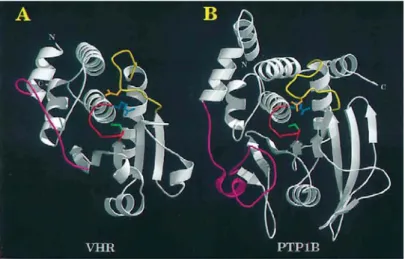
Although there is less than 5% sequence identity within the active sites of PTPs and DSPs, it has been shown that there is remarkable similarity in the overall structural fold of PTP1B, a PTP, and VHR, a DSP (Figure 8).
[30]Figure 8: Backbone representation of the catalytic domain of dual-specificity phosphatase VHR (A) and protein tyrosine phosphatase PTP1B (B).
[30]The catalytic sites of the PTPs and DSPs share a characteristic highly conserved loop structure, C(X)
5R (where C = cysteine, X = any amino acid, R = arginine), which interacts with the phosphate residue of the phosphotyrosine (shown in red in Figure 8). The major structural difference in the catalytic domains of the PTPs and DSP occurs at the insertion loop between the first α-helix (α1) and the first β-strand (β1, shown in magenta in Figure 8). This loop region contributes to the overall depth of the active-site pocket. In VHR, this loop was found to be 75% shorter than in PTP1B, with the consequence that a decrease in loop structure decreases the overall depth of the pocket.
[43]It has been thus suggested that substrate specificity between PTPs and DSPs lies within the depth of the active-site pocket.
[43]It may therefore be possible to achieve selective inhibition of PTPs and DSPs by
designing molecules that have a core structure that interacts with the conserved loop
region instead of the phosphate of the substrate, and substituents that interact with the
distinctive loop region between α1-β1. This design hypothesis was successfully tested
with a library of tetronic acids, where 3-acyltetronic acids was used as the phosphate
mimic (Figure 9).
[44]By modulating the side-chain substituents, it was possible to
tune the selectivity towards either PTPs or DSPs.
______________________________________________________________
O O
OH R2 O
R1 3 5 4
6: R1 = OH, R2 = (CH2)14CH3 11.6 µM (VHR) 7: R1 = C(=N2)-COOCH2CH3
R2 = (CH2)14CH3 0.4 µM (Cdc25B) 5
Figure 9: Chemical structure of the tetronic acid core 5.
Moreover, PTP1B possesses a vicinal second aryl phosphate binding site which lies within a region that is not conserved among protein tyrosine phosphatases.
[45]The presence of this structural feature suggests an effective strategy for the generation of potent and selective inhibitors: by tethering two small ligands, enhanced affinity and specificity ought to be achieved. Recently many industrial and academic groups have succeeded in generating such inhibitors.
[46-48]The central role of protein phosphatases in signaling make them particularly interesting targets to be studied with small molecules in a reverse chemical genetics approach.
Antibiotics
The optimism that accompanied the discovery of the first penicillin antibiotics to treat Staphylococcal infections in the 1940s has been tempered by the emergence of bacterial strains that are resistant to developed antibiotic treatment. Resistance to antibiotics has been monitored to develop rapidly, within one year to one decade of the introduction of the drug to clinical use.
[49]Clinically relevant bacteria are not only characterized today by single drug resistance but also by multiple drug resistance, making them a significant public health threat.
Although antibiotics discovery is an example of the successful mining of natural
products for therapeutic use, there is currently little advance in the discovery of novel
lead structures. Many of the antibiotics in clinical use are second or third generation
modifications of an older scaffold that still bind to the same protein. Therapeutic
scaffolds have been developed via two alternate routes: from natural products or from
synthetic sources. Some of the natural products have evolved to interact with and to
be recognized by targets in pathogenic bacteria. They have successfully served as
sources of inspiration to create some of the semi-synthetic variants of antibiotic
______________________________________________________________
natural products currently being used in the clinic. Antibiotics from synthetic sources include the sulfa antibiotics that target folic acid synthesis and the fluoroquinolone family of compounds (Ciprofloxacin) that block DNA gyrase and topoisomerase IV.
The latest structural class of antibiotics approved for use, the oxazolidinones (Linezolid), was introduced 40 years after the development of the fluoroquinolones class.
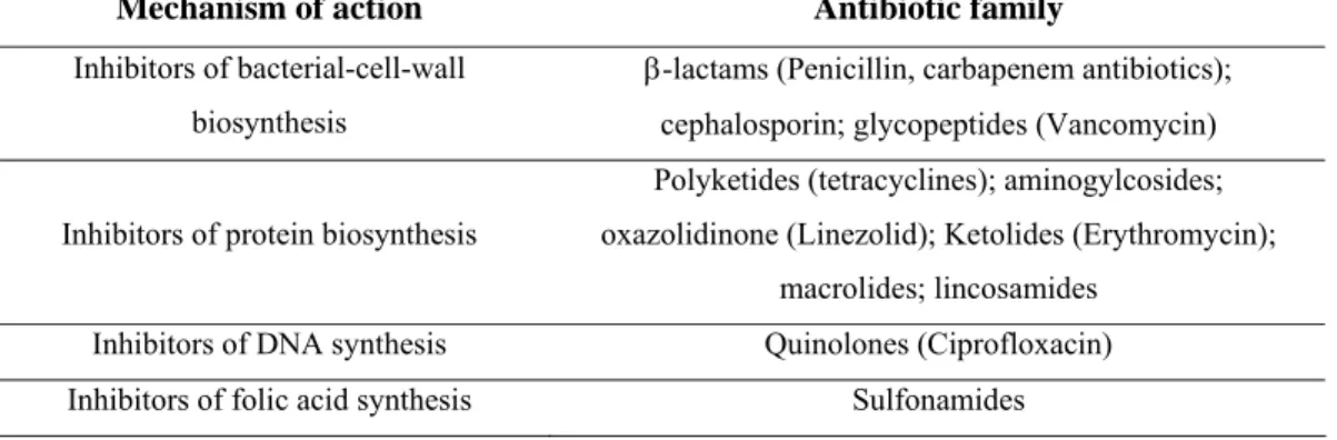
Mechanism of action Antibiotic family
Inhibitors of bacterial-cell-wall biosynthesis
β-lactams (Penicillin, carbapenem antibiotics);
cephalosporin; glycopeptides (Vancomycin) Inhibitors of protein biosynthesis
Polyketides (tetracyclines); aminogylcosides;
oxazolidinone (Linezolid); Ketolides (Erythromycin);
macrolides; lincosamides Inhibitors of DNA synthesis Quinolones (Ciprofloxacin) Inhibitors of folic acid synthesis Sulfonamides
Table 1: Major families of antibiotics and their mechanism of action.
Not only does there appear to be an innovation gap in the generation of novel antibiotic scaffolds, there are currently only four molecular targets for the main classes of clinically-used antibiotics (Table 1): bacterial-cell-wall biosynthesis;
bacterial protein biosynthesis; DNA replication; folate coenzyme synthesis. New targets may be less susceptible to existing mechanisms of bacterial resistance due to novel mechanisms of action.
2.1.3 Library Design for Chemical Genetics
To fully exploit the potentials of chemical genetics, it is necessary to have a compound library capable of modulating the function of many proteins. Additional properties of the ideal library include cell permeability and synthetic possibilities.
Not only is it currently impossible to generate a library that contains compounds that bind selectively to each protein, but for many proteins, small molecule ligands have not yet been identified. The composition of the library is of great importance, as it should increase the probability of finding interesting substances and effects.
Fortunately, many natural products have been selected to interact with a wide
assortment of proteins and other biological targets. For example, natural products
______________________________________________________________
have become effective drugs in a large range of therapeutic indications: a few prominent examples are vancomycin, an antibiotic, paclitaxel, a cytostatic drug used to treat cancer, and cyclosporine, an immunosuppressive agent. Natural products can be considered privileged structures for chemical genetics.
[50]Currently, there are multiple approaches to designing such libraries: libraries based on the general structural features of natural products, libraries based on core scaffolds of natural products and finally libraries based on specific substructures from classes of natural products. It should to be noted that these categories overlap to some extent, and that most synthesized libraries are designed to lie somewhere in between.
Libraries based on the general structural features of natural products
This approach seeks to exploit the structural characteristics of natural products in a high-throughput manner: the aims of this strategy are to generate compounds that are easy to access, that have plenty of chiral functional groups and are rich in stereochemical information, and are skeletally diverse.
[51]In general, the compounds generated are used as probes for understanding cellular processes. In other words, the substances are not biased towards specific targets. This strategy was applied to generate a 1,3-dioxane based library to identify a novel function selective inhibitor of Ure2p, a yeast nutrient responsive signaling protein.
[52]Libraries based on core scaffolds of natural products
The second approach is used primarily to exploit the activity of parent natural products. Natural products can be used as biologically validated frameworks upon which to display diverse functionalities.
Waldmann and coworkers propose that protein domain cores with similar three dimensional structures can be clustered in so-called protein similarity clusters (PSSC)
[53]and that this classification can be used as a guiding principle for the selection of biologically pre-validated starting points for compound library synthesis.
According to this principle, ligands for one of the proteins of the cluster can be used
as the starting point to obtain ligands for other members of the cluster. This approach
was used to identify inhibitors of the dual-specificity phosphatase Cdc25A 8, acetyl
cholinesterase (AchE, 9) and 11-β-hydroxysteroid dihydrogenase type 1 and 2 (11-β-
HSD-1, 10, and 11-β-HSD-2, 11) from a library of butenolides (Figure 10).
[53]______________________________________________________________
O O
HO H
O O HO
OH 11 OH
O O HO
OH O
8 O
O O HO
OH 10
F Cl
Dysidiolide Cdc25A inhibitor
IC50 9.4 µM
11-β-HSD-1 inhibitor IC50 10 µM
11-β-HSD-2 inhibitor IC50 5.3 µM AChE inhibitor
IC50 1.3 µM
8 9 10 11
Figure 10: Selected compounds from a butenolide library based on a core scaffold of the natural product Cdc25A inhibitor Dysidiolide (8).
Libraries based on specific substructures from classes of natural products
Similarly to the last described approach, here the substructures, which are generally found within a class of natural products and are also considered as biologically validated and privileged, are used as the starting point for library design. This approach offers the opportunity to generate structural diversity so that many targets can be used. One of the first examples of the application of this strategy was the synthesis of a 10,000-member library of 2,2-dimethylbenzopyrans (12),
[54;55]a structural motif found in diverse natural products with many biological activities (Figure 11). Screening of this library in diverse assays, target identification and optimization of active compounds yielded inhibitors for NADH:ubiquinone oxidoreductase, 13,
[56]anti-MRSA antibacterials, 14
[57]and farnesoid-X reporter (FXR), 15.
[58]O
2,2-Dimethylbenzopyran substructure
O
O
OMe OMe OMe
NADH:ubiquinone oxidoreductase inhibitor
O
CN
O
Anti-MRSA antibacterial
O
Cl Cl
N O
OMe O
Fexachloramide FXR agonist
12 13 14 15
Figure 11: Library based on the specific substructure of 2,2-dimethylbenzopyran (12).
Another application of this approach made use of the structural classification of
natural products (SCONP),
[59]which is a structural classification of natural products
______________________________________________________________
in a tree-like arrangement, providing a viable analysis and hypothesis-generating tool for the design of natural product-based libraries. SCONP was used as a guiding principle to generate libraries of 11-β-HSD-1 and 11-β-HSD-2 inhibitors.
[59]One class of natural products, the tetramic acids, have an interesting framework and
also biological activity and are therefore good targets for a natural product-based
compound collection for the chemical genetics study of their cellular and biological
activity. The following sections will describe the biology, biosynthesis and chemistry
of tetramic acids.
______________________________________________________________
2.2 Tetramic Acids
2.2.1 Tetramic Acid-Containing Natural Products
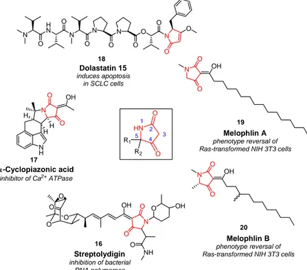
The tetramic acid (2,4-pyrrolidinedione, Figure 12) ring system was recognized in the sixties to be a reoccurring structural motif in a variety of isolated natural products (Figure 12).
O O O
O
N OH O
O NH
N O
H O H H
OH
N OH
O
O
Streptolydigin inhibition of bacterial
RNA polymerase α-Cyclopiazonic acid
inhibitor of Ca2+ ATPase
Melophlin A phenotype reversal of Ras-transformed NIH 3T3 cells N
HN
N N
O O
O
N O
O O
O N O
O
Dolastatin 15 induces apoptosis
in SCLC cells
HN O
O R1
R2 1
2 4 3 5 18
16 17
19
NH O
O OH
N OH
O
O
Melophlin B phenotype reversal of Ras-transformed NIH 3T3 cells
20
Figure 12: Representative natural products containing a tetramic acid motif.
This family of compounds has attracted significant attention in the past few years due to their synthetic challenge as well as their broad range of biological activities.
[2]A steadily increasing number of tetramic acids are being isolated from natural sources ranging from marine species to fungi and terrestrial bacteria.
[2]The biological activity is also remarkably varied, spanning from antibiotic and antiviral activity, cytotoxicity, mycotoxicity as well as inhibition of the cell cycle.
The tetramic acid antibiotic streptolydigin (16, Figure 12) inhibits initiation, elongation and pyrophosphorolysis in bacterial RNA polymerase (RNAP).
[60]Streptolydigin does not inhibit eukaryotic RNAPI, RNAPII or RNAPIII because the
______________________________________________________________
proteins have low sequence identity with bacterial RNAP, though the proteins share high three-dimensional structural similarity. The target, mechanism and structural basis of inhibition were recently disclosed by Tuske et al.
[60]and these results have implications in the design of novel bacterial RNAP-based antibiotics.
Cyclopiazonic acid (17, Figure 12) is a toxic indole tetramic acid produced by numerous Penicillium species.
[61]It is an interesting mycotoxin as it is produced by numerous fungi that infect commodities though it is toxic only at higher concentrations (ingested LC
50~30 mg/kg). Its toxicity appears to stem from its abitility to inhibit Ca
2+ATPase.
[62]The dolastatin family of natural products includes a series of linear and cyclic antineoplastic and/or cytostatic peptides. One of the most potent members of this family, dolastatin 15 (18, Figure 12) includes a tetramic acid at its terminal moiety.
Dolastatin 15 induces apoptosis and is antiangiogenic. It binds weakly to tubulin but clearly disrupts microtubule formation.
[63]Melophlin A (19) and B (20), novel tetramic acids isolated from the marine sponge Melophlus sarassinorum from the Indonesian sea, possesses the ability to reverse Ras- transformed NIH3T3 cells as well as arrest NIH3T3 cells in the G1 phase of the cell cycle at a concentration of 1 µg. mL
-1.
[64]Tetramic acids are also found in the agrochemical field: they have been patented for fungicidal and herbicidal use.
[65]Bayer CropScience has developed a series of insecticidal agents based on tetronic and tetramic acids. Spirotetr(o)amic acid 21 and 22 (Figure 13) appear to block acetyl-coenzyme A carboxylase,
[66]a protein vital to the production of fatty acids in mites. As lipids are the principle source of energy in mite eggs, disruption of their lipid utilization impairs developmental processes.
NH O
O O
O
O O O O
O
Cl Cl
21 22
Figure 13: Structure of insecticidal tetronic acid 21 and Spirotetramat 22.
______________________________________________________________
The interesting biological and structural diversity of this class of substances makes it a particularly interesting template for the design of compound libraries in search of small molecules that affect cellular signalling pathways.
2.2.2 Biosynthesis of Tetramic Acids
The acyl tetramic acid group of metabolites that have been investigated biosynthetically possess a tetramic acid ring that is derived from an acetate unit and an amino acid.
[2]The acyl group derives from a polyketide. However, the order in which these three entities are assembled is still subject to controversy.
NH N
O
H O H H
OH
17
H H
HO HN
O
O
HO 23
O O O
O
N OH O
O
HN O
O OH
H2N O
NH H2N OH
O
OH OH
H2N O
OH O 16 HO
A B
C
24
25
26
Figure 14: Biosynthetic precursors to equisetin (23, A), cyclopiazonic acid (17, B), and streptolydigin (16, C).
Thus, it is assumed that the pyrrolidine moieties in equisetin (23),
[67]α-cyclopiazonic acid (17),
[68;69]and streptolydigin (16)
[70]are derived from serine (24), tryptophan (25), and methylaspartate (26), respectively (Figure 14).
Janda and co-workers
[71]gave the biology of tetramic acids a different turn by
proposing that tetramic acid 28 is formed non-enzymatically from N-acylhomoserine
lactone, 27, produced in Gram-negative bacteria by a Claisen-like intramolecular
alkylation (Scheme 2).
______________________________________________________________
HN O
O O
O
NH O
O OH
OH
27 28
Scheme 2: Reaction of 3-oxo-AHL 27 to generate tetramic acid 28.
The formed tetramic acid 28 is postulated as being used as an interference strategy against encroachment by competing bacteria, behaving therefore as quorum sensors.
This is an intriguing hypothesis as many biologically active tetramic acids are selectively toxic against Gram-positive bacteria.
[72;73]2.2.3 Synthetic Routes to Tetramic Acids Solution-Phase Synthesis of Tetramic Acids
Due to mounting interest in tetramic acids, various synthetic routes towards their
synthesis have been established. The Lacey-Dieckmann cyclization
[74]is the most
widely adopted synthetic strategy for the generation of the 2,4-pyrrolidinedione core
(A, Scheme 3).
[75-78]It is a base-induced cyclization of N-(β-ketoacetyl)-α-amino
esters (30), that are easily obtained from α-amino acids. This is an extremely flexible
synthesis and is used to synthesize tetramic acids with various C3 substituents,
provided the N-acetoacetyl-α-amino ester can be prepared. However, complete or
partial racemization at C5 of the 2,4-pyrrolidinedione core has been frequently
observed.
[79]Recently, Schobert and coworkers reported an expedient synthesis from
α-amino esters and in which the cyclization involved a domino addition-Wittig
alkenation reaction with immobilized (triphenylphosphoranylidene)ketene (33) under
neutral non-racemizing conditions (B, Scheme 3).
[80]Acylation to 3-acyltetramic acids
was then performed with the appropriate acyl chloride and boron trifluoride-diethyl
etherate under microwave irradiation.
______________________________________________________________
Dieckmann Cyclisation
N OR
O
R1 R3
O O
R2 NHR2
CO2tBu R1
P C C O Ph
Ph
R2N O R1
OtBu P
Ph
Ph N
O R2 R1 O
OtBu R2HN
OR O
R1
N O
H2C RHN
R2 Me
RN NR Me
Me Me
O
O
RNH2
CDI R2HN
OH O
R1
O O
R2HN O H
O O
O O
N R3 OH R2 R1
O
R1 O
O
N O
H3C RHN
R2 β-keto equivalent
H2 , Pd(C) isoprenyl
chloroformate DMAP, CH2Cl2 -5 °C, 2h
2.) R3COCl BF3. Et2O Microwave 100 °C, 45 min.
1.) TFA, r.t., 3h
EtOAc reflux, 20 min.
CH2Cl2, HCO2H 25 °C, 7d
N N
O N N
1.) BF3. Et2O THF, ∆
2.) 3N aq. NaOH MeOH, 25 °C
6N aq. H2SO4 Et2O, 25 °C,7h Base
THF Microwave 120 °C, 30 min.
29 30
32
33
34 35
37
38
39
40
41
42
43 44
31
36
A
B
C
D
Scheme 3: Summary of recent solution-phase synthesis of tetramic acids.
Another method was used by Jouin for the generation of the tetramic acid core via the condensation of N-protected amino acids with Meldrum’s acid 37 in the presence of isoprenyl chloroformate and DMAP (C, Scheme 3).
[81]The cyclization of 38, by heating in an organic solvent, provided N-protected tetramic acids. Fustero and coworkers proposed an alternative asymmetric synthesis of tetramic acids in which the key step is a carbonyl transfer from carbonyldiimidazole (42, CDI) to α-diimines 41 to form 43 (D, Scheme 3).
[82]These compounds can then be transformed in tetramic acid derivatives in two additional steps. Unfortunately, the sequence is limited to the generation of “symmetric” tetramic acids due to the double condensation with initial dione 39.
Solid-Phase Synthesis of Tetramic Acids
The majority of reported solid-phase tetramic acid syntheses begin with solid-phase
bound amino acids 45 as building blocks and involve a Dieckmann cyclization in the
cleavage step (Scheme 4, A). These methods differentiate themselves from each other
by the use of different conditions and reagents to initiate the cyclative cleavage from
the resin. Matthews and Rivero
[83]report the synthesis of mostly 3-unsubstituted
tetramic acids, employing NaOEt and heating to 85 °C to cyclize from the solid
support. Ganesan and co-workers
[84]report the preparation of tetramic acids without
______________________________________________________________
substitution at the 3-position using tetrabutylammonium hydroxide as the cyclization agent. The product needs however to be treated with strongly acidic Amberlyst A-15 ion exchanger resin in order to remove this reagent. Weber et al.
[85]demonstrate the synthesis of 3-acyl tetramic acids using Meldrum’s acid to acylate at the 3-position, but by using DIPEA and heat for the cyclization the products underwent racemization.
Finally, Romoff
[86]reported a method where the cleavage is effected in the ultimate step by a Dieckmann cyclization in the presence of KOH/MeOH at room temperature, without apparent loss of chirality.
O NH2 O
R
O N
O
R R2 O R1
N O
OH R2 R R1 base
A
45 46 47
N O
R OH X
O
O O
O
DCC, DMAP CH2Cl2
EtOAc
∆
B
48
49
50 O
O O
O OH R NHX XHN
O OH R
51
O CO2
52 see text
for conditions
Scheme 4: Solid-phase synthesis of tetramic acids using Lacey-Dieckmann cyclization (A) and according to Huang (B).
Huang and co-workers (Scheme 4, B) devised an alternative synthesis based on the condensation of resin-bound Meldrum’s acid 48 with N-acylated amino acids 49.
[87]Cyclative cleavage was effected by heating in ethyl acetate. This method is restricted to the synthesis of tetramic acids without an acyl substituent at the 3-position.
All of the above-mentioned syntheses demonstrate the synthetic feasibility of the
route using aromatic or aliphatic amino acids such as phenylalanine or valine but do
not thoroughly probe the scope of their synthesis.
______________________________________________________________
3 Aim of the Dissertation
The use of small molecules as probes to investigate complex biological systems is termed chemical genetics. This is a powerful method to study such phenomena like signal transduction or for the search for novel antibiotics. Key to this approach is the design of an effective compound library. Inspiration for such a library can be taken from natural products, which are themselves pre-validated structures.
[59]An example of such a privileged scaffold is the tetramic acid (2,4-pyrrolidinedione) ring system. It is present in numerous natural products that display diverse biological activities
[2]making it a particularly suitable template for a natural-product-based library for a chemical genetics approach to study cellular signaling.
The first aim of this thesis was to develop a strategy for the synthesis of tetramic acids in a minimal number of steps, in an efficient manner and by maximizing diversity.
Ideally, this approach should allow the introduction of different functional groups at all three modifiable positions of the tetramic acid scaffold (Figure 15).
N O
R1 OH R1 R2
R3
Figure 15: Positions open for diversification on the tetramic acid scaffold.
To demonstrate the feasibility of this strategy, the synthesis of a small library of tetramic acids should be undertaken. A successful route to the tetramic acid scaffold should open up synthetic pathways for a number of analogs of the natural product Laccarin. In an interesting extension of the synthesis, the tetramic acid scaffold should be substituted with indole moieties. Therefore, the development of a synthesis of tryptophan analogs should be carried out.
In light of the documented biological activities of tetramic acid natural products,
[2]the
final objective of this thesis should be the testing of the synthesized tetramic acid
library against biological targets. First, the compound collection should be assayed to
uncover modulators of the Ras/MAPK signaling cascade in cell-based tests.
______________________________________________________________
Furthermore, their inhibitory activity for phosphatases should also be investigated.
Ultimately, the effect of tetramic acids on bacterial cell growth should be examined.
______________________________________________________________
4 Results and Discussion
4.1 Synthesis of tetramic acid libraries
4.1.1 Synthetic plan
The synthetic route chosen is based on a solid phase approach and provides access to diverse tetramic acids. The building blocks stem from the chiral pool: amino acids are used as the starting material for the synthesis.
O O
N R1
H
PG O
O N R1
H R2
O O
N R1 R2
R2 O
N OH
R3 R2
R1 O O
O N R1
HO R3
N OH
H R3
R2
R1 O deprotection
reductive amination
deprotection, β-keto acylation
β-keto acylation
cyclization
cyclization 53
54
55
56 57
58
*
*
*
*
*
*
Scheme 5: Synthetic strategy towards the synthesis of 3-acyl tetramic acids.
In the first step, N-protected α-amino acids are condensed with a hydroxyl-
functionalised resin (Scheme 5). After deprotection of the N-terminus of the amino
acid, the resulting amine can be converted to secondary amine 54 in a reductive
amination step. Next, this amine can be acylated with β-keto equivalents to generate
cyclization precursor 55. Finally, cleavage from the resin is achieved by base
catalyzed cyclization to the tetramic acid 56. Alternatively, after condensation of the
α-amino acid with the resin and deprotection of the N-terminus, the resulting amine
______________________________________________________________
can be directly acylated with a β-keto equivalent to 57. Cleavage is achieved analogously to provide 58.
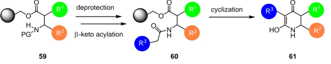
A similar strategy can be applied for the synthesis of 6-membered tetramic acid analogs 61. Instead of using α-amino acids as the starting material, β-amino acids like β-alanine or the more constrained anthranilic acid are to be used (Scheme 6).
O O
PGN H
R1
R2
O O
NH R1
R2 O
R3
NH O
HO
R1 R2 R3
deprotection cyclization
β-keto acylation
59 60 61
Scheme 6: Synthetic strategy towards the synthesis of 6-membered ring tetramic acid analogs.
Examples of natural products containing quinoline-3-carboxamide 62 (Figure 16) are Linomide (63, immunomodulator as well as anti-angiogenic activities)
[88;89]and the Militarinone
[90]class of compounds (64).
N O O
R3 OH R2 R1
R4
N O N O OH
CH3
CH3
N O O OH
H H HO
64 Militarinone D
63 Linomide
62
quinoline-3-carboxamide core
Figure 16: Structure of quinoline-3-carboxamide core 62 and representative natural products Linomide (63) and Militarinone D (64).
The modular assembly of tetramic acids by using a variety of commercially available
or easily synthesized building blocks allows the synthesis of a diverse tetramic acid
library. Furthermore, by using a solid-phase approach, not only is the carboxylic
______________________________________________________________
functionality of the amino acid temporarily protected by being bound as an ester to the resin, time consuming workup steps and the purification of synthetic intermediates are eliminated. This is realized by washing the solid-phase with a cocktail of solvents;
reactions on the solid-phase can be driven to completion by using large excesses of reagents. The cleavage method also ensures that only selected compounds are released from the solid phase: only substances with a β-keto entity will undergo concomitant cyclization and cleavage from the solid support. This synthetic strategy is amenable for larger scale solution phase synthesis of tetramic acids.
4.1.2 Implementation and Optimization of the Synthesis Immobilization of the amino acid building blocks
The first step in the synthetic sequence is the immobilization of the amino acids to the solid phase. As delineated above, the amino acid is loaded onto the linker as an ester and the cleavage of the product from the resin is effected through a base-mediated cyclization and not an acid-mediated ester cleavage. Therefore, any hydroxyl-linker may be used, for example Merrifield or Wang resin. Wang resin (65, Scheme 7) was chosen because it allows the monitoring of intermediate compounds by cleavage with TFA, that can be undertaken using conventional glassware. Cleavage of intermediates from Merrifield resin would necessitate special apparatus because of the use of HF as the cleavage reagent.
For the synthesis of a diverse tetramic acid library, a variety of different α-amino acids were used as starting material: apolar (glycine (66a), valine (66b), leucine (66c)), heterocyclic (tryptophan (66f)) and basic (ornithine (66d) and arginine (66e)) amino acids were employed (Scheme 7).
All amino acids were protected on the N-terminus with the base-labile Fmoc group,
enabling facile loading determination. Orthogonal protection of side chain
functionalities was achieved with acid-labile protecting groups, such as the tert-
butyloxycarbonyl (Boc, for lysine and ornithine), and pentamethyldihydrobenzofuran
(Pbf, for arginine) protecting groups. A protecting group was not necessary for the
indole nitrogen of tryptophan.
______________________________________________________________
NHFmoc O
HO R1
O
O H
N R1
R1 = - H = -CH(CH3)2 = -CH2CH(CH3)2 = -(CH2)3NHBoc
N H
NH NHPbf
NH
=
=
O O
NH2 R1
Fmoc
N
68 DIC, DMAP
DMF, r.t., 2 h O
OH Wang resin
20% piperidine DMF r.t., 2 x 20 min.
65
66 a - h
66a 66b 66c 66d 66e
66f
67
69 a - f 69a
69b 69c
69e 69f
69%
69%
63%
86%
80%
84%
69d
Scheme 7: Loading of Fmoc-protected amino acids onto Wang resin.
Secondary amino acids such as proline were also used and generated bicyclic tetramic acid 74 (Scheme 8). Such a bicyclic moiety was recently disclosed as part of the core structure of UCS1025A (75), an inhibitor of the telomerase enzyme.
[91-93]Wang-OH
HO O
N Fmoc
DIC, DMAP DMF, r.t., 2 h
O O
NFmoc
O O H
N
O O
N O O N
O
O OH
20 % Piperidine DMF, 2 x 20 min
diketene, Et3N, CH2Cl2
16 h, r.t.
0.1 M KOH in MeOH CH2Cl2, 1,4-dioxane
2 h, r.t.
39 %
0.459 mmol/g 70 53 %
71 72
73 74
N O
O H
H OH
O O
UCS1025A 75
Scheme 8: Proline-based tetramic acid bicycle 74 and structure of telomerase inhibitor
UCS1025A (75).
______________________________________________________________
All N-Fmoc-protected α-amino acids were condensed with Wang-functionalized resin (1% DVB crosslinking, 100-200 mesh, 1.2 mmol/g), via a Steglich esterification (DIC, 5 mol% DMAP, DMF, 2 hours, r.t.).
[94]Loading of the resin was determined by the Fmoc-method
[95]with spectrometric monitoring the absorbance of dibenzylfulvene-piperidine adduct 68 formed in the cleavage of Fmoc with piperidine in DMF (Scheme 7). The resin loadings ranged between 0.40 mmol/g and 0.75 mmol/g (63-86%), in reasonable agreement with published values.
For the synthesis of six-membered-ring tetramic acid analogs, β-amino acids were used as the starting materials. The simple β-amino acid β-alanine was loaded onto the resin using the Steglich esterification method described previously to provide Wang- β-alanine-Fmoc resin (64) in 68% yield (Scheme 11). Loading of conformationally restrained anthranilic acid (76) onto the resin to provide 78 proved to be more challenging (Scheme 9). After protecting anthranilic acid 76 with Fmoc-Cl and Na
2CO
3in 1,4-dioxane from 0 °C to room temperature overnight in 86% yield, it was not possible to load it onto the resin via the Steglich esterification method, even after double coupling. This is probably due to the steric bulk of the system.
HO O
H2N
HO O
N H Fmoc
O O
H2N
O NH O
O Fmoc-Cl
Na2CO3, dioxane
86%
1.) DIC, DMAP DMF
DMAP toluene 16 h, 80 °C
X
76 77
2.) 20% piperidine DMF
95 % 0.995 mmol/g
O O
N O O N H
H O
O
OH diketene, Et3N
CH2Cl2, 16 h, r.t.
0.1M KOH in MeOH CH2Cl2, 1,4-dioxane
2 h, r.t.
81 80
78 79
11 %
Scheme 9: Loading of anthranilic acid onto Wang resin and synthesis of quinolinine-3- carboxamide 81.
Because difficulties in loading the resin due to the steric bulk of Fmoc-anthranilic
acid, a commercially available synthon, isatoic anhydride (79, scheme 9), was
______________________________________________________________
successfully condensed to the resin with DMAP at 60 °C for 16 h leading to resin- loaded anthranilic acid 78. An advantage of this method is that no protecting group is needed. As the resin-bound anthranilic acid does not have a leaving group that can be spectroscopically monitored, the loading was determined by cleavage of a small aliquot of the resin with 90% TFA in dichloromethane. The determined loading demonstrated that the condensation reaction occurred in excellent yields (0.995 mmol/g, 95%).
Before any new functionalities were introduced, the Fmoc group was deprotected.
This was achieved using standard conditions: 20% piperidine in DMF for 20 minutes at room temperature.
Functionalization by reductive amination
Additional diversity was introduced in a reductive amination step. After Fmoc- deprotection of an α-amino acid linked to Wang resin, the resulting primary amine 82 was treated with an aldehyde and NaBH(OAc)
3in dichloromethane at room temperature for 16 h (Scheme 10). The aldehydes used were commercially available, and displayed a range of chemical properties: aliphatic, aromatic (electron-poor and – rich) and heteroaromatic aldehydes were successfully employed.
O O
NH2
R1
O O
N R1
R2 R2CHO, NaBH(OAc)3 H
R2 =
82 83
OMe
OMe MeO
Br
N O
N CH2Cl2, 16 h, r.t.
Scheme 10: Aldehydes employed to functionalize amine 82.
Generally a 15-fold excess of aldehyde was used in the reaction. The yields ranged
from 16-73%. Overalkylation to the tertiary amine accounted for the low yields,
although no trends due to the nature of the aldehyde could be observed.
______________________________________________________________
Wang-OH
HO O
N H
Fmoc
O N
H O
Fmoc O NH2
O
O N
H O N
O OH
O
DIC, DMAP DMF, 2h, r.t.
0.547 mmol/g 84 68 %
85 86
88 89
20 % Piperidine DMF, 2 x 20 min
r.t.
benzaldehyde (1 eq.) NaBH(OAc)3, CH2Cl2
16 h, r.t.
1.) diketene, Et3N, CH2Cl2, 16 h, r.t.
2.) 0.1 M KOH in MeOH CH2Cl2, 1,4-dioxane 2 h, r.t.
55 %
O N
O
87
excess benzaldehyde NaBH(OAc)3, CH2Cl2
16 h, r.t.

![Figure 6: Multiple signaling networks affecting EMT. [20]](https://thumb-eu.123doks.com/thumbv2/1library_info/3632735.1502284/13.918.283.638.571.859/figure-multiple-signaling-networks-affecting-emt.webp)