Epithelial conjunctival neoplasias – the importance of an early diagnosis and optimal treatment
Abstract
Objective:To emphasize the importance of an early diagnosis and an adequate treatment in conjunctival tumors.
Laura Tabuenca Del Barrio
1Methods: We present two clinical cases and compare the course of
each case: one of conjunctival intraepithelial neoplasia (CIN) which took
Marcos Mozo Cuadrado
1a positive course, and a fatal case of squamous cell carcinoma (SCC)
with intraocular and orbital extension.
Luiz Miguel Nova
Camacho
2Results:Epithelial conjunctival malignancies are one of the most prev- alent ocular surface tumors. Among these, CIN are the most common.
Alicia Zubicoa Enériz
1CIN have an excellent prognosis, given adequate treatment. However,
Miren Dolores Aranguren Laflin
1when the diagnosis of CIN is late, the epithelial basement membrane will be affected, resulting in SCC. SCC may have poorer results due to its capacity to infiltrate near tissues and create distant metastasis.
Araceli Alcaine Soler
1Conclusion:It is not common today to treat patients with orbital exten- sion of SCC; however, it is crucial to note the importance of an early diagnosis of conjunctival malignancies. An early diagnosis is essential to prevent the transformation to other life-threatening types.
1 Ophthalmology, Complejo Hospitalario de Navarra, Pamplona, Navarra, Spain Keywords:squamous cell carcinoma, conjunctival malignancies, CIN,
exenteration, excisional biopsy, radiotherapy 2 Pathology, Complejo
Hospitalario de Navarra, Pamplona, Navarra, Spain
Introduction
Conjunctival tumors are one of the most common ocular surface malignancies [1], [2]. Their most frequent cell origin is located on epithelial and melanocytic cells. Since in most cases it is very difficult to differentiate between premalignant and malignant lesions, a biopsy is necessary to determine their nature [1].
Epithelial neoplasms can be divided into
1. carcinoma in situ/conjunctival intraepithelial neo- plasia (CIN), which exclusively affect the epithelium;
2. invasive squamous cell carcinomas (SCC), when the epithelial basement membrane is damaged and neoplastic cells are present in the corneal stroma [3].
We report two clinical cases of conjunctival neoplasm.
The first one exhibits a patient affected by CIN with an early diagnosis and effective treatment based on topical chemotherapy and surgical excision. In contrast, the second case shows a patient with SCC with an aggressive clinical course and intraocular and orbital extension.
The fact that the cell origin was the same in both cases shows that an adequate diagnosis and optimal treatment are required to control the progression of these types of tumors. Intraocular and orbital extension is infrequent but commonly fatal.
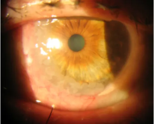
Figure 1: Slit-lamp examination; nasal limbal gelatinous mass with inferior corneal infiltration
Case descriptions
Case 1
A 70-year-old man presented with a conjunctival lesion in his left eye that had appeared four months earlier.
Visual acuity (VA) was 20/200 in both eyes. Slit-lamp examination showed a nasal limbal gelatinous mass with inferior corneal infiltration and cataract (Figure 1).
biopsy and dysplastic cells at the bottom in image 10x; dysplastic epithelium with high mitosis index in image 20x
Figure 3: A) One month later, the lesion had decreased. B) At the third month, CIN had disappeared.
Intraocular pressure and fundoscopy were unremarkable.
An excisional biopsy was performed: A CIN with free margins was confirmed by the histopathological study (Figure 2). Topical adjuvant treatment with interferon (IFN)-α2β drops/6 hours was initiated. One month later, the lesion decreased in size (Figure 3A), and topical treatment was reduced to 1 drop three times a day.
At the third month, no evidence of CIN was observed (Figure 3B), and the patient has since remained free of disease.
Case 2
A 90-year-old man was referred to us with a mass in his right eye that had first been noticed three months earlier and had been growing progressively since then.
VA was 20/200 in the right eye and hand movement in the left eye. Slit-lamp examination revealed a gelatinous temporal conjunctival mass (7 mm long, 10 mm wide) with dilated superficial vessels (Figure 4A). Fundus examination showed age-related macular degeneration (AMD) in both eyes. Ocular movements were affected in the form of abduction limitation in the right eye (Figure 4B).
Figure 4: A) Slit-lamp examination: a gelatinous temporal conjunctival mass (7x10 mm) with dilated superficial vessels without corneal involvement is exposed. B) Abduction limitation
in right eye.
Figure 5: Anatomopathological study: moderately differentiated squamous cell carcinoma was confirmed; tumour cells present in corion
Figure 6: A) Magnetic Resonance Imaging showed lateral rectus muscle involvement. B) Anterior Segment – Optical Coherence Tomography (AS-OCT): a hyperreflective lesion involving conjunctival tissue and spread over corneal surface
An incisional biopsy was performed and a moderately differentiated SCC was confirmed (Figure 5). Magnetic resonance imaging (MRI) showed malignant infiltration of the lateral rectus muscle (Figure 6A). Anterior segment optical coherence tomography (AS-OCT) displayed a hy- perreflective lesion which involved the conjunctival tissue and spread over the corneal surface (Figure 6B). Due to the advanced stage of the tumor, an exenteration or ra- diotherapy treatment was offered, but the patient rejected both. After five months, the tumor had progressed and involved ocular globe tissues and soft periorbital struc- tures (Figure 7). The patient died due to metastatic dis- ease nine months after the initial diagnosis.
Figure 7: Tumor progression involving ocular globe tissues and soft periorbital structures
Joint Committee on Cancer (AJCC) and is based on the depth of invasion as well as the size and extent of the affected adjacent structures [3].
CIN most commonly occurs in individuals in their 6thor 7thdecade of life, and is divided into three dysplasia types:
mild, moderate, and severe. It usually affects the inter- palpebral fissure area in the form of a gelatinous, sessile, or papillomatous mass with irregular margins, which can spread over the corneal epithelium [1]. It is considered as a premalignant lesion and has a low risk of metasta- sizing. However, without rapid diagnosis and the estab- lishment of appropriate treatment, there is a risk that atypical cells will invade the epithelial basement mem- brane and that the tumor will become a more malignant lesion with a worse prognosis.
Treatment is based on surgical excision with clear margins in combination with cryotherapy of the excised conjuncti- val edge. Cases with corneal involvement might also be managed with an alcohol epitheliectomy, which is in fact recommended [4]. Due to the irregular and unclear mar- gins of the tumor, its correct excisional surgery can in some cases be a challenge for the surgeon. Recurrence rates range from 5 to 53% [1], [4], [6]. Adjuvant treat- ments with topical mitomycin C (MMC), 5-fluorouracil, or IFN-α2β are commonly used in order to avoid surgical procedures [4]. Excellent outcomes have been reported with topical IFN-α2β as primary treatment for conjunctival and/or corneal disease. It also plays a role as adjuvant therapy in cases with positive margins or recurrence after surgical excision [3]. As there is no established treatment protocol in terms of the duration over which IFN-α2β is administered, treatment must be individualized [2].
One third of patients develop irritative symptoms as side effects of surgery and chemotherapy, which often resolve quickly. One third of patients show radiation-induced complications that persist in the long term and consist of dry eye syndrome, lid alopecia, rubeosis, and secondary glaucoma [4].
The finding of epidermal growth factor receptor (EGFR) overexpression was an important aspect when consider- ing targeted therapies in cases of inoperable advanced orbit and periocular SCC. EGFR inhibitors such as erlotinib have shown the effect of a significant decrease of SCC tumor size. Targeted therapy might be appropriate for non-surgical candidates, comprising patients with ad- vanced metastatic orbital disease involving regional lymph
should be closely monitored as recurrences can occur even more than 5 years after treatment [2].
Conclusion
Early diagnosis is essential in epithelial conjunctival tu- mors. Histopathological examination allows a classifica- tion of the lesions as premalignant or malignant, which is crucial for the development of a therapeutic strategy.
An excisional biopsy with cryotherapy and free margins is sufficient in most cases of CIN. Topical chemotherapy can be used additionally. In cases of SCC, surgical ex- cision may be sufficient, but may also be combined with adjuvant topical therapy, which may enhance treatment in cases of insufficient surgical removal. In addition, when SCC penetrates the intraocular or orbital septum, enucle- ation, exenteration, radiotherapy, or targeted therapy have to be performed on each specific case.
Notes
Competing interests
The authors declare that they have no competing in- terests.
Informed consent
Informed consent has been obtained from the patient for the publication of this case report.
References
1. Saornil MA, Becerra E, Méndez MC, Blanco G. Tumores de la conjuntiva [Conjunctival tumors]. Arch Soc Esp Oftalmol. 2009 Jan;84(1):7-22. DOI: 10.4321/s0365-66912009000100003 2. Miller CV, Wolf A, Klingenstein A, Decker C, Garip A, Kampik A,
Hintschich C. Clinical outcome of advanced squamous cell carcinoma of the conjunctiva. Eye (Lond). 2014 Aug;28(8):962- 7. DOI: 10.1038/eye.2014.79
3. Bellerive C, Berry JL, Polski A, Singh AD. Conjunctival Squamous Neoplasia: Staging and Initial Treatment. Cornea. 2018 Oct;37(10):1287-91. DOI: 10.1097/ICO.0000000000001651
4. Santoni A, Thariat J, Maschi C, Herault J, Baillif S, Lassalle S, Peyrichon ML, Salleron J, Caujolle JP. Management of Invasive Squamous Cell Carcinomas of the Conjunctiva. Am J Ophthalmol.
2019 Apr;200:1-9. DOI: 10.1016/j.ajo.2018.11.024
5. LLull-Tombo M, Curbelo-Gómez M, Martínez-Ojeda D, Díaz-Alfonso L, Suárez-Rodríguez B, Martínez-Díaz A. Carcinoma epidermoide de conjuntiva. Presentación de un caso Epidermoid carcinoma of the conjunctiva. Medisur. 2011;9(6). Available from: http://
www.medisur.sld.cu/index.php/medisur/article/view/1474/801 6. Galor A, Karp CL, Oellers P, Kao AA, Abdelaziz A, Feuer W, Dubovy SR. Predictors of ocular surface squamous neoplasia recurrence after excisional surgery. Ophthalmology. 2012 Oct;119(10):1974- 81. DOI: 10.1016/j.ophtha.2012.04.022
7. Yin VT, Pfeiffer ML, Esmaeli B. Targeted therapy for orbital and periocular basal cell carcinoma and squamous cell carcinoma.
Ophthalmic Plast Reconstr Surg. 2013 Mar-Apr;29(2):87-92.
DOI: 10.1097/IOP.0b013e3182831bf3
Corresponding author:
Laura Tabuenca Del Barrio
Ophthalmology, Complejo Hospitalario de Navarra, Calle Irunlarrea, 31008 Pamplona, Navarra, Spain
lauratabuillueca@gmail.com
Please cite as
Tabuenca Del Barrio L, Mozo Cuadrado M, Nova Camacho LM, Zubicoa Enériz A, Aranguren Laflin MD, Alcaine Soler A. Epithelial conjunctival neoplasias – the importance of an early diagnosis and optimal treatment. GMS Ophthalmol Cases. 2020;10:Doc30.
DOI: 10.3205/oc000157, URN: urn:nbn:de:0183-oc0001576
This article is freely available from
https://www.egms.de/en/journals/oc/2020-10/oc000157.shtml Published:2020-08-06
Copyright
©2020 Tabuenca Del Barrio et al. This is an Open Access article distributed under the terms of the Creative Commons Attribution 4.0 License. See license information at
http://creativecommons.org/licenses/by/4.0/.