Diagnosis and laparoscopic treatment of cornual ectopic pregnancy
Diagnose und laparoskopische Behandlung einer ektopischen Schwangerschaft im Uterushorn
Abstract
Cornual (interstitial) ectopic pregnancy is an uncommon variant of ec- topic pregnancy which often poses a diagnostic and therapeutic chal-
M. Sami Walid
1Richard L. Heaton
2lenge with a significant risk of rupturing and bleeding. We present a ruptured right cornual pregnancy and explain how to deal with such a
case laparoscopically. 1 Medical Center of Central
Georgia, Macon, GA, USA Keywords:cornual pregnancy, interstitial pregnancy, laparoscopy
2 Heart of Georgia Women's Center, Warner Robins, GA,
Zusammenfassung
USADie Schwangerschaft im Uterushorn ist eine ungewöhnliche Variante der ektopischen Schwangerschaft, die häufig eine diagnostische und therapeutische Herausforderung verbunden mit einem hohen Risiko auf Rupturierung und Blutungen darstellt. Es wird eine rupturierte ekto- pische Schwangerschaft im Uterushorn vorgestellt und erklärt, wie bei einem derartigen Fall laparoskopisch vorgegangen wird.
Schlüsselwörter:Schwangerschaft im Uterushorn, interstitielle Schwangerschaft, Laparoskopie
Background
Cornual (interstitial) ectopic pregnancy is an uncommon variant of ectopic pregnancy which often poses a diagnos- tic and therapeutic challenge to the clinician. These cases may rupture with massive bleeding. Rudimentary horn, previous salpingectomy and proximal intratubal adhesions are factors predisposing for this clinical entity [1], [2].
Despite the currently available diagnostic modalities for pregnancy including transvaginal ultrasonography and beta-human chorionic gonadotropin assays, early identi- fication of a cornual ectopic pregnancy remains a difficult task. Accurate dating of pregnancy can be provided by transvaginal ultrasound between 9 and 12 weeks (plus or minus 4 days as the crown-rump measurement at that gestational age is the most accurate simple single measurement to assure accurate dating). If at the same time the classic endometrial stripe was visualized with the pregnancy located laterally in the uterine fundus the diagnosis of a cornual pregnancy can easily be made by a skilled transvaginal ultrasonographer. The typical rup- ture of these ectopic pregnancies within the myometrium usually occurs later than 9 weeks and as late as 20 weeks (authors’ personal experience). Routine dating through transvaginal ultrasound scanning should diagnose and allow prevention of rupture in most of these cases. Simi- larly, the rare cervical pregnancy can also be easily iden-
tified by noting that the pregnancy is implanted beneath, at, or below the level of the lower uterine segment where the uterine arteries join the uterus so that the bulk of the pregnancy is beneath the uterine vessels. This ectopic also tends to bleed late with massive vaginal bleeding.
The frequently missed diagnosis of cornual and cervical ectopic pregnancy may result in life-threatening internal hemorrhage in the first case and life-threatening vaginal hemorrhage in the second.
In advanced interstitial pregnancies with high hCG levels, systemic methotrexate therapy is generally ineffective [3]. So early diagnosis with routine transvaginal dating at 9 to 12 weeks which allows prerupture diagnosis of both of these rare but dangerous forms of ectopic preg- nancy is essential.
Prior to rupture the cornual ectopic can be easily man- aged by any advanced gyn-laparoscopist that can perform myomectomy (the author has done several and finds them no more challenging than a similar size laparoscopic myomectomy). The recognized cervical ectopic (the author has previously diagnosed a cervical ectopic prior to hemorrhage via transvaginal ultrasound allowing pre- emergent referral to a perinatologist that had experience with their treatment) can have cerclage stitches placed prior to suction dilatation and curretage. If hemorrhage results, tying sutures should control the bleeding.
1/4 GMS German Medical Science 2010, Vol. 8, ISSN 1612-3174
Case Report
OPEN ACCESS
In spite of all diagnostic modalities we have, the rupture of a cornual pregnancy will still be encountered from time to time. Traditionally, a cornual pregnancy is dealt with via open surgery. But with the introduction of laparoscopic surgery it is now possible to manage these cases laparo- scopically, even in emergent rupture cases. A series of eleven patients with cornual ectopic pregnancy treated laparoscopically was presented by MaCrae et al.[4]. These patients were treated by laparoscopic cornuotomy or cornual resection with a single (9%) conversion to laparo- tomy. Tinelli et al. [5] reported three women who were admitted with suspicion of cornual pregnancies, one of them with a haemoperitoneum; all were treated laparo- scopically.
Case presentation
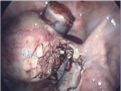
The images shown are from a 27 year old woman who presented with a ruptured right cornual pregnancy. She was admitted with an acute abdomen and hemoperi- toneum. Around 500 cc blood was evacuated from her abdomen. Based on the patient's condition, it was felt that the case could be managed laparoscopically (Figure 1). After diluted vasopressin solution was infused into the uterus around the ectopic site and excellent blanching of the uterus occurred, excision of the cornual pregnancy was began. First, the tube was taken at the midisthmic portion with the Harmonic Scalpel and fol- lowed back up along the round ligament taking the mesosalpinx toward the uterus. As we approached the junction of the tube with the uterus and the area became thick and more vascular, this dissection was stopped and the area of bleeding was coagulated with bipolar cautery.
Dissection was then taken back to the uterus, and using Harmonic Scalpel the specimen was resected down to the cornu of the endometrial cavity. Hemostasis was se- cured with the Harmonic Scalpel and with Pitressin injec- tion. The removed specimen was stored in the anterior cul-de-sac (Figure 2). The patient then had intracorporeal knot tying with 8-0 Polyglactin (Vicryl) to get a hemostatic closure of the cornu. Three figure-of-eights were required to close the cornu. Then a second 8-0 Vicryl was used to bring the round ligament up over the cornual defect to cover it to prevent scarring to the site giving excellent coverage of the closure site of the cornu (Figure 3). At this point, a slightly larger incision was made in the left lower quadrant and an endopouch was used to remove the specimen which was sent for pathology. A fascial su- turing device was used to oversew the peritoneal fascial defect in the left lower quadrant. The adipose tissue was reapproximated with 4–0 Polydioxanone (PDS) interrupted and a 4–0 PDS subcuticular closure was done then closed with a liquid bonding agent. Thorough irrigation of the abdomen was done, then the trocars were removed under vision and the gas allowed to pass from the umbilical trocar which was removed last. On removing of all trocars, the incisions were closed with a liquid bonding agent.
Conclusions
The purpose of this paper is to increase awareness and understanding of this minimally invasive modality for treating cornual pregnancies and to advocate for the routine use of transvaginal ultrasound at 9 to 12 weeks allowing confirmation of an extrauterine conception and making high risk management decisions simpler throughout pregnancy. Laparoscopic surgery for cornual ectopic is a minimally invasive, safe procedure if per- formed by a confident and experienced surgeon and has the advantage of preserving future fertility [3]. However, as with any surgery on the uterus the scar of a previous laparoscopic cornual resection may become the site of a uterine rupture in future pregnancy and therefore a prior cornual resection in our opinion contraindicates la- boring a patient and is an indication for C-section [6].
Notes
Conflicts of interest
None declared.
References
1. Chopra S, Keepanasseril A, Rohilla M, Bagga R, Kalra J, Jain V.
Obstetric morbidity and the diagnostic dilemma in pregnancy in rudimentary horn: retrospective analysis. Arch Gynecol Obstet.
2009;280(6):907-10. Epub 2009 Mar 13. DOI:
10.1007/s00404-009-1013-4
2. Pluchino N, Ninni F, Angioni S, Carmignani A, Genazzani AR, Cela V. Spontaneous cornual pregnancy after homolateral
salpingectomy for an earlier tubal pregnancy: a case report and literature review. J Minim Invasive Gynecol. 2009;16(2):208-11.
DOI: 10.1016/j.jmig.2008.11.008
3. Api M, Api O. Laparoscopic cornuotomy in the management of an advanced interstitial ectopic pregnancy: a case report. Gynecol Endocrinol. 2010;26(3):208-12. DOI:
10.3109/09513590903215524
4. MacRae R, Olowu O, Rizzuto MI, Odejinmi F. Diagnosis and laparoscopic management of 11 consecutive cases of cornual ectopic pregnancy. Arch Gynecol Obstet. 2009;280(1):59-64.
Epub 2008 Dec 16. DOI: 10.1007/s00404-008-0872-4 5. Tinelli A, Malvasi A, Pellegrino M, Pontrelli G, Martulli B, Tsin DA.
Laparoscopical management of cornual pregnancies: a report of three cases. Eur J Obstet Gynecol Reprod Biol. 2010. [Epub ahead of print]
6. Liao CY, Ding DC. Repair of uterine rupture in twin gestation after laparoscopic cornual resection. J Minim Invasive Gynecol.
2009;16(4):493-5. DOI: 10.1016/j.jmig.2009.03.025
Corresponding author:
M. Sami Walid, MD, PhD
Medical Center of Central Georgia, 840 Pine Street, Suite 880 Macon, GA 31201, USA, Tel.: (478) 743-7092 ex 266
mswalid@yahoo.com
2/4 GMS German Medical Science 2010, Vol. 8, ISSN 1612-3174
Walid et al.: Diagnosis and laparoscopic treatment of cornual ectopic ...
Figure 1: The cornual pregnancy before excision
Figure 2: The cornual pregnancy excised and stored in the anterior cul-de-sac
Figure 3: The final outcome of laparoscopic surgery
3/4 GMS German Medical Science 2010, Vol. 8, ISSN 1612-3174
Walid et al.: Diagnosis and laparoscopic treatment of cornual ectopic ...
Please cite as
Walid MS, Heaton RL. Diagnosis and laparoscopic treatment of cornual ectopic pregnancy. GMS Ger Med Sci. 2010;8:Doc16.
DOI: 10.3205/000105, URN: urn:nbn:de:0183-0001050
This article is freely available from
http://www.egms.de/en/journals/gms/2010-8/000105.shtml
Received:2010-01-29 Revised:2010-05-29 Published:2010-07-27
Copyright
©2010 Walid et al. This is an Open Access article distributed under the terms of the Creative Commons Attribution License
(http://creativecommons.org/licenses/by-nc-nd/3.0/deed.en). You are free: to Share — to copy, distribute and transmit the work, provided the original author and source are credited.
4/4 GMS German Medical Science 2010, Vol. 8, ISSN 1612-3174
Walid et al.: Diagnosis and laparoscopic treatment of cornual ectopic ...