Diagnosis and management of subclinical hypothyroidism in pregnancy
Roberto Negro,
1Alex Stagnaro-Green
2ABSTRACT
In prospective studies, the prevalence of undiagnosed subclinical hypothyroidism in pregnant women ranges from 3% to 15%. Subclinical hypothyroidism is associated with multiple adverse outcomes in the mother and fetus, including spontaneous abortion, pre-eclampsia, gestational hypertension, gestational diabetes, preterm delivery, and decreased IQ in the offspring. Only two prospective studies have evaluated the impact of levothyroxine therapy in pregnant women with subclinical hypothyroidism, and the results were mixed. Subclinical hypothyroidism is defined as raised thyrotropin combined with a normal serum free thyroxine level. The normal range of thyrotropin varies according to geographic region and ethnic background. In the absence of local normative data, the recommended upper limit of thyrotropin in the first trimester of pregnancy is 2.5 mIU/L, and 3.0 mIU/L in the second and third trimester. The thyroid gland needs to produce 50%
more thyroid hormone during pregnancy to maintain a euthyroid state. Consequently, most women on levothyroxine therapy before pregnancy require an increase in dose when pregnant to maintain euthyroidism. Ongoing prospective trials that are evaluating the impact of levothyroxine therapy on adverse outcomes in the mother and fetus in women with subclinical hypothyroidism will provide crucial data on the role of thyroid hormone replacement in pregnancy.
Introduction
The past 20 years have witnessed a dramatic increase in the understanding of the interaction between the thyroid gland and pregnancy, and its impact on adverse events in the mother and fetus. In particular, our understand‑
ing of how to interpret thyroid function results during pregnancy, as well as the prevalence of subclinical hypo‑
thyroidism, its impact, and the best way to treat it, have improved. Pregnancy is a stress test for the thyroid. The thyroid gland must produce 50% more thyroid hormone for euthyroidism to be maintained and to provide enough thyroid hormone for the developing fetus. Simultane‑
ously, the physiological changes that accompany preg‑
nancy result in marked alterations in the normal range of thyroid function. Specifically, human chorionic gon‑
adotropin, which peaks in the first trimester, crossreacts with the thyrotropin receptor, resulting in an upper limit of normal of thyrotropin of 2.5 mIU/L during the first tr imester.1‑4
The most dramatic recent findings have been the asso‑
ciation between subclinical hypothyroidism during preg‑
nancy and multiple negative outcomes in the mother and the fetus, including spontaneous abortion, gestational hypertension, gestational diabetes, pre‑eclampsia, pre‑
term delivery, and decreased IQ in the offspring.5‑7 The increased prevalence of thyroid dysfunction in pregnancy and the need for proper management to reduce obstetrical and neonatal adverse events led the American
Thyroid Association (ATA) and the Endocrine Society (ES) to release specific guidelines.8 9
The current review provides a summary of the above, recommendations for the diagnosis and treatment of subclinical hypothyroidism, and a discussion of the pros and cons of universal screening for thyroid disease during pregnancy. It concludes with a description of prospective ongoing trials.
Definition of subclinical hypothyroidism
Subclinical hypothyroidism is defined as the combination of a raised thyrotropin concentration and normal serum thyrox‑
ine (either total thyroxine or free thyroxine). In theory, the diagnosis of subclinical hypothyroidism does not contain an upper thyrotropin limit as long as thyroxine remains within
SOURCES AND SELECTION CRITERIA
We created a list of all relevant topics related to subclinical hypothyroidism in pregnancy and then performed a comprehensive literature review, carrying out a systematic PubMed and Medline search for original articles, reviews, and guidelines published from 1990 to February 2014.
Primary papers published before 1990 that were seminal in the field were also analyzed. The search terms used were thyrotropin, levothyroxine, pregnancy, subclinical hypothyroidism, adverse effects, abortion, miscarriage, iodine, thyroid antibodies, and Hashimoto’s thyroiditis. We prioritized randomized controlled trials, meta-analyses, and important studies in the field.
1Division of Endocrinology, Department of Internal Medicine,
“V Fazzi” Hospital, Lecce, 73100, Italy
2University of Illinois-College of Medicine at Rockford, Rockford, IL, USA
Correspondence to: N Roberto dr.
negro@libero.it
Cite this as: BMJ 2014;349:g4929 doi: 10.1136/bmj.g4929
thebmj.com
Use our interactive tool to explore the the definitions of SCH in key studies. See http://www.bmj.com/
content/349/bmj.g4929/
infographic
the reference range. However, The ATA, ES, and American Association of Clinical Endocrinologists (AACE) have recom‑
mended and generally accepted that any pregnant woman with thyrotropin above 10.0 mIU/L and normal free thy‑
roxine should be diagnosed with overt hypothyroidism.8‑10 Subclinical hypothyroidism is a biochemical diagnosis and cannot be based on the patient’s symptoms because these are non‑specific and often mimic some of the normal symptoms that a woman experiences during pregnancy.11
Thyrotropin and free thyroxine ranges in different trimesters
The normal reference ranges of thyrotropin and free thy‑
roxine differ markedly during pregnancy from those seen in non‑pregnant women. To complicate interpretation fur‑
ther, thyrotropin and free thyroxine are significantly differ‑
ent in each of the three trimesters, so that reference ranges need to be trimester specific. It is therefore imperative to use pregnancy derived normal ranges to avoid misdiagnosing a significant number of euthyroid pregnant women as having thyroid disease.12
Factors that alter thyroid function during pregnancy include the thyrotropic action of human chorionic gonad‑
otropin, increased iodine renal clearance, raised concen‑
trations of serum thyroxine binding globulin (TBG), and inner ring placental deiodination of thriiodothyroinine and thyroxine.13‑17 Multiple studies have shown that in iodine replete areas, thyrotropin values decrease during the first trimester, whereas concentrations progressively increase during the second and the third trimesters. By contrast, thyroxine is highest in the first trimester and decreases as pregnancy progresses.18
The ATA 2011 and the ES 2012 guidelines recommend that the normal thyrotropin reference range should be 0.1‑
2.5 mIU/L, 0.2‑3.0 mIU/L, and 0.3‑3.5 mIU/L in the first, second, and third trimesters of pregnancy, respectively.8 9 However, these reference ranges are probably not valid worldwide, because recent publications indicate that val‑
ues vary with geographic region and ethnic origin.
Thyrotropin and free thyroxine ranges in different countries Studies from China and India, for example, reported a signifi‑
cantly higher thyrotropin reference range for each trimester (table).19‑21 Specifically, the normal first trimester range in Chinese women was reported as 0.12‑5.08 mIU/L. Use of the ATA 2011 and the ES 2012 guidelines would have resulted in 28% of pregnant Chinese women being diagnosed as having subclinical hypothyroidism versus 4% if the ethnic specific reference range had been used.20 This study also found that
only 30.0% and 20.3% of the 118 pregnant women who had serum thyrotropin greater than 2.5 mIU/L in the first trimes‑
ter had a value greater than 3.0 mIU/L at the 20th and 30th week of gestation, respectively.
The Generation R prospective cohort study of 3944 people used a 2.5 mIU/L cut‑off point as the upper limit of thyrotropin. It reported a marked difference in the prevalence of subclinical hypothyroidism between four ethnic groups—people of Dutch (15.5%), Moroccan (2.7%), Turkish (10.8%), and Surinamese (17.9%) origin.
In addition, when population based or ethnicity specific reference ranges were compared, the diagnosis changed in 18% of women initially diagnosed with abnormal t hyroid function.22
Measurement of free thyroxine
Intrinsic to the definition of subclinical hypothyroidism is that serum thyroxine falls within the trimester specific ref‑
erence range. However, the diagnosis of subclinical hypo‑
thyroidism during pregnancy is complicated by doubts about the accuracy of the standard thyroxine immunoassay during pregnancy. Some investigators have concluded that increased serum TBG concentrations and reduced albumin interfere with the results and decrease the utility of this assay during pregnancy.23 However, others have concluded that the accuracy of the assay is satisfactory.24
As an alternative, dialysate or ultrafiltrate using online solid phase extraction‑liquid chromatography‑tandem mass spectrometry, has been recommended as the gold standard.23 However, this technology is expensive, tech‑
nically demanding, and is not available in most clinical laboratories.
Because of the lack of consensus regarding the optimal technique for measuring free thyroxine during pregnancy, alternative strategies have been proposed. The first alter‑
native is to multiply the non‑pregnant total thyroxine reference range (50‑150 nmol/L or 3‑8.8 µg/dL) by 1.5.
The second, the so called free thyroxine index, which has been shown to be reliable during pregnancy, is based on two estimates, the thriiodothyroinine resin uptake and the total thyroxine assay.9 Unfortunately, no trimester specific reference intervals are available for the free thyroxine index.
Furthermore, many laboratories no longer measure total thyroxine, which limits the use of this index.
Using the standard immunoassay during pregnancy, ret‑
rospective studies have indicated that the lower free thy‑
roxine concentration (2.5th centile) is about 10.3 pmol/L (0.8 ng/dL) in the first trimester.18 25‑ 28 However, given the limitations of the free thyroxine assay, the ATA 2011 and the ES 2012 guidelines suggest that, whenever feasible, the reference range for thyroid function tests during pregnancy should be assessed on a local basis, assuming that daily iodine intake is adequate.
A comparison of two longitudinal prospective cohort studies showed the importance of using local reference ranges. Different immunoassays were used to measure thy‑
rotropin and free thyroxine in two separate Danish cohorts of healthy pregnant thyroid antibody negative women.
Although thyrotropin values were comparable between the different assays and cohorts, highly significant differences in free thyroxine were seen between cohorts. Specifically, Thyrotropin reference ranges in different populations
Reference Population
Thyrotropin reference range (mIU/L) 1st trimester 2nd trimester 3rd trimester
Stagnaro-Green8 US* 0.1-2.5 0.2-3.0 0.3-3.0
De Groot9 US† 0.1-2.5 0.2-3.0 0.3-3.5
Yan19 Chinese 0.03-4.51 0.05-4.50 0.47-4.54
Li20 Chinese 0.14-4.87
Marwaha21 Indian 0.6-5.0 0.44-5.78 0.74-5.7
Korevaar22 Mixed (Dutch, Moroccan, Turkish, Surinamese) 0.06-4.51
*American Thyroid Association guideline recommendations.
†Endocrine Society guideline recommendations.
Even studies within the same country among different ethnic groups have found a great variation in prevalence.22 These discrepancies are probably the result of different daily intakes of iodine, varying prevalence of thyroid auto‑
immunity, genetic background, and environmental factors.
The marked variation in the thyrotropin range distribution seen in the literature supports the ATA 2011 and ES 2012 recommendation that during pregnancy a trimester specific reference range should be established on a local basis.
Iodine deficiency in pregnancy
During pregnancy, the iodine requirement increases by about 50% because the woman needs to produce more thyroid hor‑
mone, renal loss of iodine is exacerbated by the increased glomerular filtration rate, and the fetus needs to produce thyroid hormone during the second half of pregnancy.15‑17 The contribution of iodine deficiency to thyroid insufficiency depends on the severity of iodine deficiency, and inadequate iodine intake is seen in both developing and developed coun‑
tries.40 In 2011, nearly 45% of Europeans, including preg‑
nant women and those of child bearing age, were estimated to be iodine deficient.41
Although the deleterious effects of severe iodine defi‑
ciency on fetal development have been studied extensively, less is known about the impact of mild to moderate iodine insufficiency.42 The Avon Longitudinal Study of Parents and Children confirmed the central role that maternal iodine status plays in the development of childhood cognition.43 This British study assessed IQ at age 8 years (Wechsler intel‑
ligence scale for children) and reading speed, accuracy, and comprehension (Neale analysis of reading ability) at age 9 years in 7408 children. Each mother’s urinary iodine con‑
centration was measured in stored samples from the first trimester (≤13 weeks’ gestation; median 10 weeks). Iodine to creatinine ratios were dichotomized on the basis of World Health Organization criteria for iodine deficiency or suffi‑
ciency in pregnancy (<150 μg/g or ≥150 μg/g). The results showed that after adjustment for confounders, children of women with an iodine to creatinine ratio less than 150 μg/g were more likely to have scores in the lowest quartile for ver‑
bal IQ, reading accuracy, and reading comprehension than children of mothers with ratios 150 μg/g or more. Moreover, when the less than 150 μg/g group was subdivided, scores worsened progressively in the less than 150 μg/g, 50‑150 μg/g, and less than 50 μg/g subgroups.
A Spanish study showed that children whose mothers had received an iodine supplement of 300 μg had a more favorable psychometric assessment than those who had not (higher scores on the psychomotor development index (P=0.02) and the behavior rating scale).44
These data highlight the importance of adequate iodine status during early gestation. They also emphasize the risk of iodine deficiency even in developed countries and the need for randomized placebo controlled trials to test the effect of maternal iodine supplementation on child cogni‑
tion.
The National Health and Nutrition Examination Sur‑
vey (NHANES) has documented a marked decrease in the median urinary iodine concentration over the past three decades, with the current value for pregnant women being 125 μg/L, indicating that pregnant women in the US are use of the gestational age specific range for free thyroxine
from one cohort resulted in misclassification of all free thy‑
roxine values in the other cohort.29
In summary, thyrotropin and thyroxine concentrations change throughout the three trimesters of pregnancy and probably differ by both ethnic group and geographic area.
When feasible, local reference ranges should be used. How‑
ever, most clinicians will not have access to such a labo‑
ratory. Consequently, clinicians should use the published thyrotropin reference range that is most appropriate for their geographic area and ethnic origin of their patients.
Causes of hypothyroidism
The leading cause of hypothyroidism in developing countries is severe iodine deficiency, whereas in developed countries it is autoimmune thyroiditis. Thyroid autoantibodies are detected in about half of pregnant women with subclinical hypothyroidism and in more than 80% with overt hypothy‑
roidism.30 Antibodies directed against thyroid peroxidase (TPO‑Ab) should therefore be measured in patients with subclinical hypothyroidism to establish a diagnosis of auto‑
immune thyroid disease.8‑10
Although only positive TPO‑Ab tests have been shown to be significantly associated with hypothyroidism, antibodies to thyroglobulin (TG‑Ab) should also be measured.31 In a study of 992 unselected women who consulted a tertiary referral center for infertility, the overall prevalence of auto‑
immune thyroid disease was 16%. Of these women, 8% had both antibodies, 5% had TG‑Ab only, and 4% had TPO‑Ab only. Women with isolated TG‑Ab had significantly higher serum thyrotropin concentrations than those without auto‑
immune thyroid disease.32
If thyrotropin concentrations are raised, TPO‑Ab should be measured to establish a diagnosis of autoimmune thy‑
roid disease. If TPO‑Ab are present, the measurement of TG‑Ab should be considered. Finally, it is important to real‑
ize that because the immune system is suppressed during pregnancy, thyroid antibody titers decrease on average by 60% in the second half of pregnancy.4 Consequently, in some women with autoimmune thyroid disease, thyroid antibody test will be negative during pregnancy but posi‑
tive postpartum because the immunosuppression of preg‑
nancy yields to an immunologic rebound during the first six months postpartum.
Prevalence of subclinical hypothyroidism
Estimates of the prevalence of subclinical hypothyroidism in the first trimester of pregnancy vary. Initially, this varia‑
tion was the result of early studies using a non‑pregnancy specific upper limit of normal range (4‑6 mIU/L) rather than the current definition of 2.5 mIU/L as set by the ATA 2011 and ES 2012 guidelines.30
Although most recent studies performed in the United States have shown a 2‑3% prevalence of subclinical hypo‑
thyroidism when using a thyrotropin cut off of 2.5 mIU/L, a large study published in 2012 reported a prevalence of 15.5% using an upper limit of normal of 2.75 mIU/L.27 33‑ 37 Similarly, studies conducted outside the US and those that have included various ethnic groups report levels of sub‑
clinical hypothyroidism in excess of 15% when using the ATA 2011 and ES 2012 guidelines.20 38 39
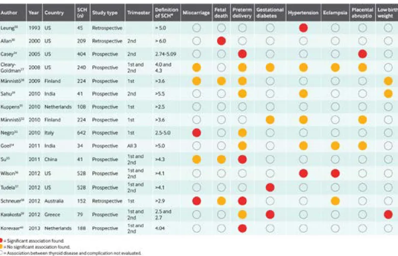
neuro‑intellectual development of the offspring. Figure 1 presents the 16 studies, all of which have been published in the past 20 years.27 30 34 36 39 50‑ 60
In these mostly prospective observational studies, the cut off used to define subclinical hypothyroidism and the point in gestation at which thyrotropin was assessed varied. Some evaluated fewer than 100 women with subclinical hypothy‑
roidism and are therefore underpowered to detect the preg‑
nancy and neonatal complications studied, which occur in a small proportion of pregnant women. Preterm delivery, miscarriage, and gestational hypertension were associated with subclinical hypothyroidism in two or more studies.
Eight further studies focused on evaluating the rate of subclinical hypothyroidism in a population of women with specific pregnancy related complications. Four studies focused on preterm delivery. One found a threefold increase in subclinical hypothyroidism (thyrotropin ≥3.0 mIU/L) in women with very preterm delivery (n=28) compared with 124 women who delivered at term.61 However, the other three studies did not report an increased prevalence of sub‑
clinical hypothyroidism in women who delivered preterm compared with those who delivered at term.62‑64
A Dutch study found a significant increase in the rate of subclinical hypothyroidism (thyrotropin ≥2.5 mIU/L) in 58 women who presented in the breech position com‑
pared with 1000 women who presented in the cephalic position.51 Another study found that, in the third trimester of pregnancy, thyrotropin concentrations were significantly probably mildly iodine deficient.45 The Institute of Medi‑
cine recommends a dietary intake of 220 μg iodine per day during pregnancy and 290 μg per day in breastfeed‑
ing women.46 Consequently, many organizations, includ‑
ing the ATA, the ES, the AACE, and the Teratology Society recommend that all pregnant and breastfeeding women take a prenatal vitamin that contains 150 μg of potassium iodide.47
Impact of subclinical hypothyroidism on pregnancy outcome and intellectual development of the fetus Studies have evaluated the impact of hypothyroidism on the outcome of pregnancy for more than 50 years, and early studies provided clear evidence of a link between overt hypo‑
thyroidism and adverse events.48 Subsequent studies have confirmed that gestational hypertension, pre‑eclampsia, increased placental weight, cretinism, low birth weight, fetal death, spontaneous abortion, and intrauterine growth retar‑
dation are all associated with overt hypothyroidism in preg‑
nancy.49 Therefore, it is now well accepted that the detection and treatment of pregnant women with overt hypothyroidism is crucial to both maternal and fetal health.
Pregnancy complications excluding neuro-intellectual development
To date, 16 observational studies have evaluated the association between subclinical hypothyroidism and complications of pregnancy that are not related to the
Fig 1 | Data from 16 studies that have evaluated an association between thyroid disease and pregnancy and maternal, fetal, neonatal, or offspring complications.
SCH=subclinical hypothyroidism
It is difficult to compare the results of the above studies because of differences in the patients’ ages and the tests used. It is plausible, for example, that subclinical hypo‑
thyroidism, rather than reducing global IQ, may exert det‑
rimental effects on specific functions, such as memory or visuospatial and orientation performance.
Studies that included women with subclinical hypothyroidism or overt hypothyriodism
Three other studies that assessed adverse outcomes in women with subclinical hypothyroidism or overt hypothy‑
roidism are worth noting. The first was a retrospective elec‑
tronic chart analysis of the US Cohort Consortium on Safe Labor data, which analyzed thyroid status and pregnancy outcome in 223 512 singleton pregnancies. Hypothyroid‑
ism (subclinical hypothyroidism was not differentiated from overt hypothyroidism) was significantly associated with pre‑eclampsia, gestational diabetes, preterm birth, cesarean section, admission of the mother to intensive care, placental abruption, and breech position.72
The second study from the same US cohort found that maternal hypothyroidism increased the risk of neonatal sepsis, respiratory distress syndrome, transient tachypnea, and apnea.73
A study of 2497 Dutch women found a positive linear association between risk of child loss, and thyrotropin values, which extended into the normal range (for exam‑
ple, the absolute risk for child loss increased from 0.8%
in women with a thyrotropin of 0.54 mIU/L to 2.2% when thyrotropin was 3.13 mIU/L).74
Finally, three further studies have evaluated pregnancy outcome in women being treated for hypothyroidism dur‑
ing pregnancy. A retrospective study of 150 women with hypothyroidism, 99 of whom were on treatment before conception, found that when treatment was inadequate (thyrotropin ≥4.0 mIU/L during pregnancy), 71% of the women with subclinical hypothyroidism aborted.75 Another reported on pregnancy outcomes in 848 468 women included in the Swedish Health Register, 9866 of whom were on thyroid hormone.76 Women treated with thyroid hormone had a significantly increased risk of congenital malformations, preterm birth, cesarean section, gestational diabetes, and pre‑eclampsia. A study of 203 American women who had previously had subclinical hypothyroid‑
ism during pregnancy found increased rates of gestational diabetes and stillbirth in a subsequent pregnancy.77
Overall, evidence published over the past 20 years sup‑
ports an association between subclinical hypothyroid‑
ism and adverse maternal, fetal, and neonatal outcomes.
Although not all studies found such associations, many of the negative studies had inadequate study size and a lack of power to find any association that might exist. Alterna‑
tively, in some, screening was performed too late in preg‑
nancy. However, some well designed studies have found no link between subclinical hypothyroidism and adverse pregnancy outcomes.
Results from studies of association should always be interpreted with caution, and this is especially so for studies of thyroid function in pregnancy. This is because hypothy‑
roidism may worsen in women with subclinical hypothy‑
roidism in the first trimester as pregnancy progresses—this higher in 100 women with pre‑eclampsia than in 50 ges‑
tation matched normotensive controls (thyrotropin 5.63 v 2.00 mIU/L).65
Only one randomized placebo controlled trial has assessed whether levothyroxine therapy would decrease the rate of adverse pregnancy and neonatal events in sub‑
clinical hypothyroidism.66 It screened 4562 women at a mean gestational age of 8.8 weeks. Tests for thyrotropin, free thyroxine, and thyroid antibodies were performed immediately in half of the women and serum samples were frozen and assayed after delivery in the other half. Women treated during pregnancy had a significant decrease in the rate of adverse obstetrical and neonatal outcomes (number needed to treat 40, 95% confidence interval 1.4 to 2.5).
Neurologic and intellectual complications
Maternal thyroxine is crucial for the normal neurodevelop‑
ment of the fetus. Animal studies have shown that mater‑
nal hypothryroxinemia, with or without raised thyrotropin, can result in adverse neurodevelopmental outcomes.67 Not surprisingly, therefore, in the past two decades most atten‑
tion has been paid to the impact of maternal subclinical hypothyroidism on the intellectual and neurologic devel‑
opment of children.
The first study to confirm the association was published in 1999.68 It compared the IQ of 64 children (age 7‑9 years) born to mothers with thyrotropin values above the 98th centile with that of 124 children born to euthyroid preg‑
nant control women. The mothers were part of a cohort of 25 216 women previously screened for Down’s syndrome in the second trimester. Of the 64 women with raised TSH concentrations during pregnancy, 48 were not treated with levothyroxine. The IQ of the offspring of these 48 women was 7 points lower than that of the offspring of the 128 controls (P=0.005).
Subsequently, two smaller studies assessed the associa‑
tion between subclinical hypothyroidism and neurologic development of the newborn. One found a significant decrease in the mean mental developmental index (MDI) in the offspring of seven women with subclinical hypo‑
thyroidism during pregnancy compared with that of the offspring of six women who were euthyroid during preg‑
nancy.69 The other screened 1268 women at gestational weeks 16‑20 for thyroid function and assessed the intel‑
lectual and motor development of their children at the age of 25‑30 months.70 Children of the nine mothers with subclinical hypothyroidism during pregnancy had signifi‑
cantly lower scores on the Bayley Scale of Infant Develop‑
ment and the MDI when compared with the offspring of mothers who were euthyroid during pregnancy.
In 2012, a prospective multi‑country randomized con‑
trolled trial in Europe assessed the impact of levothyroxine in 21 846 women with a thyrotropin concentration greater than the 97.5th centile or free thyroxine lower than the 2.5 centile (or both).71 Serum samples were obtained at a mean gestational age of 12 weeks and three days, and the pri‑
mary outcome variable was the children’s IQ at 3 years of age. Mean IQ and the proportion of children with IQ levels below 85 were not significantly different between the 390 children of the mothers treated during pregnancy and the 404 children of those who were not treated.
pregnant. The increase was seen as early as the fifth gesta‑
tional week and the average increment was 47%.82 The thyrotropin concentration before pregnancy affects if, and how much, levothyroxine will need to be increased during pregnancy. A prospective study found that before pregnancy the mean thyrotropin value was lower in 50 women who did not need additional levothyroxine during pregnancy than in the 34 women who needed an increase (1.36 v 4.63 mIU/L).83 Similarly, a prospective study found that only 17% of women with a thyrotropin less than 1.2 mIU/L before conception needed an increase in their dose during pregnancy compared with 50% of women whose preconception thyrotropin was 1.2‑2.4 mIU/L.84
A randomized trial compared two different levothyrox‑
ine regimens during pregnancy in women with pre‑existing hypothyroidism.85 The daily prepregnancy levothyroxine dose was increased by two tablets a week in one group and by three tablets a week in the second group. Thyroid function tests were performed every two weeks during preg‑
nancy. Increasing the pregestation dose, by either two or three tablets, maintained thyrotropin below 5.0 mIU/L in all women. Thyrotropin was below 0.1 mIU/L in only 8%
of women on the two extra dose regimen, whereas suppres‑
sion of thyrotropin was found in 26% of women who took three extra tablets. Most (92%) abnormal thyrotropin val‑
ues were detected by monitoring thyroid levels every four weeks.
Maintenance and monitoring of levothyroxine therapy During pregnancy the thyroid gland must produce 30‑50%
more thyroxine to maintain the euthyroid state. This results in new onset subclinical hypothyroidism in women who were euthyroid before pregnancy but had decreased thyroidal reserve (most often as a result of autoimmune thyroid dis‑
ease). Whether levothyroxine therapy should be continued postpartum, when increased thyroid hormone production is no longer necessary, is an important question. A recent retrospective study followed up 65 women who developed subclinical hypothyroidism during pregnancy but stopped taking levothyroxine postpartum.86 Most women (75%) were euthyroid after five years, whereas 25% had a persistently raised thyrotropin (>4.5 mIU/L). The presence of TPO‑Ab was the most important risk factor for a raised thyrotropin.
Recommendations
Both the ATA 2011 and the ES 2012 guidelines contain rec‑
ommendations for the initiation of levothyroxine for subclini‑
cal hypothyroidism during pregnancy and the frequency of thyrotropin monitoring once treatment is started.8 9 The ATA guidelines recommend that all pregnant women with thyro‑
tropin greater than 2.5 mIU/L and normal free thyroxine who are TPO‑Ab positive and all women with thyrotropin greater than 10.0 mIU/L, irrespective of the free thyroxine value, be treated with levothyroxine. The guidelines concluded that insufficient data were available for TPO‑Ab negative pregnant women with a thyrotropin greater than 2.5 mIU/L.
The ES guidelines recommend that all women with a thy‑
rotropin greater than 2.5 mIU/L and normal free thyroxine in the first trimester be treated with levothyroxine, irrespec‑
tive of their TPO‑Ab status. The ES guidelines recommend that thyrotropin testing is repeated every four to six weeks is particularly true for those who are TPO‑Ab positive. Con‑
versely, some women, particularly those who are TPO‑Ab negative, return to the euthyroid state as pregnancy pro‑
ceeds.4 20 Ongoing prospective studies will be invaluable in further defining the association between subclinical hypo‑
thyroidism and maternal, fetal, and neonatal outcomes.
Treatment of subclinical hypothyroidism
The decision on whether to treat subclinical hypothyroidism diagnosed during pregnancy is controversial. The ATA 2011 and the ES 2012 guidelines, but not the American College of Obstetricians and Gynecologists guidelines, recommend initiating levothyroxine therapy in these patients.8 9 78 For physicians who elect to start treatment, the evidence on the timing and dose modification is presented below.
Women without pre-existing hypothyroidism
Evidence on the appropriate dose of levothyroxine in preg‑
nant women newly diagnosed with subclinical hypothyroid‑
ism is limited. A prospective randomized controlled trial gave all women 150 µg of levothyroxine from the start of treat‑
ment.71 A high dose was used because of the need to normal‑
ize maternal thyrotropin and free thyroxine values rapidly. In 85% of women, the dose did not need to be modified during pregnancy, whereas 10% needed a decrease in dose to 125 µg and 5% required an increase to 175 µg.
In another prospective trial of levothyroxine in 56 women with subclinical hypothyroidism first diagnosed during pregnancy,79 the starting dose of levothyroxine was based on the initial thyrotropin value. Women whose thyrotropin was 2.5‑5.0 mIL/U were started on 50 μg per day, women with a thyrotropin of 5.0‑8.0 mIU/L were started on 75 μg per day, and those with thyrotropin greater than 8.0 mIU/L were given 100 μg per day. Over 80% of the women did not require any dose adjustment during pregnancy.
A retrospective study assessed 64 women with new onset subclinical hypothyroidism during pregnancy.80 The goal of treatment was to achieve a thyrotropin of 2.5 mIU/L or less in the first trimester and 3.0 mIU/L or less in the second and third trimesters. Women with a presenting thyrotropin of 2.5‑4.2 mIU/L needed 1.20 (standard deviation 0.39) µg per kg per day (mean dose 77.98 µg/day) of levothyroxine, whereas those with a presenting thyrotropin of 4.2‑10.0 mIU/L needed 1.42 (0.31) µg per kg per day (95.35 µg/day).
In 89% of the women the initial dose of levothyroxine did not need to be modified as pregnancy progressed.
Women with pre-existing hypothyroidism
Women with pre‑existing hypothyroidism often need a higher dose of levothyroxine during pregnancy to maintain euthyroidism. Several studies have investigated this impor‑
tant issue, and their main results are summarized in chron‑
ological order. A retrospective analysis of thyroid function and levothyroxine dose requirements in 12 women found that all had a rise in thyrotropin during pregnancy and 75%
needed an increase in levothyroxine dose of about 50 μg per day.81 Postpartum, the mean thyrotropin value in all women decreased, indicating a decline in the post‑pregnancy thy‑
roxine dose requirement.
A prospective study found that the dose of levothyroxine needed to be increased in 17 of 20 women who became
in four to eight weeks in women who are not planning on having more children. It is recommended that women who are planning on pregnancy within the next couple of years carry on taking levothyroxine to ensure that they are euthy‑
roid at the time of conception.
Women on levothyroxine before pregnancy with a thyrotropin value less than 1.2 mIU/L may not need an increase in levothyroxine but require ongoing monitor‑
ing. Irrespective of the prepregnancy thyrotropin value, all patients should be instructed to have thyrotropin measured as soon as pregnancy is confirmed. Figure 2 summarizes these recommendations.
Screening for thyroid disease in pregnancy
Debate about the need for universal screening for thyroid dysfunction during pregnancy is ongoing. The expert panel involved in producing the ES guidelines on thyroid and preg‑
nancy could not reach consensus—some participants con‑
cluded that universal screening is warranted, whereas others thought that a case finding strategy should be initiated.9
Guidelines from AACE, the Society of Maternal‑Fetal Medicine, the American College of Obstetrics and Gynecol‑
ogy, the Cochrane Collaboration, and the ATA all endorse a case finding strategy.8 78 90‑ 92 Those who favor universal screening cite the increased prevalence of hypothyroidism (overt and subclinical) during pregnancy, the inexpensive nature of the treatment (levothyroxine), the wide availabil‑
ity of an inexpensive screening test (thyrotropin measure‑
ment), and the cost effectiveness of a screening strategy.
Those who oppose universal screening cite the paucity of evidence that identification and treatment of pregnant women with subclinical hypothyroidism improves maternal or neonatal outcomes.
The main unresolved point in the debate about uni‑
versal screening is the lack of an agreed policy on whom to treat given the paucity of randomised controlled trials in pregnant women with subclinical hypothyroidism. As discussed earlier, many studies and meta‑analyses have documented an association between subclinical hypo‑
thyroidism and gestational diabetes, miscarriage, preterm delivery, gestational hypertension, pre‑eclampsia, and impaired neuropsychological development of the offspring.
However, only two randomised controlled trials have been published that have investigated the potential benefits of treating maternal subclinical hypothyroidism.
Case finding as a strategy for identifying women with thyroid disease during pregnancy has limitations. Firstly, data from prospective studies have shown that when risk factors for thyroid disease are used, case finding will miss between 33% and 81% of pregnant women with hypothy‑
roidism.67 93‑ 95 Secondly, a large number of risk factors need to be evaluated, which is time consuming. Thirdly, obstetri‑
cians and gynecologists provide the majority of pregnancy related care. Studies have reported that some obstetricians have limited knowledge about the association between thy‑
roid disease and pregnancy.96 97
Even the risk factors that need to be included in a case finding strategy are controversial. Both the ATA 2011 and ES 2012 guidelines recommend screening all women over the age of 30 years. Although one analysis found no significant association between age and abnormal thy‑
throughout pregnancy and the ATA guidelines recommend testing every four weeks until 16‑20 weeks and at least once between 26 and 32 weeks’ gestation.
In 2013, clinical members of the ES, the ATA, and the AACE were asked to fill in a web based survey consisting of 30 questions that dealt with testing, treatment, and modulating factors in the management of primary hypothy‑
roidism. Results showed that 96.1% of respondents used a thyrotropin target of less than 2.5 mIU/L in newly preg‑
nant women, whereas 63.5% preferred a target of less than 1.5 mIU/L. In addition, 67.7% would check thyroid hor‑
mone concentrations every four weeks during pregnancy, and 21.4% would check every eight weeks. Only 36.9% of respondents said they would immediately increase the dose of levothyroxine in a hypothyroid patient with a thyrotropin of 0.5 mIU/L who became pregnant.87
Application in clinical practice
Although the results of the survey demonstrate good adher‑
ence to current guidelines, in clinical practice pregnant women on thyroxine substitution are often dysregulated—
about 50% of women have a raised thyrotropin concentra‑
tion in the first half of gestation.88 89
Subclinical hypothyroidism has been associated with multiple adverse maternal, fetal, and neonatal outcomes, and a preliminary intervention trial suggests that treatment is beneficial.66 On the basis of current evidence, we believe it is reasonable to recommend treating women with new onset subclinical hypothyroidism during pregnancy. Levo‑
thyroxine therapy during pregnancy is inexpensive and has been shown to be safe.
We recommend adoption of the treatment algorithm rec‑
ommended by Yu and colleagues, which is based on the initial maternal thyrotropin value.79 Those with a thyro‑
tropin value of 2.5‑5.0 mIU/L, 5.0‑8.0 mIU/L, and greater than 8.0 mIU/L should be started on levothyroxine at 50 μg, 75 μg, and 100 μg per day, respectively. After delivery levothyroxine can be stopped or titrated down and retested Fig 2 | Treatment algorithm for women taking levothyroxine before pregnancy.78 TPO-Ab=anti- thyroid peroxidase antibodies
Institute of Child Health and Human Development study is screening pregnant women of less than 20 weeks’ gesta‑
tion for subclinical hypothyroidism or hypothyroxinemia.
Women will be randomized to treatment with levothyroxine or placebo until delivery (http://clinicaltrials.gov/ct2/show/
NCT00388297). The offspring will have annual developmen‑
tal testing until age 5 years to determine whether treatment improves IQ.
In the United Kingdom, the Thyroid AntiBodies and LEv‑
oThyroxine (TABLET) trial is recruiting euthyroid TPO‑Ab positive women before conception and treating half with levothyroxine and half with placebo (www.controlled‑trials.
com/ISRCTN15948785). The major outcome variables are the rate of miscarriage and preterm delivery.
Finally, in China 4800 women have been enrolled in the Subclinical Hypothyroid and Iodine Deficiency in Early Pregnancy and Women Planning for Pregnancy: Screen‑
ing and Intervention (SHEP) trial. A major factor being assessed is the impact of levothyroxine on fetal brain devel‑
opment.104 Conclusion
The past two decades have seen major advances in our understanding of the physiological changes that occur in the thyroid during pregnancy and the impact of subclinical hypothyroidism on adverse maternal and fetal outcomes. The normal upper range of thyrotropin is 2.5 mIU/L in the first trimester of pregnancy and 3.0 mIU/L in the second and third trimesters. Hypothyroidism is present in 2‑15% of pregnant women. It is mainly caused by iodine deficiency in develop‑
ing countries and autoimmune thyroid disease in developed countries. Subclinical hypothyroidism has been associated with multiple negative outcomes, including pregnancy loss, preterm delivery, gestational diabetes, and impaired neuro‑
logic development in the offspring. Women on levothyroxine before conception require careful management to ensure that the euthyroid state is maintained throughout pregnancy.
Although it is well accepted that treating overt hypothy‑
roidism decreases maternal and fetal adverse outcomes, current data on the impact of treating subclinical hypothy‑
roidism are limited and conflicting. Universal screening of all pregnant women to detect and treat overt hypothyroid‑
ism is recommended. Ongoing prospective trials will pro‑
vide future guidance on the efficacy of treating subclinical hypothyroidism during pregnancy.
Contributors: RN and AS-G contributed equally to the writing of this article and are both guarantors.
Competing interests: We have read and understood BMJ policy on declaration of interests and declare the following interests: none.
Provenance and peer review: Commissioned; externally peer reviewed.
1 Glinoer D. What happens to the normal thyroid during pregnancy? Thyroid 1999;9:631-5.
2 Soldin OP. Thyroid function testing in pregnancy and thyroid disease:
trimester-specific reference intervals. Ther Drug Monit 2006;28:8-11.
3 Mandel SJ, Spencer CA, Hollowell JG. Are detection and treatment of thyroid insufficiency in pregnancy feasible? Thyroid 2005;15:44-53.
4 Glinoer D, Riahi M, Grün JP, Kinthaert J. Risk of subclinical hypothyroidism in pregnant women with asymptomatic autoimmune thyroid disorders. J Clin Endocrinol Metab 1994;79:197-204.
5 Van den Boogaard E, Vissenberg R, Land JA, van Wely M, van der Post JA, Goddijn M, et al. Significance of (sub)clinical thyroid dysfunction and thyroid autoimmunity before conception and in early pregnancy: a systematic review. Hum Reprod Update 2011;17:605-19.
6 Reid SM, Middleton P, Cossich MC, Crowther CA, Bain E. Interventions for clinical and subclinical hypothyroidism pre-pregnancy and during pregnancy. Cochrane Database Syst Rev 2013;5:CD007752.
rotropin values, in a subgroup analysis of risk factors for hypothyroidism, the addition of women aged 30 years or more increased the proportion of women identified in a case finding screening strategy from 55.3% to 85.6%.98 Geographic considerations
The thyrotropin concentration at which women should be diagnosed with subclinical hypothyroidism varies by region.
As noted earlier, it is recommended that local guidelines be developed to determine the normal thyrotropin range for the three trimesters of pregnancy.8 9 Consequently, a patient with a thyrotropin of 4.0mIU/L would be diagnosed with subclini‑
cal hypothyroidism in the US but not in China or India.20 21 This may prove confusing to physicians and patients. Fur‑
thermore, it is unclear whether the thyrotropin value above which the incidence of a particular complication increases is the same as for other complications.
Finally, geographic differences in clinical practice affect the care that a patient receives. A 2010 survey of members of the European Thyroid Association reported a wide vari‑
ation in clinical practice regarding the diagnosis and treat‑
ment of thyroid disease in pregnancy. The survey found that 42% of responders, or their institutions, screened all pregnant women for thyroid dysfunction, 43% performed targeted screening of only high risk women, whereas 17%
carried out no systematic screening. Timing of the screen‑
ing, tests used, and criteria for starting treatment and moni‑
toring varied.99 In the US the approach to screening also varies widely. A study from Boston Medical Center showed a screening rate of 85%, whereas another national study in the US reported a screening rate of only 23%.37 100
Current evidence on subclinical hypothyroidism does not support universal screening. However, the incidence and impact of overt hypothyroidism and the ability of treat‑
ment to prevent associated adverse events is sufficient to justify universal screening for thyroid disease.48 101 102 In support of this position, a cost effective analysis showed that universal screening with the goal of identifying and treating overt hypothyroidism is cost effective.103 Because universal screening would also identify patients with sub‑
clinical hypothyroidism, these patients should be treated as indicated in current guidelines unless ongoing and future studies prove otherwise.
Future studies
Three ongoing randomized prospective trials will yield important data that should be instrumental in informing future guidelines on thyroid and pregnancy. A National
FUTURE RESEARCH QUESTIONS
Is there a thyrotropin threshold above which the rate of adverse obstetrical or fetal events is substantially increased?
Does the treatment of subclinical hypothyroidism during pregnancy decrease the risk of adverse events in the mother and the fetus?
Does the treatment of hypothyroxinemia during pregnancy decrease the risk of adverse events in the mother and the fetus?
Does maternal subclinical hypothyroidism cause a decrease in the IQ of the offspring?
31 Hollowell JG, Staehling NW, Flanders WD, Hannon WH, Gunter EW, Spencer CA, et al. Serum TSH, T(4), and thyroid antibodies in the United States population (1988 to 1994): National Health and Nutrition Examination Survey (NHANES III). J Clin Endocrinol Metab 2002;87:489-99.
32 Unuane D, Velkeniers B, Anckaert E, Schiettecatte J, Tournaye H, Haentjens P, et al. Thyroglobulin antibodies: any added value in the detection of thyroid autoimmunity in women consulting for fertility treatment. Thyroid 2013;23:1022-8.
33 Krassas GE, Poppe K, Glinoer D. Thyroid function and human reproductive health. Endocr Rev 2010;31:702-55.
34 Casey BM, Dashe JS, Wells CE, McIntire DD, Byrd W, Leveno KJ, et al.
Subclinical hypothyroidism and pregnancy outcomes. Obstet Gynecol 2005;105:239-45.
35 Vaidya B, Anthony S, Bilous M, Shields B, Drury J, Hutchison S, et al. Detection of thyroid dysfunction in early pregnancy: universal screening or targeted high-risk case finding? J Clin Endocrinol Metab 2007;92:203-7.
36 Männistö T, Vääräsmäki M, Pouta A, Hartikainen AL, Ruokonen A, Surcel HM, et al. Perinatal outcome of children born to mothers with thyroid dysfunction or antibodies: a prospective population-based cohort study. J Clin Endocrinol Metab 2009;94:772-9.
37 Blatt AJ, Nakamoto JM, Kaufman HW. National status of testing for hypothyroidism during pregnancy and postpartum. J Clin Endocrinol Metab 2012;97:777-84.
38 Moreno-Reyes R, Glinoer D, Van Oyen H, Vandevijvere S. High prevalence of thyroid disorders in pregnant women in a mildly iodine-deficient country: a population-based study. J Clin Endocrinol Metab 2013;98:3694-701.
39 Sahu MT, Das V, Mittal S, Agarwal A, Sahu M. Overt and subclinical thyroid dysfunction among Indian pregnant women and its effect on maternal and fetal outcome. Arch Gynecol Obstet 2010;281:215-20.
40 Aguayo A, Grau G, Vela A, Aniel-Quiroga A, Espada M, Martul P, et al. Urinary iodine and thyroid function in a population of healthy pregnant women in the north of Spain. J Trace Element Med Biol 2013;27:302-6.
41 Andersson M, Karumbunathan V, Zimmermann MB. Global iodine status in 2011 and trends over the past decade. J Nutr 2012;142:744-50.
42 Trumpff C, De Schepper J, Tafforeau J, Van Oyen H, Vanderfaeillie J, Vandevijvere S. Mild iodine deficiency in pregnancy in Europe and its consequences for cognitive and psychomotor development of children: a review. J Trace Element Med Biol 2013;27:174-83.
43 Bath SC, Steer CD, Golding J, Emmett P, Rayman MP. Effect of inadequate iodine status in UK pregnant women on cognitive outcomes in their children:
results from the Avon Longitudinal Study of Parents and Children (ALSPAC).
Lancet 2013;27:331-7.
44 Velasco I, Carreira M, Santiago P, Muela JA, García-Fuentes E, Sánchez-Muñoz B, et al. Effect of iodine prophylaxis during pregnancy on neurocognitive development of children during the first two years of life. J Clin Endocrinol Metab 2009;94:3234-41.
45 Caldwel KL, Makhmudov A, Ely E, Jones RL, Wang RY. Iodine status of the US population, National Health and Nutrition Examination Survey, 2005-2006 and 2007-2008. Thyroid 2011;21:419-27.
46 Food and Nutrition Board. Institute of Medicine. Dietary reference intakes.
National Academy Press, 2006:320-7.
47 Stagnaro-Green A, Sullivan S, Pearce EN. Iodine supplementation during pregnancy and lactation. JAMA 2012;23:1-2.
48 Jones WS, Man EB. Thyroid function in human pregnancy. Premature deliveries and reproductive failures of pregnant women with low serum butanol-extractable iodines. Maternal serum TBG and TBPA capacities. Am J Obstet Gynecol 1969;104:909-14.
49 Stagnaro-Green A. Overt hyperthyroidism and hypothyroidism during pregnancy. Clin Obstet Gynecol 2011;54:478-87.
50 Leung AS, Millar LK, Koonings PP, Montoro M, Mestman JH. Perinatal outcome in hypothyroid women. Obstet Gynecol 1993;81:349-53.
51 Kuppens SMI, Kooistra L, Carwford WS, Vader HL, Hasaart THM, Oei SG, et al.
Maternal thyroid function during gestation is related to breech presentation at term. Clin Endocrinol 2010;72:820-4.
52 Männistö T, Vääräsmäki M, Pouta A, Hartikainen AL, Ruokonen A, Surcel HM, et al. Thyroid dysfunction and autoantibodies during pregnancy as predictive factors of pregnancy complications and maternal morbidity in later life. J Clin Endocrinol Metab 2010;95:1084-94.
53 Negro R, Schwartz A, Gismondi R, Tinelli A, Mangieri T, Stagnaro-Green A.
Increased pregnancy loss rate in thyroid antibody negative women with TSH levels between 2.5 and 5.0 in the first trimester of pregnancy. J Clin Endocrinol Metab 2010;95:E44-8.
54 Goel P, Kaur J, Saha PK, Tandon R, Devi L. Prevalence, associated risk factors and effects of hypothyroidism in pregnancy: a study from north India.
Gynecol Obstet Invest 2012;74:89-94.
55 Su PY, Huang K, Hao JH, Xu YQ, Yan SQ, Li T, et al. Maternal thyroid function in the first twenty weeks of pregnancy and subsequent fetal and infant development: a prospective population-based cohort study in China. J Clin Endocrinol Metab 2011;96:3234-41.
56 Wilson KL, Casey BM, McIntire DD, Halvorson LM, Cunningham FG.
Subclinical thyroid disease and the incidence of hypertension in pregnancy.
Obstet Gynecol 2012;119:315-20.
57 Tudela CM, Casey BM, McIntire DD, Cunningham FG. Relationship of subclinical thyroid disease to the incidence of gestational diabetes mellitus.
Obstet Gynecol 2012;119:983-8.
58 Schneuer FJ, Nassar N, Tasevski V, Morris JM, Roberts CL. Association and predictive accuracy of high TSH serum levels in first trimester and adverse pregnancy outcomes. J Clin Endocrinol Metab 2012;97:3115-22.
7 Thangaratinam S, Tan A, Knox E, Kilby MD, Franklyn J, Coomarasamy A.
Association between thyroid autoantibodies and miscarriage and preterm birth: meta-analysis of evidence. BMJ 2011;342:d2616.
8 Stagnaro-Green A, Abalovich M, Alexander E, Azizi F, Mestman J, Negro R, et al; American Thyroid Association Taskforce on Thyroid Disease During Pregnancy and Postpartum. Guidelines of the American Thyroid Association for the diagnosis and management of thyroid disease during pregnancy and postpartum. Thyroid 2011;21:1081-125.
9 De Groot L, Abalovich M, Alexander EK, Amino N, Barbour L, Cobin RH, et al. Management of thyroid dysfunction during pregnancy and postpartum:
an Endocrine Society clinical practice guideline. J Clin Endocrinol Metab 2012;97:2543-65.
10 Garber JR, Cobin RH, Gharib H, Hennessey JV, Klein I, Mechanick JI, et al;
American Association of Clinical Endocrinologists and American Thyroid Association Taskforce on Hypothyroidism in Adults. Clinical practice guidelines for hypothyroidism in adults: cosponsored by the American Association of Clinical Endocrinologists and the American Thyroid Association. Endocr Pract 2012;18:988-1028.
11 Canaris GJ, Manowitz NR, Mayor G, Ridgway EC. The Colorado thyroid disease prevalence study. Arch Intern Med 2000;160:526-34.
12 Stricker R, Echenard M, Eberhart R, Chevailler MC, Perez V, Quinn FA, et al.
Evaluation of maternal thyroid function during pregnancy: the importance of using gestational age-specific reference intervals. Eur J Endocrinol 2007;157:509-14.
13 Moleti M, Lo Presti VP, Campolo MC, Mattina F, Galletti M, Mandolfino M, et al. Iodine prophylaxis using iodized salt and risk of maternal thyroid failure in conditions of mild iodine deficiency. J Clin Endocrinol Metab 2008;93:2616- 14 21.Lockwood CM, Grenache DG, Gronowski AM. Serum human chorionic
gonadotropin concentrations greater than 400 000 IU/L are invariably associated with suppressed serum thyrotropin concentrations. Thyroid 2009;19:863-8.
15 Zigman JM, Cohen SE, Garber JR. Impact of thyroxine-binding globulin on thyroid hormone economy during pregnancy. Thyroid 2003;13:1169-75.
16 Roti E, Fang SL, Emerson CH, Braverman LE. Human placenta is an active site of thyroxine and 3, 3′, 5-triiodothyronine tyrosyl ring deiodination. J Clin Endocrinol Metab 1981;53:498-501.
17 Dafnis E, Sabatini S. The effect of pregnancy on renal function: physiology and pathophysiology. Am J Med Sci 1992;303:184-205.
18 Soldin OP, Soldin D, Sastoque M. Gestation-specific thyroxine and thyroid stimulating hormone levels in the United States and worldwide. Ther Drug Monit 2007;29:553-9.
19 Yan YQ, Dong ZL, Dong L, Wang FR, Yang XM, Jin XY, et al. Trimester and method-specific reference intervals for thyroid tests in pregnant Chinese women: methodology, euthyroid definition and iodine status can influence the setting of reference intervals. Clin Endocrinol (Oxf) 2011;74:262-9.
20 Li C, Shan Z, Mao J, Wang W, Xie X, Zhou W, et al. Assessment of thyroid function during first-trimester pregnancy: what is the rational upper limit of serum TSH during the first trimester in Chinese pregnant women? J Clin Endocrinol Metab 2014;99:73-9.
21 Marwaha RK, Chopra S, Gopalakrishnan S, Sharma B, Kanwar RS, Sastry A, et al. Establishment of reference range for thyroid hormones in normal pregnant Indian women. BJOG 2008;115:602-6.
22 Korevaar TI, Medici M, de Rijke YB, Visser W, de Muinck Keizer-Schrama SM, Jaddoe VV, et al. Ethnic differences in maternal thyroid parameters during pregnancy: the generation R study. J Clin Endocrinol Metab 2013;98:3678- 23 86.Kahric-Janicic N, Soldin SJ, Soldin OP, West T, Gu J, Jonklaas J. Tandem mass
spectrometry improves the accuracy of free thyroxine measurements during pregnancy. Thyroid 2007;17:303-11.
24 Anckaert E, Poppe K, Van Uytfanghe K, Schiettecatte J, Foulon W, Thienpont LM. FT4 immunoassays may display a pattern during pregnancy similar to the equilibrium dialysis ID-LC/tandem MS candidate reference measurement procedure in spite of susceptibility towards binding protein alterations. Clin Chim Acta 2010;411:1348-53.
25 Dashe JS, Casey BM, Wells CE, McIntire DD, Byrd EW, Leveno KJ, et al. Thyroid- stimulating hormone in singleton and twin pregnancy: importance of gestational age-specific reference ranges. Obstet Gynecol 2005;106:753-7.
26 Lambert-Messerlian G, McClain M, Haddow JE, Palomaki GE, Canick JA, Cleary-Goldman J, et al; FaSTER Research Consortium. First- and second- trimester thyroid hormone reference data in pregnant women: a FaSTER (First- and Second-Trimester Evaluation of Risk for aneuploidy) Research Consortium study. Am J Obstet Gynecol 2008;199:62.e1-6.
27 Cleary-Goldman J, Malone FD, Lambert-Messerlian G, Sullivan L, Canick J, Porter TF, et al. Maternal thyroid hypofunction and pregnancy outcome.
Obstet Gynecol 2008;112:85-92.
28 Haddow JE, McClain MR, Lambert-Messerlian G, Palomaki GE, Canick JA, Cleary-Goldman J, et al; First and Second Trimester Evaluation of Risk for Fetal Aneuploidy Research Consortium. Variability in thyroid-stimulating hormone suppression by human chorionic gonadotropin during early pregnancy. J Clin Endocrinol Metab 2008;93:3341-7.
29 Bliddal S, Feldt-Rasmussen U, Boas M, Faber J, Juul A, Larsen T, et al.
Gestational-age-specific reference ranges from different laboratories misclassify pregnant women’s thyroid status: comparison of two longitudinal prospective cohort studies. Eur J Endocrinol 2013;170:329-39.
30 Allan WC, Haddow JE, Palomaki GE, Williams JR, Mitchell ML, Hermos RJ, et al.
Maternal thyroid deficiency and pregnancy complications: implications for population screening. J Med Screen 2000;7:127-30.