2
3
Für meine Familie
4
Danksagung
An dieser Stelle möchte ich mich bei allen bedanken, die mich während der Entstehung dieser Arbeit begleitet und unterstützt haben.
Mein besonderer Dank gilt Prof. Dr. Peter Oefner für die Möglichkeit am Institut für Funtionelle Genomik zu promovieren, seine Unterstützung während der gesamten Zeit, die Übernahme des Zweitgutachtens, die Bereitstellung des Arbeitsplatzes und die Möglichkeit mich durch die Teilnahme an verschiedensten Kursen ständig weiterzubilden.
Ein großer Dank geht an Prof. Dr. Richard Warth, für die Erstbetreuung der Dissertation und für die Möglichkeit einen Teil meiner Doktorarbeit an seinem Institut anfertigen zu können.
Für die Übernahme des Drittgutachtens bin ich Prof. Dr. Jens Schlossmann dankbar.
Dr. Jörg Reinders danke ich für die intensive, warmherzige und professionelle Betreuung, der Hilfe beim Auswerten der riesigen Datensätze und die stetige Diskussionsbereitschaft während aller Phasen meiner Doktorarbeit.
Für die Betreuung bei der Durchführung der Metabolomics-Messungen und ein stets offenes Ohr danke ich Dr. Katja Dettmer-Wilde und Prof. Dr. Wolfram Gronwald.
Bei den Sekretärinnen Sabine Botzler und Eva Engl möchte ich mich herzlich Bedanken für die Hilfe bei all den großen und kleinen Problemen.
Bei all meinen Kollegen der Proteomics-Gruppe Sophie Schirmer, Dr. Yvonne Reinders, Anja Thomas, Corinna Feuchtinger†, Johann Simbürger und Elke Perthen möchte ich mich für die stetige Unterstützung, die Hilfsbereitschaft und die enge Zusammenarbeit bedanken. Dabei gilt ein ganz besonderer Dank Sophie Schirmer, die mich schon während meiner Diplomarbeit am Institut hervorragend betreut hat, immer da war und mich unterstützt hat wenn es mal nicht so gut lief und für die enge Freundschaft die über die Zeit entstanden ist.
Ein herzliches Dankeschön geht an Nadine Nürnberger und Claudia Samol. Danke Nadine, dass du mit einer wirklich hervorragenden Musikauswahl das Arbeiten im Labor etwas aufgepeppt hast, für die unzählichen Einkaufsfahrten und deine Freundschaft. Danke, liebe Claudia, dass du mit deiner freundlichen, lieben und zuvorkommenden Art die Zeit am Institut wirklich unvergesslich gemacht hast, für die
5 Unterstützung über die ganze Zeit, die vielen aufmunternden Worte und ebenfalls für deine Freundschaft.
Nicht unerwähnt bleiben dürfen die anderen ständigen Mitglieder der Kaffeepause Christian Wachsmuth, Magdalena Waldhier, Dr. Martin Almstetter, Dr. Matthias Klein und Lisa Ellmann, die die Nachmittage wirklich unvergesslich gemacht haben.
Für die Lösung so mancher statistischer Probleme und die stetige Hilfsbereitschaft bei meinen Computerproblemen möchte ich mich ganz herzlich bei allen Mitarbeitern der Arbeitsgruppen von Prof. Dr. Rainer Spang und Dr. Claudio Lottaz bedanken.
Dabei gilt ein besonderer Dank Prof. Dr. Rainer Spang, Dr. Claudio Lottaz, Dr. Christian Hundsrucker, Dr. Katharina Meyer, Franziska Taruttis und Christian Kohler.
Vielen Dank auch an die Mitarbeiter des Lehrstuhls für Medizinische Zellbiologie Dr. Markus Reichold, Dr. Evelyn Humberg, Carsten Broeker, Christina Sterner und Ines Tegtmeier für das angenehme Arbeitsklima und die Hilfsbereitschaft bei der Arbeit am Lehrstuhl.
Vielen Dank auch an Dr. Kathrin Renner-Sattler und Stephanie Färber für die Unterstützung bei der Durchführung der respirometrischen Messungen.
Ein herzlicher Dank geht auch an unsere Kooperationspartner aus England Prof. Dr.
Robert Kleta und Dr. Enriko Klootwijk für das Bereitstellen der Zellkulturen und die gute Zusammenarbeit über all die Jahre.
Ein Dank geht auch an das Team des Kompetenzzentrums für Fluoreszente Bioanalytik Dr. Thomas Stempfl, Dr. Christoph Möhle, Jutta Schipka und Susanne Schwab. Vielen Dank für das Annehmen aller Pakete und die vielen netten Gespräche zwischendurch.
Ich möchte mich auch bei Jochen Hochrein, Helena Zacharias, Philipp Schwarzfischer, Dr. Alexander Riechers und Franziska Vogl für die Unterstützung in allen Phasen meiner Arbeit und das angenehme Arbeitsklima bedanken.
Der größte Dank gilt meinen Eltern Sieglinde und Wolfgang Aßmann, meinem Bruder David Aßmann und meiner Schwester Nicole Schädler, sowie deren Ehemann Eugen Schädler und Sohn Jakob. Vielen Dank, dass ihr mich immer Unterstützt habt, immer an meiner Arbeit interessiert wart, für die vielen aufmunternden Worte und dass ihr immer an mich geglaubt habt.
6
I. Table of contents
I. Table of contents ... 6
II. Abbreviations and Acronyms ... 10
1 Summary ... 13
2 Zusammenfassung ... 14
3 Introduction ... 16
3.1 The kidney ... 16
3.2 Transport processes in the proximal tubulus ... 18
3.2.1 Sodium transport ... 19
3.2.2 Luminal Na+-coupled symporter for the transport of bicarbonate, glucose and amino acids ... 20
3.2.3 Protein and peptide transport... 20
3.2.4 Water and chloride transport... 21
3.3 Fanconi´s syndrome ... 22
3.3.1 Inherited Fanconi´s syndrome ... 23
3.3.2 Acquired Fanconi´s syndrome ... 24
3.4 Mitochondria ... 25
3.4.1 Mitochondrial structure ... 26
3.4.2 Mitochondrial energy metabolism ... 27
3.4.3 Mitochondrial protein import... 34
3.5 Peroxisomes ... 37
3.5.1 Structure and Function ... 37
3.5.2 Peroxisomal protein import ... 41
3.6 Enoyl-coenzyme A hydratase / L-3-hydroxyacyl-coenzyme A dehydrogenase (EHHADH) ... 42
4 Aim of this work ... 44
7
5 Materials and Methods ... 45
5.1 Material ... 45
5.1.1 Cell line ... 45
5.1.2 Media ... 45
5.1.3 Buffers and Solutions ... 46
5.1.4 Antibodies ... 47
5.1.5 Kits, turnkey solution, marker ... 48
5.1.6 Consumable Material ... 49
5.1.7 Chemicals ... 50
5.1.8 Devices ... 53
5.1.9 Software ... 54
5.2 Methods ... 54
5.2.1 Cell culture work ... 54
5.2.2 Immunofluorescence staining ... 55
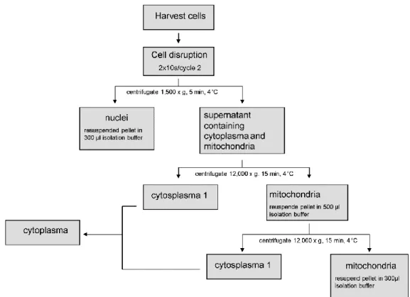
5.2.3 Isolation of mitochondria ... 56
5.2.4 BSA-fatty acid complex ... 57
5.2.5 SDS-PAGE with subsequent immunoblot analysis ... 58
5.2.6 Two-dimensional differential in-gel electrophoresis ... 59
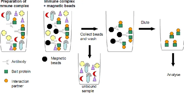
5.2.7 Co-Immunoprecipitation ... 60
5.2.8 Blue native PAGE analysis ... 63
5.2.9 Metabolic analysis ... 64
5.2.10 High resolution respirometry ... 67
5.2.11 Citrate synthase activity measurement ... 68
5.2.12 Respiratory chain supercomplex assembly ... 69
5.2.13 SWATH™ analysis ... 69
5.2.14 Statistical analysis ... 70
8
6 Results ... 71
6.1 Stable overexpression and localization of EHHADH ... 71
6.1.1 Time series for EHHADH overexpression ... 71
6.1.2 Analysis of mistargeting of EHHADH by immunoblotting ... 71
6.1.3 Control of mistargeting of EHHADHMUT by immunofluorescence staining 72 6.1.4 Two-dimensional differential in-gel electrophoresis ... 73
6.2 Incorporation of mutated EHHADH into the mitochondrial trifunctional protein ... 74
6.2.1 Co-immunoprecipitation of EHHADH and HADHB ... 74
6.2.2 Blue native PAGE analysis ... 75
6.3 Respiratory chain analysis ... 76
6.3.1 High-resolution respirometry ... 76
6.3.2 Interaction analysis of EHHADH with the respiratory chain by 2D-blue native/ SDS-PAGE with subsequent immunoblot analysis ... 79
6.3.3 Quantification of supercomplex assembly ... 80
6.4 Metabolic analysis ... 82
6.4.1 Palmitic acid uptake ... 82
6.4.2 Metabolic analysis of acetyl-CoA ... 82
6.4.3 Acylcarnitne analysis ... 83
6.4.4 Measurement of ATP content ... 84
6.5 Proteomic analysis ... 85
7 Discussion ... 88
7.1 Localization and mistargeting of EHHADHMUT ... 88
7.2 Erroneous interaction of EHHADHMUT with the mitochondrial trifunctional protein ... 89
7.3 Effects of mistargeting of EHHADHMUT on mitochondrial fatty acid β- oxidation ... 90
9
7.4 Impact of impaired mitochondrial fatty acid β-oxidation on other
cellular mechanisms ... 91
7.4.1 Uptake of exogeneous long-chain fatty acids ... 91
7.4.2 Formation of acetyl-CoA from β-oxidation ... 94
7.4.3 Generation of ATP ... 94
7.5 Global proteomic analysis of the EHHADHWT and EHHADHMUT cell lines ... 96
7.6 Effects of mistargeting of EHHADHMUT on mitochondrial respiration and supercomplex formation ... 97
7.7 Development of diseases due to the mistargeting of proteins ... 99
8 Conclusion and Outlook ... 101
III. References ... 102
10 II. Abbreviations and Acronyms
1D One-dimensional
2D Two-dimensional
2D-PAGE Two-dimensional polyacrylamide gel electrophoresis AAA ATPase associated with various cellular activities ACAD Acyl-CoA dehydrogenase
ACOX Acyl-CoA oxidase
ADP Adenosine diphosphate
APS Ammonium persulfate
ATP Adenosine triphosphate
BisTris Bis(2-hydroxyethyl)amino-tris(hydroxymethyl)methane BSA Bovine serum albumin
CACT Carnitine acylcarnitine translocase
CHAPS 3-[(3-Cholamidopropyl)-dimethylammonio]-1- propanesulfonate
CoA Coenzyme A
CoIP Co-immunoprecipitation
CPT1 Carnitine-palmitoyltransferase I CPT2 Carnitine-palmitoyltransferase II DTT 1,4-dithio-D-threitol
DMSO Dimethyl sulfoxide
DTNB 5,5’ - Dithiobis(2-nitrobenzoic acid) EDTA Ethylenediaminetetraacetic acid EGTA Ethylene glycol tetraacetic acid
EHHADH Enoyl-Coenzyme A hydratase / L-3-Hydroxyacyl-Coenzyme A dehydrogenase, L-bifunctional enzyme
ESI Electrospray ionization
11 ETC Electron transport chain
ETF Electron transfer flavoprotein
FA Fatty acid
FAD Flavin adenine dinucleotide FATP Fatty acid transport protein
FCCP Carbonylcyanide p-(trifluoromethoxy)phenylhydrazone
FCS Fetal calf serum
FDR False discovery rate FMN Flavin mononucleotide
GC Gas chromatography
GFR Glomerular filtration rate
HEPES 2-[4-(2-hydroxyethyl)piperazin-1-yl]ethanesulfonic acid HFBA 2,2,3,3,4,4,4-Heptafluorobutanoic acid
HPLC High-performance liquid chromatography HRP Horseradish peroxidase
IDA Information Dependent Acquisition IMM Inner mitochondrial membrane IPG Immobilized pH-gradient
LCAD Long-chain acyl-CoA dehydrogenase
LCHAD Long-chain 3-hydroxyacyl-CoA dehydrogenase MCAD medium-chain acyl-CoA dehydrogenase
MEMα Minimum Essential Medium Eagle , alpha modification MOPS 4-Morpholinopropanesulfonic acid
MPC Mitochondrial pyruvate carrier MSD Mass Selective Detector MS/MS Tandem mass spectrometry
NAD+ Nicotineamide adenine dinucleotide
12 OMM Outer mitochondrial membrane
PBS Phosphate buffered saline
PFA Paraformaldehyde
PMSF Phenylmethylsulfonyl fluoride PTS Peroxisomal targeting sequence PVDF Polyvinylidene difluoride
RNS Reactive nitrogen species ROS Reactive oxygen species
RPMI 1640 Roswell Park Memorial Institute Medium 1640 SCAD Short-chain acyl-CoA dehydrogenase
SDS Sodium dodecyl sulfate
SWATH-MS Sequential Windowed data independent Acquisition of the Total High-resolution Mass Spectra
TCA cycle Tricarboxylic acid cycle
TEMED N,N,N′,N′-Tetramethylethane-1,2-diamine TIM Translocase of the inner membrane
TMPD N,N,N',N'-Tetramethyl-1,4-phenylenediamine
TOF Time-of-flight
TOM Translocase of the outer membrane
Tris 2-Amino-2-hydroxymethyl-propane-1,3-diol VLCAD Very-long-chain acyl-CoA dehydrogenase
13
1 Summary
This work describes the analysis of a novel, isolated, autosomal dominant form of Fanconi´s syndrome, a disorder of the renal proximal tubule associated with decreased reapsorption of solutes from the primary urine. This yet unknown Fanconi´s syndrome is evoked by a mutation in the third codon of the peroxisomal protein enoyl-CoA hydratase / L-3-hydroxyacyl-CoA dehydrogenase (EHHADH), also called “Fanconi-associated protein”, which results in the substitution of a glutamic acid residue with lysine (p.E3K). By complementing proteomic and metabolomic analyses of wildtype- and mutant-EHHADH-expressing proximal tubular cell lines (LLC-PK1) with different biochemical and cell biological investigations, the underlying pathomechanism is elucidated. The E3K-mutation leads to the erroneous localization of peroxisomal EHHADH into mitochondria causing a mitochondriopathy. Upon mistargeting of EHHADHMUT into mitochondria, it replaces an alpha subunit of the mitochondrial trifunctional protein (MTP). The MTP normally builds a heterooctamer consisting of four alpha and four beta subunits and is involved in mitochondrial fatty acid β-oxidation. The incorporation into MTP impairs both mitochondrial β-oxidation and respiratory supercomplex assembly, leading to a decreased oxidative phosphorylation capacity. Impairment of the former is shown by the characteristic accumulation of hydroxyacyl-, enoyl- and acylcarnitines in the cell culture supernatant, thus resembling the situation in patients with MTP and/or LCHAD deficiency. The impaired mitochondrial β-oxidation consequently decreases cellular long-chain fatty acid uptake and the acetyl-CoA production in EHHADHMUT cell line.
In addition, EHHADHMUT is also incorporated into respiratory supercomplexes, thereby disturbing their assembly, as shown by blue native PAGE. As a result of impaired mitochondrial β-oxidation and diminished supercomplex assembly the EHHADHMUT cell line shows a decreased oxidative phosphorylation capacity and reduced ATP generation. This mitochondriopathy results in the decreased tubular reabsorption of electrolytes and low-molecular-weight proteins, leading to the Fanconi´s syndrome.
14
2 Zusammenfassung
Diese Arbeit beschreibt die Analyse einer neuen Form eines isolierten, autosomal dominanten Fanconi Syndroms, einer Erkrankung des proximalen Tubulus der Niere, die mit einer verringerten Absorption verschiedener Komponenten aus dem Primärharn einhergeht. Dieses bisher unbekannte Fanconi Syndrom wird durch eine Mutation am N-terminalen Ende des peroxisomalen Proteins Enoyl-CoA Hydratase / L-3-Hydroxyacyl-CoA Dehydrogenase (EHHADH), auch „Fanconi-assoziertes Protein“ genannt, hervorgerufen. In dieser Arbeit werden proteomische und metabolomische Analysen einer renalen proximalen tubulären Zelllinie (LLC-PK1) durch verschiedene biochemische und zellbiologische Untersuchungsmethoden ergänzt, um den zugrundeliegenden Pathomechanismus aufzuklären. Die E3K- Mutation führt zu einer fehlerhaften Lokalisierung von EHHADH in die Mitochondrien, wodurch eine Mitochondriopathie hervorgerufen wird. In den Mitochondrien wird EHHADHMUT ins mitochondriale trifunktionelle Protein (engl.: „mitochondrial trifunctional protein / MTP“) an Stelle einer alpha-Untereinheit eingebaut. Das MTP ist an der mitochondriellen β-Oxidation von Fettsäuren beteiligt und besteht normalerweise aus je vier alpha- und beta-Untereinheiten, welche ein Hetero- Oktamer bilden. Der Einbau ins MTP beeinträchtigt dabei sowohl die mitochondriale β-Oxidation von Fettsäuren als auch die Zusammensetzung der Superkomplexe der Atmungskette. Beide Vorgänge führen zu einer verringerten Aktivität der oxidativen Phosphorylierung.
Die Störung der mitochondrialen β-Oxidation wird durch die charakteristische Akkumulation von Hydroxyacyl-, Enoyl- und Acylcarnitinen im Zellkulturmedium experimentell bestätigt; damit weisen die Medien ein ähnliches Muster wie Seren von Patienten mit MTP- und/oder LCHAD-Defizienz auf. Die gestörte mitochondriale β- Oxidation von Fettsäuren führt nachfolgend zu einer erniedrigten zellulären Aufnahme langkettiger Fettsäuren und zu einer erniedrigten Produktion von Acetyl- CoA in der EHHADHMUT Zelllinie. Zusätzlich zu der gestörten β-Oxidation ist auch die Bildung von Superkomplexen der Atmungskette gestört. Aufgrund dessen sind die oxidative Phosphorylierung und ATP-Produktion in den betroffenen Zelllinien erniedrigt. Die durch die Mislokalisation entstandene Mitochondriopathie ist der Grund für die erniedrigte tubuläre Resorption von Glukose, Aminosäuren, Phosphat,
15 Kalium und niedermolekularen Proteinen, welche in erster Linie zu Minderwuchs und Vitamin-D-resistenter Rachitis führt.
16
3 Introduction
3.1 The kidney
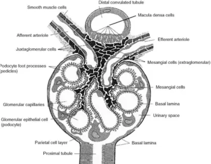
The kidneys are paired, bean-shaped organs, that are located in human behind the retroperitoneum in the abdominal cavity, one on either side of the spine, between the twelfth thoracic vertebra and the third lumbar vertebra 1. They have three main functions: they are major excretory organs, they regulate the salt- and water balance, and they have an endocrine function. Each kidney contains about 0.5 – 1 million nephrons that are divided in cortical and juxtamedullary nephrons. Each nephron is composed of a glomerulus and a tubular apparatus 2. The glomerulus is located in the cortex of the kidney and fed with blood by the afferent arteriole, which splits into the glomerular capillary loops and exits through the efferent ateriole. Along the glomerular capillary wall the blood is filtered, by which the primary urine is produced into the Bowman´s space.
Figure 1: Schematic representation of a glomerulus.
(Figure from Avner E. D., H.W.E., Niaudet P., Yoshikawa N., Pediatric Nephrology, Springer Verlag Berlin Heidelberg, Sixth Edition.)
Every day about 170 L of primary urine are filtered in the glomerula. The transudation along the capillary wall happens across three layers: the endothelial cells of the capillary wall with endothelial pores, the circumambient basement membrane, and
17 the glomerular epithelial cells (podocytes) with foot protrusions on the side of the Bowman´s space. The glomerular filtration is a permselective and pressure dependent process due to the hydrostatic and oncotic pressure difference between capillary lumen and the Bowman´s space 1. The selective permeability depends on both the size and the charge of molecules. Only molecules with a diameter < 4 nm and a molecular weight < 50 kDa are filtered across the capillary wall, and the basement membrane is nearly impermeable to negatively charged macromolecules 1-
3 due to its high content of negatively charged heparan sulfate.
Subsequent to the urinary pole, the tubular apparatus arises. The main function of the tubular apparatus is the production of urine from the primary urine. The tubular apparatus is divided into different segments: proximal tubule, Henle´s loop, distal tubule, and the collecting duct.
The mitochondria-rich proximal tubule is responsible for the reabsorption of nearly all the filtered water and solutes, approximately two thirds of the previously filtered NaCl, 95% of the bicarbonate, and the entire glucose and amino acids 2. Characteristic for the proximal tubule cells is the brush-border membrane, which creates a large luminal surface, as well as the “leaky” tight junctions, which are permeable for small ions and water 3. The pars recta, the straight segment of the proximal tubule, the descending and ascending thin limb segment and the thick ascending limb are parts of the loop of Henle. The loop of Henle is responsible for the formation of an osmotic gradient for the urinary concentration. The pars recta shows the same transport systems as the proximal tubule, and the thin descending limb segment shows nearly no active transport, however a passive transport of cations takes place through the
“leaky” tight junctions. The thin ascending limb segment and the thick ascending limb are the important parts of the loop of Henle. They show active transport of NaCl from the luminal to the basolateral side, while they are impermeable to water, resulting in a higher osmolarity in the interstitial space. Water is transported through the loop-like arrangement of the tubule and the higher interstitial space osmolarity from the thin descending limb to the interstitial space, which leads to a concentrated luminal fluid in the descending limb segment. In the ascending limb segment more solutes are transported into the interstitital space, so that the osmolarity of the tubular fluid drops.
Thus, the tubular fluid is hypotonic at the end of the loop of Henle 2. The thick ascending limb comes in contact with the vas afferens of the own glomerulum at the end of the loop of Henle (Figure 1). To be more precise, the macula densa cells of
18 the thick ascending limb touch the extraglomerular mesangial cells and the renin producing juxtaglomerular cell of the vas afferens. Together, the three cell types build the juxtaglomerular apparatus, which regulates the function of each nephron. In case of an elevated blood pressure, which is accompanied with an elevated glomerular filtration rate (GFR), this can only partly be rescued by the autoregulation of the kidney. This elevated GFR leads to an increased concentration of NaCl in the ascending limb segment of the loop of Henle. The registered elevated NaCl concentration is counteracted by a constriction of the afferent arteriole, thereby decreasing the GFR. A decrease in NaCl concentration on the other hand, has the inverse effect. Subsequent to the juxtaglomerular apparatus the tubular fluid reaches the distal tububule ending in the collecting duct. The fine tuning of the urine occurs in the distal nephron segment. In contrast to the “leaky” tight junctions of the proximal tubulus and the loop of Henle, the tight junctions of the distal nephron are tight. The nadir of luminal fluid osmolality is achieved in the distal nephron, where NaCl is also resorbed form the luminal fluid and the membrane is impermeable to water. The urine has an osmolality of ~ 50 mOsm/kg water entering the collecting duct 4. The urine produced by the nephrons is initially collected in the renal pelvis, to be then transported over the ureter to the urinary bladder.
3.2 Transport processes in the proximal tubulus
The proximal tubule is the first segment of the tubular component of the nephron. It is responsible for the reabsorption of nearly all the filtered water and solutes. This means that in the proximal tubule 60% of the sodium, as well as 60% of the potassium, water and chloride, 95 % of the bicarbonate, and nearly all of the filtered glucose and amino acids are reabsorbed. For a detailed overview of the reabsorbed solutes and water see also Table 1.
19 Table 1: Transport of substances in the proximal tubulus segment 2.
Substances Transport in the proximal tubulus in %
Water 60
Creatinine 0
Sodium 60
Chloride 55
Potassium 60
Bicarbonate 95
Calcium 60
Phosphate 70
Magnesium 30
Glucose 99
Amino acids 99
Urea 50
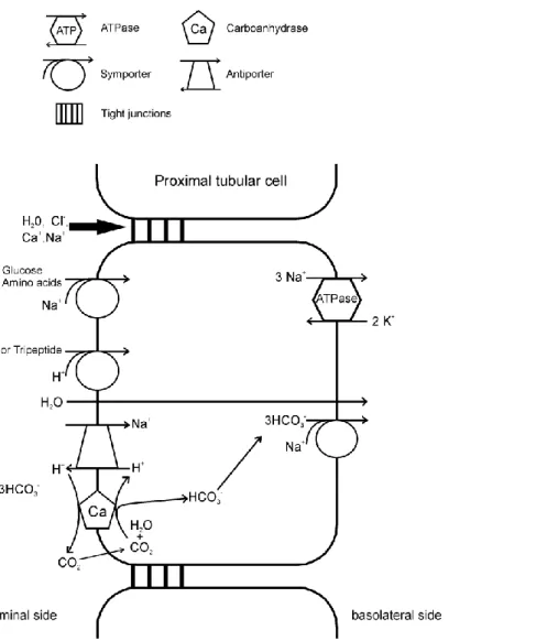
In the proximal tubule four main transport mechanisms are at work, the primary- active transport, the secondary-active transport, endocytosis of large molecules, and passive transport across the “leaky” tight junctions. These transport processes are shown schematically in Figure 2.
3.2.1 Sodium transport
The steep electrochemical gradient of sodium is generated by the Na+/K+-ATPase, which is imbedded in the basolateral membrane. The Na+/K+-ATPase exports three sodium ions and imports two potassium ions for every ATP consumed. It is an example for a primary active transport of the proximal tubular cell. This primary-active transport of sodium and the resulting concentration gradient is a requirement for the secondary-active transport of sodium on the luminal side, where it is transported into the cell through the Na+-coupled symporter, or the Na+/H+-antiporter. On the basolateral side sodium is transported out of the cell by the Na+,3HCO3-
-symporter.
In addition to the primary- and secondary-transport of sodium, sodium is also passively transported into the interstitium over the “leaky” tight junctions.
20
3.2.2 Luminal Na+-coupled symporter for the transport of bicarbonate, glucose and amino acids
At the luminal membrane, different Na+-coupled symporters are responsible for the secondary-active transport of bicarbonate, glucose and amino acids.
Bicarbonate is produced in the proximal tubular cells by the carboanhydrase, which is imbedded in the luminal membrane. This bicarbonate is exported into the interstitium by the basolateral Na+,3HCO3-
-symporter.
Glucose is imported into the proximal tubular cells via a luminal Na+-coupled symport.
Two different symporters, SGLT1 and SGLT2, are responsible for the import of glucose at the luminal membrane. SGLT1 couples the transport of glucose and galactose to the transport of two sodium ions, while SGLT2 couples the transport of glucose to one sodium ion. SGLT1 has a higher affinity than SGLT2. In the early proximal tubulus 95 % of the glucose is reabsorpted by the action of SGLT2.
Together with the action of SGLT 1 at the end of the proximal tubulus, nearly all of the glucose is reabsorbed.
3.2.3 Protein and peptide transport
Di - and tripeptides are reabsorbed at the luminal membrane by peptide-H+- symporters. Peptides with disulfide bridges and proteins are reabsorbed through endocytosis. First, these proteins are bound to specified receptors at the luminal membrane. Then the protein-receptor complexes are enclosed in small vesicles that are invaginated from the cellular plasma membrane 5. Through the action of Na+/H+- antiporter and H+-ATPase, protons are pumped into the protein containing vesicles.
This leads to the dissociation of the protein-receptor complex. Finally, the vesicles are fused with lysosomes. The proteins are degraded to their corresponding amino acids and the receptors are recycled back to the luminal membrane. The resulting amino acids are transported through the cytosol and the basolateral membrane into the interstitium 2. Less than 1% of proteins and peptides remain in the primary urine.
21
3.2.4 Water and chloride transport
Primary- and secondary active transport of solutes from the luminal fluid in the proximal tubular cells lead to a passive transport of water through water channels in the cell membrane and across the tight junctions. This transport of water drags along the water-solved ions such as chloride (solvent drag). In addition to the solvent drag, chloride is also transported through the “leaky” tight junctions.
Figure 2: Schematic representation of the transport processes in the proximal tubular segment.
22
3.3 Fanconi´s syndrome
The Fanconi´s syndrome is named after the swiss pediatrician Guido Fanconi (1892- 1979) and is a generalized disorder of the proximal tubule, leading to excessive urinary wasting of water, amino acids, glucose, electrolytes, and low-molecular- weight proteins.
First Abderhalden 6 described in 1903 a 21 month-old infant with cystine crystals infiltrated the inner organs at autopsy. Abderhalden called the disease familiar cystine diathesis, which is a common cause of Fanconi´s syndrome in children 7,8. In 1924 Lignac 9 described cystinosis in three children who also presented severe rickets and growth retardation. Fanconi described in 1931 10 a child with glucosuria and albuminuria in addition to cystinosis, which are additional aspects of Fanconi´s syndrome. Both, Lignac and Fanconi, also described degenerative changes in the proximal tubules 11. Later on, De Toni 12 found hypophosphatemia to be a further clinical issue and Debré 13 determined also elevated levels of organic acids in urine in an 11-year old girl. In 1936, Fanconi 14 determined that all of the former described patients show similarities and described it as the nephrotic-glucosuric dwarfism with hypophosphatemic rickets 15,16. McCune et al 17 and Stowers and Dent 18 confirmed the finding of Fanconi that the amino-aciduria and the other clinical issues originated within the kidney 11.
The Fanconi´s syndrome is manifested by a global disruption of sodium-coupled transporter systems. A low intracellular Na+ concentration is established at the basolateral membrane by the action of the Na+-K+-ATPase. This low intracellular concentration is required for the maintenance of the lumen-to-cell gradient, which promotes the Na+-coupled solute entry at the luminal membrane. An inhibition of the basolateral Na+-K+-ATPase or a decrease in the cellular ATP-ADP ratio will exert a tremendous influence on the reabsorption in the proximal tubule segment. In addition to the reabsorption of most solutes from the primary urine, the proximal tubule segment is also responsible for the uptake of low molecular weight proteins from the glomerular filtrate via receptor-mediated endocytosis. The receptors for this process are megalin and cubulin, which are present at the luminal membrane. Possible causes for proteinuria are a lack of the receptors at the luminal membrane, defective endocytosis, such as impaired acidification of early endosomes, and accumulation of toxic agents.
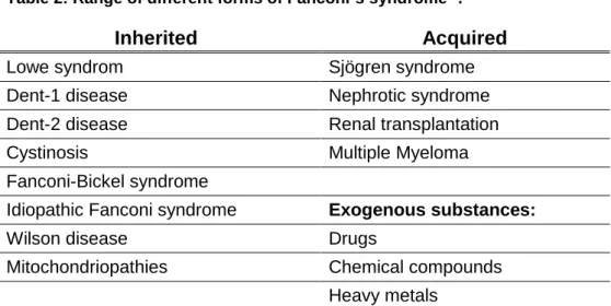
23 The Fanconi´s syndrome is a class of inherited or acquired diseases, or is caused by the action of exogenous substances.
Table 2: Range of different forms of Fanconi´s syndrome 4.
Inherited Acquired
Lowe syndrom Sjögren syndrome
Dent-1 disease Nephrotic syndrome
Dent-2 disease Renal transplantation
Cystinosis Multiple Myeloma
Fanconi-Bickel syndrome
Idiopathic Fanconi syndrome Exogenous substances:
Wilson disease Drugs
Mitochondriopathies Chemical compounds Heavy metals
Understanding of the development of the different forms of inherited and acquired Fanconi´s syndrome provides important insights in proximal tubular transport.
3.3.1 Inherited Fanconi´s syndrome
Dent´s disease is a rare X-linked proximal tubulopathy with a full-blown Fanconi´s syndrome, but rare extrarenal symptoms except for rickets 4,19. Depending on the genetic cause and pattern of signs and symptoms, two forms of Dent’s disease are distinguished.
Dent-1 disease is caused by mutations in the CLCN5 gene, which encodes for a renal specific voltage-dependent electrogenic chloride/proton antiporter. The CLC-5 antiporter is coexpressed with the vacuolar H+-ATPase 4 and is involved in the acidification of early endosomes. The decreased acidification of the early endosomes during endocytosis leads to a diminished recycling of megalin and cubulin back to the luminal membrane, causing the low-molecular-weight proteinuria. In addition, patients with Dent-1´s disease also present with increased urinary excretion of phosphate and calcium, which lead to kidney stones, nephrocalcinosis and, eventually, progressive renal failure 20.
Dent-2 disease is caused by mutations in the classical Lowe oculocerebrorenal syndrome gene, OCRL 21. Compared to classical Lowe syndrome, patients suffering
24 from Dent-2 disease present with milder extrarenal symptoms. Nephrocalcinosis is also seen less frequently than in Dent-1 disease 22.
Lowe syndrome is also a rare X-linked disorder. It is characterized by a complex phenotype involving major abnormalities of the eyes, central nervous system and an incomplete renal Fanconi´s syndrome 4. The OCRL gene encodes for phosphatidylinositol 4,5-bisphosphate phosphatase (PIP2 5-phosphatase), which colocalizes in proximal tubular cells with clathrin and megalin at the luminal membrane 22. It interferes with the actin cytoskeleton, and is involved in the inositol phosphatase signaling pathway 4. The involvement of PIP2 5-phosphatase in endocytosis explains the similarities seen in renal involvement between Lowe syndrome and Dent´s disease. The decreased activity of PIP2 5-phosphatase leads to an accumulation of phosphatidylinositol 4,5-bisphosphate (PIP2) and actin stress fibres, which have an tremendous effect on epithelial function4.
Mitochondriopathies are other inherited causes for Fanconi´s syndrome, as is the case with the Fanconi´s syndrome studied here. In proximal tubular cells, the major source of energy is fatty acid oxidation 23. Proximal tubular cells reabsorb solutes and water from the glomerular filtrate close to the cellular energy demand as Beck et al. 24 showed an intracellular decrease in ATP after stimulation of sodium-dependent transport. Therefore, it is not surprising, that inherited mitochondriopathies, which are caused by mutation of mitochondrial or nuclear DNA encoding for functional or structural mitochondrial proteins, are often associated with Fanconi´s syndrome.
3.3.2 Acquired Fanconi´s syndrome
Fanconi´s syndrome can also occur secondary to certain diseases or after the administration of exogenous substances. The development of a Fanconi´s syndrome secondary to a disease is rarely seen. In patients with Sjögren syndrome, only 3 % manifest Fanconi´s syndrome 25. Equally uncommon is the development of a Fanconi´s syndrome 4 after renal transplantation. In multiple myeloma, renal involvement mostly manifests as proteinuria and only rarely as full-blown Fanconi´s syndrome 26.
Exogenous substances such as drugs, chemicals and heavy metals, can also cause Fanconi´s syndrome. Drugs like valproic 27 acid, tenofovir 28, Chinese herbs 29,30 or
25 expired tetracycline 31 have been reported to cause a Fanconi´s syndrome. The decreased transport of proximal tubular cells after the administration of valproic acid originates from the fact that this drug causes respiratory chain defects and decreases lysosomal enzyme activity 4. The major degradation products of expired tetracycline, namely epitetracycline, anhydrotetracycline and 4-epianhydrotetracycline, are also toxic to proximal tubular cells 32. Antiviral agents, for example adefovir, which acts a reverse transcriptase inhibitor, interact with organic anion transporter and lead to mitochondrial damage and tubular toxicity. The non-specific herbicide paraquad, toluene and 6-mercaptopurine may also lead to Fanconi´s syndrome and renal failure. The kidney is the first organ of heavy metal toxicity, including lead, cadmium, mercury, chromium and platinum. Lead poisoning leads to the development of a Fanconi´s syndrome, with aminoaciduria and glucosuria which can persist for up to 13 years 4,33. Lead thereby disrupts mitochondrial respiration, phosphorylation and can directly inhibit SLC3A1, which is a renal amino acid transporter for the transport of neutral and basic amino acids across the renal brush border 34. The threshold for proximal tubular injury by lead is a blood lead level of 60 µg/dL 33. Cadmium, on the other hand, causes Fanconi’s syndrome via production of free radicals that alter mitochondrial activity or induce mitochondrial gene deletion following long time exposure and inhibits H+-ATPase which leads to a Fanconi-like syndrome 4.
3.4 Mitochondria
The mitochondrion is a membrane bound organelle found in most eukaryotic cells.
Mitochondria are mostly shaped like a rod. They are between 2 - 8 µm in length and 0.2 - 1 µm in diameter 5. Especially cells with high energy-demand are rich in mitochondria, as seen in cardiac cells, muscle cells, cells of the central nervous system, sensory cells, ovocytes, sperm and cells of the proximal tubule. Furthermore, mitochondria are localized to specific cytoplasmic areas 35 for efficient provision of energy where it is needed, which is the basolateral membrane in the case of proximal tubule cells. The main function of mitochondria is to provide energy to the cell in the form of adenosine triphosphate (ATP). In mitochondria the main energy producing pathways are located: they include the oxidation of pyruvate, the mitochondrial fatty acid β-oxidation, tricarboxylic acid cycle and the oxidative phosphorylation.
Biogenesis of mitochondria is a dynamic process, with constant mitochondrial fusion
26 and fission. When the energy demand of a cell increases, the number of mitochondria will be increased by mitochondrial fission. Mitochondria are inherited normally solely from the maternal side, however there are cases where also paternal mitochondria are found 36. Healthy paternal mitochondria are eliminated usually during embryogenesis through a process that is not well understood. Consequently, the mitochondria found are solely of maternal origin. The endosymbiotic theory from 1883 postulates, that mitochondria originate from prokaryotes, which were incorporated into a so-called primordial cell as endosymbionts and, subsequently, reduced to organelles. Facts supporting this theory are that the phospholipid cardiolipin present in the inner mitochondrial membrane (IMM) is normally only seen in prokaryotes and that mitochondria have their own cyclic DNA.
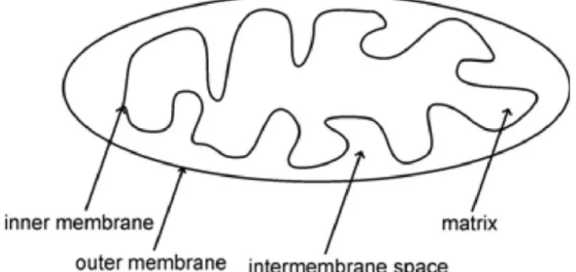
3.4.1 Mitochondrial structure
Mitochondria are composed of four main components, the outer membrane, the intermembrane space, the inner membrane and the miochondrial matrix (Figure 3).
The mitochondrium is separated from the cytosol by the outer mitochondrial membrane (OMM). The inner mitochondrial membrane (IMM) separates the intermembrane space from the mitochondrial matrix.
Figure 3: Schematic illustration of the mitochondrial structure.
The mitochondrial membranes are mainly composed of phosphatidyl choline, phosphatidyl ethanolamine, cardiolipin and phosphatidyl inositol 37. Each component has a specific assignment in mitochondrial function. The OMM separates the intermembrane space from the cytosol and is freely permeable for small proteins up to 5,000 Da through protein complexes called porines 38. Larger proteins, in contrast,
27 must be actively transported through the outer mitochondrial membrane by a transport complex called TOM (translocase of the outer membrane). The main function of the intermembrane space is oxidative phosphorylation. Other functions include the exchange of proteins, lipids, or metal ions between the matrix and the cytosol, the regulated initiation of apoptotic cascades, signalling pathways that regulate respiration and metabolic functions, and the prevention of reactive oxygen species produced by the respiratory chain 39. The inner mitochondiral membrane (IMM) separates the intermembrane space from the mitochondrial matrix. The IMM is extensively folded in so-called cristae to increase its surface area, which is about five times as large as that of the OMM. The IMM is responsible for the import of proteins, lipids and other important metabolites into the mitochondrial matrix. In contrast to the OMM, it is completely impermeable, so that everything has to be transported actively.
The impermeability is also a requirement for the maintenance of the proton gradient (∆pH) and membrane potential (∆ψ), which are a requirement for the electrochemical gradient (∆μH+). The mitochondrial matrix contains the cyclic double-stranded DNA and the whole machinery needed for the transcription and translation of the mitochondrial-encoded proteins. In addition, the enzymes for the important energy generating pathways are localized in the mitochondrial matrix or the IMM.
3.4.2 Mitochondrial energy metabolism
In mitochondria the main energy generating pathways are located, oxidation of pyruvate to acetyl-CoA, tricarboxylic acid cycle (TCA) cycle, mitochondrial fatty acid β-oxidation and oxidative phosphorylation. Mitochondria are also important for the controlling of the cellular redox state, Ca2+ homeostasis and apoptosis 40.
Oxidation of pyruvate
Pyruvate generated in glycolysis is transported into the mitochondria by the mitochondrial pyruvate carrier (MPC). In the mitochondrial matrix, pyruvate is irreversibly decarboxylated by the action of the pyruvate dehydrogenase complex to form acetyl-CoA, which enters the tricarboxylic acid cylce. Cofactors of this reaction are thiamine pyrophosphate, coenzyme A, α-lipoic acid, FAD, and NAD+. This reaction links glycoylsis and the TCA cylce.
28 Tricarboxylic acid cycle
Enzymes of the TCA cycle are located in the mitochondrial matrix. In the first step of the TCA cycle, acetyl-CoA generated by the decarboxylation of pyruvate or the ß- oxidation of even-numbered saturated fatty acids reacts with oxaloacetate to from citrate. Citrate is transformed into isocitrate by isomerisation. In two subsequent oxidative decarboxylation reactions, first α-ketoglutarate and then succinyl-CoA are formed, with each reaction yielding NADH and H+. Through the reaction of succinyl- CoA to succinate, the high phosphate transfer potential compound GTP is formed.
Succinate is then FAD-dependent oxidized to fumarate, and through the addition of water malate is formed. In the last step, malate is oxidized to oxaloacetate in a reaction catalyzed by malate dehydrogenase, which also yields NADH and H+. Oxalacetate reacts again with acetyl-CoA, providing citrate for another round of the TCA cycle.
The net reaction is:
acetyl-CoA + 3 NAD+ + FAD + GDP+ Pi + 2 H2O
→ 2 CO2 + CoA + 3 NADH+ 3H+ + FADH2 + GTP
The reducing equivalents NADH and FADH2 directly enter oxidative phosphorylation.
Mitochondrial fatty acid β-oxidation
For mitochondrial β-oxidation, fatty acids are activated in the cytosol, generating membrane-impermeable acyl-CoAs, which are subsequently transported into the mitochondrial matrix by the carnitine carrier system, which is schematically depicted in Figure 4. Upon release into the mitochondrial matrix, the acyl-CoAs are broken down to generate acetyl-CoA, which enters the TCA cycle, and NADH and FADH2, which are used by the electron transport chain.
29 Figure 4: Scheme of the carnitine carrier system and subsequent fatty acid β-oxidation. The activated long-chain fatty acids (LCFA-CoA) are transported by the carnitine carrier system, which consists of the CPT1, CACT and CPT2, into the mitochondrial matrix. The LCFA-CoA are converted into their corresponding acylcarnitines by the CPT 1, transported across the IMM by CACT and, ultimately, reconverted into the activated long-chain fatty acids by CPT2. In the mitochondrial matrix, the LCFA-CoA subsequently undergoes the first round in the β-oxidation spiral. Chain shortening of LCFA-CoAs takes place by the repeated action of four enzymatic reactions, oxidation of an acyl-CoA by the acyl-CoA dehydrogenase (AD) with flavin adenine dinucleotide (FAD) as cofactor, hydration of enoyl-CoA by the enoyl-CoA hydratase, a second oxidation step using the cofactor nicotineamide adenine dinucleotide (NAD+) and in the end a thiolytic cleavage by 3-keto-thiolase.
(Illustration taken from Sim, K.G:, Hammond, J., Wilcken, B. Strategies for the diagnosis of mitochondrial fatty acid beta-oxidation disorders. Clin Chim Acta 2002; 323:37-58)
Long-chain fatty acids represent the majority of the dietary fat undergoing ß-oxidation
41. The latter plays an essential role in the energy metabolism of proximal tubular cells, which derive almost their entire energy from β-oxidation of mostly long-chain fatty acids 42. The enzymes of mitochondrial fatty acid β-oxidation are located at the inner face of the inner mitochondrial membrane as well as are distributed in the
30 mitochondrial matrix 43. Chain shortening of long-chain fatty acids takes place by the repeated action of four enzymatic reactions (Figure 4).
In the first reaction, an acyl-CoA is oxidized to a trans-2,3-enoyl-CoA by acyl-CoA dehydrogenases (ACADs). This reaction produces a FADH2, which directly enters the oxidative phosphorylation at the electron transfer flavoprotein (ETF). The electrons form FADH2 are transferred to ETF, the reduced ETF is then oxidized by the ETF:ubiquinone oxidoreductase transferring the electrons to ubiquinone, which is then reduced to ubiquinol. By the action of complex II of the respiratory chain ubiquinol is reoxidized to ubiquinone.
The second enzyme of the β-oxidation spiral is enoyl-CoA-hydratase, which forms L- 3-hydroxyacyl-CoA by the addition of water to the double bond. The hydroxy-group is in the next step NAD+-dependent oxidized to the corresponding ketoacyl-CoA. The reducing equivalent NADH is oxidized by the respiratory chain complex I.
In the last step of the β-oxidation, which is catalyzed by keto-thiolase, the terminal acetyl-CoA is cleaved off to yield an acyl-CoA shortenend by two carbon atoms. The cleaved acetyl-CoA can then enter into the TCA and electron transport chain (ETC).
In fatty acid β-oxidation, fatty acids are with each round shortened by two carbon atoms, which are released as acetyl-CoA, until the entire fatty acid is cleaved into acetyl-CoAs. The complete oxidation of palmitate yields 8 acetyl-CoA, 7 FADH2, 7 NADH, yielding in total 106 ATP molecules 44.
In mitochondria, several enzymes are present for the different steps of β-oxidation, which vary in their chain-length specificity. For the acyl-CoA dehydrogenase (ACAD) four enzymes with overlapping chain-length specificity are known: short-chain acyl- CoA dehydrogenase (SCAD) for C4 and C6 fatty acids, medium-chain acyl-CoA dehydrogenase (MCAD) with high specificity for fatty acids of a chain length of C4 to C12, long-chain acyl-CoA dehydrogenase (LCAD) C8 to C20 fatty acids, which is important for unsaturated fatty acids, and very-long-chain acyl-CoA dehydrogenase (VLCAD) for fatty acids with a chain-length between C12 and C24 43. Of the known short-, medium- and long-chain keto-thiolases, only the latter two are important for β- oxidation 43. Four subunits each of the long-chain enoyl-CoA hydratase / long-chain L-3-hydroxyacyl-CoA dehydrogenase (alpha-subunit) and the long-chain ketothiolase (beta-subunit) are assembled in the hetero-octameric mitochondrial trifunctional protein (MTP), which is bound to the inner face of the inner mitochondrial membrane.
31 Oxidative phosphorylation
Glycolysis, fatty acid β-oxidation, TCA cycle and the oxidative phosphorylation are tightly coupled processes, as reducing equivalents produced by glycolysis, β- oxidation and TCA cycle directly enter the oxidative phosphorylation. Oxidative phosphorylation is the main source for ATP production. The electrons from the reducing equivalents are transferred onto the respiratory complexes, and are used to reduce molecular oxygen together with protons from the mitochondrial matrix to water. In parallel protons are pumped from the matrix into the intermembrane space, leading to the formation of a pH-gradient and a membrane potential (voltage gradient). Both, the pH-gradient and the membrane potential, build up the electrochemical proton gradient 38. The respiratory complex V (ATP synthase) drives protons back into the mitochondrial matrix by the electochemical gradient, which as a result generates ATP from ADP. The respiratory chain complexes, complex I - IV, the ATP synthase (complex V), and the adenine nucleotide translocase (ANT) are embedded in the IMM. The complete oxidation of glucose yields 30 ATP molecules, from which alone 26 ATP molecules are generated by oxidative phosphorylation. The complete oxidation of palmitate yields 106 ATP molecules through oxidative phosphorylation 44. Four enzyme complexes are responsible for the electron flow and at the end for the reduction of oxygen to water.
NADH:ubiquinone-reductase (complex I)
Complex I is the biggest respiratory chain complex and consists of 44 subunits 45. It needs the flavoprotein FMN and Fe-S-cluster as prosthetic groups. It is localized in the inner mitochondrial membrane in an L-shaped form, where the horizontal arm lies in the inner mitochondrial membrane and the vertical arm protrudes into the mitochondrial matrix. Complex I oxidizes NADH to NAD+. The reaction leads in parallel to the pumping of four H+ out of the mitochondrial matrix.
The net reaction is:
NADH/H+ + ubiquinone + 4 H+matrix → NAD+ + ubiquinole + 4 H+intermembrane
Succinate:ubiquinone reductase (Complex II):
Complex II is the smallest of the four respiratory complexes and consists of four subunits and equals the succinate dehydrogenase acting in the tricarboxylic acid
32 cylce. Succinate dehydrogease catalyzes the oxidation of succinate to fumarate, with the formation of a FADH2. Complex II does not pump protons out of the mitochondrial matrix.
The net reaction is:
Succinate + ubiquinone → Fumarate + ubiquinole
During mitochondrial β-oxidation also FADH2 is formed. These electrons are not transferred onto complex II, but onto the electron transfer flavoprotein (ETF), which is reoxidized by the action of ETF:ubiquinone oxidoreductase. The electrons are transferred onto complex III.
Ubiquinole:cytochrome c reductase (complex III):
Complex III consists of 10 subunits and contains as electron carrier cytochrome b, cytochrome c1 and one Fe-S-cluster 46. Through the transfer of the electrons from ubiquinole to cytochrom c, ubiquinone is reoxidized. The mechanism of the transfer of electrons from ubiquinole to cytochrome c and the coupled transport of protons, is also called the Q cycle 44. Thereby, in total four protons are pumped into the intermembrane space.
The net reaction is:
ubiquinole + 2 cyt cox + 2 H+matrix → ubiquinone + 2 cyt cred + 4 H+intermembrane
Cytochrome c oxidase (complex IV):
Complex IV consists of 19 subunits and is the final proton pumping complex of the respiratory chain. It catalyzes the electron transport from cytochrome c to elemental oxygen (O2). For electron transport, complex IV contains the prosthetic groups cytochrome a, cytochrome a3 and two copper centers. The electron is transferred from cytochrome c to the first copper centre (CuA), and via cytochrome a and cytochrome a3 to the second copper centre (CuB). Four molecules of cytochrome c are bound successively to complex IV. In the end, four electrons are transferred onto one molecule O2, which is then completely reduced to water. In parallel, four protons are pumped into the intermembrane space.
33 The net reaction is:
4 Cyt cred + 8 H+Matrix + O2 → 4 Cyt cox + 2 H20 + 4 H+intermembrane
Through the complexes I, III, and IV protons are pumped from the matrix into the intermembrane space, leading to the formation of the electrochemical proton gradient that drives ATP synthesis.
ATP synthase (complex V):
The ATP synthase consists of 19 subunits, which build two main units, the FO-unit, which forms a proton channel across the inner mitochondrial membrane and the F1- unit, which constitutes the catalytic unit of complex V. Protons flow through the FO- unit down the electrochemical gradient back into the mitochondrial matrix and the F1- unit catalyzes the reaction from ADP and organic phosphate to ATP. The function of the proton backflow is not the synthesis of ATP, but the release of ATP from the synthase 44. The mechanism of the ATP synthesis with the F1-unit is a binding- change mechanism, with three successive steps: the binding of ADP and organic phoshate, the synthesis of ATP and the release of the synthesized ATP from the F1- unit 44. The rotation of the γ-subunit of the F1-unit drives these three steps into each other and is required for the release of the ATP from the F1-unit. The rotation of the γ- subunit coheres with the rotation caused by the c-subunit of the FO-unit, through the binding of proton from the proton-rich intermembrane space. The FO-unit consists of 10 c-subunits and with every binding of a proton to the c-subunit, the subunits are rotated one step further. The c-subunit is firmly connected to the γ-subunit, so the active rotation of the c-subunit also rotates the γ-subunit. With each 360° rotation of the c-units, the γ-subunit comes to a full rotation, which correlates to the binding of 10 protons and leads to the synthesis and release of 3 ATP molecules 44.
The net reaction is:
ADP3- + HPO42-
+ 3 H+intermembrane → ATP4- + H20 + 2 H+matrix
Respiratory chain supercomplexes:
The organization of the respiratory chain complexes within the IMM has been the subject of intense debate 47-52. The first proposed model was the “solid state model”,
34 which suggested the arrangement of the respiratory chain complexes in an orderly sequence within the IMM 48. This model was soon replaced by the “liquid state model”, which hypothesized that the individual respiratoy chain complexes diffuse freely and are randomly distributed within the lipid bilayer 47,49. Today, based on the ovservations made by Bruet et al. 53, Boumans et al. 54 and Schägger et al. 55 in yeast S. cerevisiae and bovine it is believed that the respiratory chain complexes are organized into supercomplexes 52. The two major supercomplexes in human mitochondria are I1III2 and I1III2IV1 56
.
3.4.3 Mitochondrial protein import
Of the approximately 1,500 57 mitochondrial proteins, only 13 are mitochondrially encoded. The remainder is encoded in the nucleus and imported into mitochondria during or after synthesis. These proteins are synthesized as preproteins and may feature either an N-terminal mitochondrial targeting sequence, also called presequence, which is cleaved post-import, or multiple internal mitochondrial targeting sequences. Mitochondrial matrix proteins feature the N-terminal targeting sequences, whereas hydrophobic proteins, which are implemented in the IMM, feature multiple internal targeting signals. In contrast to peroxisomal targeting sequences, mitochondrial targeting sequences do not have a known amino acid sequence, so the import machinery recognizes many different forms of different targeting sequences. The N-terminal presequence of proteins consists of about 20 – 40 amino acids residues, dotted with some positively charged amino acids (lysine, histidine or arginine) 58. It is suggested that the presequence forms an amphipathic α- helix, with the positively charged amino acids lying on one side of the helix and the hydrophobic amino acids on the other, albeit the latter are not essential for mitochondrial targeting 58. Hydrophobic proteins do not contain a cleavable N- terminal targeting sequence, but rather contain multiple internal targeting sequences, which are spread over the whole length of the protein. Three major translocase complexes are involved in the import of mitochondrial proteins, one located in the outer mitochondrial membrane, called translocase of the outer membrane (TOM) and two translocases in the inner mitochondrial membrane, called translocases of the inner mitochondrial membrane (TIMs), one for the import of matrix proteins (TIM23) and the other is responsible for the insertion of complexes in the inner membrane
35 (TIM22). The proteins are transported in an unfolded, linear state or a loop formation through the mitochondrial membranes, and the transport is driven by the membrane potential (∆ψ), which is built across the inner mitochondrial membrane and the ATP hydrolysis-driven action of the mitochondrial heat shock protein 70 (mtHsp70) 59.
Import of proteins with an N-terminal presequence:
For the import of proteins with a cleavable presequence, the proteins are first located to the OMM, where the presequence is first recognized by TOM20, then transferred to TOM22, TOM5 and translocated in a linear, unfolded state across OMM by the general import pore (GIP), which is formed by the pore-building protein TOM40. The translocation across the OMM is illustrated in Figure 5. In the intermembrane space, the preprotein is bound to the C-terminal end of TOM22 before further transport. The TIM23 protein exposes a domain into the intermembrane space to which the preprotein is bound after the translocation through the OMM. The membrane potential activates the channel formed by the TIM23 complex and exerts and electrophoretic effect on the positively charged presequence, which leads to the transport of the preprotein across the IMM. In the matrix, mtHsp70 binds to the unfolded preprotein in transit and drives the preprotein into the matrix, by the hydrolysis of ATP. The import of preproteins across the IMM is therefore driven by these two energy sources, the mitochondrial membrane potential (∆ψ) and the ATP hydrolysis-driven action of mtHsp70. In the matrix a mitochondrial processing peptidase removes the presequence and the mature matrix protein is folded. The heterodimeric mitochondrial processing peptidase (MPP) removes the N-terminal presequence proteolytically 59. The process of translocation across the IMM and the processing in the matrix are shown in Figure 5.
36 Figure 5: Mitochondrial protein import.
(Figure from Pfanner, N., Wiedemann, N. Mitochondrial protein import two membranes, three translocases. Current opinion in cell biology 2002; 14: 400-11)
Import of proteins with an internal targeting sequence:
Hydrophobic proteins, which feature internal targeting sequences are guided during their transport to the OMM by chaperons to prevent misfolding and aggregation in the aqueous environment. After their transport to the OMM, the internal targeting sequences are recognized by TOM70, which in an ATP-dependent manner transports the preprotein to TOM22 and TOM5, before the preprotein is translocated across the OMM by the GIP in a loop formation (Figure 5). Subsequent to the translocation across the OMM, the protein binds to the TIM9-TIM10-complex in a loop formation, which has chaperone-like characteristics, to prevent the aggregation and misfolding of proteins with internal targeting sequences. The TIM9-TIM10 complex then transports the protein across the intermembrane space to the IMM and binds to the TIM22 complex. The subsequent import of the protein into the IMM is carried out by the TIM22 complex, consisting of TIM22, TIM54 and TIM18, which is activated by the presence of a targeting signal. The mitochondrial membrane
37 potential (∆ψ) is the only energy source during this process. The schematic illustration of the transport and insertion at the IMM is shown in Figure 5.
3.5 Peroxisomes
3.5.1 Structure and Function
Peroxisomes are small spherical organelles, which are between 0.1 - 1 µm in diameter and are surrounded by a single membrane 60. Peroxisomes are present in all eukaryotic cells and compared with mitochondria do not contain DNA or ribosomes, i.e., all peroxisomal proteins are encoded in the nucleus and have to be transported into the peroxisomes. Rhodin was the first to describe peroxisomes in 1954. He termed them microbodies 61, and later on the term peroxisome was used.
Peroxisomes and mitochondria are replicated by the fission of already present peroxisomes or mitochondria, respectively. The important functions of peroxisomes are oxidation and detoxification metabolism, synthesis of plasmalogen and oxidation of fatty acids via α- and β-oxidation.
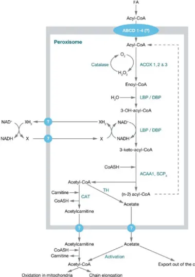
Oxidation fatty acids via α- and β-oxidation:
In plants cells, yeast and most fungal cells β-oxidation of fatty acids take place only in peroxisomes, whereas in mammalian cell the β-oxidation of fatty acids take place in both mitochondria and peroxisomes. Peroxisomes and mitochondria both have their own sets of enzymes for fatty acid β-oxidation, where peroxisomes are responsible for the shortening of very-long-chain fatty acids, medium- and long-chain dicarboxylic acids, bile acid precursors, branched chain fatty acids, prostaglandins, leukotrienes, xenobiotics and certain mono- and polyunsaturated fatty acids 40,62 and mitochondria are responsible for the oxidation of short-, medium- and long-chain fatty acids. The shortened fatty acids acyl-CoAs from the peroxisomes are exported to the mitochondria for complete oxidation, because the mitochondrial fatty acid β-oxidation is more efficient concerning energy generation. The enzymes for peroxisomal β- oxidation are distributed within the peroxisomal matrix. Only saturated very-long- chain fatty acids and 2-methyl-branched fatty acids can be directly activated and are available for peroxisomal β-oxidation. All other fatty acids have to be converted via different peroxisomal enzymes into suitable substrates for peroxisomal β-oxidation (Figure 6).