A rare case of CNS hemangiopericytoma presenting with papilledema
Abstract
Hemangiopericytoma rarely affects the central nervous system (CNS) and usually presents to neurologists with neurological symptoms. We
Sanjana B. Singh
1Narayanaswamy Vanathi
1report a rare case of large CNS hemangiopericytoma which presented to an ophthalmologist with only signs of mild defective vision and
papilledema.
Virna M. Shah
1Keywords:hemangiopericytoma, central nervous system, papilledema
1 Department of Neuro Ophthalmology, Aravind Eye Hospital & Postgraduate Institute of Ophthalmology, Coimbatore, India
Introduction
Hemangiopericytoma is a rare aggressive type of soft tissue sarcoma which arises from pericytes of the capil- laries [1] mostly involving the musculoskeleton system.
Presentation in the central nervous system (CNS) is very rare, accounting for less than 1% of all intracranial tumors and 2–4% of meningeal tumours, and can mimic a men- ingioma [2]. We report a case of a large intracranial hemangiopericytoma in the left temporal region that presented to us with blurring of vision, headache, and papilledema. There were no sensory or motor disturb- ances. This is one of the few cases of rare intracranial hemangiopericytoma presenting to an ophthalmologist with only ocular symptoms and no systemic signs.
Case description
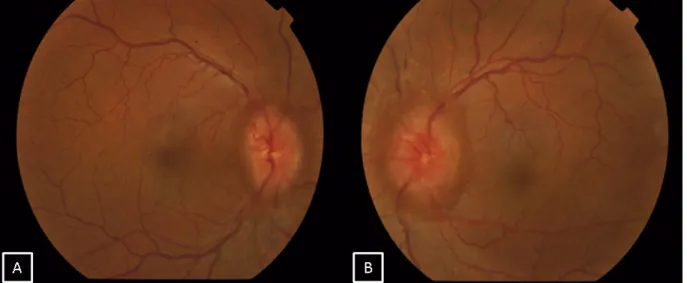
A forty-one-year old male presented with defective vision in the right eye since 2 months. He also gave a history of transient obscuration of vision in the right eye with asso- ciated headache. There was no history of associated vomiting, loss of consciousness, weakness in any part of the body, diplopia, or seizures. On examination, his best corrective visual acuity was 20/30 in the right eye and 20/20 in the left eye. Anterior segment examination of both eyes was within normal limits. Fundus examination of both eyes showed disc edema suggestive of papille- dema (Figure 1). Colour vision and extraocular movements were normal in both eyes. Humphrey visual field analyzer showed an enlargement of the blind spot in both eyes.
Higher functions and other cranial nerves examinations were within normal limits. The patient was diagnosed to have bilateral papilledema and was advised magnetic
resonance imaging (MRI) of the brain and orbit. MRI showed a large mass lesion measuring about 7x6x6 cm in maximum dimension over the left petrous temporal bone with prominent vascular channels within the lesion suggestive of hemangiopericytoma (Figure 2). Mass effect in the form of midline shift to the right was noted with mild dilatation of the right ventricle suggestive of intra- cranial hypertension. Spectroscopy revealed elevated myoinositol and choline peaks. Limited computerized tomography showed erosion of the anterior aspect of the petrous portion of the temporal bone on the left side. The patient was referred to a neurosurgeon for further man- agement. The patient decided against treatment and deceased within two months.
Discussion
Hemangiopericytoma is encountered in adults in their fifth decade of life with a slight male predilection [3].
Based on histology, hemangiopericytoma of the CNS are of two types, namely differentiated and anaplastic, of which the latter is more aggressive. The differentiated type of intracranial hemangiopericytoma is considered a World Health Organization grade II neoplasm, whereas the anaplastic variant is classified as grade III neoplasm [4]. Hemangiopericytoma mimics meningioma, but has a much more agressive course [2]. One third of the cases exhibit malignant features like metastasis or local inva- sion and recurrence. Bone, liver, CNS, and abdominal cavity are the most common sites of metastasis [5].
However, the prognosis depends upon location and size of the tumor as well as the presence of metastasis.
Tentorial and posterior fossa tumors are found to be more aggressive than supratentorial tumors. Large tumours
1/3 GMS Ophthalmology Cases 2019, Vol. 9, ISSN 2193-1496
Case Report
OPEN ACCESS
Figure 1: Fundus pictures of the right (A) and left eye (B) showing bilateral optic disc edema suggestive of papilledema
Figure 2: A) Coronal T2-weighted MRI brain showing an iso-hyperintense large mass lesion in the tempo-parietal lobe (white arrows). B,C): Post contrast sagittal and axial cuts showing an intense heterogeneous enhancement of the lesion (white arrows).
(≥6 cm) and non-skull base lesions have an increased risk of early recurrence after treatment. A treatment modality of intracranial hemangiopericytoma is total sur- gical excision with pre-operative catheter embolisation and adjuvant radiotherapy to reduce the incidence of re- currence [5].
Our case presented to us with defective vision and headache of 2 months duration without any motor or sensory disturbances, though the size of the tumor was large on MRI. We report this case to lay emphasis on the fact that intracranial hemangiopericytoma can present to an ophthalmologist which requires timely referral.
Notes
Competing interests
The authors declare that they have no competing in- terests.
References
1. Koch M, Nielsen GP, Yoon SS. Malignant tumors of blood vessels:
angiosarcomas, hemangioendotheliomas, and
hemangioperictyomas. J Surg Oncol. 2008 Mar 15;97(4):321-9.
DOI: 10.1002/jso.20973
2. Kanesen D, Kandasamy R, Idris Z. Occipital Falcine Anaplastic Hemangiopericytoma Mimicking Meningioma. J Neurosci Rural Pract. 2016 Dec;7(Suppl 1):S95-S98. DOI: 10.4103/0976- 3147.196463
3. Kim JH, Jung HW, Kim YS, Kim CJ, Hwang SK, Paek SH, Kim DG, Kwun BD. Meningeal hemangiopericytomas: long-term outcome and biological behavior. Surg Neurol. 2003 Jan;59(1):47-53;
discussion 53-4. DOI: 10.1016/S0090-3019(02)00917-5 4. Louis DN, Ohgaki H, Wiestler OD, Cavenee WK, Burger PC, Jouvet
A, Scheithauer BW, Kleihues P. The 2007 WHO classification of tumours of the central nervous system. Acta Neuropathol. 2007 Aug;114(2):97-109. DOI: 10.1007/s00401-007-0243-4 5. Rutkowski MJ, Jian BJ, Bloch O, Chen C, Sughrue ME, Tihan T,
Barani IJ, Berger MS, McDermott MW, Parsa AT. Intracranial hemangiopericytoma: clinical experience and treatment considerations in a modern series of 40 adult patients. Cancer.
2012 Mar 15;118(6):1628-36. DOI: 10.1002/cncr.26411
2/3 GMS Ophthalmology Cases 2019, Vol. 9, ISSN 2193-1496
Singh et al.: A rare case of CNS hemangiopericytoma presenting with ...
Corresponding author:
Dr. Virna M. Shah
Department of Neuro Ophthalmology, Aravind Eye Hospital
& Postgraduate Institute of Ophthalmology, Coimbatore, Tamil Nadu, India, Phone: +91 422 4360400, Fax: +91 422 2593030
virna@cbe.aravind.org
Please cite as
Singh SB, Vanathi N, Shah VM. A rare case of CNS hemangiopericytoma presenting with papilledema. GMS Ophthalmol Cases. 2019;9:Doc32.
DOI: 10.3205/oc000121, URN: urn:nbn:de:0183-oc0001216
This article is freely available from
https://www.egms.de/en/journals/oc/2019-9/oc000121.shtml Published:2019-08-20
Copyright
©2019 Singh et al. This is an Open Access article distributed under the terms of the Creative Commons Attribution 4.0 License. See license information at http://creativecommons.org/licenses/by/4.0/.
3/3 GMS Ophthalmology Cases 2019, Vol. 9, ISSN 2193-1496
Singh et al.: A rare case of CNS hemangiopericytoma presenting with ...