Intracranial extension of Schneiderian inverted papilloma:
a case report and literature review
Intrakranielle Ausbreitung des invertierten Papilloms: Fallvorstellung und Literaturübersicht
Abstract
Inverted papilloma is an uncommon primary nasal tumor. Despite its benign nature, this tumor represents three typical characteristics: a
Poramate Pitak-Arnnop
1high propensity of recurrence, local aggressiveness and association
Julia Bertolini
2with malignancy. Inverted papilloma can reduce the patient’s quality of
Kittipong Dhanuthai
1,3life due to compromised nasal function, extension to the orbit and brain.
The authors reported the unusual case of a 72-year-old male patient
Jörg Hendricks
1with inverted papilloma, which fatally extended to the intracranial tem-
Alexander Hemprich
1poral fossa after multiple recurrences. To the authors’ knowledge, this
Niels Christian Pausch
1is the twelfth case in the literature of inverted papilla extending into the temporal fossa. The current and pertinent literature in English, French and German was reviewed, and an algorithm for managing inverted
papilloma was also proposed. 1 Department of Oral,
Craniomaxillofacial and Keywords:inverted papilloma, tumor extension, paranasal sinus tumor,
intracranial involvement
Facial Plastic Surgery, Scientific Unit for Clinical and Psychosocial Research, Evidence-Based Surgery and
Zusammenfassung
Innerhalb der Tumoren der Nasenhaupt- und Nasennebenhöhlen sind invertierte Papillome vergleichsweise seltene Neubildungen. Trotz ihrer
Ethics in Oral and
Maxillofacial Surgery, Faculty of Medicine, University Hospital of Leipzig, Leipzig, Germany
benignen Natur weist diese Tumorentität drei wesentliche Besonderhei- ten auf: die ausgesprochene Rezidivfreudigkeit, das lokal aggressiv-in-
2 Institute for Pathology, Faculty of Medicine, filtrative Wachstum und das beträchtliche Potential zur malignen Entar-
tung. Invertierte Papillome können Symptome im Bereich der Nasen- University Hospital of Leipzig, Leipzig, Germany
und Nasennebenhöhlen verursachen, aber auch Orbita und Schädelba- sis infiltrieren. Berichtet wird über den ungewöhnlichen Fall eines
3 Department of Oral Pathology, Faculty of 72-jährigen männlichen Patienten mit invertiertem Papillom, bei wel-
chem es nach multiplen Rezidiven durch Schädelbasisarrosion und
Dentistry, Chulalongkorn University, Bangkok, Thailand -infiltration zu einem fatalen Ausgang kam. Nach Kenntnis der Autoren
handelt es sich um den bisher zwölften publizierten Fall einer solchen Tumorausbreitung.
Neben einer aktuellen Literaturübersicht, die englisch-, französisch- und deutschsprachige Publikationen einschließt, wird außerdem ein Therapiealgorithmus vorgeschlagen.
Schlüsselwörter:invertiertes Papillom, Tumorausbreitung, Nebenhöhlentumor, Hirninfiltration
Introduction
The World Health Organization (WHO) defined Schneiderian papillomas as a benign epithelial neoplasia composed of well-differentiated columnar or ciliated respiratory epithelium with various degree of squamous differentiation [1], [2]. These tumors arise from the Schneiderian membrane that lines the nasal cavity and paranasal sinuses. This membrane is derived from ecto-
derm of the nasal placode and suffers from many struc- tural changes, predisposing neoplastic differentiation.
Schneiderian papillomas can be classified in descending order of frequency into 3 types: exophytic (everted or fungiform), inverted, and oncocytic (columnar or cylindric- al) [1], [2], [3], [4], [5], [6], [7], [8], [9].
Inverted papilloma (IP) is an uncommon lesion that ac- counts for 0.5–4% of all primary nasal tumors. It affects all ages, most commonly white males in the fifth to the
seventh decades of life (average age, 53 years). There is a strong male predominance of 3:1 to 5:1. The most fre- quent sites are the lateral nasal wall near the middle turbinate or ethmoid recesses and the maxillary sinuses.
The nasal septum, frontal and sphenoid sinuses are rarely affected. The tumor may extend beyond the nasal fossa and paranasal sinuses into the nasopharynx, pterygoid fossa, nasolacrimal duct, retrobulbar region and brain in descending order of frequency. Despite being be- nign, 3 typical characteristics of this tumor include its relatively high recurrence rate, local aggressiveness and potential for malignant transformation: atypia, dysplasia, carcinomain situ, and frank squamous cell carcinoma (SCC) [1], [2], [3], [4], [5], [7], [8], [10], [11], [12], [13], [14], [15], [16], [17], [18], [19], [20], [21], [22], [23], [24], [25], [26], [27], [28].
The aims of this article were (1) to report a case of IP with multiple recurrences and fatal extension into the intracra- nial temporal fossa; (2) to review the current and pertin- ent literature in English, French and German; and (3) to establish an algorithm for managing sinonasal IP lesions.
Case report
A 72-year-old German man was referred to our craniomax- illofacial plastic surgical department. He reported a few- month history of left-sided facial pain and pressure, ret- robulbar headache, nasal fullness, purulent nasal dis- charge, posterior nasal drip, epiphora and proptosis. The patient was otherwise healthy, with the exception of senile dementia. He stayed in a nursing home for the elderly.
His caregiver denied history of tobacco smoking, alcohol consumption and allergy of the patient. Over the past 12 years, the patient received 2 endonasal resections with radiotherapy and one tumor extirpation for nasal lesions at the same location at otolaryngology clinics elsewhere.
Histopathological diagnosis of the first operation was unspecific inflammation of the nasal mucosa, while the pathological diagnosis of the other 2 surgeries was IP.
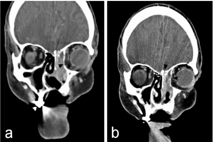
Clinical examination, anterior rhinoscopy and endoscopy revealed purulent secretions and a papilloma-like tumor occupying and obstructing the entire left nasal cavity, and proptosis of the left eye. There was no diplopia, cranial nerve palsy nor palpable cervical lymphadenopathy. Since recurrent IP was suspected, the patient was elected to computed tomogram (CT) of the head and neck area, which showed a heterogeneous opacification of an ex- pansile mass in the left nose and the ethmoid sinus. The medial orbital wall, the anterior wall of the sphenoid sinus and the roof of the left ethmoid sinus were eroded with protrusion of the mass to the left orbit. Nasal septal per- foration due to prior endonasal resections was also ob- served (Figure 1 and Figure 2).
Under general anesthesia, the patient underwent an ex- tensive resection with orbital preservation via a left lateral rhinotomy approach. There was no contamination of the cerebrospinal fluid (CSF) during the surgery. Histopatho- logical examination of the resected specimens confirmed
the diagnosis of IP without evidence of malignancy (Figure 3).
Our patient was hospitalized for a week after the surgery and recovered well postoperatively. Two weeks later, he regained a well formed naso-orbital area with minor de- formity. The patient remained disease-free after one year before loss to the follow up. Two years after the resection from our institution, the nursing home reported us about the patient’s acute neurological deterioration and death of disease with no response to treatment at the nursing home. The autopsy result showed tumor extension to the intracranial temporal fossa without malignant changes of the tumor. There was no attempt to do postmortem imaging for scientific purposes.
The patient’s family gave the written consent before our chart reviewing. The requirement for ethical approval was waived by the institutional research ethics committee.
Discussion and literature review
Etiology
To date, the etiology of IP remains unknown. Many factors may cause IP, including allergy, chronic rhinosinusitis, proliferation of nasal polyps, human papilloma virus (HPV) 6/11 and 16/18, environmental agents, carcinogens, tobacco smoking, hepatocyte growth factor (HGF) and its receptor, c-Met, as well as several enzymes, such as metalloproteinase-9 (MMP-9) [2], [4], [5], [8], [9], [10], [11], [13], [15], [22], [23], [24], [29], [30], [31].
Based on the“hit-and-run”mechanism in the oncogenesis of Schneiderian papillomas by HPV, sinonasal HPV 6/11 infection occurs in an early event during the multistep tumorigenesis process. Types of the papillomas depend on locations of the infection: HPV infection of nasal septal mucosa in exophytic papilloma, and HPV infection of the lateral nasal wall and/or paranasal sinus mucosa in IP.
For unknown reasons, IP epithelium tends to be nonker- atinized. Hence, viral replication and reinfection rarely or never occur. As the superficial epithelial cells are shed, HPV can be lost from the lesion. This partly explains why HPV 6/11 infection rate in IP is lower than in exophytic papilloma. The progression of IP to dysplasia and malig- nancy results from (1) secondary infection of HPV 16/18, (2) loss of tumor suppressor genes, (3) integration of HPV 6/11 and/or HPV 16/18, or (4) a combination of these 3 factors [31]. It is possible that the development of car- cinomas in IP is heralded by a reduced cellular apoptosis, which is triggered by HPV infection [32]. In addition to HPV, studies have showed thatp21and p53, probably coupled with epidermal growth factor receptor (EGFR;
ErbB-1), transforming growth factor alpha (TGF-α), des- moglein 3, and the muscle segment homeobox gene Msx2, are predictive of malignant transformation [1], [4], [5], [9], [11], [13], [20], [26], [31]. The role of HPV in the pathogenesis of IP was extensively reviewed by Lawson et al. [31].
Figure 1: Axial CT scan showing destruction of the lateral wall of the maxillary sinus by a heterogeneous tumor in the left nasal cavity. Bone dehiscence of the anterior wall of the sphenoid sinus is also noted (white arrow).
Figure 2: Coronal CT scans showing (a) a lobulated tumor in the left nasal cavity and ethmoid sinus with extension to the periorbita (black arrow). The nasal septal perforation results from prior endonasal resections;
(b) bone erosion of the roof of the ethmoid sinus and the medial orbital wall (black arrows).
Figure 3: Photomicrograph showing an endophytic epithelial growth into the underlying connective tissue. The epithelium is multilayer and the majority of which is non-keratinizing squamous epithelium. There is no malignant transformation.
(Hematoxylin & eosin stain, original magnification a: 50x, b: 100x)
Differential diagnosis
While inflammatory nasal polyps are typically bilateral, most IP lesions are unilateral at diagnosis [7], [16], [33].
Small IP lesions are usually asymptomatic and detected incidentally on radiographic imaging performed for non- rhinologic purposes. When the tumor reaches a grotesque size, it can cause nasal congestion, posterior nasal drip, epistaxis, hyposmia or anosmia, frontal headaches, epi- phora, proptosis, diplopia, facial numbness and swelling.
Pain and epistaxis associated with IP are rare, but they may indicate secondary infection or malignant changes of the lesion [2], [3], [4], [5], [14], [15], [20], [21]. These
symptoms overlap with a myriad of other conditions, such as allergy, chronic rhinosinusitis and migraine [14]. Under the endoscopic view, the tumor is often seen as one or more polypoid masses with multiple digitations and a papillary surface located laterally to the middle turbinate [5].
Differential diagnosis of IP includes antral choanal polyp, nasal cavity squamous polyp, allergic fungal sinusitis, fibrous dysplasia, giant cell granuloma, juvenile an- giofibroma, nasal glioma, meningoencephalocele, muco- cele, mucus retention cyst, Tornwaldt’s cyst, grossly en- larged adenoids, SCC, lymphoma, adenocarcinoma and esthesioneuroblastoma [5], [8], [21], [34]. It is generally
accepted that unilateral tumor localization involving the lateral nasal wall and the middle meatus is a diagnostic clue to IP. However, even experienced head and neck radiologists also have difficulty in diagnosing IP without a histopathological report because of the absence of pathognomonic radiographic appearance of this tumor [14], [16], [19], [20].
Radiological aspects
Radiographic features of IP depend on the extent of dis- ease. Early lesions appear as soft-tissue density masses, which are heterogeneously enhanced with contrast on CT, within the nasal cavity and/or paranasal sinuses. With progression, the lesions increase opacification and thickening of one or more of the sinuses. Expansion and displacement of adjacent structures associated with the tumor may be observed. However, the nasal septum is usually preserved until late in the disease course. Al- though focal osteosclerosis is a common hyperplastic reaction to chronic rhinosinusitis, and bone erosion can be secondary to benign nasal polyposis, both focal bone sclerosis and erosion can be found in IP patients. There are 2 forms of focal hyperostosis of IP: (1) plaque-like bone thickening, often at the lateral nasal wall; and (2) cone-shaped bone thickening, commonly at the walls of paranasal sinuses or the nasal bony septum. IP and es- thesioneuroblastomas are the 2 most common nasal tu- mors associated with intralesional calcification [2], [3], [4], [13], [14], [15], [16], [20], [26], [33].
CT of IP usually shows a mass extending from the middle meatus to the adjacent maxillary antrum, through an ex- panded maxillary ostium. Although CT is helpful in determ- ining tumor location and extension, it cannot differentiate tumor from inflammatory changes or diseases within the sinus, such as retained secretion, entrapped debris and scar tissue. Hence, tumor size and extension are often overestimated, leading potentially to overtreatment and increased subsequent morbidity. Administration of cor- ticosteroid and antibiotics prior to CT imaging can reduce the inflammatory polyps and thereby decreases overes- timation of the lesion. Alternatively, this drawback can be resolved by using magnetic resonance imaging (MRI):
either T2-weighted images or T1-weighted images with contrast or both [4], [5], [6], [7], [15], [16], [19], [21], [29], [33], [35].
IP is hypodense to isodense on T1-weighted images and isodense to hyperdense on T2-weighted images. T2- weighted MRI can distinguish IP (low to intermediate signal intensity with heterogeneous enhancement throughout the tumor post-gadolinium) from inflammatory tissue and inspissated mucus (bright signal intensity due to high water content). On MRI, IP lesions frequently present a convoluted cerebriform or columnar pattern or a septate-striated appearance. However, T2-weighted MRI cannot differentiate between postoperative scarring and a recurrent tumor. Moreover, destruction of sinus walls is more difficult to recognize by MRI, compared to CT. To date, there has been no adequate evidence to
prove that MRI is better than CT in identifying IP preopera- tively. A combination of CT and MRI is usually recommend- ed before treatment, although its cost-effective analysis remains unclear [2], [5], [8], [14], [15], [19], [21], [26], [33].
Preoperatively, MRI is invaluable in assessing intracranial or orbital extension of the tumor, which will impact treat- ment decisions [2], [13]. When IP appears hypointense with/without bone erosion instead of a hyperintense, heterogeneous enhancement on MRI, it tends to have a malignant transformation [2], [33].
Pathological aspects
Classification or staging systems for IP remain unsettled [4], [5]. Recently, Cannady et al. [36] proposed the new prognostic staging system based on tumor locations/ex- tension [36].
Microscopic findings of IP are distinctive. Literally, the name “inverted papilloma” describes an endophytic or inverted growth pattern of the epithelium into the under- lying stroma rather than outward proliferation from the surface. The epithelial basement membrane is intact [9], [13], [17]. The tumor is composed mainly of hyperplastic basement membrane-enclosed epithelium into the under- lying stroma. The epithelium is multilayer, up to 30 cells in thickness. The squamous or respiratory epithelium is usually mixed with mucocytes. Non-keratinizing squamous or transitional-type epithelium is the predominant cell type and frequently covered by a single layer of ciliated coloumnar epithelium. The stroma has a variable morpho- logy, ranging from dense and fibrous to loose and myxoid with/without inflammatory infiltration. However, it lacks eosinophils, which is common in allergic polyps. Minimal nuclear atypia is frequently found together with basal and parabasal mitoses [3], [5], [9].
Only on rare occasions when IP exhibits focal surface changes similar to verruca vulgaris: focal papillary squamous epithelial hyperplasia with marked keratosis and/or parakeratosis with prominent granular cell layer and numerous koilocytes, WHO recommends that, the patient be diagnosed as‘IP with focal verrucous hyper- plasia’and followed closely for possible development of verrucous carcinoma or SCC [3]. When an area of moder- ate to severe dysplasia or atypia and/or focal surface keratinization is found, microscopic evaluation of all specimen tissue is obligatory to avoid overlooking small foci of malignant transformation [3], [31], [33]. The most reliable criteria for carcinomatous changes include the loss of polarity, anaplasia of the cells and the lack of maturation. Squamous epithelium with marked atypia, an increased nuclear to cytoplasmic ratio, conspicuous nucleoli, atypical mitosis in the middle and upper layers, a loss of polarity, presence of dyskeratotic cells and the absence of eosinophils, is common in the malignant areas of IP [5], [9]. Conversely, benign behavior is linked to the presence of inflammatory polyp and the absence of hyper- keratosis [9]. Histopathological examination of all sino- nasal polypoid specimens is mandatory since unexpected
pathologies are common, especially in cases of unilateral polyps [5], [34].
There has been evidence that a combination of histopath- ological parameters including the presence of hyperkera- tosis and squamous epithelial hyperplasia, a high mitotic index (>2 mitoses per high power field) and an absence of inflammatory polyps may be negative prognostic indi- cators [1], [2], [5]. However, recent studies showed no strong correlation between the degree of atypia/dysplasia or the mitotic index, and recurrence or transformation into carcinomas [11], [17], [33]. Therefore, some authors suggest treating IP with atypia or dysplasia in the same manner as in benign diseases [17].
Surgical aspects
IP is a slow-growing, locally invasive tumor that runs a benign course in most cases. However, if neglected, the persistent and infiltrative tumor can cause gross morbidity or even kill the patient by progressive spread to involve vital structures such as orbits (1–8%) or brain (0.34%) [3], [10]. Intracranial extension in the absence of malig- nancy is usually seen in recurrent cases especially when the lesion involves the cribriform plate, frontal sinus, fo- vea ethmoidalis and orbits [23], [37]. It may be due to the technical difficulties of undertaking complete resec- tion in these anatomical areas [2], [33].
Anterior cranial fossa invasion occurs after destruction of the posterior table of the frontal sinus or the roof of the nasal cavity. Moreover, iatrogenic tumor seeding due to prior extracranial surgery can lead to intracranial and dural involvement [10]. As previously mentioned, HPV is frequently found in IP and can result in recurrence [31], [33], [37], any perioperative contamination of the CSF can spread the virus and/or tumor to the subdural space.
Some authors suggest that the periorbita and dura be preserved; the tumor instead be treated with bipolar electrocautery. It is believed that preservation of the dura and orbital septum provides an effective barrier to tumor spread [2], [13].
When the dura is opened foren blocresection or partially resected during the operation, care must be taken not to contaminate the CSF because this endangers the patient with possible diffuse tumor seeding [37]. It is therefore no surprise that once the disease transgresses the dura, the patient will have a poor prognosis [23], [37]. In the study by Vural et al. [37], 3 (25%) of 12 IP patients with intracranial invasion died with disease after an average follow-up of 9 months (range, 2–17); all of them had dural involvement. Therefore, IP approaching the anterior skull base should be aggressively treated to reduce the risk of local recurrence and intracranial invasion [37].
Until now, only 12 IP cases with extension to intracranial temporal fossa, including our patient, have been reported in the literature. It has been hypothesized that temporal bone or fossa involvement results from (1) direct exten- sion from the sinonasal cavity via the Eustachian tube or directly through the skull base, (2) a primary middle ear involvement secondary to metaplastic changes of the
middle ear mucosa, or (3) tumor seeding in extranasal regions due to surgical manipulation during sinonasal tumor removal [22], [29]. Bui et al. [11] reported a patient with IP of the nasal fossa who developed multiple recur- rences in the middle ear before intracranial extension of the auricular lesion. In our case, local invasion of the le- sion may cause bony destruction and give access for in- tracranial spread.
The treatment of choice for IP is wide excision using either an open or endoscopic approach. Rhinologic/head and neck surgeons today typically tailor their approaches, based on the extent and location of the tumor as deter- mined preoperatively and intraoperatively, the skill and expertise of the surgeon and the available technology [2], [3], [4], [5], [6], [13], [19]. Surgical approaches used for IP resection can be categorized as followed: (1) nonendo- scopic endonasal approach; (2) limited external nasal approach (e.g. Caldwell-Luc); (3) radical external nasal approach (e.g. medial maxillectomy via a lateral rhinotomy or midfacial degloving approach); (4) endonasal endoscop- ic approach; and (5) a combination of these approaches [2], [12].
External approaches provide good exposure and a favor- able tumor control rate, but they are associated with sig- nificant morbidity and a prolonged recovery period [12], [26]. In contrast, endoscopic tools enhance illumination, magnification and angled visualization of the operative field and allow the use of powered instrumentation. This technique can be used in selected cases with a limited disease encroaching on the ethmoid sinuses, the lateral nasal wall, and the medial maxillary wall, including diffi- cult-access locations such as the anterior or lateral wall of the maxillary sinus and the alveolar recess. Other per- tinent benefits are decreased resection of healthy tissues and postoperative morbidity, such as bleeding/epistaxis, crusting, facial swelling, facial pain/neuralgia, epiphora, dacryocystitis, blepharitis, diplopia, mucocele, Eustachian tube dysfunction, CSF leak and external scarring.
Moreover, endoscopic resection can be performed in an ambulatory setting and offers advantages in terms of patient comfort and health care cost [3], [4], [5], [12], [14], [15], [18], [25], [37], [38], [39], [40].
An adjunct to endoscopic resection may be considered if the extranasal component of the tumor cannot be ad- equately visualized endoscopically, for example, a Cald- well-Luc approach when the lateral, inferior or anterior walls of the maxillary sinus are involved; the osteoplastic frontal flap or Draf II- or III-type approaches (median drainage procedure) for the frontal sinus and/or supraor- bital involvement. Temporary septotomy may be used when the tumor confines to the posterior nasal cavity and/or sphenoid sinus [6], [13], [26], [33], [37], [40].
Hence, the patient should be advised that adding or conversion to an open technique may be necessary if the endoscopic approach proves unsatisfactory during the procedure, such as limited access to the entire tumor [33], [39]. Contraindications to endoscopic excision in- clude massive skull base erosion, dural or extensive frontal or infratemporal involvement, intraorbital extension
(as seen in our patient), abundant scar tissue from previ- ous surgery and presence of malignancy [2], [4], [15], [39].
Extensive tumors usually require a lateral rhinotomy and medial maxillectomy, which comprise the following 5 steps: (1) uncinectomy, (2) mega-antrostomy, (3) inferior turbinectomy, (4) debridement of mucosa overlying the medial maxillary wall, and (5) drilling the bony wall with a cutting or diamond burr, or osteotomizing with a chisel.
This radical procedure allows en bloc resection of the lateral nasal wall, ethmoid labyrinth, and medial portion of the maxilla, which are the common tumor locations.
The stalk or attachment of the tumor should be identified and completely removed along with the surrounding mu- cosa to reduce the risk of further recurrence [4], [5], [13], [25], [40]. It has been our experience that the Le Fort I approach to the tumor at the lateral nasal wall should be avoided because the osteotomy line may cut through the tumor or its stalk or site of attachment directly, increasing the risks of tumor spread and recurrences. Using the Caldwell-Luc approach alone provides limited surgical access and hence contributes to high recurrence rates.
To avoid external scarring and morbidity, Rouge-Denker’s rhinotomy, septal translocation or a midfacial degloving or sublabial vestibular approach can be used [2], [5], [10], [15]. IP affecting the anterior skull base, an aggress- ive form, may require a craniofacial resection [10], [37].
However, the frontal sinus should not be obliterated after removing the tumor in order to be able to assess the sinus postoperatively by endoscopy or radiography [13], [14].
Although some authors advocated the benefit of postoper- ative radiation for preventing tumor progression or recur- rence in aggressive IP, side effects of radiotherapy should not be underestimated. The patients may suffer from ra- diation retinopathy, glaucoma, radiation-induced cataract or central retinal artery obstruction [41]. Radiation to the anterior cranial fossa can cause radiation-induced enceph- alomalacia and frontal lobe dysfunction [10]. Adjuvant radiotherapy with or without chemotherapy, therefore, reserves only for patients with multiple recurrences, un- operatable or incompletely resectable tumors or those associated with malignancy [2], [4], [8], [9], [11], [17], [22], [23], [33]. Radiation techniques and dose fraction- ation schedules for patients with IP-associated malig- nancy follow those for carcinomas of the nasal cavity and paranasal sinuses [4]. As with other cancer patients, the treatment plan should be decided by the interdisciplinary tumor board/committee together with the patient [7], [33].
Multiple factors influence recurrence rates of IP, including tumor location, extent, histology, multicentricity, method of removal, primary versus secondary resection, follow up and biological variability of the tumor [17]. Recently, smoking and tumor with extranasal/sinus extension have been found to be associated with the recurrence of IP after surgical resection [33], [42]. However, most recur- rences are related to incomplete tumor removal rather than to metachronous disease [5], [11], [15], [17], [26], [33], [37]. Conservative surgery (polypectomy, Caldwell-
Luc or simple excision) yields 67–78% recurrences, while recurrence rates are 0–14% after a lateral rhinotomy and medial maxillectomy or midfacial degloving [5]. Hence, there is general consensus that IP is best managed and controlled by radical resection. Intraoperative frozen section should be performed when feasible, to ensure adequate tumor resection with tumor-free margins [25].
Parallel with this, the risk of malignant transformation in metachronous lesions can be reduced when complete excision is accomplished [1].
Although many initial IP lesions are unifocal, inadequate tumor removal promotes multifocality [18]. A unilateral mass with a lobulated pattern is a common radiographic feature of tumor recurrence [19]. Recurrence occurring despite radical resection may result from the multicentric nature of the papilloma [17]. Most recurrences occur at the margins of previous surgical resections within the first few years of therapy, but 17% of the patients have late recurrences after 5 years [1], [3], [8], [9], [10], [17], [23], [37]. Hence, the patients should be followed up for at least 6 years [8], [35]. Careful endoscopic follow-up examination, including nasal endoscopy and biopsy of suspected areas, and serial CT scans/MRI postoperatively would help detect early recurrence and allow timely inter- vention [10], [15], [23]. Many authors recommend MRI as the best follow-up imaging because it is more sensitive and specific in differentiating soft tissue mass from un- derlying inflammation and other postoperative changes such as scar tissue, and because of no radiation risk [29], [33].
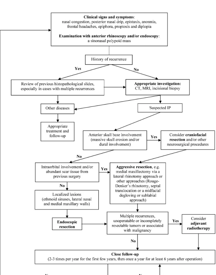
Based on the current literature and our experience, we established an algorithm for managing sinonasal IP (Figure 4).
Conclusions
This case report presents an IP case with fatal extension to the temporal fossa. This is a typical case of intracranial spread of IP after multiple recurrences. Although IP is benign, it reduces the patient’s quality of life due to compromised nasal function, extension to the orbit and brain [6]. Three typical characteristics of IP include a high propensity of recurrence, local aggressiveness and asso- ciation with malignancy. Hence, this benign tumor re- quires radical surgery. Strict attention must be paid to identifying the site of tumor attachment and aggressive resection of a margin of mucosa and bone at this location [13]. Any tumor attached to periorbita or dura should be dealt with by bipolar electrocautery rather than by excision of the dura or periorbita because they provide an effective barrier to tumor spread [2], [13]. Long-term follow up of the patients is also mandatory, as disease can be quite extensive before it becomes symptomatic [17].
Figure 4: Algorithm for managing sinonasal inverted papilloma (IP) Abbreviations:CT, computed tomogram; MRI, magnetic resonance imaging
Notes
Competing interests
The authors declare that they have no competing in- terests.
Acknowledgement
The authors thank Dr. Popchai Ngamskulrungroj MD, PhD, for his help with the literature search.
References
1. von Buchwald C, Bradley PJ. Risks of malignancy in inverted papilloma of the nose and paranasal sinuses. Curr Opin Otolaryngol Head Neck Surg. 2007 Apr;15(2):95-8. DOI:
10.1097/MOO.0b013e3280803d9b
2. Anari S, Carrie S. Sinonasal inverted papilloma: narrative review.
J Laryngol Otol. 2010 Jul;124(7):705-15. DOI:
10.1017/S0022215110000599
3. Barnes L, Tse LLY, Hunt JL. Inverted papilloma (Schneiderian papilloma, inverted type). In: Barnes L, Eveson JW, Reichart P, Sidransky D, eds. WHO Classification of Tumours. Pathology and Genetics of Head and Neck Tumours. Lyon: IARC Press; 2005.
p. 28-31.
4. Mendenhall WM, Hinerman RW, Malyapa RS, Werning JW, Amdur RJ, Villaret DB, Mendenhall NP. Inverted papilloma of the nasal cavity and paranasal sinuses. Am J Clin Oncol. 2007
Oct;30(5):560-3. DOI: 10.1097/COC.0b013e318064c711 5. Sauter A, Matharu R, Hörmann K, Naim R. Current advances in
the basic research and clinical management of sinonasal inverted papilloma (review). Oncol Rep. 2007 Mar;17(3):495-504.
6. Karkos PD, Fyrmpas G, Carrie SC, Swift AC. Endoscopic versus open surgical interventions for inverted nasal papilloma: a systematic review. Clin Otolaryngol. 2006 Dec;31(6):499-503.
DOI: 10.1111/j.1365-2273.2006.01333.x
7. Percodani J, Rose X, Vergez S, Pessey JJ, Serrano E. Voie endonasale endoscopique dans le traitement des papillomes inverses naso-sinusiens [Endonasal endoscopic approach in the treatment of sino-nasal inverted papillomas]. Ann Otolaryngol Chir Cervicofac. 2006 Dec;123(6):312-8. DOI: 10.1016/S0003- 438X(06)76680-4
8. Salomone R, Matsuyama C, Giannotti Filho O, Alvarenga ML, Martinez Neto EE, Chaves AG. Bilateral inverted papilloma: case report and literature review. Braz J Otorhinolaryngol. 2008 Mar- Apr;74(2):293-6.
9. Sandison A. Common head and neck cases in our consultation referrals: diagnostic dilemmas in inverted papilloma. Head Neck Pathol. 2009 Sep;3(3):260-2. DOI: 10.1007/s12105-009-0136- z
10. Miller PJ, Jacobs J, Roland JT Jr, Cooper J, Mizrachi HH.
Intracranial inverting papilloma. Head Neck. 1996 Sep- Oct;18(5):450-3; discussion 454. DOI: 10.1002/(SICI)1097- 0347(199609/10)18:5<450::AID-HED8>3.0.CO;2-4 11. Bui M, Calès V, Barthelmé A, Deminière C, Darrouzet V.
Papillomes inversés nasal et auriculaire [Nasal and auricular inverted papillomas]. Ann Pathol. 2004 Jun;24(3):274-7. DOI:
10.1016/S0242-6498(04)93965-X
12. Busquets JM, Hwang PH. Endoscopic resection of sinonasal inverted papilloma: a meta-analysis. Otolaryngol Head Neck Surg.
2006 Mar;134(3):476-82. DOI: 10.1016/j.otohns.2005.11.038
13. Lane AP, Bolger WE. Endoscopic management of inverted papilloma. Curr Opin Otolaryngol Head Neck Surg. 2006 Feb;14(1):14-8. DOI: 10.1097/01.moo.0000193175.54450.1f 14. Melroy CT, Senior BA. Benign sinonasal neoplasms: a focus on
inverting papilloma. Otolaryngol Clin North Am. 2006 Jun;39(3):601-17. DOI: 10.1016/j.otc.2006.01.005
15. Eggers G, Mühling J, Hassfeld S. Inverted papilloma of paranasal sinuses. J Craniomaxillofac Surg. 2007 Jan;35(1):21-9. DOI:
10.1016/j.jcms.2006.10.003
16. Head CS, Sercarz JA, Luu Q, Collins J, Blackwell KE. Radiographic assessment of inverted papilloma. Acta Otolaryngol. 2007 May;127(5):515-20. DOI: 10.1080/00016480600895144 17. Mirza S, Bradley PJ, Acharya A, Stacey M, Jones NS. Sinonasal
inverted papillomas: recurrence, and synchronous and metachronous malignancy. J Laryngol Otol. 2007 Sep;121(9):857-64. DOI: 10.1017/S002221510700624X 18. Harvinder S, Rosalind S, Mallina S, Gurdeep S. Management of
sinonasal inverted papillomas: endoscopic medial maxillectomy.
Med J Malaysia. 2008 Mar;63(1):58-60.
19. Karkos PD, Khoo LC, Leong SC, Lewis-Jones H, Swift AC.
Computed tomography and/or magnetic resonance imaging for pre-operative planning for inverted nasal papilloma: review of evidence. J Laryngol Otol. 2009 Jul;123(7):705-9. DOI:
10.1017/S0022215109004575
20. Reh DD, Lane AP. The role of endoscopic sinus surgery in the management of sinonasal inverted papilloma. Curr Opin Otolaryngol Head Neck Surg. 2009 Feb;17(1):6-10. DOI:
10.1097/MOO.0b013e32831b9cd1
21. Breining T, Lindemann J, Pauls S. Das invertierte Papillom – eine seltene Ursache der nasalen Obstruktion [Inverted papilloma – a rare cause of nasal obstruction]. Rofo. 2009 Jun;181(6):599- 600. DOI: 10.1055/s-0028-1109253
22. Acevedo-Henao CM, Talagas M, Marianowski R, Pradier O.
Recurrent inverted papilloma with intracranial and temporal fossa involvement: A case report and review of the literature.
Cancer Radiother. 2010 Jun;14(3):202-5. DOI:
10.1016/j.canrad.2010.01.012
23. Visvanathan V, Wallace H, Chumas P, Makura ZG. An unusual presentation of inverted papilloma: case report and literature review. J Laryngol Otol. 2010 Jan;124(1):101-4. DOI:
10.1017/S0022215109990703
24. Salima K, Anissa S, Samah T, Ines C, Najeh B, Samia S, Ghazi B. Le papillome inversé dégénéré des fosses nasales [Malignant degeneration of an inverted papilloma of the nasal cavity]. Tunis Med. 2010 May;88(5):369-70.
25. Bathma S, Harvinder S, Philip R, Rosalind S, Gurdeep S.
Endoscopic management of sinonasal inverted papilloma. Med J Malaysia. 2011;66(1):15-8.
26. Carta F, Verillaud B, Herman P. Role of endoscopic approach in the management of inverted papilloma. Curr Opin Otolaryngol Head Neck Surg. 2011 Feb;19(1):21-4. DOI:
10.1097/MOO.0b013e3283425213
27. Kim K, Kim D, Koo Y, Kim CH, Choi EC, Lee JG, Yoon JH. Sinonasal carcinoma associated with inverted papilloma: a report of 16 cases. J Craniomaxillofac Surg. 2012 Jun;40(4):e125-9. DOI:
10.1016/j.jcms.2011.07.007
28. Dominas N, Sandalcioglu E, Lang S, Mattheis S. Invertiertes Papillom der Nasennebenhöhlen mit intrakranieller Beteiligung [Inverted papilloma of the paranasal sinuses with intracranial extension]. Laryngorhinootologie. 2011 Jun;90(6):371-2. DOI:
10.1055/s-0031-1275306
29. Shen J, Baik F, Mafee MF, Peterson M, Nguyen QT. Inverting papilloma of the temporal bone: case report and meta-analysis of risk factors. Otol Neurotol. 2011 Sep;32(7):1124-33. DOI:
10.1097/MAO.0b013e31822a2b16
30. Poomsawat S, Punyasingh J, Vejchapipat P, Larbcharoensub N.
Co-expression of hepatocyte growth factor and c-met in epithelial odontogenic tumors. Acta Histochem. 2012 Jul;114(4):400-5.
DOI: 10.1016/j.acthis.2011.07.010
31. Lawson W, Schlecht NF, Brandwein-Gensler M. The role of the human papillomavirus in the pathogenesis of Schneiderian inverted papillomas: an analytic overview of the evidence. Head Neck Pathol. 2008 Jun;2(2):49-59. DOI: 10.1007/s12105-008- 0048-3
32. Tanvetyanon T, Qin D, Padhya T, Kapoor R, McCaffrey J, Trotti A.
Survival outcomes of squamous cell carcinoma arising from sinonasal inverted papilloma: report of 6 cases with systematic review and pooled analysis. Am J Otolaryngol. 2009 Jan- Feb;30(1):38-43. DOI: 10.1016/j.amjoto.2008.02.005 33. Jankowski R, Coste A, Verdalle P. Papillome inversé rhinosinusien
[Inverted nasosinusal papilloma]. Ann Otolaryngol Chir Cervicofac.
2008 Sep;125(4):224-33. DOI: 10.1016/j.aorl.2008.07.004 34. Yaman H, Alkan N, Yilmaz S, Koc S, Belada A. Is routine
histopathological analysis of nasal polyposis specimens necessary? Eur Arch Otorhinolaryngol. 2011 Jul;268(7):1013-5.
DOI: 10.1007/s00405-011-1534-x
35. Delank KW. Die endonasale Resektion inverser Papillome der Nasen- und Nasennebenhöhlen: „faux-pas” oder „dernier crie”?
[Endonasal Resection of Sinunasal Inverted Papilloma: "faux- pas" or "dernier crie"]. Laryngorhinootologie. 2006 Sep;85(9):633- 4. DOI: 10.1055/s-2006-944518
36. Cannady SB, Batra PS, Sautter NB, Roh HJ, Citardi MJ. New staging system for sinonasal inverted papilloma in the endoscopic era. Laryngoscope. 2007 Jul;117(7):1283-7. DOI:
10.1097/MLG.0b013e31803330f1
37. Vural E, Suen JY, Hanna E. Intracranial extension of inverted papilloma: An unusual and potentially fatal complication. Head Neck. 1999 Dec;21(8):703-6. DOI: 10.1002/(SICI)1097- 0347(199912)21:8<703::AID-HED4>3.0.CO;2-H
38. Ridder GJ, Behringer S, Kayser G, Pfeiffer J. Malignome auf dem Boden invertierter Papillome der Nase und Nasennebenhöhlen [Malignancies arising in sinonasal inverted papillomas].
Laryngorhinootologie. 2008 Nov;87(11):783-90. DOI: 10.1055/s- 2008-1077292
39. Facon F, Dessi P. Chirurgie endonasale micro-invasive: apport de l'endoscopie en chirurgie maxillo-faciale [Microinvasive endonasal surgery: contribution of endoscopy to maxillofacial surgery]. Rev Stomatol Chir Maxillofac. 2005 Sep;106(4):230- 42. DOI: 10.1016/S0035-1768(05)85852-8
40. Minovi A, Kollert M, Draf W, Bockmühl U. Endonasale mikro- endoskopische Resektion invertierter Papillome der Nase und ihrer Nebenhöhlen [Endonasal micro-endoscopic resection of sinonasal inverted papilloma]. Laryngorhinootologie. 2006 Jun;85(6):421-5. DOI: 10.1055/s-2006-925059
41. Sauter A. Focal malignancy in sinonasal inverted papilloma – is postoperative radiotherapy recommendable? Oral Oncol. 2011 Sep;47(9):779.
42. Moon IJ, Lee DY, Suh MW, Han DH, Kim ST, Min YG, Lee CH, Rhee CS. Cigarette smoking increases risk of recurrence for sinonasal inverted papilloma. Am J Rhinol Allergy. 2010 Sep- Oct;24(5):325-9. DOI: 10.2500/ajra.2010.24.3510
Corresponding author:
Dr. rer. med. Dr. med. dent. Poramate Pitak-Arnnop, DDS, PGDipClinSc (OMS), MSc, PhD, DSc
Klinik und Poliklinik für Mund-, Kiefer- und Plastische Gesichtschirurgie, Universitätsklinikum Leipzig AöR, Nürnberger Str. 57, 04103 Leipzig, Germany, Tel: +49 341 97 21 100, Fax: +49 341 97 21 109
poramate.pitakarnnop@gmail.com
Please cite as
Pitak-Arnnop P, Bertolini J, Dhanuthai K, Hendricks J, Hemprich A, Pausch NC. Intracranial extension of Schneiderian inverted papilloma:
a case report and literature review. GMS Ger Med Sci. 2012;10:Doc12.
DOI: 10.3205/000163, URN: urn:nbn:de:0183-0001633
This article is freely available from
http://www.egms.de/en/journals/gms/2012-10/000163.shtml
Received:2012-01-11 Published:2012-06-18
Copyright
©2012 Pitak-Arnnop et al. This is an Open Access article distributed under the terms of the Creative Commons Attribution License (http://creativecommons.org/licenses/by-nc-nd/3.0/deed.en). You are free: to Share — to copy, distribute and transmit the work, provided the original author and source are credited.