Wound infection caused by Photobacterium damselae in a 32-year-old woman: case report and review of the
literature
Abstract
The case of a 32-year-old woman is reported, who was affected by a persisting wound infection caused byPhotobacterium damselaeafter
Percy Schröttner
1Eric Tille
2an accident in the Mediterranean Sea. Besides the clinical case, micro-
Christian Lück
1biological characteristics based on the phenotypic and genotypic de-
Boyke Bunk
3scription of the isolate (including whole genome data) are presented and discussed.
Keywords:Photobacterium damselae, wound infection, whole genome data, MALDI TOF MS, antimicrobial profile
1 Institut für Medizinische Mikrobiologie und Hygiene, Medizinische Fakultät Carl Gustav Carus, Technische
Zusammenfassung
Wir berichten über den Fall einer 32-jährigen Frau, die nach einem Ba- deunfall im Mittelmeer von einer persistierenden Wundinfektion, welche
Universität Dresden, Germany
2 UniversitätsCentrum für Orthopädie, Unfall- und durchPhotobacterium damselaeverursacht wurde, betroffen war. Neben
der Beschreibung des klinischen Falls werden mikrobiologische Charak- Plastische Chirurgie (OUPC), Universitätsklinikum teristika des Isolats vorgestellt und diskutiert. Diese beinhalten phäno-
Dresden, Medizinische typische und genotypische Beschreibungen (auch unter Bezugnahme
des gesamten bakteriellen Genoms). Fakultät Carl Gustav Carus
Dresden, Germany 3 Leibniz-Institut DSMZ –
Deutsche Sammlung für Mikroorganismen und Zellkulturen GmbH, Braunschweig, Germany
Introduction
Photobacterium damselaebelongs to the family of the Vibrionaceae. The species was first described by Love et al. asVibrio damselain 1981 [1]. The first reclassification followed in 1985 and the species was included into the genus Listonella [2]. In 1991, Smith et al. undertook a reevaluation of the genus Listonella andP. damselaewas finally introduced into bacterial taxonomy [3].
P. damselaehas been detected in sea water and is a well-known fish pathogen [4], [5], [6]. The two hae- moylsins damselysin (Dly) and phobalysin (PhlyP) have been identified as the main virulence factors for fish. The corresponding genes (thedlyrespectivelyhlyAgene) are encoded by the virulence plasmid pPHDD1 [5].
Besides this, infections in humans, occasionally with a fatal outcome, have also been described. In most of these cases, a previous contact with sea water or fish was re- ported and infections often originated from minor injuries, which many patients could not remember. Fatal courses
of the disease were usually caused by a rapidly progress- ing necrotizing fasciitis, sepsis or are mediated by bacteri- al toxins. However, there are also localized infections of the skin, which mostly resulted in a complete healing.
Here we report on a persisting wound infection of a 32- year-old woman, which was caused byP. damselaeafter an accident in the Mediterranean. Furthermore, we also describe the bacterial isolate both phenotypically and genotypically.
Case description
In August 2019, a 32-year-old female presented to our emergency department and reported that she had injured herself at a rotor leaf of a boat engine after falling off a dinghy into the salt water of the Mediterranean Sea on a trip to Spain 10 days before. Initially, the patient had received medical treatment by wound cleaning, disinfec- tion and surgical stapling. Unfortunately, there is no in-
formation available on an antimicrobial therapy that has already been given in Spain.
Clinically we saw four laceration wounds (each approxim- ately 4–5 cm) to the lateral thigh and calf of the left leg.
While the two proximal wounds (thigh) were inconspicu- ous, the distal wounds on the calf displayed a local hyper- emia, swelling and pressure pain. Additionally, there was slight bleeding and purulent secretion. The peripheral sensitivity, strength and mobility were unaffected.
Moreover, the patient showed no systemic signs of infec- tion. Except for a marginal elevation of the inflammation parameter CRP (14.7 mg/L) all further laboratory findings were normal. Radiographic imaging was not altered either.
Apart from a nicotine (20 packyears) and alcohol abuse (2–3 drinks per day) the medical history of the patient was empty.
The patient was admitted to the hospital and treated surgically. Intraoperatively, the swelling revealed to be an infected hematoma. After collection of microbiological samples, the hematoma was removed and the wound cavity was lavaged thoroughly. A drainage was inserted and the wound was then closed layer by layer. After the surgical treatment the patient received an immediate, empirical, intravenous antibiotic treatment with a ceph- alosporin (cefuroxime 4x 1.5 g per day). After confirmation of infection byP. damselae, the antibiotic treatment was adjusted to a combination of ampicillin (1 g) and sul- bactam (2 g). The antibiotic was administered three times a day. After seven days of intravenous treatment, the patient was discharged from the hospital. We recommen- ded an additional oral antibiotic treatment with amoxicillin (875 mg) and clavulanic acid (125 mg) for seven further days. The antibiotic was administered three times a day.
A scheduled appointment for clinical reevaluation was not met by the patient.
Microbiological methods and results
Cultivation
In total, three specimens were sent to the Institute for Medical Microbiology and Hygiene of the Technical Uni- versity Dresden: the first sample was collected from a wound swab (collected one day prior to the operation, isolate DSM 110633). The second (intraoperative wound swab, isolate DSM 110632) and the third sample (biopsy, isolate DSM 110634) were obtained in the course of the operation. The samples were cultured on Columbia agar with 5% sheep blood (Oxoid, Wesel, Germany), bile chrysoidin glycerol agar (Oxoid, Wesel, Germany), brain heart infusion (Becton Dickinson, Heidelberg, Germany) and Schaedler broth (bioMérieux, Nürtingen, Germany).
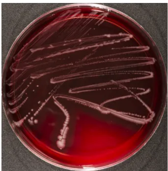
The agar plates were incubated for 18 hours at 37ºC (without CO2infusion). After incubation, smooth, glossy and slightly transparent bacterial colonies showing a strong beta haemolysis were detected on all Columbia
in brain heart infusion (indicated by turbidity) and bile chrysoidin glycerol agar.
Figure 1:Photobacterium dampselae DSM 110634 growing on Columbia blood agar containing 5% sheep blood (Oxoid, Wesel, Germany). The bacteria were incubated for 18 hours.
Identification
MALDI-TOF MS (Bruker Daltonik, Bremen, Germany) was used for primary species identification. All isolates were identified asP. damselaewith score values >2.0, which indicates a high confidence identification (DSM 110632:
2.131; DSM 110633: 2.334; DSM 110634: 2.395). The results were additionally confirmed by sequencing of the 16S rRNA gene using 27F (agagtttgatcmtggctcag) as forward and 1498R (cggttaccttgttacgactt) as reverse primer. The data were analysed using the BLAST algorithm (https://blast.ncbi.nlm.nih.gov/Blast.cgi). The species P. damselaewas confirmed in all isolates (99% identity).
The PCR product covers a length of 1,378 bases. A total of four mismatches were found.
Antimicrobial susceptibility testing
Bacterial colonies originating from the third specimen (DSM 110634, biopsy) were inoculated in physiological sodium chloride solution (Fresenius, Bad Homburg, Germany) and a McFarland standard of 0.5 was created.
The bacterial suspension was plated on Mueller-Hinton agar (bioMérieux, Nürtingen, Germany) using a plate ro- tator (bestbion dx, Köln, Germany). Gradient diffusion test strips (bestbion dx, Köln, Germany) were then placed on the agar plates and incubated at 37ºC for 18 hours.
The interpretation of the MIC values was performed ac- cording to the EUCAST guidelines (PK/PD breakpoints) published in 2020 [7]. The isolate was susceptible to- wards ampicillin (0.25 mg/L), ampicillin sulbactam (0.25 mg/L), amoxicillin-clavulanate (0.5 mg/L), pipera-
cefotaxime (≤0.016 mg/L), ceftazidime (0.125 mg/L), imipenem (0.5 mg/L), meropenem (0.032 mg/L), cipro- floxacin (0.008 mg/L), levofloxacin (0.004 mg/L) and moxifloxacin (0.016 mg/L). However, it showed resistance towards the aminogylcosides gentamicin (1.0 mg/L) and amikacin (4.0 mg/L). There are no breakpoints available for fosfomycin since there is insufficent evidence for its usefulness in the clinical setting.
Next generation sequencing and whole genome data analysis
It could be assumed that all the isolates were identical sinceP. damselaeis only extremely rarely detected as a pathogen in humans and beyond that, only pure cultures were found. For this reason, only one isolate (DSM 110634) was chosen for whole genome sequencing.
Sequencing of a Nextera XT DNA Library (Nextera XT DNA Library Prep Kit; Illumina, San Diego, CA, USA) was per- formed on an Illumina NextSeq 550 instrument (Illumina, San Diego, CA, USA) followed by genome assembly using SPAdes 3.14 and an automated annotation applying NCBI Prokarytic Genome Annotation Pipeline [8], [9]. Screening for resistance genes was performed using ResFinder 2.1 as recently described and CARD5 [10], [11]. Pylogenomic analyses were carried out performing digital DNA-DNA hybridization (dDDH). Type (Strain) Genome Server (TYGS) was used as database [12]. The results showed an identity of 88.2% toP. damselaeCIP 102761, thus con- firming the species [13]. Resistance gene analysis using both ResFinder 2.1 and CARD 5 did not lead to an explan- ation for the observed resistance against aminoglycos- ides. The genome sequence was submitted to NCBI GenBank under accession number JAATTX000000000.
In total three genes encoding for haemoylsis were detect- ed:dly,hlyAand (chromosomally encoded)hylA. Interest- ingly, the complete virulence plasmid pPHDD1 was not found.
Discussion
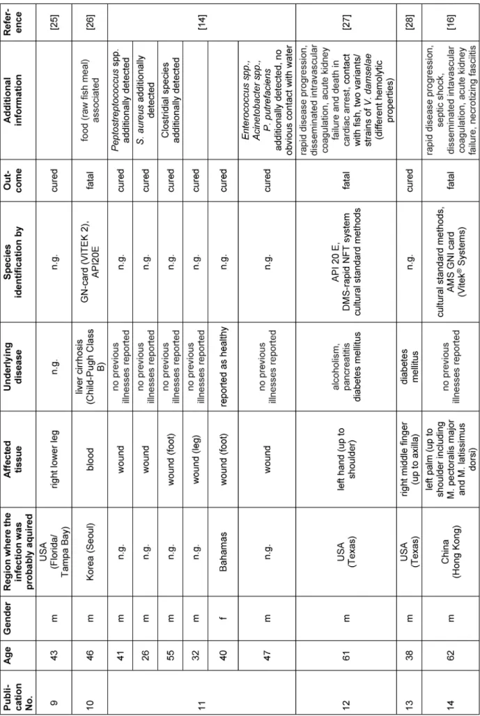
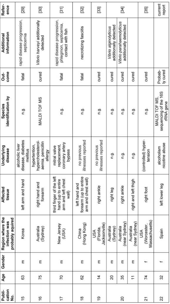
To the best of our knowledge, a total of 29 case descrip- tions ofP. damselaeinfections in humans (including the present report) are described so far (Table 1). All reports (except one) have in common that an infection has always been preceded by contact with sea water (the natural reservoir ofP. damselae), fish or other sea animals (see Table 1) [14]. Additionally, a closer look at the available literature reveals that fatal cases tend to affect (but are not exclusive to) older patients (n=10; average age=63.1 years). In contrast, there are young patients who have recovered from the infection (n=19; average age=38.9 years) (Table 1). The patient in the present case was 32 years old. Based on our current knowledge, the course of the disease was also positive, which is congruent with the observations mentioned before.
Basically, there are two possible explanations for a worse course of the infection. On the one hand, the deteriorating function of the immune system with age (immunosenes- cence) should be mentioned [15]. On the other hand, the different virulence properties of the strains must be taken into account. So far, the two haemolysins damselysin (Dly) and phobalysin (PhlyP) have been described as the most important virulence factors [5]. Our isolates also showed strong β-haemolysis. Genome analysis of the entire genome of the P. damselaestrain DSM 110634 showed both haemolysins being present with 100% amino acid identity (Swissprot IDs D1J6Q4 and D1J6Q5). Both genes were found in one cluster similar to their assembly in pPHDD1. Moreover, the corresponding assembled contig, namely NODE_21, carried the parA replication gene showing clearly both haemolysin genes are encoded on a plasmid. However, the complete plasmid pPHDD1 (RefSeq ID NC_014653) was not found as such. Obvi- ously, the virulence plasmid structure differs within our isolate. Surprisingly, a second chromosomally encoded haemolysin gene was found with 92% amino acid identity tohlyA(NODE_13:122725.120917).
We were also able to reliably identify our isolate using the MALDI TOF MS since it was in accordance to results obtained from sequencing of the 16S rRNA gene and the dDDH. However, a general statement about the suitability of MALDI TOF MS for the identification of P. damselae can only be made using a larger collection of well-charac- terized strains derived from clinical specimens.
The data currently available are insufficient to make general statements about the antimicrobial resistance ofP. damselae. However, previous reports suggest that the species is sensitive to most antibiotics (including most of the β-lactams, cotrimoxazole, aminoglycosides, fluoroquinolones) [16]. However, resistance to amino- glycosides (as in the present case report) has also been described [17]. Unfortunately, a search for genes providing aminoglycoside resistance using both ResFinder 2.1 and CARD 5 did not obtain any results.
There are basically three possible explanations for the development of resistance to aminoglycosides:
1. reduction of the concentration of aminoglycosides within the bacterial cell (e.g. efflux pump),
2. changes in the target structure for aminoglycosides (e.g. 16S methylation or ribosomal mutations) and 3. enzymatic inactivation (e.g. aminoglycoside acetyl-
transferases, aminoglycoside nucleotidyltranserases, aminoglycoside phosphotransferases) [18].
It is important to note that amikacin is not inactivated by enzymes, which act on gentamycin or tobramycin [18].
However, both gentamycin and amikacin are resistant in our case. For this reason, a resistance mechanism is suggested in our isolate, which affects the entire class of aminoglycosides (e.g. an efflux pump).
Table 1: Overview of reports on human infections caused byPhotobacterium damselae
(Continued)
Table 1: Overview of reports on human infections caused byPhotobacterium damselae
(Continued)
Table 1: Overview of reports on human infections caused byPhotobacterium damselae
Conclusion
We describe the 29thcase of a human infection caused by the marine bacteriumP. damselae. Infections affecting younger patients (like the patient described here) seem to show a more favorable course of healing than those affecting older patients. Since the patient in this report did not keep a follow-up visit at the hospital after the therapy, we assume the healing process has been uncom- plicated. Using whole genome sequencing, we could de- tect three haemolysins serving as virulence factors.
However, the issue whether those proteins really contrib- ute to human infections as well needs to be elucidated in more detail in further studies. Moreover, a larger strain collection is needed to be able to create a reliable resis- tance profile.
Notes
Competing interests
The authors declare that they have no competing in- terests.
Acknowledgements
The authors thank Franziska Klann and Stefan Tiede for excellent technical assistance and Thomas Riedel for support regarding strain deposition.
References
1. Love M, Teebken-Fisher D, Hose JE, Farmer JJ 3rd, Hickman FW, Fanning GR. Vibrio damsela, a Marine Bacterium, Causes Skin Ulcers on the Damselfish Chromis punctipinnis. Science. 1981 Dec;214(4525):1139-40. DOI: 10.1126/science.214.4525.1139 2. MacDonell MT, Colwell RR. Phylogeny of the Vibrionaceae, and
Recommendation for Two New Genera, Listonella and Shewanella. Syst Appl Microbiol. 1985;6(2):171-82. DOI:
10.1016/S0723-2020(85)80051-5
3. Smith SK, Sutton DC, Fuerst JA, Reichelt JL. Evaluation of the genus Listonella and reassignment of Listonella damsela (Love et al.) MacDonell and Colwell to the genus Photobacterium as Photobacterium damsela comb. nov. with an emended description. Int J Syst Bacteriol. 1991 Oct;41(4):529-34. DOI:
10.1099/00207713-41-4-529
4. Fouz B, Toranzo AE, Milán M, Amaro C. Evidence that water transmits the disease caused by the fish pathogen
Photobacterium damselae subsp. damselae. J Appl Microbiol.
2000 Mar;88(3):531-5. DOI: 10.1046/j.1365- 2672.2000.00992.x
5. Terceti MS, Ogut H, Osorio CR. Photobacterium damselae subsp.
damselae, an Emerging Fish Pathogen in the Black Sea: Evidence of a Multiclonal Origin. Appl Environ Microbiol. 2016
Jul;82(13):3736-45. DOI: 10.1128/AEM.00781-16 6. Andreoni F, Magnani M. Photobacteriosis: prevention and
diagnosis. J Immunol Res. 2014;2014:793817. DOI:
10.1155/2014/793817
7. European Committee on Antimicrobial Susceptibility (EUCAST).
Breakpoint tables for interpretation of MICs and zone diameters.
Version 10.0. 2020. Available from: https://www.eucast.org/
clinical_breakpoints/
8. Zerbino DR, Birney E. Velvet: algorithms for de novo short read assembly using de Bruijn graphs. Genome Res. 2008 May;18(5):821-9. DOI: 10.1101/gr.074492.107
9. Seemann T. Prokka: rapid prokaryotic genome annotation.
Bioinformatics. 2014 Jul;30(14):2068-9. DOI:
10.1093/bioinformatics/btu153
10. Gunzer F, Rudolph WW, Bunk B, Schober I, Peters S, Müller T, Oberheitmann B, Schröttner P. Whole-genome sequencing of a large collection of Myroides odoratimimus and Myroides odoratus isolates and antimicrobial susceptibility studies. Emerg Microbes Infect. 2018 Apr;7(1):61. DOI: 10.1038/s41426-018-0061-x 11. Alcock BP, Raphenya AR, Lau TTY, Tsang KK, Bouchard M,
Edalatmand A, Huynh W, Nguyen AV, Cheng AA, Liu S, Min SY, Miroshnichenko A, Tran HK, Werfalli RE, Nasir JA, Oloni M, Speicher DJ, Florescu A, Singh B, Faltyn M, Hernandez- Koutoucheva A, Sharma AN, Bordeleau E, Pawlowski AC, Zubyk HL, Dooley D, Griffiths E, Maguire F, Winsor GL, Beiko RG, Brinkman FSL, Hsiao WWL, Domselaar GV, McArthur AG. CARD 2020: antibiotic resistome surveillance with the comprehensive antibiotic resistance database. Nucleic Acids Res. 2020 Jan;48(D1):D517-25. DOI: 10.1093/nar/gkz935 12. Meier-Kolthoff JP, Göker M. TYGS is an automated high-
throughput platform for state-of-the-art genome-based taxonomy.
Nat Commun. 2019 May;10(1):2182. DOI: 10.1038/s41467- 019-10210-3
13. Richter M, Rosselló-Móra R. Shifting the genomic gold standard for the prokaryotic species definition. Proc Natl Acad Sci U S A.
2009 Nov;106(45):19126-31. DOI: 10.1073/pnas.0906412106 14. Morris JG Jr, Miller HG, Wilson R, Tacket CO, Hollis DG, Hickman FW, Weaver RE, Blake PA. Illness caused by Vibrio damsela and Vibrio hollisae. Lancet. 1982 Jun;1(8284):1294-7. DOI:
10.1016/s0140-6736(82)92853-7
15. Pawelec G. Age and immunity: What is “immunosenescence”?
Exp Gerontol. 2018 May;105:4-9. DOI:
10.1016/j.exger.2017.10.024
16. Yuen KY, Ma L, Wong SS, Ng WF. Fatal necrotizing fasciitis due to Vibrio damsela. Scand J Infect Dis. 1993;25(5):659-61. DOI:
10.3109/00365549309008557
17. Yamane K, Asato J, Kawade N, Takahashi H, Kimura B, Arakawa Y. Two cases of fatal necrotizing fasciitis caused by
Photobacterium damsela in Japan. J Clin Microbiol. 2004 Mar;42(3):1370-2. DOI: 10.1128/jcm.42.3.1370-1372.2004 18. Jana S, Deb JK. Molecular understanding of aminoglycoside
action and resistance. Appl Microbiol Biotechnol. 2006 Mar;70(2):140-50. DOI: 10.1007/s00253-005-0279-0 19. Fraser SL, Purcell BK, Delgado B Jr, Baker AE, Whelen AC. Rapidly
fatal infection due to Photobacterium (Vibrio) damsela. Clin Infect Dis. 1997 Oct;25(4):935-6. DOI: 10.1086/597647
20. Asato J, Kanaya F. Fatal infection of the hand due to Photobacterium damsela: a case report. Clin Infect Dis. 2004 May;38(10):e100-1. DOI: 10.1086/383468
21. Hundenborn J, Thurig S, Kommerell M, Haag H, Nolte O. Severe Wound Infection with Photobacterium damselae ssp. damselae and Vibrio harveyi, following a Laceration Injury in Marine Environment: A Case Report and Review of the Literature. Case Rep Med. 2013;2013:610632. DOI: 10.1155/2013/610632 22. Alvarez JR, Lamba S, Dyer KY, Apuzzio JJ. An unusual case of
urinary tract infection in a pregnant woman with Photobacterium damsela. Infect Dis Obstet Gynecol. 2006;2006:80682. DOI:
10.1155/IDOG/2006/80682
23. Goodell KH, Jordan MR, Graham R, Cassidy C, Nasraway SA.
Rapidly advancing necrotizing fasciitis caused by Photobacterium (Vibrio) damsela: a hyperaggressive variant. Crit Care Med. 2004 Jan;32(1):278-81. DOI:
10.1097/01.CCM.0000104920.01254.82
24. Knight-Madden JM, Barton M, Gandretti N, Nicholson AM.
Photobacterium damsela bacteremia in a child with sickle-cell disease. Pediatr Infect Dis J. 2005 Jul;24(7):654-5. DOI:
10.1097/01.inf.0000168845.26758.e3
25. Nakamura Y, Uchihira M, Ichimiya M, Morita K, Muto M.
Necrotizing fasciitis of the leg due to Photobacterium damsela.
J Dermatol. 2008 Jan;35(1):44-5. DOI: 10.1111/j.1346- 8138.2007.00412.x
26. Barber GR, Swygert JS. Necrotizing fasciitis due to
Photobacterium damsela in a man lashed by a stingray. N Engl J Med. 2000 Mar;342(11):824. DOI:
10.1056/NEJM200003163421118
27. Kim HR, Kim JW, Lee MK, Kim JG. Septicemia progressing to fatal hepatic dysfunction in an cirrhotic patient after oral ingestion of Photobacterium damsela: a case report. Infection. 2009 Dec;37(6):555-6. DOI: 10.1007/s15010-009-9049-8 28. Clarridge JE, Zighelboim-Daum S. Isolation and characterization
of two hemolytic phenotypes of Vibrio damsela associated with a fatal wound infection. J Clin Microbiol. 1985 Mar;21(3):302-6.
DOI: 10.1128/JCM.21.3.302-306.1985
29. Coffey JA Jr, Harris RL, Rutledge ML, Bradshaw MW, Williams TW Jr. Vibrio damsela: another potentially virulent marine vibrio.
Infect Dis. 1986 Apr;153(4):800-2. DOI:
10.1093/infdis/153.4.800-a
30. Shin JH, Shin MG, Suh SP, Ryang DW, Rew JS, Nolte FS. Primary Vibrio damsela septicemia. Clin Infect Dis. 1996 May;22(5):856- 7. DOI: 10.1093/clinids/22.5.856
31. Akram A, Stevens RP, Konecny P. Photobacterium damselae and Vibrio harveyi hand infection from marine exposure. Med J Aust.
2015 Sep;203(5):224-5.e1. DOI: 10.5694/mja15.00179 32. Perez-Tirse J, Levine JF, Mecca M. Vibrio damsela. A cause of
fulminant septicemia. Arch Intern Med. 1993 Aug;153(15):1838- 40. DOI: 10.1001/archinte.153.15.1838
33. Tang WM, Wong JW. Necrotizing fasciitis caused by Vibrio damsela. Orthopedics. 1999 Apr;22(4):443-4.
34. Aigbivbalu L, Maraqa N. Photobacterium damsela wound infection in a 14-year-old surfer. South Med J. 2009 Apr;102(4):425-6.
DOI: 10.1097/SMJ.0b013e31819b9491
35. Dryden M, Legarde M, Gottlieb T, Brady L, Ghosh HK. Vibrio damsela wound infections in Australia. Med J Aust. 1989 Nov;151(9):540-1.
36. Sahu KK, Sherif AA, Davaro R. A Rare Cause of Cellulitis:
Photobacterium damselae. J Microsc Ultrastruct. 2020 Jan- Mar;8(1):25-26. DOI: 10.4103/JMAU.JMAU_63_18
Corresponding author:
Dr. Percy Schröttner
Institute of Medical Microbiology and Hygiene, Faculty of Medicine, TU Dresden, Fetscherstr. 74, 01307 Dresden, Germany, Phone: +49 351 458-16585, Fax: +49 351 458-6310
percy.schroettner@tu-dresden.de
Please cite as
Schröttner P, Tille E, Lück C, Bunk B. Wound infection caused by Photobacterium damselae in a 32-year-old woman: case report and review of the literature. GMS Infect Dis. 2020;8:Doc23.
DOI: 10.3205/id000067, URN: urn:nbn:de:0183-id0000675
This article is freely available from
https://www.egms.de/en/journals/id/2020-8/id000067.shtml Published:2020-11-17
Copyright
©2020 Schröttner et al. This is an Open Access article distributed under the terms of the Creative Commons Attribution 4.0 License. See license information at http://creativecommons.org/licenses/by/4.0/.