Retinoschisis transposition following a retinal detachment repair
Abstract
Objective:The authors have observed this phenomenon of translocation of the schisis cavity in a few previous cases and aim to report this un- usual finding.
Katherine McVeigh
1Johannes Keller
1Richard J. Haynes
1Method:A patient with known superotemporal retinoschisis developed a distinctly separate inferotemporal retinal detachment in his left eye.
This was repaired with a vitrectomy, cryotherapy and C2F6 tamponade 1 Retina Unit, Bristol Eye Hospital, Bristol University under local anaesthetic. Following surgery, the retinoschisis was found
in the inferonasal quadrant of the eye and remained stable as the gas dispersed.
Hospitals NHS Trust, Bristol, United Kingdom
Result:We hypothesise that the tamponading agent compressed the viscous fluid within the area of schisis, displacing the area of schisis circumferentially.
Conclusion:This case emphasises that as long as the retinal breaks are properly sealed, no intervention is required with the schisis during rhegmatogenous retinal detachment surgery.
Keywords:retinoschisis, retinal detachment, vitrectomy, transposition
Introduction
Degenerative retinoschisis (RS) is a common condition characterised by the splitting of the retinal layers, occur- ring between the outer plexiform and inner nuclear layer, creating a cavity filled with hyaluronidase-sensitive acid mucopolysaccharide presumably hyaluronic acid ([1], p. 422) leading to elevation of the inner leaf often in a tense balloon-like configuration. This tends to arise from areas of peripheral microcystoid degeneration and occurs more commonly in hypermetropes, usually affecting the inferotemporal quadrants. Degenerative retinoschisis is found in 5% of the population. Rarely, RS may progress to a rhegmatogenous retinal detachment (RRD) if holes develop in both the outer and inner leafs allowing fluid to pass from the vitreous cavity into the subretinal space [2], [3]. This condition is then treated following the prin- ciples of RRD repair.
Case description
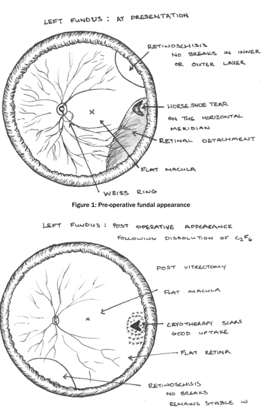
A 72-year-old patient, previously diagnosed with RS, presented to the emergency clinic complaining of floaters, photopsia and a visual field defect in the left eye. Exam- ination revealed a macula-on inferotemporal RRD with associated posterior vitreous detachment, originating form a horseshoe tear on the horizontal meridian. A dis- tinctly separate area of RS was found superotemporally with no identifiable retinal breaks in either the inner or outer layer on indentation examination (Figure 1).
The following day the patient underwent pars plana vitrec- tomy, cryotherapy and internal tamponade with C2F6.
Flattening of the detached retina was achieved intraoper- atively with perflourodecalin heavy liquid, yet the schitic area remained formed. Prone posturing on the opposite side of the break after logrolling was instructed and su- pervised by the nursing staff. Postoperatively, an 80%
gas fill was achieved and a retinal elevation with an ap- pearance in keeping with RS was noted inferonasally.
One week later the visual acuity was 6/12 with a 70%
gas fill and a flat retina except for the persistence of the raised area of RS inferonasally. Upon full reabsorption of the gas the retina remained flat with an adequate ret- inopexy scar and the visual acuity improved to 6/9. The RS remained stabilised in the inferonasal quadrant with no further changes (Figure 2).
Discussion
We present a case in which a pre-existing RS was found to have translocated inferiorly following otherwise uncom- plicated RRD surgery. We hypothesise that following fluid- air exchange and subsequent tamponading gas injection, the relative higher density of the schitic fluid within the schisis cavity compared to the gas in the vitreous cavity, resulted in migration of the schitic fluid under the influ- ence of gravity to the most dependent location, in much the same way as sub-retinal fluid will migrate to the most dependent position in a gas filled eye. However, it is hy- pothesised that in this case the intra-retinal schitic fluid migrated ‘horizontally’ within the outer plexiform layer plane to the most dependent position below the gas bubble. It is postulated that this migration is possible because the underlying abnormality or ‘weakness’ within
1/3 GMS Ophthalmology Cases 2015, Vol. 5, ISSN 2193-1496
Case Report
OPEN ACCESS
Figure 1: Pre-operative fundal appearance
Figure 2: Post-operative fundal appearance
the outer plexiform layer plane that causes retinoschisis to form in the first place is probably present throughout the peripheral retina and if sufficient force is applied to the viscous fluid within this plane, it will ‘dissect’ its way laterally within the cleavage plane between the outer plexiform and inner nuclear layers until the force is no longer applied i.e. when the schitic fluid has migrated away from the source of the motive force (below the gas bubble).
Accepting that there is potential for the post vitrectomy gas tamponade to induce a change in position of the RS, patients with RS could be asked about previous subjective field defects (although patients are not usually aware of such field defects) and visual field testing could be con- sidered pre surgery to gain an objective measure of the size of defect. In this case, visual field testing pre and post surgical intervention could be used to confirm the transposition of a visual field defect related to the schisis from the inferonasal quadrant to the superotemporal
2/3 GMS Ophthalmology Cases 2015, Vol. 5, ISSN 2193-1496
McVeigh et al.: Retinoschisis transposition following a retinal detachment ...
quadrant. However, the patient was not subjectively aware of any schisis-related field defects prior to surgery. The RS did not display breaks in either leaf and it was noted to be an entirely separate entity to the RD. A PVD was present and it was questionable in this case as to whether a buckle would have adequately supported the tear, hence a vitrectomy was performed.
Although a stable RS is not usually treated with a scleral buckle, this could be considered as a treatment option instead of vitrectomy in cases where the patient or sur- geon had concerns regarding the schisis, if breaks were situated anteriorly and in both leaflets of the schisis res- ulting in a rhegmatogenous schisis detachment.
There may be doubt whether the post-operative appear- ance indicates failure or recurrence of the detachment.
Close observation or specific testing for schisis may be performed in these cases to decide the course of action.
Indentation of the raised area characteristically leads to a tense elevation of a schisis and outer leaf whitening whereas a flaccid flattening would occur with a retinal detachment due to forced evacuation of the sub-retinal fluid from beneath the neurosensory retina via the breaks in a RRD.
Alternatively a laser test can be used to distinguish a retinal detachment from a retinoschisis. In this test a ti- trated application of laser to the outer leaf of a schisis leads to whitening of the outer leaf due to the coagulation of the thin layer of outer leaf retina adjacent to the RPE, by contrast no such whitening occurs in the case of a retinal detachment due to the absence of the retinal tis- sue adjacent to the RPE in a RRD [3].
Conclusion
Transposition of a retinoschisis may occur following a vitrectomy with use of an internal gas tamponade. The proposed mechanism is ‘horizontal’ fluid dissection in the susceptible cleavage plane between the outer plexi- form layer and inner nuclear layer, secondary to forces generated by the differential density of schisis fluid compared to gas in the vitreous cavity, leading to migra- tion of the denser schisis fluid to the most dependent location (below the gas bubble) with compression from the gas tamponade and posturing. No additional interven- tion to the schisis is warranted after surgery as long as new retinal breaks are excluded.
There were no major concerns noted in this case that would encourage surgeons to strongly consider buckling over vitrectomy when a schisis is present, as this did not affect the surgery or the post-operative outcome. Close monitoring is advised when this is noted, to ensure a further detachment is not overlooked.
There are several options in dealing with RS associated with a retinal detachment [4]. However, in the case of an incidental finding during retinal detachment repair, and when the spaces are not connected, the RS may be left alone as long as the retinal breaks within the retinal de- tachment are appropriately treated.
Notes
Competing interests
The authors declare that they have no competing in- terests.
References
1. Yanoff M, Sassani J. Ocular pathology. 6th ed. St Louis, MO:
Mosby/Elsevier; 2009.
2. Grigoropoulos VG, Williamson TH, Kirkby GR, Laidlaw DA.
Outcomes of surgery for progressive symptomatic retinal detachment complicating retinoschisis. Retina (Philadelphia, Pa). 2006 Jan;26(1):37-43. DOI: 10.1097/00006982- 200601000-00007
3. Lincoff H, Kreissig I, Stopa M. A modified laser test for the identification of retinoschisis. Am J Ophthalmol. 2003 Nov;136(5):925-6. DOI: 10.1016/S0002-9394(03)00485-9 4. Muqit MM, Xue K, Patel CK. National survey of progressive
symptomatic retinal detachment complicating retinoschisis in the United Kingdom. Eye (Lond). 2013 Dec;27(12):1425-6. DOI:
10.1038/eye.2013.230
Corresponding author:
Katherine McVeigh, MRCOphth
Retina Unit, Bristol Eye Hospital, Bristol University Hospitals NHS Trust, Lower Maudlin Street, Bristol, BS1 2LX, United Kingdom, Phone: +44 7971262818 Katherine.mcveigh@nhs.net
Please cite as
McVeigh K, Keller J, Haynes RJ. Retinoschisis transposition following a retinal detachment repair. GMS Ophthalmol Cases. 2015;5:Doc07.
DOI: 10.3205/oc000029, URN: urn:nbn:de:0183-oc0000291
This article is freely available from
http://www.egms.de/en/journals/oc/2015-5/oc000029.shtml Published:2015-09-15
Copyright
©2015 McVeigh et al. This is an Open Access article distributed under the terms of the Creative Commons Attribution 4.0 License. See license information at http://creativecommons.org/licenses/by/4.0/.
3/3 GMS Ophthalmology Cases 2015, Vol. 5, ISSN 2193-1496
McVeigh et al.: Retinoschisis transposition following a retinal detachment ...