characterization of its interactions with the membrane skeleton in
Drosophila melanogaster
DISSERTATION ZUR ERLANGUNG DES DOKTORGRADES DER NATURWISSENSCHAFTEN (DR. RER. NAT.) DER
FAKULTÄT FÜR BIOLOGIE UND VORKLINISCHE MEDIZIN DER UNIVERSITÄT REGENSBURG
vorgelegt von
Christian Volker Steffen Thiele aus
Osterode am Harz im Jahr
2014
Das Promotionsgesuch wurde eingereicht am:
01.04.2014
Die Arbeit wurde angeleitet von:
Junior Prof. Dr. Dr. Michael Krahn
Unterschrift:
Contents
1 Summary ... 2
2 Introduction ... 3
2.1 Cell polarity ... 3
2.2 Drosophila cell types as a model for cell polarity ... 3
2.3 Epithelial cell polarity in vertebrates and Drosophila ... 4
2.4 Drosophila neuronal stem cells (neuroblasts) as a model for asymmetric cell division and stem cell induced tumors ... 5
2.5 The PAR proteins ... 6
2.6 LKB1 (STK11, PAR-4) is a multifunctional protein kinase with tumor suppressor activity ... 7
2.6.1 The heterotrimeric complex LKB1-STRAD-Mo25 is the active unit of LKB1 ... 8
2.6.2 The LKB1-STRAD-Mo25 complex is probably conserved in lower model organisms ... 8
2.6.3 Posttranslational modification and regulation of LKB1 activity ... 9
2.6.4 LKB1 in Drosophila ... 10
2.6.5 AMPK as a downstream target of LKB1... 11
2.6.6 LKB1 in the regulation of cell polarity ... 13
2.6.7 Localization of LKB1 ... 13
2.6.8 The C-terminus of LKB1 is involved in membrane targeting of LKB1 .. 15
2.6.9 Potential LKB1 interaction partners ... 17
2.7 The membrane skeleton ... 18
2.7.1 Spectrin ... 18
3 Material and Methods ... 20
3.1 Material ... 20
3.1.1 Reagents ... 20
3.1.2 Solutions ... 22
3.1.3 Commercial kits ... 27
1.1 MBP 7G4 ... 29
3.1.6 Oligonucleotides ... 31
3.1.7 Plasmids ... 35
3.2 Molecular biology methods ... 36
3.2.1 Polymerase chain reaction (PCR) ... 36
3.2.2 Agarose gel electrophoresis ... 37
3.2.3 Measurement of DNA concentration ... 37
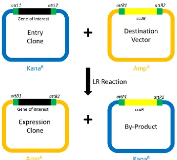
3.2.4 Gateway cloning ... 37
3.2.5 pENTR/D-TOPO cloning ... 38
3.2.6 Cloning of potential LKB1 interaction partners ... 38
3.2.7 Cloning of lkb1::gfp-lkb1 genomic ... 38
3.2.8 Gateway LR recombination reaction ... 39
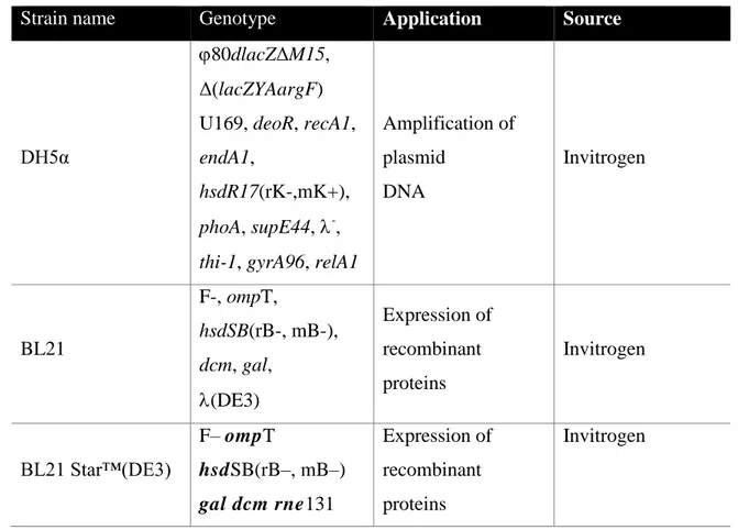
3.2.9 Transformation of chemically competent E. coli cells ... 40
XL1-Blue 41 Stratagene 41 3.2.10 Isolation of plasmid DNA by Alkaline Lysis with SDS ... 41
3.2.11 Site-directed mutagenesis ... 42
3.2.12 Sequencing of DNA ... 43
3.2.13 Isolation of genomic DNA from flies ... 44
3.3 Drosophila cell culture ... 44
3.3.1 Culture and transfection of Schneider 2 cells ... 44
3.3.2 Leptomycin B assay ... 45
3.4 Histology ... 46
3.4.1 Fixation and immunostaining of Schneider 2 cells ... 46
3.4.5 Detection of apoptosis in imaginal discs ... 48
3.4.6 Confocal microscopy ... 48
3.4.7 Preparation and imaging of wings ... 48
3.5 Biochemical methods ... 49
3.5.1 Protein extraction from embryos ... 49
3.5.2 Measurement of protein concentration ... 49
3.5.3 Co-Immunoprecipitation ... 49
3.5.4 SDS-polyacrylamide gel electrophoresis... 50
3.5.5 Western Blot ... 51
3.5.6 Protein purification ... 51
3.5.7 GST-Pulldown assay ... 52
3.5.8 Lipid overlay assay ... 52
3.6 Fly genetics ... 53
3.6.1 Fly breeding ... 53
3.6.2 Generation of transgenic flies ... 53
3.6.3 The UAS-GAL4 system ... 55
3.6.4 FLP/FRT- mediated recombination ... 55
3.6.5 Fly lines ... 56
3.6.6 Lethality assay ... 60
4 Results ... 61
4.1 Subcellular localization of LKB1 ... 61
4.1.1 LKB1 localizes to the cortex of epithelial cells and embryonic neuroblasts 61 4.1.2 Farnesylation is not crucial for the cortical localization of LKB1 and its physiological function ... 64
membrane ... 67
4.2 Investigation of α-Spectrin and β-Spectrin as potential interaction partners of LKB1 ... 71
4.2.1 The lipid binding domain of LKB1 is also involved in binding of α/β- Spectrin ... 71
4.2.2 β-Spectrin is involved in lateral localization of LKB1 in follicle cells .... 73
4.2.3 The N-terminus of β-Spectrin interacts with LKB1 ... 73
4.3 Three NLS regulate nuclear localization of LKB1 ... 74
4.3.1 Nuclear localization supports fertility and embryonic survival... 75
4.3.2 Nuclear localization signals are involved in the activation of AMPK ... 75
4.4 Effects of LKB1 overexpression ... 78
4.4.1 LKB1 overexpression in embryonic neuroblasts and ubiquitous expression in the embryo lead to embryonic lethality independent of farnesylation, the lipid binding motif and kinase activity ... 78
4.4.2 LKB1 overexpression leads to a slight reduction of eye size dependent on kinase activity ... 80
4.4.3 The reduction of wing size is minimized in the triple NLS mutant ... 82
4.5 Examination of phosphospecific antibodies against LKB1 ... 84
5 Discussion ... 86
5.1 Cortical localization of LKB1 ... 87
5.1.1 LKB1 localizes to the cortex of epithelial cells and embryonic neuroblasts 87 5.1.2 Farnesylation is not crucial for the cortical localization of LKB1 and its physiological function ... 88
5.1.3 Both farnesylation and a polybasic motif target LKB1 to the plasma membrane ... 89
5.1.4 β-Spectrin is involved in lateral localization of LKB1 in follicle cells .... 91
5.4 Kinase dependent and independent effects of overexpression of LKB1 .. 96
6 References ... 99
7 Appendix ... 114
7.1 Table of Figures ... 114
7.2 List of tables ... 115
7.3 Abbreviations ... 116
1
Danksagung
Ich möchte mich bei allen bedanken, die mir in den letzten Jahren in Göttingen und Regensburg zur Seite standen.
Ich danke Professor Michael Krahn für die Möglichkeit diese Promotion durchzuführen und die zahlreichen Anregungen, um dieses Projekt zu gestalten.
Professor Witzgall und seinem Lehrstuhl danke ich für die Möglichkeiten, die uns sein Lehrstuhl geboten hat und seine interessanten und kritischen Fragen.
I thank Arnab for all his help, patience and the company during our three years.
Professor Sprenger und Professor Klein danke ich für ihre Rolle in meinem Mentoring Team.
Ich möchte mich auch bei Florian, Gudrun, Laura und Giada bedanken für die gemeinsame Zeit und die gute Zusammenarbeit.
Besonders möchte ich mich bei meiner Freundin Stefanie bedanken, die mich begleitet und unterstützt.
2
1 Summary
Germ line mutations in the human lkb1 gene are the main cause of the Peutz-Jeghers syndrome (Hemminki, 1999) and somatic lkb1 mutations are associated with mainly epithelial cancers (Sanchez-Cespedes, 2007). The serine/threonine kinase LKB1 (STK11) is involved in different cellular processes like cell proliferation, energy homeostasis and cell polarity (Martin-Belmonte and Perez-Moreno, 2012). Especially the observation that activation of mammalian LKB1 can polarize cells in absence of cell-cell contacts (Baas et al., 2004) has drawn attention to the role of LKB1 in cell polarity. Using Drosophila melanogaster and other model organisms various potential downstream targets have been identified, while little is known about its upstream regulation and the mechanisms by which LKB1 controls cell polarity. It has been suggested that LKB1 might be mostly constitutively active and that its involvement in specific responses depends on localization of it to specific subcellular compartments (Sebbagh et al., 2011).
In order to study the role of LKB1 in cell polarity an antibody against LKB1 has been raised for this work that could detect LKB1 in different tissue of Drosophila.
Remarkably, endogenous LKB1 localizes cortically in asymmetrically dividing embryonic neuroblasts but cytoplasmic in larval neuroblasts. LKB1 has been described to control asymmetric divisions in both of these cell types. How it can exert its functions with different localizations remains to be answered. Furthermore, endogenous LKB1 localizes to the basolateral cortex in embryonic epithelial cells. I observed that a farnesylation deficiency does not alter the localization of LKB1 remarkably in epithelial cells and did not affect its activity towards its downstream target AMPK (AMP- activated protein kinase). A polybasic motif was identified in this work, which interacts with phospholipids found in the plasma membrane and potentially with the newly identified interaction partners α-Spectrin and β-Spectrin. Furthermore, three nuclear localization signals (NLS) of Drosophila LKB1 were identified. A construct carrying mutations in all three NLS had a reduced ability to rescue an lkb1-KO mutant and revealed a lower basal activity towards its substrate AMPK in embryonic lysates.
Moreover, the mutation of the three NLS decreased the phenotype of LKB1 overexpression in the negative regulation of organ size compared to its wild type counterpart.
3
2 Introduction
2.1 Cell polarity
Cells in tissues of multicellular organisms, but also cells of single-cell organisms, show asymmetries in shape, protein and lipid distribution and cell function, defined as cell polarity. Polarization is initiated by either external or internal polarity cues. Contact to other cells or the extracellular matrix can, for example, serve as external cues for epithelial cells to form junctions and give rise to an epithelia tissue (Nelson, 2003). The establishment and maintenance of cell polarity is fundamentally important for diverse complex cellular functions, like formation of epithelial barriers, cell migration and asymmetric cell division. Most of the key components of cell polarity found in metazoa are well conserved throughout evolution.
2.2 Drosophila cell types as a model for cell polarity
Several cell types of Drosophila melanogaster have been established as model systems for in vivo studies on different aspects of cell polarity:
- The oocyte, similar to the early Caenorhabditis elegans (C. elegans) embryo, displays an anterior-posterior polarity.
- The mesodermal follicle cell epithelium surrounding the egg chamber and the ectodermal epithelium surrounding the embryo. Both form monolayers of cuboidal polarized cells. A peculiarity of the follicle cells is that their apical side is not directed towards a lumen or the external environment, instead it forms cell-cell contacts with the germline cells.
- Drosophila neuronal stem cells (neuroblasts) are a common model for studying asymmetric cell division.
The use of Drosophila as a model system for cell polarity offers diverse tools to study mechanisms and effects of cell polarity in vivo. One important question for the understanding of tumor progression is, whether the loss of epithelial polarity observed
4
in cancer is an epiphenomenon or a cause of cancer (Partanen et al., 2013). Experiments in Drosophila suggest a causal role of polarity proteins in tumor progression (Bilder, 2004; Martin and St Johnston, 2003)).
2.3 Epithelial cell polarity in vertebrates and Drosophila
Sheets of epithelial cells line the cavities and surfaces throughout the body of multicellular organisms. They function as barriers between compartments and are essential for the transport of molecules between them. In order to function, epithelial cells have to be polarized into apical and basolateral membranes and form junctions with each other. A key feature of all epithelial cells is the polarized assembly of actin filaments at the cytosolic side of the apical and the basal cytoplasmic membrane. In both Drosophila and vertebrates, the apical identity of epithelia cells is maintained and regulated by two conserved polarity modules: the Crumbs module and the Par module.
The Crumbs module is composed of Crumbs, PALS1 (Stardust in Drosophila) and Patj (PALS1 associated tight junction protein), the Par module is composed of Par3 (Bazooka in Drosophila), Par-6 and atypical protein kinase C (aPKC). On the basolateral sides of the cells the Scribble polarity module composed of Scribble (Scrib), Dlg (Discs large) and Lgl (Lethal (2) giant larvae) defines basolateral identity. The polarity modules show mutually antagonistic interactions, restricting the activity of each module and regulate positioning of the adherens junctions (Humbert et al., 2008).
While many basic mechanisms of cell polarity are conserved, the junctions along the lateral side of epithelial cells are organized differently in vertebrates and Drosophila (Knust and Bossinger, 2002).
In vertebrates the apical domain contains a brush border of microvilli with a network of actin and Spectrin filaments beneath on the cytosolic side. The primary cilium protrudes from the apical surface and has a unique membrane and protein composition. Tight junctions mark the border of the apical and lateral domains; they include homophilic adhesion molecules like Occludin, JAMs (Junctional Adhesion Molecules) and Claudins that form a paracellular diffusion barrier. Adherens junctions are found basally of the tight junctions and are the main connection between neighboring cells. Nectins
5
and Cadherins localize to the adherens junctions and mediate these homophilic connections (Nelson et al., 2013). Only the basal side of the cell is connected to the extracellular matrix and has integrins and dystroglycan as receptors (Johnston and Ahringer, 2010).
Epithelial cells of Drosophila lack primary cilia. The adherens junctions of most Drosophila epithelial cells are apical of the septate junctions and contain DE-Cadherin and Nectin (Baumann, 2001). Instead of tight junctions septate junctions function as the paracellular diffusion barrier in Drosophila and contain Sinuous (a claudin family member), Cor and Megatrachea (Wu et al., 2004). Unlike in vertebrates, the Spectrin cytoskeleton of Drosophila epithelia cells is polarized, with α2βH2-Spectrin at the apical and α2β2-Spectrin at the basolateral membrane (Thomas and Kiehart, 1994). The mammalian ortholog of βH–Spectrin shows no distinct apical distribution (Stabach and Morrow, 2000).
2.4 Drosophila neuronal stem cells (neuroblasts) as a model for asymmetric cell division and stem cell induced tumors
During stage 9 of embryogenesis, neural stem cells called neuroblasts (NBs) delaminate from the neuroectodermal epithelium into the interior of the embryo. NBs divide asymmetrically, giving rise to another NB (stem cell renewal) and a ganglion mother cell (GMC), that divides once more to form a pair of neurons or glia cells.
The asymmetric division of a NB requires apical-basal polarity, which is partly inherited from the epithelium (Wodarz and Huttner, 2003). During metaphase the Par module, as well as the Insc/Pins/Gαi complex, is localized to the apical cortex, while cell fate determinants like Prospero, Brat and Numb and their adaptor proteins Miranda and Partner of Numb are localized to the basal cortex. The mitotic spindle is first oriented in parallel to the surface of the epithelium but rotates by 90° during metaphase.
As the NB divides, the apically localized daughter cell inherits the apically localized proteins and retains stem cell fate. The smaller basal daughter cell inherits the proteins of the basal cortex and becomes a GMC (Wodarz, 2005).
6
The apical-basal polarity of the NB cortex is adjusted with the spindle orientation during metaphase and essential for asymmetric division (Lee et al., 2006). Defects in cell polarity of NBs can lead to symmetric divisions, resulting in formation of ectopic NB-like stem cells, which proliferate and finally lead to tumor formation (Wodarz and Näthke, 2007).
2.5 The PAR proteins
The PAR proteins were discovered as regulators of cytoplasmic partitioning in the early embryo of C. elegans. PAR stands for ”abnormal embryonic PARtitioning of cytoplasm”, describing the phenotype of par mutants, which is a mislocalization of germ line-specific P granules (Kemphues et al., 1988). Following the nomenclature for genes and proteins in C. elegans, these six proteins required for early polarity and asymmetric cell divisions were named PAR-1 to PAR-6. During one cell stage PAR-3 and PAR-6 become enriched in the anterior cortex, while PAR-1 and PAR-2 become enriched in the posterior cortex. PAR-4 and PAR-5 are symmetrically localized cortical and cytoplasmic (Goldstein and Macara, 2007).
The biochemical roles of the PAR proteins differ remarkably. PAR-1 and PAR-4 are serine/threonine kinases, PAR-3 and PAR-6 contain PDZ domains (named after the three proteins Psd95, Discs large and ZO-1, that contain this domain) and PAR-2 is a protein with a “ring finger” zinc binding domain (Kemphues, 2000). With the exception of PAR-2, which seems to be a nematode-specific protein, homologs of the PAR protein have been identified in all bilateral animals investigated (examples in Table 2-1) and have been found to regulate cell polarization in multiple contexts in various animals (Goldstein and Macara, 2007).
7 Table 2-1: Homologs of PAR proteins
Caenorhabditis elegans Drosophila melanogaster Homo sapiens
PAR-1 Par-1 MARK1
PAR-2 - -
PAR-3 Bazooka PAR3
PAR-4 LKB1 STK11/LKB1
PAR-5 14-3-3ε and 14-3-3ζ 14-3-3 protein family
PAR-6 Par-6 PAR6
2.6 LKB1 (STK11, PAR-4) is a multifunctional protein kinase with tumor suppressor activity
Germline mutations of human LKB1 (also known as STK11) are the main cause of Peutz-Jeghers-Syndrome (PJS) (Hemminki et al., 1998), a rare autosomal cancer disease. PJS is characterized by intestinal hamartomatous polyposis and mucocutaneous melanin pigmentation (Jeghers, 1949). Patients with PJS have a very high risk of developing cancer (Giardello et al., 2000). Somatic mutations of lkb1 are associated with a wide variety of mainly epithelial cancers (Sanchez-Cespedes, 2007), especially lung adenocarcinomas (Conde et al., 2007). Some cancer cell lines show severly reduced mRNA levels of LKB1, probably due to hypermethylation of the promoter region of lkb1. In HeLa S3 cells (a cervical cancer cell line) and in G361 cells (a melanoma cell line) reintroduction of functional LKB1 suppresses proliferation by growth arrest in G1 phase (Tiainen et al., 1999). On the other hand many malignant tumors appear to have elevated LKB1 expression levels (Rowan et al., 2000).
LKB1 is a ubiquitously expressed serine/threonine kinase (also called STK11), which is known to phosphorylate the AMPK-family of 13 kinases and therefore termed a “master kinase” (Lizcano et al., 2004). In humans there are two LKB1 isoforms, resulting from alternative splicing. The long LKB1 form (50 kDa) is ubiquitously expressed in adult tissues, with higher expression in epithelia (Rowan et al., 2000). In the shorter form (48 kDa) the C-terminus is replaced by a shorter and unique amino acid sequence (Towler et al., 2008). Both isoform are expressed in human tissues, but the shorter form is especially prevalent in testis, where it has been identified to be crucial for
8
spermiogeneis (Towler et al., 2008). The catalytic domain of LKB1 is poorly related to other protein kinases, the N-terminal and C-terminal non-catalytic regions are not related to any other proteins and posess no idenfiable domains (Boudeau et al., 2003b).
Diverse effects of LKB1 have been observed on the cellular level, such as inducing cell cycle arrest (Tiainen et al., 1999), mediating apoptosis through p53 (Karuman et al., 2001 and Cheng et al., 2009), AMPK activation in energy metabolism (Shackelford and Shaw, 2009) and cell migration (Zhang et al., 2008). The observation that activation of mammalian LKB1 can polarize cells even in the absence of cell-cell contacts (Baas et al., 2004) has drawn attention to the role of LKB1 in cell polarity (2.6.6). While there are several downstream processes described, upstream regulation is poorly understood and might be mediated by localization rather than activation of kinase activity (Sebbagh et al., 2011).
2.6.1 The heterotrimeric complex LKB1-STRAD-Mo25 is the active unit of LKB1
LKB1 activity is highly increased by binding to the pseudo kinase STRAD (STe-20 Related Adaptor) (Baas, 2003). This interaction is stabilized by Mo25 (mouse protein25) (Boudeau, 2003). Together they form the LKB1-STRAD-Mo25 heterotrimeric complex, which is generally considered as the biologically active unit of LKB1. In the presence of STRADα and Mo25, LKB1 relocalizes from the nucleus to the cytoplasm as observed in several overexpression studies on mammalian cell cultures (Baas et al., 2003; Boudeau et al., 2003; Dorfman and Macara, 2008; Dension et al., 2009). The structure of the LKB1-STRAD-Mo25 complex has been determined, showing that STRAD and Mo25 promote the active conformation of LKB1 (Zeqiraj et al., 2009).
2.6.2 The LKB1-STRAD-Mo25 complex is probably conserved in lower model organisms
Lower organisms also express STRAD and Mo25 homologs, which contain the critical residues for the interaction with LKB1, like the STRAD C-terminal WEF motif (Zeqiraj
9
et al., 2009). In C. elegans the STRAD homolog STRD-1 is required for the phosphorylation of AMPK by the LKB1 homolog PAR-4 under reduced insulin signaling conditions to regulate cell growth and proliferation, but the phosphorylation of key proteins like PAR-1 by PAR-4, which is needed for establishment of early embryonic polarity, is independent of STRD-1 (Narbonne et al., 2010). Mo25 has been shown to interact with LKB1 in Drosophila neuroblasts and both could be co- immunoprecipitated reciprocally from embryonic lysates (Yamamoto et al., 2008). The Drosophila STRAD homolog Ste20-like kinase has not yet been investigated, but lacks key residues for kinase activity, indicating that it is also a pseudokinase (Anamika et al., 2009). Although the LKB1-STRAD-Mo25 complex is not established for these organisms yet, these findings suggest that it is probably conserved.
2.6.3 Posttranslational modification and regulation of LKB1 activity
The observation that LKB1 can phosphorylate and activate almost all known members of the AMPK kinase family (Lizcano et al., 2004) suggests that it has to be tightly regulated to facilitate specific responses.
Several phosphorylation sites have been found on mammalian LKB1. Four threonines (T) were identified as autophosphorylation sites, which are known to be positively correlated with activation of its catalytic activity (Baas et al., 2003). ATM mediates phosphorylation of LKB1 at T366 (Sapkota et al,, 2002b), which has no effect on the catalytic activity of LKB1, but seems to be required for inactivation of CRTC2, a coactivator of CREB, in B cell proliferation (Sherman et al., 2010). Phosphorylation of S428 by p90(RSK) and PKA (cAMP-dependent protein kinase) does not affect LKB1 kinase activity but has been described to be essential for LKB1 to suppress cell growth (Sapkota et al., 2001). Other phosphorylation sites have been described, but the respective kinases and their function are not known. Their substitution with alanine, however, had no observable effect on LKB1 kinase activity (Sapkota et al., 2002a).
Until now little knowledge is established about the regulation of the kinase activity of LKB1. Remarkably, there is no LKB1 complex activating factor known (Sebbagh et al., 2011), which lead to the hypothesis that the LKB1 complex could be constitutively
10
active and that specific responses are regulated by its intracellular localization (Fogarty and Hardie, 2009; Sebbagh et al., 2009). Two chaperone complexes have been suggested to control the cellular level of LKB1 protein: The chaperone heat shock protein 90 (Hsp90) and its co-chaperone Cdc37 inactivate LKB1, while Hsp/Hsc70 and CHIP trigger LKB1 degradation, which finally controls the cellular level of LKB1 protein (Gaude et al., 2012).
2.6.4 LKB1 in Drosophila
The lkb1 gene (also known as dlkb1) of Drosophila was first identified in a genetic screen for mutants that disrupt the localization of Staufen in germline clones (Martin &
St Johnston 2003). Due to its stronger homology to the human tumor-suppressor LKB1 than to C. elegans PAR-4 it was named lkb1. In the same study, lkb1 is described to be important for the establishment of cell polarity in the follicle cell epithelium (Martin &
St Johnston 2003). The oocyte phenotype resembled that of hypomorphic mutants in Drosophila par-1 and a strong genetic interaction between these genes has been observed. A par-1 hypomorphic mutation could be rescued by GFP-LKB1 overexpression, but not by a kinase dead version of GFP-LKB1, while lkb1 mutant germline clones could not be rescued by PAR-1 overexpression. This and in vitro experiments indicated PAR-1 is not a substrate for LKB1. Instead, the amino-terminal half of LKB1 was identified to be phosphorylated by PAR-1, which led to the idea that LKB1 acts downstream of PAR-1 (Martin & St Johnston 2003). But Wang et al. could identify that LKB1 phosphorylates PAR-1 at the threonine residue at position 408 (T408) in vitro and in vivo (Wang et al., 2007). Through biochemical experiments as well as genetic studies of Drosophila retinal phenotypes they have shown that LKB1 acts upstream of PAR-1 and tau in a cytotoxic pathway in a model for neurodegenerative diseases (Wang et al., 2007).
LKB1 has been reported to phosphorylate AMPK (AMP-activated protein kinase) to control cell polarity and mitosis under energetic stress in the embryo (Lee et al., 2007) In the retina, LKB1 has been observed to act on an array of targets to regulate polarity remodeling probably independent of AMPK, possibly through PAR-1 and several
11
AMPK-like kinases (Amin et al., 2009). It has been reported that Drosophila LKB1 negatively regulates organ size by inducing caspase-dependent apoptosis without affecting cell size or cell cycle progression (Lee et al., 2006). Silnoon, a monocarboxylate transporter, was identified as a modifier enhancing LKB1- dependent apoptosis, and is reported to be transported to the apical side of polarized cells depending on the kinase activity of LKB1 (Jang et al., 2008). In larval neuroblasts lkb1 mutations are reported to lead to defects in apical polarity (Bazooka, Par-6 and aPKC localization are not properly localized), suppress asymmetric cell division and disrupt spindle formation resulting in polyploid cells in larval brains (Bonaccorsi et al., 2007).
Overexpression of a GFP-tagged LKB1 fusion protein has been observed to reduce the size of embryos and localizes to the cortex of ectodermal embryonic epithelial cells and NBs. In NBs, the overexpression of LKB1 causes a broader distribution of Miranda along the cortex. This effect has been observed to be independent of the kinase activity of LKB1 (Yamamoto et al., 2008). A gain-of-function screen identified that overexpression of LKB1 reduced organ size and extended lifespan (Funakoshi et al., 2011).
2.6.5 AMPK as a downstream target of LKB1
LKB1 has been shown to be the dominant regulator of AMPK (AMP-activated protein kinase) activation in several mammalian cell types by phosphorylating a critical phosphorylation site in its T-loop (T172) (Shaw et al., 2004). Low energy conditions increase the level of AMP and ADP, which interact with the CBS motifs of the AMPKγ subunit of AMPK (Hardie, 2011). This causes conformational changes that activate the AMPKα subunit and promote phosphorylation of the phosphorylation site T172 by inhibiting its dephosphorylation (Sanders et al., 2007). The activation of the AMPKα1 and AMPKα2 catalytic subunits functions to restore energy levels by phosphorylating an immense number of proteins, which are involved in multiple processes like metabolism, cell growth and proliferation (Carling et al., 2011). The net result of AMPK activation is a shift of the balance from anabolic to catabolic function and thereby restoring the cellular ATP levels of the cell.
12
The tumor suppressor activity of LKB1 is thought to be at least partly mediated by its activation of AMPK, which in turn inhibits mTOR (mammalian target of rapamycin), which is a known regulator of diverse cellular processes including cell growth and proliferation (Shackelford and Shaw., 2009). In ciliated MDCK cells, LKB1 has been found to colocalize with AMPK at the basal body, where it regulates the phosphorylation of AMPK in response to urine flow, which regulates cell size through mTOR (Boehlke et al., 2010).
In spite of its role as a tumor suppressor, LKB1 protects cells from apoptosis in response to elevated AMPK levels (Shaw et al., 2004). The LKB1-AMPK pathway can have a positive role in tumorigenesis by acting to maintain metabolic homeostasis and attenuate oxidative stress and thus enabling the survival of tumor cells by maintaining NADPH levels (Jeon et al,. 2012). This might also explain why many malignant tumors display elevated levels of LKB1 (Rowan et al., 2000).
ampk-null mutant Drosophila embryos are reported to be lethal with abnormal cell polarity and mitosis, similar to those in lkb1-null mutants and a phosphomimetic mutant of myosin II regulatory light chain (MRLC) rescued the phenotype of ampk-null mutants to some extent (Lee et al., 2007). This indicates that the actin-myosin cytoskeleton might be regulated by AMPK under energetic stress conditions. Although Lee et al. could show that AMPK phosphorylates MRCL in Drosophila and in the human epithelial cell line LS174T, in mammalian pancreatic cells the inhibition of LKB1 did not affect the phosphorylation status of MRLC (Hezel et al., 2008) and inhibition of AMPK in vascular smooth muscle cells even increased MRLC phosphorylation (Horman et al., 2008). This indicates that MRLC may not be directly phosphorylated by AMPK but that it may mediate the polarity signal from AMPK to the actin cytoskeleton (Mirouse and Billaud 2011). Many aspects regarding an involvement of AMPK in cell polarity like its direct downstream targets associated with this function remain unclear. The cytoplasmic linker protein of 170 Da (CLIP-170) has been proposed to be a target of AMPK involved in cell polarity by affecting the stability of microtubules (Nakano et al., 2010).
13 2.6.6 LKB1 in the regulation of cell polarity
Activation of LKB1 by overexpression of its adaptor protein STRAD was reported to induce polarization even in the absence of cell-cell contacts in human intestinal epithelial cells, as seen by the formation of an actin-rich brush border on one side of the cell, indicating an apical-like surface (Baas et al., 2004). This suggests that LKB1 is a major regulator of cell polarity. Mst4 and the actin filament binding protein ezrin were later identified as downstream targets of LKB1 in the induction of brush border formation, but had no effect on other polarity events downstream of LKB1, like the formation of lateral junctions (Klooster et al., 2009). The role of LKB1 in actin filament assembly has been further investigated in HeLa cells, which lack endogenous LKB1. In this process LKB1 expression leads to an activation of Rho, mediated by the guanine nucleotide exchange factor Dbl, although the exact mechanism remains unknown.
Interestingly, the kinase activity of LKB1 is not required for the induction of stress fibers, but the kinase domain is (Xu et al., 2010). It has also been reported that LKB1 regulates the expression of E-Cadherin and thereby intercellular junction stability through Salt-inducible kinase 1 (Eneling et al., 2012). On the other hand LKB1/STRAD localization has been reported to be localized to the adherens junctions of polarized
epithelial cells (MDCK cells) under control of E-Cadherin (Sebbagh et al., 2009).
2.6.7 Localization of LKB1
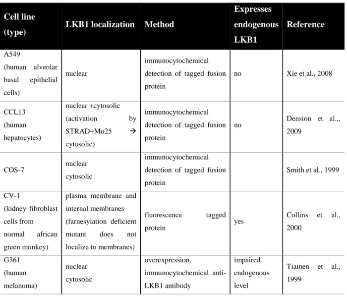
A nuclear localization signal (NLS) has been identified in the N-terminus of mammalian LKB1 (Nezu et al., 1999; Smith et al., 1999; Tiainen et al., 2002).
Localization of LKB1 has mostly been studied in mammalian overexpression systems in cell culture (Table 2-2). In most of these studies, LKB1 has been observed predominantly in the nucleus and to a lower degree in the cytosol (Nezu et al., 1999;
Smith et al., 1999; Tiainen et al., 1999; Baas et al., 2003; Boudeau et al., 2003; Denison et al., 2009; Dorfman and Macara et al., 2008; Xie et al., 2008). STRADα has been reported to regulate export of LKB1 through inhibition of the nuclear import of LKB1 and by serving as an adaptor between LKB1 and exportins CRM1 and exportin 7 (Dorfman and Macara, 2008). Some studies have also found localization of LKB1 to the cytocortex (Collins et al., 2000; Sapkota et al, 2001; Xu et al., 2010).
14
The localization of LKB1 in cells expressing endogenous levels of LKB1 has not been investigated much, probably due to low expression levels and lack of suitable antibodies (Sebbagh et al., 2009). Surprisingly, in MDCK cells, endogenous LKB1 is reported to localize mainly to the cytosol and membrane and not to the nucleus (Sebbagh et al., 2009). A fractionation study of HEK293 cells, which express moderate amounts of endogenous LKB1, was only able to detect LKB1 in the cytosolic and membrane fractions, but not in the nuclear fraction (Denison et al., 2009). Fractionation of polarized Caco-2 cells revealed an absence of LKB1 in the nuclear fraction, but showed abundance in the cytosolic and membrane fractions (Sebbagh et al., 2009). It can be assumed, that the examined cell lines express enough STRAD to export the endogenous amounts of LKB1 from the nucleus.
Table 2-2: Localization of LKB1 in mammalian cell culture lines
Cell line
(type) LKB1 localization Method
Expresses endogenous LKB1
Reference
A549
(human alveolar basal epithelial cells)
nuclear
immunocytochemical detection of tagged fusion protein
no Xie et al., 2008
CCL13 (human hepatocytes)
nuclear +cytosolic (activation by
STRAD+Mo25
cytosolic)
immunocytochemical detection of tagged fusion protein
no Dension et al.,, 2009
COS-7 nuclear
cytosolic
immunocytochemical detection of tagged fusion protein
Smith et al., 1999
CV-1
(kidney fibroblast cells from normal african green monkey)
plasma membrane and internal membranes (farnesylation deficient mutant does not localize to membranes)
fluorescence tagged
protein yes Collins et al.,
2000
G361 (human melanoma)
nuclear cytosolic
overexpression,
immunocytochemical anti- LKB1 antibody
impaired endogenous level
Tiainen et al., 1999
15 Cell line
(type) LKB1 localization Method
Expresses endogenous LKB1
Reference
HeLa (cervical carcinoma)
nuclear cytosolic
(coexpression of STRAD cytosolic)
fluorescence tagged
protein no
Boudeau et al., 2003;
Dorfman and Macara, 2008 HeLa-S3
(cervical carcinoma)
nucleus, fraction at cell membrane
immunocytochemical detection of tagged fusion protein
no Xu et al., 2010
HUVEC (Human
Umbilical Vein Endothelial Cells)
nuclear
(treatment with
metformin
cytosolic)
immunocytochemical detection of tagged fusion protein
yes Xie et al., 2008
LS174T
(human intestinal epithelial cancer)
predominantly nuclear (coexpression of STRAD cytosolic)
immunocytochemical detection of tagged fusion protein
Probably not (author statement)
Baas et al., 2003
Rat-2
(fibroblast-like)
mainly cytosolic, small but significant amount at membrane
fractionation
yes Sapkota et al., 2000
2.6.8 The C-terminus of LKB1 is involved in membrane targeting of LKB1
Numerous mutations that affect only the C-terminus have been identified in tumors, making it an interesting area of investigation (Boudeau et al., 2003b). The C-terminal CAAX-sequence, which is only present in the long isoform of mammalian LKB1 and conserved in most model organisms including Drosophila (Figure 2-1), is a farnesylation motif (Collins et al., 2000; Sapkota et al., 2001; Martin and St Johnston, 2003). In CV-1 cells and Drosophila oocytes a farnesylation-deficient mutant of LKB1 strongly impairs the cortical localization of LKB1 (Collins et al., 2000; Martin and St Johnston, 2003). The overexpression of a farnesylation-deficient LKB1 mutant in mammalian cell culture, however, had no effect on the ability to suppress cell growth (Sapkota et al., 2001). With a farnesylation-specific antibody the majority of LKB1 was
16
identified to be farnesylated in wild-type mouse tissues and cultured cells (Houde et al., 2014). A farnesylation deficient version of LKB1 (LKB1C433S) was investigated using mouse knockin analysis (Houde et al., 2014).
Another motif at the C-terminus that is conserved in model organisms including Drosophila and C. elegans is the PKA/p90RSK phosphorylation motif “RKLS” (Figure 2-1). In human endothelial cells it has been reported, that PKC-ζ phosphorylates LKB1 at S428 and that this would lead to a nuclear export of LKB1 and hence AMPK activation (Xie et al., 2008). Others describe that this phosphorylation site is essential for mammalian LKB1 to suppress cell growth (Sapkota et al., 2001), but is not required for regulation of AMPK or cell cycle arrest (Fogarty and Hardie, 2009). In agreement with the latter mentioned findings, AMPK is activated normally in knockin mice carrying the phosphodeficient mutant LKB1S431A (Houde et al., 2014). Co-expression and mobility shift assays of Drosophila LKB1 and PKA in S2 cells revealed a conservation of this phosphorylation site (Martin and St Johnston, 2003). Moreover, a GFP-LKB1 with a phosphodeficient version of this site, when expressed in low amounts, does not rescue localization of Staufen to the posterior oocyte of Drosphila, while a phosphomimetic version rescues even more efficiently than the wild type control, indicating a positive regulation of LKB1 by phosphorylation of this site (Martin and St Johnston, 2003).
STK11[Homo sapiens] 1…409-R---APNPARKACSASSKIRRLSACKQQ--- 433 LKB1[Mus musculus] 1…411-RPG----TANPARKVCS-SNKIRRLSACKQQ--- 436 STK11[Danio rerio] 1…415-SS---SSNPSRKGLSAASKIRKLSTCKQQ--- 440 XEEK1[Xenopus laevis] 1…411-SS---QRKASTTGSKVRKLSACKQQ--- 432 LKB1[Drosophila melanogaster] 1…537-PVKKKGSALKRRAKKLTSCISVRKLSHCRTS--- 567 PAR-4[Caenorhabditis elegans] 1…581-GVASASDPPPTAAPGAPPRRRKRNFFSCIFRSRTDSA 617
Figure 2-1: Alignment of the C-termini of LKB1 homologs. Blue color indicates the PKA phosphorylation motif and green color the farnesylation motif conserved between human LKB1 and homologs in model organisms.
17 2.6.9 Potential LKB1 interaction partners
Apart from an activation of of the AMPK family of kinases (Licano et al., 2004), several potential LKB1 effectors have been identified by mass spectrometry and yeast two hybrid assays (Sebbagh et al., 2011). Interactions with the chaperone complexes Hsp90/Cdc37 and Hsp/Hsc79-CHIP have been reported to regulate activity and stability of LKB1 (Gaude et al., 2012). LKB1 has also been reported to interact with LIP1 (LKB1 interacting protein 1), but the functional consequences of this interaction remain unknown (Smith et al., 2001). Similar to this, the interaction of LKB1 with PTEN was observed but no distinct functional consequences were found (Mehenni et al., 2005).
Furthermore, an interaction of LKB1 with the transcription factors Estrogen receptor-α and Brahma related gene-1 has been described, but phosphorylation of these partners by LKB1 could not be observed, indicating a potential indirect effect of LKB1 (Marignani et al., 2001; Nath-Sain and Marignani, 2009).
In order to find new potential interaction partners of LKB1, they were co- immunoprecipitated by expressing GFP-LKB1 in Drosophila embryos and identified via mass spectrometry (Krahn, so far unpublished). The co-immunoprecipitation of the LKB1 substrate and the homologs of the components of the LKB1 complex (Stlk and Mo25), indicate that this approach is promising for the identification of interaction partners. Among the potential interaction partners are α-Spectrin and β-Spectrin, which are components of the membrane skeleton. In this work the potential interaction of these proteins with LKB1 were analyzed.
Table 2-3: Proteins identified to bind to GFP-LKB1
Protein # of
peptides Cellular function
-Spectrin 5 Cytoskeleton linker protein
-Spectrin 2 Cytoskeleton linker protein
AMP-activated kinase 7 Regulation of cellular energy homeostasis Ste20-like kinase (Stlk) 24 Co-factor for LKB1 (STRADa homolog)
Mo25 26 Co-factor for LKB1
18 2.7 The membrane skeleton
Actin has critical roles in establishing and maintaining cell morphology, cell motility, cell division and intracellular transport (Pollard and Cooper, 2009). Most actin filaments in animal cells are nucleated at the plasma membrane, where they form a layer at the cytoplasmic side called the cell cortex or membrane skeleton, which is important for the shape and movement of the cell surface.
2.7.1 Spectrin
Spectrin has been studied mostly in human erythrocytes, were it forms a protein network by binding to actin and peripheral membrane proteins (Bennett, 1985; Byres and Brandon, 1985), which builds stiff structures that are important for the flexibility of this cell type. But it is found in all metazoan species and in almost all cells examined (Baines, 2009). In erythrocytes it is forming the Spectrin-based membrane cytoskeleton by crosslinking transmembrane proteins, signaling proteins, membrane lipids and the actin cytoskeleton, while the cortical composition of other cell types appears to be more complex (reviewed in Bennett and Baines, 2001). In the Drosophila neuromuscular junction an erythrocyte-like polygonal lattice structure has been reported (Pielage et al., 2006), which has also been proposed for other cell types (Baines, 2010).
Three types of Spectrins exist in invertebrates: α-Spectrin, β-Spectrin and βH-Spectrin.
α-Spectrin is forming heterotetrameric complexes with β-Spectrin and βH-Spectrin (α2β2
and α2βH2) (Dubreuil et al., 1990). The Spectrin cytoskeleton of Drosophila epithelial cells is polarized, with α2βH2 found in the apical cortex of polarized cells and α2β2 in the basolateral cortex (Thomas and Kiehart, 1994). In a Spectrin dimer, α- and β-chains lie side-by-side and antiparallel. The Spectrin proteins are linking actin fibers and other proteins by numerous interaction motifs. Both α-Spectrin and β-Spectrin are mostly made of triple-helical repeats. α-Spectrin contains a middle SH3 domain and a C- terminal EF-hand binding motif in addition to multiple Spectrin repeats. SH3 domains are protein interaction domains that bind to proline-rich ligands, that play several different roles in the cell including the regulation of enzymes, changing the subcellular
19
localization of signaling pathway components, and mediating the formation of multiprotein complex formations (Macheler-Bauer et al., 2011).The β-Spectrin N- terminal region contains a pair of CH domains that binds actin, adducin and Phosphatidylinositol-4,5-bisphosphate (PIP2). Triple helical repeats 14–15 contain an ankyrin binding site. Other triple helices in both chains bind a variety of different ligands, including the region between repeats 9 and 11 (including the SH3 domain) which can bind both proteins and phospholipids (Baines, 2009).
In human epithelial cells loss of β-Spectrin or ankyrin has been shown to lead to loss of the lateral membrane (Kizhatil, 2007). Apical α2βH2 is needed in the Drosophila follicle cell epithelium for epithelial morphogenesis but not for apicobasal polarity (Zarnescu and Thomas, 1999). Genetic experiments in C. elegans suggest a requirement for Spectrins in muscle and nerves (Hammarlund et al., 2000; Moorthy et al., 2000).
Analysis in Drosophila Spectrin mutants indicate the need of Spectrin and ankyrin in the consolidation and maintenance of synaptic structures (Pielage et al., 2005, 2006 and 2008) and distinct roles of α-Spectrin and β-Spectrin in axonal pathfinding (Hülsmeier et al., 2007).
20
3 Material and Methods
3.1 Material
Chemicals (Table 3-1) and kits (Table 3-3) were bought from following companies:
Agilent Technologies (Böblingen, Germany), BioVision (San Francisco, USA), Chromotek (Planegg, Germany), Invitrogen (Groningen, Netherlands), Machery-Nagel (Düren, Germany), Merck Chemicals Ltd. (Nottingham, UK), PAN biotech (Aidenbach, Germany), Promega (Mannheim, Germany), Roche Diagnostics Deutschland GmbH and Roche Applied Science (Mannheim, Germany), Roth (Karlsruhe, Germany), Sigma-Aldrich (Steinheim, Germany) and Thermo Fisher Scientific. Chemicals were generally of analytical grade.
Instruments and other material were purchased from Carl Zeiss (Jena, Germany), Echelon Biosciences (Salt Lake City, USA), Eppendorf (Hamburg, Germany), Intas (Göttingen, Germany), Machery-Nagel (Düren, Germany) and Thermo Fisher Scientific.
3.1.1 Reagents
Table 3-1: Reagents
Reagent Utilization Note Company
Albumin fraction
V Blocking Solutions Roth
Amlyose Resin Protein Purification of MBP-tagged proteins
New England BioLabs
Aprotinin Protease inhibiton
used
concentration: 2 µg/ml
Roth
Bradford Rothi®
Quant Coomassie Brilliant Blue-G250
Protein concentration measurement
Roth
21
Reagent Utilization Note Company
FuGENE®HD Transfection Reagent
Transfection of Cells Blend of Lipids in
80% Ethanol Promega
GFP-Trap Co-IP
GFP-Trap® coupled to agarose beads particle size ~80 µm; stored in 20% EtOH
Chromotek
Leptomycin B Inhibition of CRM1
(exportin 1, embargoed) Used 1:100 Sigma-Aldrich
Leupetin Protease inhibiton
used
concentration : 2 µg/ml
Roth
Pepstatin A Protease inhibiton
used
concentration : 2 µg/ml
Roth
PfuS Polymerase PCR Homemade -
Phosphatase Inhibitor Cocktail Set, IV
Phosphatase Inhibitors Phosphatase
Inhibitors Merck
PMSF Protease inhibiton Roth
Protein A
sepharose Co-IP
Supplied as 50%
slurry in 20%
Ethanol/H2O
BioVision
Protino Glutathione Agarose 4B
Protein Purification of
GST-tagged proteins Macherey-Nagel
T4 DNA Ligase Ligation Thermo Fisher
Scientific
22 3.1.2 Solutions
Solutions were prepared with distilled water and sterilized by autoclaving or sterile filtration. Instruments and other material were purchased from Carl Zeiss (Jena, Germany), Echelon Biosciences (Salt Lake City, USA), Eppendorf (Hamburg, Germany), Intas (Göttingen, Germany), Machery-Nagel (Düren, Germany) and Thermo Fisher Scientific.
Table 3-2: Solutions
Name Composition Usage
Apple juice agar plates (2%)
1980 ml H2O 60 g Agar
1020 ml Apple juice 51 g Sugar
10% Nipagin in ethanol
Egg collection, feeding
Blocking Buffer (for Western Blots)
500 ml TBST;
3% skim milk powder;
1% BSA
Western Blot
Blocking Buffer
TBS (50 mM Tris-HCl, pH 7.4, 150 mM NaCl), 3% BSA
Lipid overlay assay
Buffer A
100 mM Tris-HCl, pH 7.5
100 mM EDTA 100 mM NaCl 1% SDS
Isolation of genomic DNA from adult flies
Buffer G (10x)
10 mM Tris-HCl (pH 7.5 at 37°C); 10 mM MgCl2; 50 mM NaCl;
0.1 mg/ml BSA
Buffer System for restriction enzymes;
Thermo Fisher Scientific
23
Name Composition Usage
Buffer O (10x)
50 mM Tris-HCl (pH 7.5 at 37°C); 10 mM MgCl2;100 mM NaCl;
0.1 mg/ml BSA
Buffer System for restriction enzymes;
Thermo Fisher Scientific
Buffer R (10x)
10 mM Tris-HCl (pH 8.5 at 37°C); 10 mM MgCl2; 100 mM KCl;
0.1 mg/ml BSA
Buffer System for restriction enzymes;
Thermo Fisher Scientific
Buffer Tango (10x)
33 mM Tris-acetate (pH 7.9 at 37°C); 10 mM Mg-acetate; 66 mM K- acetate; 0.1 mg/ml BSA
Buffer System for restriction enzymes;
Thermo Fisher Scientific
DAPI 5 µg/ml DNA staining
Embryo-Glue adhesive tape; heptane
Mounting of embryos for injection
Fixation Solution PBS (1x); 4%
Formaldehyde Fixation
Freezing Medium
45% conditioned medium; 45% fresh medium;
10% DMSO
Freezing Cells
LB Medium
10 g Bacto-Tryptone;
5 g Bacto-Yeast Extract;
10 g NaCl
E.coli liquid culture medium
LBAmp Medium
10 g Bacto-Tryptone;
5 g Bacto-Yeast Extract;
10 g sodium chloride;
500 µg/ml Ampicillin
Selective liquid culture medium for E.coli
24
Name Composition Usage
LB0 Plates 10% tryptone; 5%
Yeast extract;
5% sodium chloride;
15% Agar Agar; pH 7.0
LBamp Plates LB0 Plates + 100 mg/l ampicillin
selective ampicillin plates
LBkana Plates LB0 Plates + 50 mg/l kanamycin
selective kanamycin plates
LBchl Plates LB0 Plates + 30 mg/l chloramphenicol
selective
chloramphenicol plates
LiCl/KAc Solution
4,29 M lithium chloride 1,43 M potassium acetate
Isolation of genomic DNA from adult flies
Loading Buffer (6x)
2.5% Ficoll 400; 11 mM EDTA; 3.3 mM Tris- HCl; 0.017% SDS;
0.015%
Bromophenolblue (pH 8.0)
Agarose Electrophoresis
Lysis Buffer
TNT Buffer;
Leupeptin; Pepstatin;
Pefabloc/PMSF;
Aprotinin (1:500 each)
Co
Immunoprecipitation
Methylene Blue solution
Methylene blue powder
ddH2O Agarose gel staining
Mowiol
0.4 g/ml Mowiol;
1 g/ml Glycerol;
2% 0.2 M Tris-HCl
Embedding Medium
25
Name Composition Usage
NP-40-lysis-buffer
50 mM Tris, pH7.5 140 mM NaCl 1% NP40/Igepal 1 mM CaCl2 1 mM MgCl2
Lysis of embryos
P1-Buffer
50 mM Tris-HCl (pH 8.0); 10 mM EDTA;
100 µg/ml RNase A Storage 4°C
Mini-Preparation
P2-Buffer 200 mM NaOH; 1%
SDS Mini-Preparation
P3-Buffer 3.0 M potassium
acetate, pH 5.5 Mini-Preparation
PBS (10x)
58.44 g/mol NaCl;
74,55 g/mol KCl;
141.96 g/mol Na2HPO4; 136 g/mol KH2PO4
Washing Buffer
PBT PBS (1x); 0.1%
Tween20 Immunohistology
Schneider’s
Drosophila Medium
With L-Glutamine;
0.40 g/l NaHCO3
Transfection Medium, PAN biotech
Schneider’s
Drosophila Medium (Complete)
With L-Glutamine;
0.40 g/l NaHCO3; 5%
Penicillin/Streptomycin;
10% FCS
Liquid culture medium for Drosophila
Schneider Cells, PAN biotech
2X SDS Loading Buffer (Laemmli Buffer)
126 mM Tris (pH 6,8);
4% SDS; 0.2%
bromophenol blue; 20%
glycerol; 200 mM DTT
SDS-PAGE
26
Name Composition Usage
6X SDS Loading Buffer
375 mM Tris (pH 6,8) 9% SDS
0.03% bromophenol blue 50% glycerol 600 mM DTT
SDS-PAGE
SDS Running Buffer (10x)
1.92 M Glycine 250 mM Tris 1% SDS
SDS-PAGE
TE
10 mM Tris-HCl, pH 8.0
1 mM EDTA
TAE (1x) 40 mM Tris-Base 1 mM
EDTA 0.1 % acetic acid Agarose Electrophoresis
TBS (1x) 50 mM Tris-HCl, pH
7.4 150 mM NaCl Western Blot
TBST 0,1%
1 mM Tris-HCl; pH 8.0;
150 mM NaCl;
0.1% Tween 20
Western Blot
TNT Lysis Buffer
150 mM NaCl;
50 mM Tris pH 7.5;
1% Triton X-100
Co
Immunoprecipitation Transfer Buffer
(10x)
250 mM TRIS-base
1.9 mM Glycine Western Blot
27 3.1.3 Commercial kits
Table 3-3: Commercial Kits
Kit Utilization Company
BM Chemiluminescence Western Blotting Substrate (POD)
Western Blot Analysis Roche
In situ Cell Death Detection Kit, TMR red
TUNEL-assay for
apoptosis detection Roche pEntr/D-Topo Cloning Kit Gateway Cloning Invitrogen Nucleo Bond® PC 100 Midi-Preparation Macherey-Nagel Nucleo Spin®Gel and PCR
Clean-Up PCR Purification Macherey-Nagel
QuikChange Site-directed mutagenesis Agilent Technologies Super Signal West Pico
Chemiluminescence Substrate
Western Blot Analysis Thermo Scientific
3.1.4 Instruments and other material
Table 3-4: Instruments and other material
Name Utilization Company
Mikro-Dialysierkapsel QuixSep
(max. Volume 1 mL)
Dialysis Roth
Slide-A-Lyzer Dialysis
Cassettes Dialysis Thermo Scientific
PIP Strips (P-6001) Lipid binding assay Echelon Biosciences Digital-Microscope VHX- Imaging of Drosophila Keyence
28
Name Utilization Company
500FC eyes
NanoDrop 1000
Spectrophotometer
Measuring DNA
concentration Thermo Scientific
LSM 510 Meta Microscopy Zeiss
LSM 710 Meta Microscopy Zeiss
UV Transilluminator Intas
Femptotips®II Injection for germline
transformation Eppendorf
3.1.5 Antibodies
Table 3-5: Primary antibodies
Target Species Utilization Designation Origin/References
aPKC (aPKCξ) Rabbit IF (1:500) sc-216 Santa Cruz
Actin Rabbit IF (1:100) A2066 Sigma
Phospho-AMPKα
(Thr172) Rabbit WB(1:200)
40H9 Cell Signaling Technology
α-Spectrin Mouse IF (1:20)
WB (1:10) 3A9
Developmental Studies
Hybridoma Bank (DSHB)
Baz Nterm Rabbit IF (1:1000) DE99646-2 Wodarz et al., 1999
β-Spectrin Rabbit IF (1:1000) Christian Klämbt
c-Myc Mouse IF (1:100)
IF (1:50) 9E 10 DSHB
Crb Mouse IF (1:10) Tepass
DE-Cadherin Rat IF (1:10) DCAD 2 DSHB
29
Target Species Utilization Designation Origin/References
Dlg Mouse IF (1:25) 4F3 (DSHB
elav Mouse IF (1:20) 9F8A9
DSHB
GFP Rabbit IF (1:500) A11120 Molecular probes
(Invitrogen)
HA Rat IF (1:1000)
(1:100) DSHB
MBP Rat
WB (lipid overlay experiments) 1:5000
1.1 MBP
7G4 Sigma Aldrich
MBP Mouse WB (1:500) sc73416 Santa Cruz
LKB1 Guinea Pig IF/WB/Co-IP
(1:500)
SAC 288
SAC 289 Homemade LKB1 phospho Thr-
312 Rabbit IF (1:500) Homemade
LKB1 phospho Thr-
460 Rabbit IF (1:500) Homemade
Lgl Guinea Pig IF (1:500)
Mir Rat IF (1:1000) Homemade
PATJ Guinea Pig IF (1:500) Homemade
α-tubulin Mouse WB (1:1000) 12G10 DSHB
30 Table 3-6 Secondary antibodies
Antibody Species Utilization
(Dilution) Origin/References Alexa Fluor 488-anti Guinea
Pig Goat IF (1:200) Life Technologies
Alexa Fluor 488-anti Rat Goat IF (1:200) Life Technologies Alexa Fluor 488-anti Rabbit Goat IF (1:200) Life Technologies Alexa Fluor 488-anti Mouse Goat IF (1:200) Life Technologies Alexa Fluor 568-anti Guinea
Pig Goat IF (1:200) Life Technologies
Alex Fluor a 568-anti Rabbit Donkey IF (1:200) Life Technologies Alexa Fluor 568-anti Rat Goat IF (1:200) Life Technologies Alexa Fluor 568-anti Mouse Donkey IF (1:200) Life Technologies Alexa Fluor 647-anti Guinea
Pig Goat IF (1:200) Life Technologies
Alexa Fluor 647-anti Rabbit Goat IF (1:200) Life Technologies Alexa Fluor 647-anti Rat Goat IF (1:200) Life Technologies Alexa Fluor 647-anti Mouse Goat IF (1:200) Life Technologies
HRP-anti Guinea Pig Goat WB (1:10000) Roche
HRP-anti Rabbit Goat WB (1:10000) Roche
HRP-anti Rat Goat WB (1:10000) Roche
HRP-anti Mouse Goat WB (1:10000) Roche
31 3.1.6 Oligonucleotides
Oligonucleotides were designed with DNADynamyo (BlueTractorSoftware, UK) and synthesized by Biotez (Berlin, Germany) or Metabion (Martinsried, Germany).
Plasmids were obtained from Amersham Pharmacia Biotech (now GE Healthcare Life Sciences), Invitrogen, Murphy lab (Carnegie Institution for Science, Department of Embryology, Baltimore, USA) and New England Biolabs (Frankfurt am Main, Germany).
Table 3-7: Oligonucleotides
Name Sequence 5’ → 3’ Description
GST for CAGCAAGTATATAGCATGGC
Sequencing of destination vectors
LKB1-F CACC ATGCAATGTTCTAGCTCTCGG
LKB1-N-R CTACGAAGTTCGGCAGTGG
LKB1-C-R CGAAGTTCGGCAGTGGCT
LKB1-EGFP-C1-F AAAAGATCTATGCAATGTTCTAGCTCTCGG
LKB1-EGFP-C1-R AAAGAATTCG CTACGAAGTTCGGCAGTGG
LKB1-302-R TGGCTTGATATCCTTGTGGA
LKB1Exon5-seq-F GAACACGACGTAAATC Sequence
primer
LKB1 C564A-F GTG CGC AAG CTT AGC CAC GCC CGA ACT TCG TAG
Mutagenesis of farnesyl acceptor cysteine
32
Name Sequence 5’ → 3’ Description
LKB1DeltaNLS-F ATCATCTATCAGCAGGCCGCGGCGAGCATTAA
GATGGTG Mutagenesis
of NLS???
LKB1Delta1-162-F GCCCCCTTCACCATGATCTATCAGCAGAAA N-teminal truncation LKB1Delta1-162-
R TTTCTGCTGATAGATCATGGTGAAGGGGGC N-teminal
truncation
LKB1 K201M-F AACCTGTGCCGGCTGGCCGTCATGATCCTGAC
TAAG Mutagenesis
LKB1genomic-F CACC CACTAGCGTAATTTGACGG 2830before
Start
LKB1genomic-R CTC GAG CAGCAGTACGGTCATCTC + XhoI
3kbp
LKB1genomic2nd
Half-F CACC CATCTACATCATCCCACGG
LKB1genomic2nd
Half-R CTC GAG GACATTTCCAGATTGCCCT + XhoI
2800bp
LKB1genomic+Xb aI-F
GGCTCCGCGGAGGTTT TCT AGA CAATGTTCTAGCTCTC
deletes Start codon
LKB1GFP-F AAA TCT AGA ATGGTGAGCAAGGGC
???
LKB1genomic+Xb aI-F2
GGGGCTCCGCGGAGGTTTTCTAGATGTTCTAG CTCTCGGCCA
Deletes 2aa
33
Name Sequence 5’ → 3’ Description
LKB1-552-C-F CACCATGCTGACGTCCTGCATCTCCGTGCGCA
AGCTGAGCCACTGCCGAACTTCGTAG
LKB1-552-C-R CTACGAAGTTCGGCAGTGGCTCAGCTTGCGCA
CGGAGATGCAGGACGTCAG CAT GGTG
LKB1K548A-F TCGGCACTGAAGAGGGCCGCCAAGAAGCTGA
CGTCC
LKB1Delta537- 551-F
GGTAGCAGAGAGGAGGCG CTGACGTCCTGCATCTCC
LKB1R547AK548 A-F
TCGGCACTGAAGGCGGCCGCCAAGAAGCTGA CGTCC
LKB1K539AK540 AK541A-F
GAGGAGGCGCCCGTCGCCGCGGCGGGATCG GCA CTG
LKB1K550AK551 A-F
GCA CTG AAG AGG CGC GCT GCG GCG CTG ACG TCC TGC
LKB1R547AR548 AK550AK551A-F
TCG GCA CTG AAG GCG GCC GCC GCG GCG CTG ACG TCC TGC
LKB1 335-F CACC ATGACGGGCCAAGGTTCT
LKB1 512-F CACC ATG CACACCTACGAACCGCC
LKB1-536-C-F CACC ATG GCGCCCGTCAAGAAG
34
Name Sequence 5’ → 3’ Description
LKB1R547AR548 AK550AK551A-F
TCG GCA CTG AAG GCG GCC GCC GCG GCG CTG ACG TCC TGC
LKB1K546AR547 AR548AK550AK5 51A-F
TCG GCA CTG GCG GCG GCC GCC GCG GCG CTG ACG TCC TGC
LKB1
K205K206K207A- F
GTCAAGATCCTGACTGCCGCGGCGTTGCGCCG GATT
LKB1 R206A-F GTCAAGATCCTGACCAAGGCGAAGTTGCGCC
GGATT
LKB1 K205-210A- F
CAAGATCCTGACTGCCGCGGCGTTGGCCGCGA TTCCCAACGGCG
M13 for GTAAAACGACGGCCAG
Sequencing of inserts in pENTR vector
M13 rev CAGGAAACAGCTATGAC
Sequencing of inserts in pENTR vector
MBP for GCGTGCTGAGCGCAGGTATTAACGCCGC
Sequencing of destination vectors
35 3.1.7 Plasmids
Plasmids were obtained from Amersham Pharmacia Biotech (now GE Healthcare Life Sciences), Invitrogen, Murphy lab (Carnegie Institution for Science, Department of Embryology, Baltimore, USA) and New England Biolabs (Frankfurt am Main, Germany).
Table 3-8: Plasmids
Plasmid Description Source/Reference
pENTR/D-TOPO Entry vector for Gateway cloning,
kanamycin resistance Invitrogen
pGEX-4T-1
Vector for expression of GST fusion
proteins in E. coli, ampicilin resistance
Amersham Pharmacia Biotech
pGGWA
Destination vector for expression of GST
fusion proteins in E. coli, ampicilin resistance
Invitrogen
pMAL-c2X Expression of Maltose binding
protein, ampicilin resistance New England Biolabs
pMGWA
Destination vector for expression of MBP
fusion proteins in E. coli, ampicilin resistance
Invitrogen
pPGW
Expression vector for Drosophila cells, UASp promoter, N-terminal GFP tag, ampicilin resistance
Murphy lab
pPWH (attB)
GAL4-driven somatic and female
germline expression in vivo Murphy lab
36
Plasmid Description Source/Reference
pTGW (attB)
Expression vector for Drosophila cells, UASt promoter, N-terminal GFP tag, ampicilin resistance, GAL4-driven somatic expression
Murphy lab, Baltimore, USA
3.2 Molecular biology methods
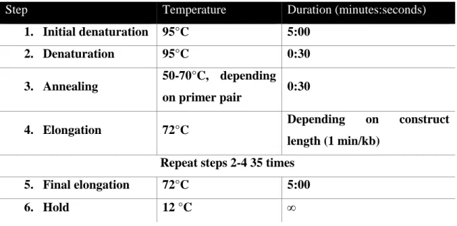
3.2.1 Polymerase chain reaction (PCR)
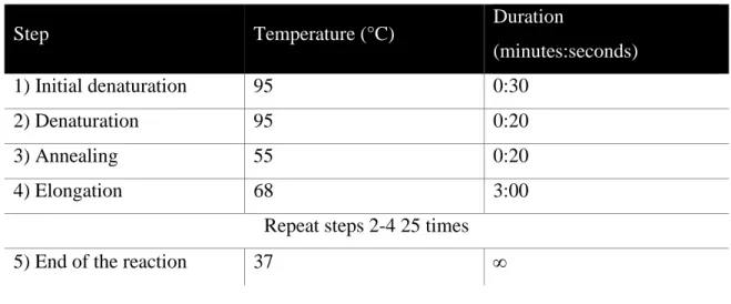
DNA fragments were amplified by the PCR method (Mullis and Faloona, 1987) according to standard protocols. PCR reactions were done in 25 µl or 50 µl total reaction volume. Typically 20-100 ng/µl of plasmid DNA were mixed with 200 nM of forward/reverse primer, 250 µM of each dNTP (Bioline) and 0,02 µl polymerase per µl of total volume in the corresponding reaction buffer. For most applications Pfu S polymerase (lab internal) was used, for site-directed mutagenesis Accuzyme (Bioline, London, UK) was utilized.
Table 3-9: Standard PCR program
Step Temperature Duration (minutes:seconds)
1. Initial denaturation 95°C 5:00
2. Denaturation 95°C 0:30
3. Annealing 50-70°C, depending on primer pair 0:30
4. Elongation 72°C Depending on construct
length (1 min/kb) Repeat steps 2-4 35 times
5. Final elongation 72°C 5:00
6. Hold 12 °C