• Keine Ergebnisse gefunden

„Characterization of tumor-antigen-specific T cells – insights into WT1- and HPV16-specific T cell repertoires“

N/A
N/A
Protected

Academic year: 2021

Aktie "„Characterization of tumor-antigen-specific T cells – insights into WT1- and HPV16-specific T cell repertoires“"

Copied!
131
0
0

Wird geladen.... (Jetzt Volltext ansehen)

Volltext

(1)

„Characterization of tumor-antigen-specific T cells – insights into WT1- and HPV16-specific T cell repertoires“

Inaugural Dissertation

zur

Erlangung des Doktorgrades Dr. nat. med.

der Medizinischen Fakultät und

der Mathematisch-Naturwissenschaftlichen Fakultät der Universität zu Köln

vorgelegt von

Sabine Schmied, MSc. Biol.

aus Waldshut

November 2014

(2)

Berichterstatter/Berichterstatterin: Prof. Dr. Hinrich Abken PD Dr. Thomas Wunderlich

Tag der letzten mündlichen Prüfung: 23. Februar 2015

(3)

Eidesstattliche Erklärung

Ich versichere, dass ich die von mir vorgelegte Dissertation selbstständig angefertigt, die benutzen Quellen und Hilfsmittel vollständig angegeben und die Stellen der Arbeit – einschließlich Tabellen, Karten und Abbildungen -, die anderen Werken im Wortlaut oder dem Sinn nach entnommen sind, in jedem Einzelfall als Entlehnung kenntlich gemacht habe;

dass diese Dissertation noch keiner anderen Fakultät oder Universität zur Prüfung vorgelegen hat; dass sie – abgesehen von unten genannten Teilpublikationen – noch nicht veröffentlicht worden ist sowie, dass ich eine solche Veröffentlichung vor Abschluss des Promotionsverfahrens nicht vornehmen werde. Die Bestimmungen dieser Promotionsordnung sind mir bekannt. Die von mir vorgelegt Dissertation ist von Prof. Dr.

Hinrich Abken, PD Dr. Thomas Wunderlich, Dr. Mario Assenmacher und Dr. Anne Richter betreut worden.

Übersicht der Publikationen:

Schmied S., Gostick E., Price D., Abken H., Assenmacher M., Richter A. Analysis of the functional WT1-specific T cell repertoire in healthy donors reveals a discrepancy between CD4 + and CD8 + memory formation. Submitted to Immunology.

Schmied S., Preuss SF., Wolber P., Huebbers CU., Abken H., Assenmacher M., Richter A.

Functional HPV16-reactive T cells in periphery and tumor tissue of HPV16 pos HNSCC patients. Submitted to The International Journal of Cancer.

Ich versichere, dass ich alle Angaben wahrheitsgemäß nach bestem Wissen und Gewissen gemacht habe und verpflichte mich, jedmögliche, die obigen Angaben betreffenden Veränderungen, dem Promotionsausschuss unverzüglich mitzuteilen.

Köln, den 05. März 2015 ______________________

Unterschrift

(4)

Abstract

This thesis provides characterization of rare tumor-associated antigen (TAA)-specific T cells concerning their in vivo prevalence, functionality, and phenotype in healthy and diseased individuals. Such knowledge on the existing in vivo repertoire is crucial for the understanding of T cell immunity towards tumors and furthermore for the utilization of TAA-specific T cells in immunotherapeutic approaches. Thus far, low frequencies of such TAA-specific T cells in blood and tissues hindered an in-depth characterization, even in tumor patients. Here, we establish and implemented highly sensitive methods, based on magnetic enrichment of TAA- specific T cells in combination with a multiparameter flow cytometry analysis to overcome existing sensitivity hurdles and allow ex vivo description of the T cells. Two different activation markers were implemented, CD154 (CD40L) for ex vivo enumeration and characterization of blood CD4 + T cells, and CD137 (4-1BB) for subsequent characterization of rare antigen-specific CD4 + and CD8 + T cell subsets from blood and tumor tissue after a short-term in vitro expansion.

On the one hand, T cells specific for the self-antigen WT1 and, on the other hand, T cells specific for the viral antigens HPV16 E6 and E7 were investigated.

The Wilms’ tumor 1 (WT1) protein is a transcription factor expressed at high levels in several hematological malignancies and some solid tumors and is considered a prime target for cancer immunotherapy. Remarkably, in this study, WT1-specific CD4 + and CD8 + T cell populations were detected in the vast majority of healthy individuals. Memory responses specific for WT1 were commonly present in the CD4 + T cell compartment, whereas WT1- specific CD8 + T cells almost universally displayed a naïve phenotype. Moreover, WT1- specific memory CD4 + and naïve CD8 + T cells were found to co-exist in healthy individuals.

Collectively, these findings suggest a natural discrepancy between the CD4 + and CD8 + T cell lineages responsive to self-antigens with respect to memory formation. Nonetheless, WT1- specific T cells from both lineages were readily activated ex vivo and expanded in vitro, supporting the general use of strategies to exploit this expansive reservoir of self-reactive T cells for immunotherapeutic purposes.

Furthermore, characteristics of HPV16-specific T cells were investigated in head and neck

squamous cell carcinoma (HNSCC) patients, as HNSCC is frequently induced by HPV16

infection. This study revealed that HPV16-specific T cells in the blood of HPV16 pos HNSCC

patients exhibit profound differences in prevalence, functionality, and phenotype between

individuals. While all patients harbored HPV-specific T cells in peripheral blood, we identified

a subgroup of patients with significantly elevated frequencies of functional effector memory

HPV16-specific CD4 + T cells compared to other patients. Moreover, these high responder

patients had a more active anti-tumor immunity in comparison to low responders. Despite

(5)

these variations, HPV16-specific T cells with functional capacities could be isolated and propagated both from blood and tumor tissue of all HPV16 pos HNSCC patients. Furthermore, this data supports the notion that HPV16-reactive tumor infiltrating lymphocytes were suppressed by intratumoral T reg cells, the depletion of which restored T cell effector functions and facilitated expansion of reactive T cells for therapeutic use.

In summary, the techniques established in this study permit detection of rare antigen-specific

T cells and therefore are superior to common immune monitoring methods in terms of

sensitivity and determination of T cell properties. In particular, these techniques were

implemented to provide novel insights into the naturally occurring repertoire of WT1- and

HPV16-directed T cells. These findings support the use of WT1- and HPV16-specific T cells

in immunotherapy and provide knowledge crucial for the optimal design of WT1- and HPV16-

directed immunotherapeutic interventions.

(6)

Zusammenfassung

Diese Arbeit umfasst eine Charakterisierung in Bezug auf Prävalenz, Funktionalität und Phänotyp von den sehr seltenen T-Zellen, die spezifisch für Tumor-assoziierte Antigene (TAA) sind. Wissen über das in vivo existierende T-Zellrepertoire ist für das Verständnis der Immunität gegenüber dem Tumor und darüber hinaus für die Entwicklung von immuntherapeutischen Ansätzen notwendig. Eine solche detaillierte Charakterisierung war bislang nicht möglich, da TAA-spezifische T-Zellen im Blut und im Tumor nur in sehr geringer Frequenz vorliegen. In dieser Arbeit werden hoch sensitive Methoden etabliert, basierend auf magnetischer Anreicherung von TAA-spezifischen T-Zellen in Kombination mit einer Analyse mittels Multiparameter-Durchflußzytometrie, die die bisher bestehenden Sensitivitätsgrenzen überwinden und damit die ex vivo Charakterisierung der T-Zellen zulassen. Zwei Aktivierungsmarker kamen für die Zellanreicherung zum Einsatz; zum einen wurde CD154 (CD40L) zur ex vivo Enumeration und Charakterisierung von CD4 + T-Zellen aus dem Blut benutzt und zum anderen wurde CD137 (4-1BB) für die Charakterisierung von antigen- spezifischen CD4 + und CD8 + T-Zellpopulationen aus Blut und Tumorgewebe nach einer kurzen in vitro Expansion eingesetzt.

Im Speziellen wurden T Zellen untersucht, die spezifisch für das Selbstantigen WT1 sind, und T Zellen, die spezifisch für das E6 und E7 Antigen des humanen Papillomavirus Typ 16 (HPV16) sind.

Das Wilms’ Tumor 1 (WT1) Protein ist ein Transkriptionfaktor, der im hohen Maße in hämatologischen Tumoren und einigen soliden Tumoren exprimiert wird. WT1 gilt als geeignetes Zielantigen für die Immuntherapie. Erstaunlicherweise konnten in dieser Studie WT1-spezifische CD4 + und CD8 + T-Zellen im Blut von der Mehrheit der gesunden Spender detektiert werden. Eine Gedächtnisantwort, die spezifisch gegen WT1 gerichtet ist, war häufig im CD4 + T-Zellkompartiment zu finden, während WT1-spezifische CD8 + T-Zellen fast ausschließlich einen naiven Phänotyp zeigten. Darüber hinaus wurde die Koexistenz von WT1-spezifischen Gedächtnis CD4 + und naïve CD8 + T-Zellen in einem Individuum nachgewiesen. Zusammenfassend implizieren die Resultate, dass eine Diskrepanz in der Entwicklung von Gedächtnisantworten zwischen den CD4 + and CD8 + T-Zellkompartimenten, die auf Selbstantigen reagieren, vorliegt. Dennoch ließen sich WT1-spezifische T-Zellen aus beiden Kompartimenten in vitro aktivieren und expandieren. Dieses spricht für die Umsetzung von Strategien, die das Repertoire von Selbstantigen-spezifischen T-Zellen für immuntherapeutische Ansätze nutzen.

Des Weiteren wurden HPV16-spezifische T-Zellen in Patienten mit Kopf-Hals-Tumor (head

and neck squamous cell carcinoma, HNSCC) untersucht, da HNSCC häufig durch HPV16

Infektion induziert wird. Die vorliegende Arbeit zeigt auf, dass die Häufigkeit, die

(7)

Funktionalität und der Phänotyp von HPV16-spezifischen T-Zellen im Blut sich deutlich zwischen den HPV16 pos HNSCC Patienten unterscheiden. Während in allen Patienten spezifische T-Zellen nachweisbar sind, konnte ein Gruppe von HPV16 pos HNSCC Patienten identifiziert werden, die eine auffällig hohe Frequenz an HPV16-spezifischen Effektor- Gedächtnis-CD4 + T-Zellen mit hoher Funktionalität aufweist. In diesen ‚high responder’

Patienten war darüber hinaus eine aktivere Anti-Tumorimmunität im Tumorgewebe zu finden als in den ‚low responder’ Patienten. Trotz dieser Unterschiede konnten HPV16-spezifische T-Zellen sowohl aus Blut als auch aus Tumorgewebe von HPV16 pos HNSCC Patienten gewonnen werden. Dennoch verdeutlichen die Daten, dass T reg im Tumorgewebe die Aktivität der HPV16-spezifischen Tumor-infiltrierender Lymphozyten abschwächen, denn das Eliminieren von T reg aus den in vitro gewonnenen tumor-infiltrierenden Lymphozyten führte zur einer deutlich verstärkten Effektorfunktion und Expansion von HPV16-spezifischen T-Zellen.

Die in dieser Arbeit etablierten Methoden ermöglichen eine detaillierte Charakterisierung von

sehr seltenen tumor-spezifischen T-Zellen und sind damit bislang üblichen Methoden des

Immunmonitoring an Sensitivität und Informationsgehalt überlegen. Insbesondere wurden

mittels der etablierten Methoden neue, tiefreichende Erkenntnisse über die T-Zell-Immunität

gegenüber WT1 und HPV16 gewonnen. Die erzielten Daten sprechen für die

immuntherapeutische Nutzung von WT1- und HPV16-spezifischen T Zellen und stellen

wichtige Erkenntnisse für die optimale Gestaltung von WT1- und HPV16-basierten

Immuntherapien bereit.

(8)

Content

1 INTRODUCTION ... 1

1.1 T LYMPHOCYTES ... 1

1.1.1 Target antigen recognition by T cells ... 1

1.1.2 Development of T cells ... 3

1.1.3 Primary activation of T cells ... 4

1.1.4 Differentiation of T cells ... 5

1.2 A NTI - TUMOR IMMUNITY ... 7

1.2.1 Tumor-associated antigens ... 8

1.2.2 Implementation of anti-tumor T cell immunity ... 8

1.3 D ETECTION OF ANTIGEN - SPECIFIC T CELLS ... 10

1.4 WT1 ... 11

1.5 HPV16 E6/E7 ... 12

1.6 O BJECTIVES ... 15

2 MATERIALS AND METHODS ... 16

2.1 R EAGENTS , MEDIA , SUPPLEMENTS , AND ANTIGENS ... 16

2.1.1 Staining antibodies ... 16

2.1.2 MHC/peptide multimers ... 16

2.1.3 Antibodies for cell culture ... 16

2.1.4 Reagents for cell isolation ... 17

2.1.5 Media and supplements ... 17

2.1.6 Recombinant cytokines ... 17

2.1.7 Buffer ... 17

2.1.8 Antigens ... 17

2.1.9 Other ... 18

2.2 C ONSUMABLES , DEVICES , AND SOFTWARE ... 18

2.3 C ELL SEPARATION AND PROCESSING ... 19

2.3.1 Patients’ characteristics ... 19

2.3.2 PBMC preparation ... 21

2.3.3 Tumor dissociation ... 21

2.3.4 Separation of T cell subsets ... 21

2.3.5 Stimulation of T cells ... 22

2.3.6 Enrichment of antigen-specific T cells ... 22

2.3.7 Expansion of antigen-specific T cells ... 23

2.3.8 Cultivation of primary tumor cells ... 24

2.3.9 Freezing and thawing of cells ... 24

2.4 D ETECTION METHODS ... 24

2.4.1 Determination of HPV16 Status of HNSCC tumors ... 24

2.4.2 Cell Surface marker staining ... 24

2.4.3 pHLA tetramer staining ... 25

2.4.4 Staining of intranuclear FoxP3 ... 25

2.4.5 Intracellular cytokine staining ... 25

2.4.6 Detection of released cytokines by Multiplex... 26

2.4.7 Determination of CD107a expression ... 26

2.4.8 Cytotoxicity assay ... 26

2.4.9 Determination of functional avidity ... 27

2.5 S TATISTICS ... 27

3 RESULTS ... 28

3.1 E STABLISHMENT OF ENRICHMENT STRATEGIES BASED ON ACTIVATION MARKERS FOR THE DETECTION OF RARE ANTIGEN - SPECIFIC T CELLS ... 28

3.1.1 Enrichment of CD154

+

T cells ... 28

3.1.2 Enrichment of CD137

+

T cells ... 32

3.1.2.1 CD137 is co-expressed with other T cell activation markers ... 33

3.1.2.2 CD137

+

cells are accurately detected after enrichment with anti-CD137-PE ... 34

(9)

3.1.2.3 CD137

+

effector and regulatory T cells are enriched via CD137 ... 36

3.1.2.4 Low frequent T cells can be enriched via CD137 ... 38

3.1.2.5 Antigen-specific T cell lines can be generated after CD137 enrichment ... 40

3.2 C HARACTERIZATION OF WT1- SPECIFIC T CELLS IN HEALTHY INDIVIDUALS ... 43

3.2.1 Ex vivo characterization of WT1-specific T cells ... 43

3.2.1.1 Majority of healthy donors have a pool of WT1-reactive CD4

+

memory T cells ... 43

3.2.1.2 CD8

+

WT1-specific T cells derive predominantly from the naïve T cell repertoire in healthy donors ... 46

3.2.1.3 Disparity of phenotype in CD4

+

and CD8

+

T cells specific for WT1 ... 48

3.2.2 In vitro generation of WT1-specific T cell cultures ... 51

3.2.2.1 In vitro propagation of WT1-specific T cells by enrichment of CD137

+

cells ... 51

3.2.2.2 WT1-specific T cell cultures can be efficiently generated using CD137 enrichment ... 52

3.2.2.3 Expanded WT1-specific T cell cultures are polyfunctional ... 53

3.2.2.4 Expanded WT1-specific T cell cultures show a non-exhausted phenotype ... 57

3.2.2.5 Expanded WT1-specific CD8

+

T cells have a functional avidity comparable to CMV-specific T cells ... 58

3.3 C HARACTERIZATION OF THE HPV16- SPECIFIC T CELL IMMUNITY IN HNSCC PATIENTS ... 61

3.3.1 Magnitude of HPV16-specific immunity in blood of HPV16

pos

HNSCC patients varies ... 61

3.3.2 HPV16-reactive T cells detected in high responder HPV16

pos

HNSCC patients are functional ... 65

3.3.3 Favorable cell composition of the tumor tissue in high responder HPV16

pos

HNSCC patients ... 68

3.3.4 HPV16-directed T cell immunity in blood of HNSCC patients reflects intratumoral immunity ... 69

3.3.5 T cells expanded from blood and tumor tissue exhibit a similar phenotype ... 73

3.3.6 T cells expanded from blood and tumor tissue show HPV16-specific cytotoxic potential ... 74

3.3.7 Immunosuppressive tumor microenvironment in HPV16

pos

HNSCC impairs T cell functions ... 76

4 DISCUSSION ... 78

4.1 E STABLISHMENT OF ACTIVATION MARKER - BASED ENRICHMENT STRATEGIES FOR THE DETECTION OF RARE ANTIGEN - SPECIFIC T CELLS ... 78

4.1.1 Ex vivo enumeration and characterization of rare antigen-specific T cells via CD154 enrichment . 78 4.1.2 Detection of rare antigen-specific T cells via CD137 enrichment ... 79

4.1.3 Influence of T

reg

on enrichment of antigen-specific effector T cells via CD137 ... 80

4.1.4 Generation of TAA-specific T cell cultures ... 81

4.1.5 Peptide pools for stimulation of the full repertoire of antigen-specific T cells ... 82

4.2 WT1- SPECIFIC T CELL REPERTOIRE IN HEALTHY DONORS ... 84

4.2.1 Prevalence and functionality of WT1-specific T cells ... 84

4.2.2 Discrepancy in differentiation status of CD4

+

and CD8

+

WT1-specific T cells ... 86

4.2.3 In vitro generation of WT1-specific T cell lines ... 88

4.2.4 Expanded WT1 specific T cells for adoptive anti-cancer therapy ... 90

4.2.5 Employing WT1-specific T cells for therapeutic anti-cancer vaccination ... 90

4.3 HPV16- SPECIFIC T CELL IMMUNITY IN HNSCC PATIENTS ... 92

4.3.1 Prevalence and functionality of HPV16-reactive T cells in HPV16

pos

HNSCC patients ... 92

4.3.2 HPV16-reactive T cells in healthy individuals ... 94

4.3.3 Correlation between HPV16-specific naïve and memory T cell frequencies ... 94

4.3.4 In vitro generation of HPV16-reactive T cell populations from blood and tumor ... 95

4.3.5 Cytotoxicity of expanded HPV16-reactive T cells from blood and tumor ... 98

4.3.6 Role of T

reg

in activation and expansion of HPV16-specific T cells ... 98

4.3.7 Quantity and quality of T cell immunity in tumor lesions ... 99

4.3.8 Comparison of HPV16-reactive T cell immunity in the blood and tumor ... 99

4.3.9 Immunosuppressive conditions in HNSCC tumors ... 100

4.3.10 Implications on immunotherapeutic approaches ... 101

4.4 J UXTAPOSITION OF WT1- SPECIFIC T CELLS AND HPV16- SPECIFIC T CELLS ... 104

5 REFERENCES ... 105

6 PUBLICATIONS ... 120

7 CURRICULUM VITAE ... 121

(10)

Abbreviations

Ab antibody / antibodies

APC antigen presenting cells

CD cluster of differentiation

CMV cytomegalovirus

cTEC cortical thymic epithelial cells

CTL cytotoxic T cell

DC dendritic cell

DLI Donor lymphocyte infusion

E6 early protein 6

E7 early protein 7

EBV Epstein-Barr virus

EC 50 half maximal effective concentration

FSC forward scatter

GvHD Graft-versus-host disease

GvL Graft-versus-leukemia

Gy gray

HLA human leukocyte antigen

HNSCC Head and neck squamous cell carcinoma

HPV human papilloma virus

HSCT hematopoietic stem cell transplantation

IE-1 immediate early protein 1

IFN-γ interferon-gamma

IL interleukin

L1 late protein 1

MART-1 melanoma antigen recognized by T cells 1

MB MicroBeads

MHC major histocompability complex

MNC mononuclear cells

mTEC medullar thymic epithelial cells

NK natural killer

PBMC peripheral blood mononuclear cells

PI propidium iodid

pp65 phosphoprotein 65

TAA tumor-associated antigen

T CM central memory T cells

T eff Effector T cells

T EM effector memory T cells

T EM-RA end-differentiated effector memory T cells

T H helper T cells

TIL tumor-infiltrating lymphocytes

TNF-α Tumor necrosis factor alpha

T reg regulatory T cells

WT1 Wilms tumor 1

(11)

1

1 Introduction

The human immune system fights invading pathogens. Furthermore, the immune system is able to eliminate aberrant, genetically altered, mutated, autologous cells which are potential precursors of tumor cells. Human immunity is built on two major pillars, innate immunity and adaptive immunity. The innate immunity, including neutrophilic, eosinophilic and basophilc granulocytes, monocytes, macrophages, dendritic cells, and natural killer cells provides a general first defense against invading pathogens. A more delicate defense is generated by the adaptive immunity, built by B- and T-lymphocytes. B- and T-lymphocytes are capable of specific recognition of foreign antigens, brought in by pathogens, with the help of highly specific antigen receptors. The vast repertoire of antigen receptors allows for elimination of pathogens that evade or overwhelm the innate immunity. Furthermore, adaptive immunity provides an immunological memory to protect the body from reinfection.

The human immune system plays a major role in preventing the development of cancer.

Especially T lymphocytes, which are essential for elimination of aberrant cells of the body. In case of cancer formation, however, the anti-tumor immune response is insufficient.

1.1 T lymphocytes

T lymphocytes, or T cells, originate from lymphoid progenitor cells in the bone marrow and mature in the thymus; the “T” in T cell stands for thymus. After maturation T cells circulate in the blood, lymph, and secondary lymphoid tissues, such as the lymph nodes, the spleen, and the mucosal lymphoid tissues, where they scan for the emergence of foreign peptides.

Main functions of T cells are direct killing of target cells, i.e. infected or tumor cells, and activation of other immune cells via cell surface molecules or by the secretion of soluble mediators like cytokines upon antigen encounter.

1.1.1 Target antigen recognition by T cells

T cells recognize their targets via their T cell receptor (TCR). A minor population of T cells

carries a TCR consisting of γ:δ chains, however, the majority of T cells express an α:β chain

TCR. Each TCR chain is composed of a constant region and a variable region, the latter

forming the antigen binding-site (Fig. 1.1). The α:β TCR binds to peptide loaded major

histocompatibility (MHC) complexes on professional antigen-presenting cells (APC) or other

cells of the body. Classically T cells fall into two major categories, which are CD4 + T helper

(12)

2 cells and CD8 + cytotoxic T cells, defined by the expression of the co-receptors CD4 and CD8, which are engaged in TCR-MHC binding. CD4 + T cells recognize peptides presented on MHC class II molecules and CD8 + T cells recognize peptides presented on MHC class I molecules. Distinct pathways of antigen processing are responsible for loading of MHC class I and MHC class II molecules. Specifically, antigens taken up from the extracellular fluid by endocytosis are digested in the endosome and loaded onto MHC class II molecules. In contrast, peptides loaded onto MHC class I molecules are typically derived from intracellular proteins introduced by viral or bacterial infection of the cell. Cytosolic proteins are degraded via the proteasome and transported to the ER where they are loaded onto MHC class I molecules (Neefjes, Jongsma et al. 2011). An exception to this rule is exerted by DC, as they are able to process antigens from the extracellular space via the ER onto MHC class I molecules. This process is called cross-presentation (Joffre, Segura et al. 2012). Additionally, peptides loaded onto MHC class I and MHC class II differ in length. MHC class I molecules have a distinctly defined binding pocket, which binds peptides of 8 to 10 amino acids in length. Peptide binding is stabilized by specific anchor residues at the end of the peptide. In contrast, the binding groove of MHC class II molecules is more flexible as it can bind peptides of 13 to 17 amino acids in length. The ends of these peptides are not bound, but instead overlap the groove, and anchor residues for MHC class II are less defined. MHC class I molecules are constitutively expressed on all cells of the body, whereas MHC class II expression is restricted to professional APC of the immune system, such as DCs, macrophages, and B cells. Presence of IFN-γ can trigger the expression and upregulation of MHC class I and MHC class II molecules.

Figure 1.1 Structure of the T cell receptor

complex. The T cell receptor itself is a

heterodimer, each chain consisting of a

constant and variable section. The variable

section includes the CDR3 binding region,

which accounts for the huge diversity in

antigen-specificity of TCRs. The TCR co-

localizes with CD3γ, CD3δ, and CD3ε

molecules and the ζ chains, which carry

immunoreceptor tyrosine-based activation

motifs (ITAM) intracellularly. Through

phosphorylation of these ITAMs the TCR

signal is transmitted to the cell (Figure taken

from (Murphy 2011).

(13)

3 1.1.2 Development of T cells

During maturation of the T cell in the thymus TCR rearrangement takes place. Here, different variable (V), joining (J), and diversity (D) segments are combined in a stochastic manner in both the  chain and  chain locus. This rearrangement and the combination of  and  chains creates the diversity of TCR binding specificities, which is estimated to comprise

>10 11 possible specificities (Robins, Srivastava et al. 2010). Positive selection in the thymus ensures that only T cells carrying a TCR, that is able to bind self-peptide:self-MHC complexes, survive. This applies only to about 10% of T cells Positively selected T cells recognizing self-MHC complexes on cortical thymic epithelial cells (cTEC) do become single positive for either CD8 or CD4 depending on whether they recognize MHC class I or MHC class II complexes, respectively. ‘Single positive’ cells migrate from the thymic cortex to the medulla. In the thymic medulla negative selection takes place, which deletes the T cells bearing a strong affinity against self-peptide:self-MHC complexes to protect the body from strong autoimmunity (Fig. 1.2).

Figure 1.2 Affinity model of T cell selection. The strength of interaction of the TCR with peptide:self-MHC during development within the thymus determines the fate of the T cell. No binding leads to elimination during positive selection, likewise very strong binding also leads to elimination in the course of negative selection. Strong recognition can also lead to the development of regulatory T cells (T

reg

). Recent studies provide evidence that there is a stochastic overlap in affinity, which can either lead to the development of a T

reg

or deletion of the T cell. Likewise, similar affinities can exist in the T

reg

and naïve T cell compartment (Bains, van Santen et al. 2013) (Figure adapted from (Klein, Kyewski et al. 2014)).

To enable negative selection against the full repertoire of self-antigens of the body, medullar

thymic epithelial cells (mTEC) are able to express antigens normally only expressed on

specific tissues or during embryonic development. This feature of mTEC is called

promiscuous gene expression. However, there have been reports that the negative selection

is incomplete for certain antigens. Reasons for this insufficient negative selection can be very

low expression of the antigen in mTEC (Gotter, Brors et al. 2004, Lv, Havari et al. 2011),

(14)

4 differential splicing of mRNA (Klein, Klugmann et al. 2000, Pinto, Sommermeyer et al. 2014), or missing post-translational modifications of proteins in mTEC (Scally, Petersen et al. 2013, Van Lummel, Duinkerken et al. 2014). Strong recognition of self-antigens cannot just lead to deletion of T cells, but also to development of these CD4 + T cells into T cells with regulatory functions (T reg ) (Hsieh, Lee et al. 2012). Only 2 - 4% of all thymocytes will leave the thymus as functional naïve T cells.

1.1.3 Primary activation of T cells

After leaving the thymus the naïve lymphocyte will circulate via lymph and blood between lymphoid organs until encountering its respective antigen. Naïve T cells express the lymph node homing receptor CD62L and the chemokine receptor CD197 (CCR7), which enable them to migrate to the secondary lymphoid tissues such as lymph nodes, spleen, and mucosal lymphoid tissues. Professional APC take up antigen at sites of infection or aberrant tissue formation and get activated following inflammation as part of innate immunity. This activation triggers the migration of APC to the secondary lymphoid organs. The T cell becomes activated when it meets such an activated professional APC, including DC, but also activated macrophages and B cells, presenting the respective antigen within the T cell zone of a lymphoid organ. This process is called T cell priming. The primed T cell starts to proliferate and differentiate into an effector T cell. For a successful priming of a naïve T cell, co-stimulatory signals mediated by molecules on the professional APC and the T cell are needed, in addition to TCR signaling through peptide:MHC binding. One crucial co- stimulatory signal is the interaction of B7 molecules (CD80, CD86) with CD28 on the T cell.

Primed T cells leave the secondary lymphoid organs to migrate to the site of infection.

T cells specific for self-antigens, which were not negatively selected during their development

in the thymus, are usually not activated in the periphery, as peripheral tissues do not express

co-stimulatory molecules. Similarly, professional APC express very few co-stimulatory

molecules in the absence of inflammatory signals. This peripheral self-tolerance provides a

further safety mechanism to prevent autoimmunity. When pro-inflammatory signals such as

pro-inflammatory cytokines produced by innate immune cells and/or co-stimulatory

molecules on APC are missing, T cells can be anergized or converted into T reg .

(15)

5 Figure 1.3 Priming of naïve T cells.

Professional APC need to deliver three different signals to a naïve T cell to activate and therefore prime it. First, the TCR of the T cell needs to specifically bind the peptide:MHC complex. In addition, the co-receptor CD4 or CD8 bind to MHC. Second, the co-stimulatory B7 molecules need to be expressed on the APC and bind to CD28 expressed on the T cell. Also other co- stimulatory molecules, like members of the TNF receptor family, can provide such a signal. Third, the APC provides cytokines to trigger the differentiation of the primed T cells. In a non- inflammatory setting APC lack co-stimulatory molecules and T cell contact leads to anergizing of the T cell (Figure taken from (Murphy 2011)).

1.1.4 Differentiation of T cells

Cytokine signals released by the professional APC determine the fate of the primed T cell.

Especially for CD4 + T cells several subclasses are known, differing in the cytokines needed for differentiation and also in cytokines secreted by those CD4 T cells (Fig. 1.4). Different CD4 + T cell subsets exert different effector functions.

Figure 1.4 Major T cell subsets and their effector functions.

The main function of CD8

+

T cells is the killing of target cells presenting the respective peptide on

MHC class I molecules by the release of cytolytic granules into the immunological synapse. CD4

+

T

cells bear diverse functions. Main subclasses of CD4

+

T cells include T

H

1, T

H

1, T

H

17, and T cells

bearing regulatory functions (T

reg

) (Figure taken from (Murphy 2011)).

(16)

6 T H 1 cells are specialized to stimulate innate immune responses, they activate macrophages and are also able to kill infected macrophages. Main effector cytokines of the T H 1 subset are IFN-γ and TNF-α. T H 1 cells are especially important for anti-tumor immunity as they can exert anti-tumor functions, both by direct tumor cell killing or by activation of other immune cells by secretion of pro-inflammatory cytokines (Haabeth, Tveita et al. 2014). T H 2 cells promote the immune response to extracellular pathogens mainly by activating B cells. Effector cytokines of T H 2 cells include IL-4, IL-5, and IL-13 (Wan and Flavell 2009). In addition to T H 1, T H 2, and T reg cells also T H 9, T H 17, T H 22, and T follicular helper cells (T FH ) exist. Recent studies have discovered even more distinct subsets and have also shown the plasticity of T H cells, adding to the complexity of human adaptive immunity (Lee, Turner et al. 2009, Wei, Wei et al. 2009).

In an activated state, T H cells express CD40 ligand (CD154) on their surface, which is important for activation of professional APC and CD8 T cell priming.

CD4 + T cells with regulatory functions can evolve from the thymus (natural T reg = nT reg ) and be induced in the periphery (induced T reg = iT reg ). Both T reg subsets are characterized by the constitutive expression of the transcription factor FoxP3 and the cell surface molecule IL-2R α-chain (CD25). T reg cells are able to suppress effector T cell function through contact- dependent inhibition of T cell activation as well as inhibition of proliferation owing to release of soluble factors such as IL-10 and TGFβ (Corthay 2009) or competition for IL-2 (de la Rosa, Rutz et al. 2004).

Priming of CD8 T cells requires CD4 T cell help to APC, especially when inflammatory signals are lacking to activate the APC sufficiently. CD40 ligand (CD154) on activated CD4 + T H cells binding to CD40 on DC results in enhanced co-stimulatory signals to the naïve CD8 + T cell. An activated CD8 + effector T cell can kill antigen-presenting target cells specifically via release of cytolytic granules. Perforin-containing granules form holes in the targeted cells and allow for granzyme-containing granules to enter the target cells and trigger apoptosis (de Saint Basile, Menasche et al. 2010). As MHC class I molecules are present on virtually all cells of the body, CD8 + T cells can kill autologous cells, which present non-self-peptides, as in the case of virus infected cells. However, the clear functional distinction between CD4 + helper T cells and CD8 + cytotoxic T cells is not as strict as previously thought, as cytolytic CD4 + T cells and CD8-expressing T cells with helper cell functions have also been identified in recent years (Paludan, Bickham et al. 2002, Frentsch, Stark et al. 2013).

Once a T cell has differentiated into an effector cell, the T cell exerts its effector functions

upon antigen encounter without the need for a professional APC providing co-stimulatory

signals. Additionally, the T cell’s homing behavior drastically changes, as it now needs to

leave the lymphoid tissue and enter the sites of infection. Effector cells loose the expression

of CCR7 and CD62L, which mediate homing to the secondary lymphoid organs. CCR7 and

(17)

7 CD62L are therefore markers, which can be used to distinguish naïve from effector T cells.

Furthermore, the protein tyrosine phosphatase CD45, involved in TCR signaling, is expressed in different isoforms during differentiation. Naïve T cells expressing the long CD45RA isoform and memory T cells expressing the shorter CD45RO isoform. According to the expression patterns of these molecules, the differentiation status of the T cell can be phenotypically determined. Naïve T cells being CD45RO - CCR7 + , central memory T cells CD45RO + CCR7 + , effector memory T cells CD45RO + CCR7 - , and exhausted T cells being CD45RO - CCR7 - . After clearance of the infection or prolonged antigen-stimulation, an effector T cell eventually progresses to an exhausted state, its effector functions decrease and the T cell eventually dies. But central memory T cells exist also which retain the expression of CD62L and CCR7 (Sallusto, Geginat et al. 2004). Initial models of T cell differentiation suggested a unidirectional progression from naïve to effector T cells to exhausted effector T cell to apoptosis in which central memory T cells are cells arrested at intermediate stages of differentiation preceding effector T cells. Newer models suggest the existence of a memory T cell population bearing stem cell-like properties. This stem T cell population is marked, amongst other, by the expression of the IL-7 receptor α-chain (CD127) in humans. CD127 is also expressed on naïve T cells but normally rapidly lost upon activation (Gattinoni, Lugli et al. 2011). These putative stem T cells are able to self-renew and give rise to central memory and effector memory T cells (Bondanza, Hambach et al.

2011, Mackall, Fry et al. 2011).

1.2 Anti-tumor immunity

The adaptive immunity is able to conduct a directed immune response against cancerous

cells. It is believed that many aberrant cells, which are potential precursors of tumor cells, are

cleared by these immunological defense mechanisms, a process called immunosurveillance

(Corthay 2014). In case of tumor formation these immunological defense mechanisms are

impaired or not sufficient. Then the tumor is able to overcome immunosurveillance. The

hypothesis of immunoediting states that the immunosurveillance alters the composition of

tumors towards more aggressive forms, which are able to circumvent recognition by the

immune defense mechanism or actively suppress it (Kim, Emi et al. 2007). It is clear that

many tumor entities exhibit such tumor escape features. Tumor escape mechanisms include,

on the one hand, defects in antigen processing or presentation components and therefore

loss of antigen presentation; on the other hand, formation of a immunosuppressive

microenvironment by attraction of suppressive cell types, such as T reg , myeloid suppressor

cells, or release of suppressive cytokines (Kim, Emi et al. 2007). However, in the last decade

it has been clearly shown that tumor escape can be overcome by immunotherapeutic

(18)

8 interventions even in quite aggressive forms of cancer by different strategies, e.g. increasing the number of anti-tumor T cells or modulating immunosuppressive features of the tumor.

1.2.1 Tumor-associated antigens

Tumor cells have an aberrant protein expression profile compared to normal cells of the body, which can result in presentation of distinct antigens. This feature renders them sensitive to recognition by cells of the adaptive immune system. Antigens especially expressed on tumor cells are referred to as tumor-associated antigens (TAA). T cells specific for TAA can mediate such an anti-tumor response. TAA fall into different classes and can be of self,

“altered-self”, or foreign origin: (1) Foreign antigens derived from viral oncogenes such as the human papilloma virus (HPV) E6 and E7 protein and (2) “altered-self” antigens, e.g. mutated antigens such as Bcl-Abl expressed in some forms of myeloid leukemia. On the other hand, TAA can also be classical self-antigens as in the case of: (3) Oncofetal antigens normally only expressed during embryonic and fetal development or in germ-line tissues (NY-ESO-1, MAGE-A3), (4) tissue differentiation antigens (MART-1, gp100) or (5) antigens overexpressed in tumor cells, such as WT1, overexpressed in myeloid leukemia and many solid tumors.

1.2.2 Implementation of anti-tumor T cell immunity

Several treatment approaches have proven the potential of naturally existing immunity, especially T cells, for the treatment of cancer.

Firstly, prophylactic vaccination has been successfully implemented for the protection against tumor formation in the case of HPV16 (and HPV18)-induced cervix carcinoma. Therapeutic vaccination has not been clinically established thus far; however, numerous studies have shown that an anti-tumor immunity can be triggered against multiple cancer entities, including HPV16-induced vulvar neoplasias (Kenter, Welters et al. 2009, Daayana, Elkord et al. 2010) and WT1-expressing tumors (Van Driessche, Berneman et al. 2012).

Secondly, so called checkpoint inhibitors have been used to re-activate existent immunity

against tumors. Successfully used in the clinic is a CTLA-4 inhibitor (ipilimumab) used for

treatment of melanoma, which blocks the inhibitory CTLA-4:B7 interaction and leads to

enhanced general activation of T cells (Hodi, O'Day et al. 2010, Ott, Hodi et al. 2013). The

clinical effect of re-activation of intratumoral T cells underlines that naturally occurring T cells

have a good anti-cancer potential.

(19)

9 Thirdly, adoptive transfer of naturally derived TAA-specific T cells has directly demonstrated their therapeutic potential. The feasibility and efficacy of adoptive T cell transfer was successfully proven in melanoma where tumor-infiltrating lymphocytes are expanded in vitro with the use of IL-2 and administered to lympho-depleted patients. This therapy has led to shrinkage and also clearance of major tumor lesions in metastatic melanoma patients (Rosenberg, Yannelli et al. 1994). Additionally to naturally derived TAA-specific T cells, genetically engineered T cells specific for a TAA or T cells transduced with a chimeric antigen receptor specific for a surface protein of the tumor cell are successfully used in the clinic (Porter, Levine et al. 2011, Kershaw, Westwood et al. 2013).

For adoptive T cell therapy, several studies have highlighted that the quality of the administered T cells is crucial for clinical success (reviewed in (Riddell 2007) and (Klebanoff, Gattinoni et al. 2012). Therefore, attempts were made to design the best in vitro expansion protocol to achieve a good in vivo performance and persistence of T cells. Firstly, extensive in vitro cultivation leads to an exhausted state of T cells, characterized by loss of expression of co-stimulatory markers and expression of exhaustion markers such as CD57. Exhausted T cells were shown to have decreased effector functions compared to non-exhausted T cells in vivo and to have a short in vivo persistence and hence are not preferred for adoptive transfer (Powell, Dudley et al. 2005, Hinrichs, Borman et al. 2009). The recently identified stem T cell population (see section 1.1.4) is favored for the use in adoptive T cell therapy, as this population is characterized by good in vivo persistence (Zhang, Joe et al. 2005). Cytokines used for expansion are a critical factor. Use of IL-7 and IL-15 has been reported to preserve a non-exhausted phenotype of in vitro expanded T cell cultures, which harbor superior in vivo antitumor functions compared to cells expanded with IL-2 (Cieri, Camisa et al. 2013, Xu, Zhang et al. 2014). Secondly, polyfunctionality of the applied T cells has been shown to provide enhanced protection, as they combine a broader repertoire of effector functions, i.e.

the ability of one individual T cell to secrete more than one different cytokine (Seder, Darrah et al. 2008). Thirdly, early adoptive transfer studies used mostly CTL clones, but within the last decade it became clear that CD4 + T cells are also important for anti-tumor efficacy of a cell product, both due to cytolytic functions of the CD4 + T cells themselves (Quezada, Simpson et al. 2010), help for anti-tumor CD8 + T cells (Wong, Bos et al. 2008), and recruitment of other cells with anti-tumor functions (Perez-Diez, Joncker et al. 2007).

Therefore, a CD4 + and CD8 + mixed TAA-specific T cell product might have the best efficacy.

A combined phenotypic and functional analysis of the T cell product can give important

insights into whether a T cell population is effective in therapy. To date, the knowledge

regarding the effective tumor-specific T cell response is incomplete. However, such

knowledge is crucial in order to, on the one side, develop immunotherapies such as adoptive

(20)

10 T cell transfer or vaccination and, on the other side, monitor and predict clinical outcome of such an intervention.

1.3 Detection of antigen-specific T cells

Antigen-specific T cells can be detected with various techniques. The most common tool for direct detection of antigen-specific T cells are pHLAI multimers, where multimerized, peptide loaded MHC class I molecules are conjugated to a fluorophore, which enables detection of peptide-specific CD8 + T cells via flow cytometry. Recently, pHLAII multimers have also been successfully introduced for detection of peptide-specific CD4 + T cells (Moon, Chu et al. 2007, Obar, Khanna et al. 2008). Owing to the facts that only a limited amount of cells can be measured by flow cytometry and that multimers can produce unspecific background staining, which increases with increased numbers of measured cells, pHLA multimers have an approximate detection limit of 10 -5 within PBMC.

Another commonly used method is the enzyme-linked immuno spot assay (ELISPOT), which allows for detection of a released cytokine upon antigen stimulus with an estimated detection limit of 10 -5 within PBMC (reviewed in (Schmittel, Keilholz et al. 1997)). The ELISPOT is an indirect method, it is therefore only semi-quantitative and does not allow for a further characterization of single T cells. Cytokine-producing T cells after stimulation can also be detected in a direct method via intracellular staining and flow cytometric analysis. However, only a subset of activated T cells produce a certain cytokine upon stimulation, therefore enumeration of the entire pool of antigen-specific T cells is not feasible.

More recently, surface molecules upregulated after antigenic stimulation (activation markers) have been successfully used for visualization of antigen-specific T cells. For example, CD154 (CD40L) is expressed within a few hours after antigen-specific stimulation of CD4 + T cells. CD137 (4-1BB) is upregulated on both CD4 + and CD8 + T cells after longer stimulation times (>24 h). Both marker have been shown to be expressed both on activated naïve and memory T cells and thus enable detection of antigen-specific T cells independent of their differentiation status. Furthermore, when peptide pools are used for T cell stimulation a previous knowledge of epitope specificities and HLA status is not needed. Thus, activation markers, in principle, provide a compelling opportunity to detected the full repertoire of antigen-specific T cells.

T cells specific for one particular antigen are commonly present in very low frequencies, as

the human T cell repertoire comprises approximately 10 6 - 10 7 different antigen specificities

(Arstila, Casrouge et al. 1999). Therefore, self-antigen-specific and tumor-associated-antigen

(TAA)-specific T cells in healthy individuals cannot be detected with commonly used

(21)

11 technologies such as ELISPOT and pHLAI multimer staining, as they are not sensitive enough. An exception are T cells specific for the melanoma-associated self-antigen MelanA/MART-1, where rather high frequencies of specific T cells (up to 10 -3 within CD8 + T cells), exhibiting a naïve phenotype, in healthy individuals were reported (Pittet, Valmori et al.

1999, Zippelius, Pittet et al. 2002). Apart from this exception normally only T cells specific for immune-dominant recall antigens can be readily detected, as for example CMV pp65-specific T cells in CMV-infected donors, where frequencies of up to 5% pp65-specific T cells within the CD8 + T cell compartment can occur.

To circumvent detection limitations for rare antigen-specific T cells, enrichment strategies can be employed. Here, cells of interest are magnetically labeled and applied to a column placed in a magnetic field where they are retained. Non-labeled cells are washed away, and the enriched, magnetically labeled cells are eluted (Miltenyi, Muller et al. 1990). Magnetic enrichment of pHLA multimer + T cells was successfully used to detect CD4 + and CD8 + antigen-specific at very low frequencies from the memory and naïve repertoire (Barnes, Ward et al. 2004, Obar, Khanna et al. 2008, Chu, Moon et al. 2009, Alanio, Lemaitre et al.

2010, Kwok, Tan et al. 2012). Recently, an elegant magnetic enrichment technique based on CD154-expression after antigen stimulation was established, which allows a simultaneous enumeration and functional characterization of antigen-specific T cells directly ex vivo, this method was named antigen-reactive T cell enrichment (ARTE) (Bacher, Schink et al. 2013).

CD137 was also used to enrich antigen-specific T cells (Wolfl, Kuball et al. 2007). However, stimulation periods >24 h, which are needed for the expression of CD137 on naïve T cells, render CD137 only limited suitable for ex vivo characterization, as prolonged stimulation can alter the expression of phenotypic markers on activated T cells.

With the help of these magnetic enrichment techniques rare TAA-specific T cells were detected and characterized. Two model antigens of current clinical interest were chosen, the self-antigen WT1 and the viral antigens HPV16 E6 and E7. Apart from providing crucial information on the TAA-specific T cell repertoire for each TAA separately, investigation on T cells specific for these two distinct TAA antigens could unravel potential principle differences in the T cell repertoire for self- and virus-specific T cells, due to divergent negative selection mechanisms during T cell development (introduced in 1.1.2).

1.4 WT1

The Wilms’ tumor 1 (WT1) protein is a transcription factor, which is expressed at high levels

in several types of hematological malignancies as well as in some solid-tumors. WT1

expression in adult humans is restricted to early hematopoietic precursor cells (Baird and

Simmons 1997) and is rapidly lost upon differentiation of these precursors (Maurer, Brieger

(22)

12 et al. 1997). WT1 was ranked top in a list of potential tumor antigens to be used in anti- cancer immunotherapy because of its oncogenic potential and presumptive immunogenicity (Cheever, Allison et al. 2009). Indeed, spontaneous T cell responses towards WT1 are induced in leukemia patients after allogeneic stem cell transplantation (SCT) or donor lymphocyte infusions (Rezvani, Yong et al. 2007, Kapp, Stevanovic et al. 2009, Tyler, Jungbluth et al. 2013) and are associated with disease regression ((Tyler, Jungbluth et al.

2013). In order to design WT1-directed immunotherapies, knowledge about the in vivo existence of WT1-specific T cells is a prerequisite. However, until now, such detailed insight into the naturally occurring WT1-specific T cell repertoire is vastly limited. This is due to the fact that ex vivo detection and hence also the characterization of WT1-specific T cells so far is hindered owing to their exceedingly low frequencies, especially in blood of healthy donors.

WT1-specific CD8 + T cells have been detected ex vivo in myeloid leukemia patients via WT1 126 -HLAI multimers (Sloand, Melenhorst et al. 2011) and via qPCR of IFN-γ mRNA after stimulation, with raised prevalence after SCT (Rezvani, Grube et al. 2003, Rezvani, Brenchley et al. 2005). Using highly sensitive IFN-γ mRNA detection Rezvani et al. also gave a first hint, that CD8 + WT1-specific T cells indeed do exist in low frequencies even in healthy individuals, but an in depth characterization was not feasible with the used approach (Rezvani, Grube et al. 2003, Rezvani, Brenchley et al. 2005).

WT1 is under current investigation for the use in immunotherapeutic approaches. On the one side, several clinical WT1 vaccination trials have been done and showed promising results.

Here, a clinical response was detectable in 45% of patients bearing solid tumors and even 63% in patients with hematopoietic tumors and also immunological responses could be detected in some cases (Van Driessche, Berneman et al. 2012). However, the design of vaccines was not ideal as HLA-restricted peptides have been used in some trials.

Furthermore, the immunomonitoring was also limited as it was restricted to the detection of WT1-HLAI tetramer + CD8 + T cells. Further knowledge about the characteristics of WT1- specific T cells, as provided in this study, will hopefully improve vaccine design to achieve optimal clinical efficiency. On the other side, usage of WT1-specific T cells for adoptive transfer after hematopoietic stem cell transplantation (HSCT) is currently under investigation.

A first clinical trial has recently proven the safety of WT1 126 -specific CD8 + T cells in this setting (Chapuis, Ragnarsson et al. 2013).

1.5 HPV16 E6/E7

The human papilloma virus (HPV) is a DNA virus infecting the basal layer of epithelia by

entering wounds or lesions of skin and mucosal tissues. Only after differentiation of the

epithelial cells to cells of the squamous cell layer virus particles are assembled and released.

(23)

13 It is believed that more than 50% of people are infected by any type of HPV once during their life time. Certain high risk types of HPV, including HPV16 and HPV18, however, can induce lesions which can eventually progress to cancer if not cleared.

The E6 and E7 proteins of HPV16 are oncogenes, which induce cell-cycle progression. The E6 protein of high risk HPV acts as an E3 ligase for the p53 tumor suppressor protein and therefore triggers its degradation (Scheffner, Werness et al. 1990). The E7 protein binds and destabilizes the retinoblastoma (Rb) tumor suppressor protein and related proteins (Boyer, Wazer et al. 1996). The expression of E6 and E7 oncoproteins is mandatory for the uncontrolled growth of infected cells resulting in tumor formation. Therefore, these proteins are suggested to be ideal targets for immunotherapeutic approaches (Cheever, Allison et al.

2009).

The most prominent example for HPV-induced cancer is cervix carcinoma, which is induced by the high risk types HPV16 or HPV18 in about 70% of cases (Munoz, Bosch et al. 2004, Schiffman, Castle et al. 2007). Also head and neck squamous cell carcinoma (HNSCC) can be induced by HPV high risk types. A subgroup of HNSCC, the oropharynx carcinoma, is induced by high risk type HPV16 in more than 50% of cases (Ramqvist and Dalianis 2010, Jayaprakash, Reid et al. 2011). The incidence of HPV induced HNSCC is rising (Chaturvedi, Engels et al. 2011).

HNSCC patients bearing an HPV16 pos tumor have a better prognosis than HPV16 neg HNSCC patients. The reasons for better prognosis of HPV16 pos HNSCC patients are unknown so far.

However, there is evidence that the better outcome results from the formation of an anti- tumor immunity. In general, the presence of CD3 + and CD8 + tumor-infiltrating lymphocytes (TIL) was found to be stronger in HPV16 pos HNSCC tumors (Jung, Guihard et al. 2013) and HPV16 pos tumors have a higher CD8 + to regulatory T cell (T reg ) ratio (Russell, Angell et al.

2013). Both findings were correlated with prolonged disease-specific survival. In specific, HPV16-specific T cells have also been detected in tumor tissue of HPV16-related HNSCC (Heusinkveld, Goedemans et al. 2012).

Furthermore, the peripheral HPV16-directed immunity was investigated. For HPV-related

cervical carcinoma the systemic HPV-immunity was linked to the intratumoral HPV16-

directed immunity and improved prognosis (Piersma, Jordanova et al. 2007). However, the

overall functionality of peripheral HPV-specific T cells in cervical cancer seems to be

impaired, either due to skewing to a T H 2 phenotype (Clerici, Merola et al. 1997, Bais,

Beckmann et al. 2005, Sharma, Rajappa et al. 2007) or due to impaired functionality of the

T H cells (de Jong, van Poelgeest et al. 2004). For HNSCC, a systemic HPV16-specific T cell

immunity was also found in the periphery of HPV16 pos HNSCC patients. MHC class I

tetramer analysis revealed higher frequencies of CD8 + HPV16-specific T cells in the blood of

(24)

14 HPV16 pos HNSCC patients compared to HPV16 neg patients and healthy individuals (Albers, Abe et al. 2005, Hoffmann, Arsov et al. 2006). Similar to cervix carcinoma, the peripheral HPV16-specific immunity in HNSCC patients revealed a partial functional impairment of T cells and the absence of a systemic HPV16-directed T cell immunity in some HPV16 pos HNSCC patients (Heusinkveld, Goedemans et al. 2012). A detailed characterization in terms of frequency, phenotype, and functionality of such systemic HPV16-specific T cell immunity in HNSCC patients, however, has been missing until now.

Two phase II clinical trials for therapeutic vaccination for patients with vulvar intraepithelial

neoplasia, a pre-stage of cervix carcinoma, are ongoing. Both studies use a HPV16 vaccine,

consisting of either HPV16 E6 and E7 peptides or an HPV16 E6E7L2 fusion protein, and

showed promising results, as both achieved clinical response rates >60% (Kenter, Welters et

al. 2009, Daayana, Elkord et al. 2010). For therapeutic HPV16 vaccination of HNSCC the

first clinical phase I trials have been started (Center SKCC 2013 and Inovio Pharmaceuticals

2014, ClinicalTrials.gov). Adoptive transfer of HPV16-specific T cells is also under current

investigation (Ramos, Narala et al. 2013), but has not been implemented in the clinic as of

yet.

(25)

15 1.6 Objectives

In this study, unbiased and in-depth knowledge about rare tumor antigen-specific T cells is provided. This knowledge allows for conclusions on the feasibility and efficacy of immune- based anti-cancer therapies, such as vaccination and adoptive T cell transfer.

In the first part of this thesis the establishment of the highly-sensitive strategies, which were used to characterize the rare antigen-specific T cells, is outlined. Main objectives were: (i) Establishment of method(s) for ex vivo detection and characterization of rare antigen-specific T cells and (ii) establishment of an efficient method to generate antigen-specific T cell cultures starting from low frequent, presumably naïve T cells. The established techniques were then used to provide in-depth characterizations of TAA-specific T cells. Here, T cells specific for two distinct TAAs were investigated, the self-antigen WT1 and the viral antigens E6 and E7 derived from HPV16.

In the second part of this thesis the in-depth characterization of WT1-specific T cells is described. This second part aims at the investigation of quantity and quality of WT1-specific T cells in healthy donors to provide crucial knowledge for immunotherapeutic interventions in leukemia and other solid tumor entities.

In the third and final part of this thesis HPV16-specific T cells are characterized in HPV16 pos and, for comparison, in HPV16 neg patients and healthy individuals. Both blood and tumor derived HPV16-specific T cells of HNSCC patients were investigated to obtain so far missing knowledge on HPV16-directed peripheral and intratumoral immunity in those patients, which is valuable for the design of immunotherapeutic interventions in HPV16 pos HNSCC.

The approach used and the results obtained in this study are unique, as a characterization of these tumor-antigen specific T cells was so far not feasible due to their low frequencies. The established highly sensitive enrichment methods, for the first time, enable the detection and comprehensive functional and phenotypical characterization of these rare tumor-antigen specific T cells. Additionally, in contrast to other studies focusing on WT1- or HPV16-specific T cells this study provides a characterization of the full repertoire of antigen-specific T cells including both CD4 + and CD8 + T cells, naïve as well as memory T cells.

The consequences of the findings relating to the design of immunotherapeutic interventions

will be discussed.

(26)

16

2 Materials and Methods

2.1 Reagents, media, supplements, and antigens

2.1.1 Staining antibodies

The following antibody reagents were used for staining and flow cytometric measurements:

anti-CD3-VioGreen (BW264/56), anti-CD4-allophycocyanin-Vio770 (VIT4), anti-CD8-PE- Vio770 (BW135/80), anti-CD14-PerCP (TÜK4), anti-CD20-allophycocyanin, anti-CD20- PerCP (LT20), anti-CD27-FITC (M-T271), anti-CD28-FITC (15E8), anti-CD31-FITC (AC128), anti-CD44-allophycocyanin-Vio770 (DB105), anti-CD45-VioGreen (5B1); anti-CD45RA- VioBlue (T6D11), anti-CD45RO-FITC (UCHL1), anti-CD57-VioBlue (TB03), anti-CD62L-FITC (145/15), anti-CD107a-PE (1D4B), anti-CD127-FITC (MB15-18C9), anti-CD137- allophycocyanin, anti-CD137-biotin, anti-CD137-PE (4B4-1), anti-CD154-allophycocyanin, anti-CD154-VioBlue (5C8), anti-CD178-PE (NOK-1), anti-CD279-allophycocyanin (PD1.3.1.3), anti-CD197 (CCR7)-allophycocyanin, anti-CD197 (CCR7)-PE (150503), anti- CD324-allophycocyanin (67A4), anti-CD326-VioBlue (HEA-125), anti-FoxP3-PE (3G3), anti- HLA-ABC-PE (REA230), anti-IL-2-PE-Vio770 (N7.48A), anti-IL-4-PE (7A3-3), anti-IL-17-FITC (CZ8-23G1), anti-IFN-γ-FITC, anti-IFN-γ-PE-Vio770 (45-15), anti-TIM-3-allophycocyanin (F38-2E2), anti-TNF-α-PE, and anti-TNF-α-VioBlue (cA2) (all Miltenyi Biotec, Bergisch Gladbach, Germany).

2.1.2 MHC/peptide multimers

Soluble biotinylated pHLA-A*0201 molecules loaded with WT1 37-45 (VLDFAPPGA), WT1 126-134

(RMFPNAPYL), WT1 187-195 (SLGEQQYSV), WT1 235-243 (CMTWNQMNL), or cytomegalovirus (CMV) pp65 495-503 (NLVPMVATV) were produced as described previously (Price 2005 JExpMed). Tetramerization was done by incubating 2 nmol pHLA molecules with 0.5 nmol streptavidin-PE or streptavidin-allophycocyanin (both BioLegend, San Diego, USA) at 4°C, PBS supplemented with Protease Inhibitor (Roche, Basel, Switzerland) was used for dilution.

2.1.3 Antibodies for cell culture

Pure anti-CD28 (clone 15E8), anti-CD40 (clone HB14), and anti-MHC class I (clone W6/32)

were obtained from Miltenyi Biotec.

(27)

17 2.1.4 Reagents for cell isolation

All antibody bead reagents, separation, and isolation kits used in this study were obtained from Miltenyi Biotec.

2.1.5 Media and supplements

RPMI1640 (Miltenyi Biotec) was supplemented with 2mM L-Glutamine (PAA Laboratories GmbH, Pasching, Austria) and 5% human AB-Serum (Lonza, Basel, Switzerland). X-Vivo15 (Lonza) was supplemented with 5% human AB-serum (Lonza).

2.1.6 Recombinant cytokines

Recombinant human IL-7, IL-15, IL-21 and IFN-γ were obtained from Miltenyi Biotec and recombinant human Proleukin S was obtained from Novartis Pharma, Basel, Switzerland.

2.1.7 Buffer

0.1 M phosphate-buffered saline containing 137 mM NaCl, 2.6 mM KCl, 8.1 mM Na 2 HPO 4 , and 1.4 mM KH 2 PO 4 (all chemicals purchased from Sigma Aldrich, St. Louis, USA) was stored as 10x stock solution and freshly diluted. Sterile 0.1 M phosphate-buffered saline containing 2 mM EDTA was obtained from Miltenyi Biotec and optionally supplemented by sterile 0.5% human AB-Serum (PEB-buffer). Buffers for MACSPlex Cytokine 12 Kit and Tumor Dissociation Kit (both Miltenyi Biotec) was used as provided and indicated within kits.

InsideFix solution and 10x InsidePerm solution are provided with the Rapid Cytokine Inspector Kit (Miltenyi Biotec).

2.1.8 Antigens

PepTivator CMV pp65, EBV EBNA-1, EBV LMP2a, HPV16 E6, HPV16 E7, and WT1 were

obtained from Miltenyi Biotec. PepTivator peptide pools consist of 15-mer peptides

overlapping by 11 amino acids and spanning the complete protein sequence. Peptides

WT1 37-45 , WT1 126-134 , WT1 187-195 , WT1 235-243 were synthesized by O. Braun and S. Kramer,

Miltenyi Biotec.

(28)

18 2.1.9 Other

Propidium Iodide solution (PI) was obtained from Mitlenyi Biotec. CellTrace Violet was obtained from Invitrogen, Carlsbad, USA. Monensin was obtained from eBioscience, San Diego, USA. Mitomycin C and Brefeldin A and antibiotic antimycotic solution were obtained from Sigma-Aldrich.

2.2 Consumables, devices, and software

MACSQuant Analyzer 10 Miltenyi Biotec

Cell culture hood, Hera Safe KS Thermo Fischer Scientific, Waltham, USA CO2 Incubator, Hera Cell® Thermo Fischer Scientific

Hematologic analyzer, Sysmex KX-21 Sysmex Corporation, Japan Centrifuge, Eppendorf 5415D Eppendorf, Hamburg, Germany Centrifuge, Multifuge X3R Heraeus Instruments, Hanau Centrifuge, Multifuge 4KR Heraeus Instruments

Centrifuge 4515R Eppendorf

MACSmix Tube Rotator Miltenyi Biotec

pH-Meter, pH-Meter 765 Calimatic Elektronische Messgeräte GmbH, Berlin, Germany

Irradiator RS 2000 Rad Source Technologies, Inc., Suwanee, USA

MACS Separator (Mini, Midi, Octo, Quadro) Miltenyi Biotec MACS separation columns (MS, LS, LD) Miltenyi Biotec

gentleMACS Dissociator Miltenyi Biotec

gentleMACS C Tubes Miltenyi Biotec

Microscope, Leica DM IL LED Leica Microsystems, Wetzlar, Germany Orbital shaker Titramax 100 Heidolph, Schwabach, Gemany

Conical bottom tubes, 50 ml, 15 ml Falcon (BD Biosciences), New Jersey, USA 96/48/24/12-well flat bottom cell culture plates Falcon (BD Biosciences)

75/175 cm 2 cell culture flasks Falcon (BD Biosciences)

Cell Strainer 70 µm Falcon (BD Biosciences)

96-well filter plate, 0.2 µm PVDF membrane Corning, Corning, USA

Cell Scraper MIDSCI, St. Louis, USA

Surgical Scalpell Aesculap AG, Tuttlingen, Germany

NALGENE® Cryogenic vials Nalge Nunc International, Rochester, USA

Dispenser Tips Th. Geyer, Renningen, Germany

Microcentrifuge tubes STARLAB, Hamburg, Germany

MACSQuantify, version 2.5 Miltenyi Biotec

Abbildung

Figure  1.1  Structure  of  the  T  cell  receptor  complex.  The  T  cell  receptor  itself  is  a  heterodimer,  each  chain  consisting  of  a  constant  and  variable  section
Figure  1.2  Affinity  model  of  T  cell  selection.  The  strength  of  interaction  of  the  TCR  with  peptide:self-MHC during development within the thymus determines the fate of the T cell
Figure 1.4 Major T cell subsets and their effector functions.
Table 2.2: Characteristics of HNSCC patients included at follow-up. n.d.: not determined, m: male,  f: female, n: no,  y: yes, t.d.: technical difficulties, tumor were classified according to the TNM system  accepted  by  the  American  Joint  Committee  o
+7

Referenzen

ÄHNLICHE DOKUMENTE

[r]

To exclude that the reduced number of virus-specific T cells in Bis VIII–treated mice at day 7 after viral infection was due to inhibition of T cell activation and reduced expansion,

Among aged donors, statistical differences in basal glycolysis, compensatory glycolysis and the ratio basal mitoOCR/glycoPER between memory CD4 + T cells and naive CD4 + T. cells

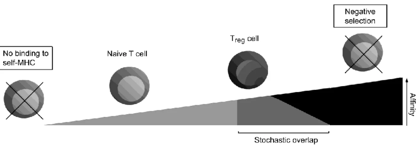
fewer# OT6I# T# cells# survive# negative# selection# in# RIP6variant# mice# expressing#

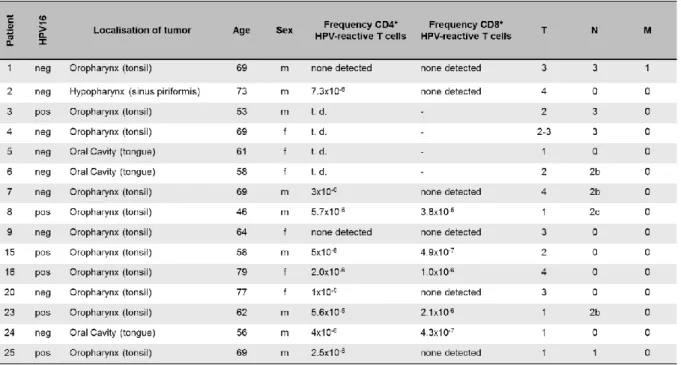
RESULTS: We show for the first time that EC present a quantitatively different peptide repertoire with abundance of certain peptides, compared with leukocytes. The abundance of

In a clinical trial, transferring CMV-specific T cell from the stem cell donor in CMV-reactivating HSCT recipients, patients with proliferating CMV-specific T

The cell-surface binding and internalization process of human papillomaviruses is a complex multi-step process that is not yet fully understood. To date, numerous

Here it is shown for the first time that in vitro conversion of CD4 + CD25 - T cells into CD4 + CD25 + Foxp3 + Tregs was significantly reduced in cells derived from