• Keine Ergebnisse gefunden

Tumorantigen-Specific CD40B Cells: Combining Enhanced Antigen-Presentation and Antibody-Secretion for Tumor Targeting

N/A
N/A
Protected

Academic year: 2021

Aktie "Tumorantigen-Specific CD40B Cells: Combining Enhanced Antigen-Presentation and Antibody-Secretion for Tumor Targeting"

Copied!
171
0
0

Wird geladen.... (Jetzt Volltext ansehen)

Volltext

(1)

Combining Enhanced Antigen-Presentation and Antibody-Secretion for Tumor Targeting

Inaugural-Dissertation zur

Erlangung des Doktorgrades

der Mathematisch-Naturwissenschaftlichen Fakultät der Universität zu Köln

vorgelegt von

Kerstin Wennhold

aus Köln

(2)

Berichterstatter: Prof. Dr. Thorsten Hoppe Prof. Dr. Hinrich Abken

Tag der mündlichen Prüfung: 16. April 2015

(3)
(4)
(5)
(6)
(7)

Danksagung

Bedanken möchte ich mich bei allen, die zum Erfolg dieser Arbeit beigetragen und mich unterstüzt haben.

Mein Dank gilt Prof. Hallek, unter dessen Schirmherrschaft diese Arbeit entstand. Ebenso möchte ich Prof. Hoppe danken, dass er so kurzfristig meine Betreuung übernommen hat.

Vielen Dank an Prof. Hinrich Abken, der meine Arbeit über die Jahre inhaltlich begleitet und unterstützt hat.

Dank geht an die Else-Kröner Fresenius Stiftung, den NZT.NRW Verbund und Köln Fortune für die Finanzierung dieses Projekts. Ebenso danke ich dem Graduiertenprogramm der Uniklinik Köln und der Bayer Health Care AG für finanzielle Unterstüzung bei Fortbildungen und Konferenzen und den wissenschaftlichen Austausch.

Mein größter Dank gilt Prof. Michael von Bergwelt-Baildon, der mir die Möglichkeit gab in seinem Labor zu forschen und meine Doktorarbeit anzufertigen. Vielen Dank für Deine Betreuung und Unterstüzung, die enthusiastischen Diskussionen und die lehrreichen Herausforderrungen. Besonderer Dank gilt Dr. Alexander Shimabukuro-Vornhagen, der mir gutes wissenschaftliches Arbeiten und die Fähigkeit meine Ergebnisse zu hinterfragen beibrachte und mir immer mit Rat zur Seite stand. Mit Deiner zuversichtlichen Art und Deinen schlauen Lösungansätzen hast Du mich immer aufgemuntert, wenn meine Experimente nicht so wollten wie ich.

Maria y Juliane, les agradezco por su amistad y el intercambio cultural. Tener que trabajar es mucho más divertido cuando se puede pasar con amigos. No importa dónde estemos en el futuro; ustedes siempre tienen un lugar especial en mi corazón.

Ich möchte mich ganz herzlich bei Martin und Sabrina für ihre tatkräftige, maustechnische und moralische Unterstützung im letzten Jahr bedanken. Ohne Eure große Hilfe im Labor hätte die Krebszelle viel häufiger auf der schlechten Seite gestanden. Vielen Dank an Anne;

durch Dich habe ich endlich gelernt zu kompensieren. Michael, Maria, Juliane, Martin, Sabrina, Anne, Hans S., Hans B., Sebastian, Alex, Sasha, Geothy, Steffi und Claudia, ich danke Euch für die tolle Zeit im Labor, für die unterhaltsamen Mittagspausen, gemeinsamen Abendessen und spektakulären Laborausflüge. Ich werde von dieser besondere Erfahrung vor allem die Zeit mit Euch immer in guter Erinnerung behalten.

Ein ganz großer Dank gilt auch Hannah, Manuel, Verena und Carola. So viele Jahre begleitet Ihr mich schon durch alle Höhen und Tiefen mit Lachen, Zuhören, Quatschen und Quatsch machen, Trösten, Freuen und Fröhlich sein.

Ganz besonders bedanken möchte ich mich bei meiner Familie. Ihr seid in allen Lebenslagen

bedingungslos für mich da, um Euch mit mir zu freuen oder mir mit Rat zur Seite zu stehen.

(8)
(9)

Zusammenfassung

Voraussetzung für die Induktion einer effizienten Immunantwort ist neben dem richtigen Antigen eine effiziente Präsentation von Antigen in vitro und in vivo. Dendritische Zellen (DC) gelten als die am besten charakterisierten antigen-präsentierenden Zellen. Allerdings können sie kaum in großen Mengen und ausreichender Reinheit hergestellt werden, was einen entscheidenden Nachteil bei der Nutzung von DCs in der klinischen Anwendung dar stellt. Deshalb wurde ein alternativer Ansatz entwickelt, bei dem polyklonale B-Zellen durch die Behandlung mit stimulierenden Zytokinen und dem CD40-Liganden als antigenpräsentierende Zellen genutzt werden können. Die Aktivierung durch den CD40- Liganden induziert entscheidend die Antigen-präsentation, was zu einer effizienten Induktion von CD4+ und CD8+ T-Zellen in vitro führt. Darüber hinaus wandern diese CD40- aktivierten (CD40)B-Zellen nicht nur in die peripheren lymphatischen Organe, sondern induzieren auch eine anti-tumorale Immunität in Mäusen.

Die Antigen-präsentation und -prozessierung durch antigen-spezifische B-Zellen ist bis zu tausendfach effektiver als bei polyklonalen B-Zellen. Deshalb wurden in der vorliegenden Arbeit antigen-spezifische B-Zellen genutzt, um die antigen-präsentierende Funktion von CD40B-Zellen zu steigern. Nach CD40-Stimulation regulieren aufgereinigte, antigen- spezifische B-Zellen kostimulatorische Moleküle wie CD80 und CD86 signifikant hoch, was zu einer verstärkten Antigen-präsentation und der Induktion einer spezifischen T-Zellantwort in vitro und in vivo führt. Antigen-spezifische CD40B-Zellen induzieren eine signifikant stärkere T-Zellanwort als polyklonale CD40B-Zellen, die mit einer DC-induzieren Reaktion vergleichbar ist. Eine präventive Vakzinierung mit antigen-spezifischen CD40B-Zellen alleine oder in Kombination mit antikörper-sezernierenden Plasmazellen führt zu einer Anti-Tumor Immunität in vivo, bei der das Tumorwachstum komplett unterdrückt wird. Darüber hinaus migrieren antigen-spezifische CD40B-Zellen in den Tumor, was neue Anwendungsmöglichkeiten als Trägerstoff bei Imagingverfahren oder zum ‚Drug Delivery‘

ermöglicht.

(10)
(11)

Abstract

Efficient antigen presentation is a prerequisite for the development of a T-cell-mediated immune response in vitro and in vivo. As “nature’s adjuvant”, dendritic cells (DCs) are the most prominent professional antigen-presenting cells (APCs). However, they have several significant disadvantages concerning application as an adjuvant in cancer immunotherapy.

They are rare in peripheral blood and the isolated population is not homogenous, since it also contains unwanted tolerogenic DCs. Therefore, an alternative approach to DCs was developed, in which polyclonal B cells can serve as potent APCs by treatment with the inflammatory cytokine IL-4 and the CD40 ligand. CD40-activation dramatically improves antigen presentation by normal and malignant B cells, efficiently inducing naïve and memory CD4+ and CD8+ T-cell responses. Moreover, these CD40-activated (CD40) B cells do not only home to secondary lymphoid organs, but also induce anti-tumor immunity in mice.

However, antigen-processing and -presentation by antigen-specific B cells is 1000-fold more

effective compared to polyclonal B cells. Therefore, tumorantigen-specific B cells were used

in the present study to improve the antigen-presenting function of CD40B cells. Purified

tumorantigen-specific B cells highly upregulate activation markers upon CD40-stimulation

resulting in an enhanced antigen-presentation and a specific T-cell response in vitro and in

vivo. The T cell response elicited by antigen-specific CD40B cells is significantly stronger than

that induced by polyclonal CD40B cells and comparable to the stimulation induced by

mature DCs. Alone or in combination with antibody-secreting plasma cells, preventive

vaccination with these antigen-specific CD40B cells leads to an anti-tumor immune response

in vivo resulting in complete inhibition of tumor growth. Moreover, antigen-specific B cells

home to the tumor site, thereby offering new application perspectives as vehicle for tumor

(12)
(13)

Abbreviations

A

500

Absorbance at 500 nm

APC antigen-presenting cells

BCR B cell receptor

β-ME beta-mercaptoethanol

BSA bovine serum albumin

°C degrees Celsius

CCR C-C-chemokine receptor

CD cluster of differentiation

CD40B CD40-activated B cells

CD40L CD40 ligand

CEA cell adhesion molecule

CFSE 5-(and-6)-carboxyfluorescein diacetate succinimidyl ester

cm centimeter

CO

2

carbon dioxide

CTLA-4 cytotoxic T-lymphocyte-associated Protein 4

CXCR CXC-motiv-chemokine receptor

DC dendritic cell

DMSO dimethylsulfoxid

EDTA ethylenediaminetetraacetic acid

ELISA Enzyme Linked Immunosorbent Assay

ER endoplasmatic riticulum

FACS fluorescence activated cell sorting

FcR Fc receptor

FDA Food and Drug Administration

FITC Fluorescein isothiocyanate

FBS fetal bovine serum

g gram

GC germinal center

GM-CSF granulocyte/macrophage colony stimulating factor

Gy gray

h hour

H

2

O water

HBV hepatitis-B-antigen

HIV Human Immundeficient-Virus

HLA human leukocyte antigen

HSC Hematopoietic stem cells

IFA Incomplete Freund’s adjuvant

IFN interferon

Ig immunoglobulin

i.p. intraperitoneal

i.v. intravenous

IL interleukin

(14)

Abbreviations

LPS Lipopolysaccharide

Luc Luciferase

M molar

mAb monoclonal antibody

MAGE melanoma-antigen family

MFI mean fluorescent intensity

mg milligram

MHC major histocompatibility complex

min minutes

MLR mixed-lymphocyte reaction

mmol millimol

mM millimolar

ml milliliter

mm millimeter

MW molecular weight

MZ marginal zone

µg microgram

µl microliter

µm micrometer

µM micromolar

ng nanogram

NK natural killer

nm nanometer

nM nanomolar

OVA chicken ovalbumin

PBMC peripheral blood mononuclear cell

PBS phosphate buffered saline

PC plasma cell

PD-L1 Programmed death-ligand 1

PE phycoerythrin

PE-Cy phycoerythrin- cyanine

Pen/Strep Penicillin/ Streptomycin

PerCp-Cy peridinin chlorophyll protein complex-cyanine dye

PFA paraformaldehyde

rh recombinant human

rm recombinant murine

RT room temperature

s.c. subcutaneous

SD standard deviation

SDS sodium dodecylsulfate

SEM standard error of mean

TCR T cell receptor

TGFβ transforming growth factor-beta

T

h

T helper cell

TNF tumor necrosis factor

Treg regulatory T cell

U units

(15)

Table of Contents

Danksagung ………..…………...….…....……I Zusammenfassung ………....…….II Abstract ………...….III Abbreviations ……….………...….IV Table of Contents ..………....….VI

INTRODUCTION 1

1.1 The Adaptive Immune System ..………...….3

1.1.1 T Cells ………..…….………....….4

1.1.1.1 CD8

+

T Cells ………..………..………....…..4

1.1.1.2 CD4

+

T Cells ………..………....….5

1.1.2 Immunological Tolerance .………....…....5

1.1.3 B Cells and Humoral Immune Response ………...6

1.1.4 Antigen-Presentation to T cells ………....….8

1.2 Basic Principles of Cancer Immunology ………...10

1.2.1 Cancer Immunosurveillance ………...10

1.2.2 Immunoediting ………...11

1.2.3 Tumor Evasion Strategies ………....…11

1.3 Targeted Immunotherapy ………....……12

1.3.1 Tumor Antigens ………....….12

1.3.1.1 Tumor-Specific Antigens ………....………12

1.3.1.2 Tumor-Associated Antigens ………....…………..13

1.3.2 Passive Immunotherapy ………....………13

1.3.3 Active Immunotherapy ………....……14

1.3.4 Dendritic Cell Vaccination ………....…15

1.4 B Cells as Antigen-Presenting Cells ………...17

1.4.1 CD40-CD40L Interaction ………....17

1.4.2 CD40-Activated B Cells ………....19

1.4.3 Antigen-Specific B Cells ………....21

1.5 Purpose of this Study ………...21

MATERIAL AND METHODS 23 2.1 Material ………....…25

2.1.1 Chemicals ………....……..25

2.1.2 Consumables ………....………….25

2.1.3 Kits ………....………26

2.1.4 Devices ………....………..26

2.1.5 Cell Culture ………....……..26

(16)

Table of Contents

2.1.7 Solutions and Buffers ………...…28

2.1.8 Software ………....………..28

2.2. Methods ………...28

2.2.1 Cell Subset Enrichment ………....….…28

2.2.1.1 Purification of Murine Lymphocytes from Spleen ………....……28

2.2.1.2 Purification of Murine Lymphocytes from Lymph Nodes ..………....……29

2.2.1.3 Purification of Human Lymphocytes from Blood ………....………29

2.2.1.4 Purification of Murine CD3

+

T Cells ………..………...…...……...29

2.2.1.5 Purification of CD34

+

Bone Marrow Progenitor Cells ………....………29

2.2.1.6 Purification of Murine and Human CD19

+

B Cells ………....………30

2.2.1.7 Purification of Murine and Human Antigen-Specific B Cells ………....……..31

2.2.1.7.1 Generation of Protein-Biotin Conjugates for Antigen Tetramers ...31

2.2.1.7.2 Quantification of Biotinylation ………..…………..…...…...31

2.2.1.7.3 Enrichment of Antigen-Specific B Cells ...………...…..32

2.2.1.8 Purification of Murine Memory B Cells ...………...…..32

2.2.2 Cell Culture ...………...33

2.2.2.1 Counting of Cells ...………...33

2.2.2.2 Cryopreservation and Thawing of Cells ...………...…..33

2.2.2.3 Trypsinization of Adherent Cells ...………...34

2.2.2.4 Assessment of Morphology by Microscopy ...………...…....34

2.2.2.5 Assessment of Morphology by Pappenheim Staining ...………...…...34

2.2.2.6 Cell Lines ...………...34

2.2.2.6.1 Culture of the tmuCD40L HeLa Cell Line ...………...34

2.2.2.6.2 Culture of the NIH3T3/tCD40L Cell Line ...………...….35

2.2.2.6.3 Culture of the E.G7 Lymphoma Cell Line ...………...35

2.2.2.7 Generation of Murine and Human CD40B Cells ...………....….….35

2.2.2.8 Generation of Murine Dendritic Cells ...………....…...36

2.2.2.9 Generation of Antibody-Secreting Plasma Cells ...………....…...36

2.2.3 Phenotypical and Functional Analyses ...………....…...37

2.2.3.1 Fluorescence Activated Cell Sorting ...………....…...37

2.2.3.1.1 Surface Staining with Monoclonal Antibodies ...………....….….37

2.2.3.1.2 Staining of Antigen-Specific B Cells with Antigen Tetramers ...38

2.2.3.2 Mixed-Lymphocyte Reaction ...………...39

2.2.3.3 Enzyme-Linked Immunosorbent Assay ...……….…....…..39

2.2.4 In Vivo Experiments ...………...40

2.2.4.1 Vaccination Strategies ...………...41

2.2.4.1.1 Immunization with Peptide-Loaded Cell Subsets ...…………...41

2.2.4.1.2 Immunization with Incomplete Freund’s Adjuvant ...…………....….41

2.2.5 In Vivo Cytotoxicity Assays ...………...41

2.2.5.1 Injection of Target Cells ...………...42

2.2.5.2 Detection of Specific Cytolysis ...………...42

2.2.6 In Vivo Homing Studies ...………...42

2.2.7 Tumor Formation ...………...43

2.2.8 Statistics ...………...43

(17)

RESULTS 45

3.1 Antigen-Specific B Cells can be Isolated by Antigen Tetramers ...47 3.1.1 Murine Antigen-Specific B Cells can be Purified by Antigen-Tetramers ...47 3.1.2 Murine Antigen-Specific B Cells Show a Class-Switched Phenotype ...50 3.1.3 Efficient Purification of Human Antigen-Specific B Cells Depends on

Initial HBV-Specific B Cell Frequency ...56 3.1.4 Human Antigen-Specific B Cells Show a Class-Switched Phenotype ...58 3.2 Stimulation with the CD40 Ligand Activated Antigen-Specific B Cells ...65

3.2.1 Murine Antigen-Specific B Cells Show a Proliferative Disadvantage

in the CD40L System ...65 3.2.2 Murine Antigen-Specific B Cells Upregulate Activation Markers

upon CD40L Stimulation ...73 3.2.3 Human Antigen-Specific B Cells are Activated by Stimulation with the CD40L...78 3.3 Murine Antigen-Specific CD40B Cells Efficiently Present Antigen to T Cells In Vitro...83 3.4 Antigen-Specific CD40B Cells Migrate to Secondary Lymphoid Organs

and to the Tumor ...97 3.5 Antigen-Specific CD40B Cells Induce an Antigen-Specific Immune Response In Vivo....100 3.6 Antigen-Specific B Cells Differentiate into Antibody-Secreting B Cells

upon Stimulation ...102 3.7 The Combined Vaccination with Antigen-Specific CD40B Cells and

Plasma Cells Induces Anti-Tumor Immunity and Prolongs Survival ...106

DISCUSSION 111

4.1 Murine Antigen-Specific B Cells are Class-Switched and Develop

an APC Phenotype when Stimulated with the CD40L ...113 4.2 Human Antigen-Specific B Cells can be Isolated from PBMCs

and Develop an APC Phenotype when Stimulated with the CD40L ...116 4.3 Murine Antigen-Specific CD40B cells Induce an Antigen-Specific

T Cell Response In Vitro and In Vivo ...119 4.4 Tumorantigen-Specific CD40B Cells Home to Secondary Lymphoid

Organs and to the Tumor ...121 4.5 Antigen-Specific B Cells can be Stimulated to Differentiate into

Antibody-Secreting Plasma Cells ...122 4.6 Immunotherapy with Antigen-Specific CD40B Cells in Combination

With Plasma Cells Leads to Anti-Tumor Immunity and Prolongs Survival ...124

4.7 Concluding Remarks ...125

(18)
(19)
(20)
(21)

1. Introduction

The immune system plays an essential role in the development and progression of tumors.

Several different immune cells interact with tumor cells and the surrounding non-malignant cells of the tumor micromilieu. On the one hand, the effector immune cells prevent tumor growth and destroy established tumors. On the other hand, tolerogenic immune cells inhibit the anti-tumor immunity and enhance tumor growth by secretion of growth factors and expression of inhibitory surface molecules (Popi et al. 2004).

Immunotherapy of malignant diseases is one of the most promising therapeutic approaches in modern cancer treatment. Beside the clinically established and routinely applied therapy with antibodies, the use of tumor vaccines could further improve treatment of malignant diseases. Tumor vaccines aim to enhance preexisting immune responses or induce novel tumor-directed effector mechanisms, thereby tipping the balance towards immunological enhanced tumor control.

CD40-activated B cells provide a promising source of antigen-presenting cells for the use as cellular adjuvant in cancer immunotherapy (Schultze et al. 1997). Most published B cell- based immunotherapeutic approaches use polyclonal B cells which are loaded with tumor antigens (Wennhold et al. 2013). However, this strategy does not exploit the full potential of B cell-based cancer immunotherapy, since it does not fully exploit the advantage of antigen- specificity conferred by the B cell receptor, i.e. enhanced antigen-uptake and production of antigen-specific antibodies.

1.1 The Adaptive Immune System

The immune system consists of specialized systems, which provide protection against

invading pathogens and degenerated endogenous cells. The first line of defense is

represented by the innate immune system. It is phylogenetically selected and provides

immediate defense against infections. Macrophages and natural killer (NK) cells make up the

cellular defense mechanism of the innate immune system. Recognition of conserved

patterns on pathogens by their diverse receptors triggers macrophage activation resulting in

phagocytosis of the pathogens and release of cytokines and chemokines for the attraction of

other leukocytes (Mantovani and Sica 2010). NK cells on the other hand recognize the

(22)

Introduction

4

absence of MHC molecules as it occurs in transformed or stressed cells (Herberman et al.

1986). However, unlike the adaptive immune system it does not provide a long-lasting or protective immunity. The adaptive immune system, on the other hand, is acquired during lifetime. Unlike innate immunity, adaptive immunity is characterized by the development on immunologic memory and improves with repeated exposures to pathogens. An adaptive immune response consists of two components: the humoral and the cellular mediated immunity. The humoral immunity is mediated by antibodies produced by B cells (Kearney et al. 1997). They are soluble molecules that bind to specific antigens on the surface of their target, thereby neutralizing them or targeting them for destruction by phagocytes. Cellular immunity involves direct effector functions of T cells directed against intracellular pathogens, which are inaccessible to the humoral system.

1.1.1 T Cells

After developing in the thymus, T cell recirculate in the bloodstream and the lymphatic tissues as mature naïve T cells until they encounter their specific antigen. The antigen is presented to them in combination with major histocompatibility complex (MHC) and co- stimulatory molecules (Bretscher 1999, Bevan 2004) leading to a cascade of signaling events that result in the activation of naïve T cells. The activated T cells rapidly proliferate, migrate through the tissues to the sites of antigen presence and perform effector functions, which depend on the subtype of T cells (Broere et al. 2011).

1.1.1.1 CD8

+

T Cells

CD8

+

cytotoxic T cells recognize 8-10 amino acid long peptides presented on the surface of

their target cells in combination with self-MHC class I molecules (Klein and Sato 2000). Naïve

CD8

+

T cells become initially activated (‘primed’) when their T-cell receptor binds to its

specific antigen presented by activated antigen presenting cells (APCs). The effector function

of CD8

+

T cells includes secretion of interferon (IFN)-γ and the apoptosis inducing molecules

perforin and granzyme B and the upregulation of the Fas ligand. Furthermore, interaction

with the specific antigen leads to differentiation into memory T cells, which respond faster

upon re-exposure to the antigen (Weninger et al. 2002).

(23)

1.1.1.2 CD4

+

T Cells

In contrast to CD8

+

T cells, CD4

+

helper T cells recognize 10-34 amino acid long peptides from exogenous proteins presented in combination with MHC class II molecules by APCs (Klein and Sato 2000). Depending on the context of antigen encounter CD4

+

T cells can differentiate into several distinct subgroups, which can be distinguished by their cytokine profile.

Type 1 cytokine-secreting CD4

+

helper T cells (T

h

1) develop in the presence of IL-12 and a strong antigen-TCR interaction or high density of antigen (Fishman and Perelson 1999). T

h

1 cells by production of IFN-γ are responsible for directing cell-mediated immune responses (Mosmann et al. 1986). Furthermore, they enhance activation of APCs through engagement of the CD40 ligand (CD40L) and its receptor CD40 (Bourgeois et al. 2002). Interleukin (IL)-4 and IL-6 secretion and a weak antigen-T cell receptor (TCR) interaction or low density of antigen stimulates development of T

h

2 T cells, which secrete IL-4, IL-5, IL-6, IL-10 and IL-13.

T

h

2 cells thereby favor a humoral immune response by B cells, while inhibiting T

h

1 cellular immune responses. Another subset of T helper cells develops in the presence of IL-23 into IL-17A secreting T cells (Th17 cells). Th17 cells are the first T cells that are generated during infection and their interaction with fibroblasts, epithelial cells and keratinocytes leads to the recruitment of neutrophils and macrophages (Steinman 2007, Stockinger and Veldhoen 2007). They also play a major role in autoimmune diseases such as rheumatoid arthritis, psoriasis and Crohn’s disease.

Regulatory CD4

+

T cells (Tregs) are characterized by the constitutive expression of CD25 and the transcription factor FoxP3 (Wing and Sakaguchi 2010). They build one arm of immunological tolerance mechanisms.

1.1.2 Immunological Tolerance

Activation of self-reactive T cells and thus failure of the immune system to tolerate the

body’s own tissues can result in tissue-specific autoimmunity. Two essential mechanisms

account for self-non-self discrimination by the immune system, which allows productive

immunity to pathogens while minimizing the risk of autoimmunity. Central tolerance

includes clonal deletion of autoreactive T cells in the thymus according to their recognition

pattern (positive selection) and affinity (negative selection). This results in a repertoire of

(24)

Introduction

6

T-cells that recognize foreign antigens presented by self-MHC molecules (Kappler et al. 1987, Kisielow et al. 1988). Peripheral tolerance deals with auto-reactive T cells, which escape clonal deletion. One mechanism is the induction of anergy in naïve T cells to avoid autoimmune responses to tissue-specific proteins, which are not presented in the thymus and thus do not lead to clonal deletion of self-reactive T cells (Albert et al. 2001). Anergy is induced by antigen recognition of the TCR in the absence of costimulatory signals, as it occurs with antigens expressed on the surface of tissue cells in the periphery, and is characterized by the induction of a hyporesponsive state of self-reactive naïve T cells. More recently, evidence for the existence of a second mechanism of immune tolerance was provided by the identification and characterization of regulatory T cells (Fontenot et al.

2003, Sakaguchi 2005). Tregs suppress T-cell responses through direct contact with APCs or through production of anti-inflammatory cytokines such as transforming growth factor (TGF)-β, IL-10 and IL-35.

1.1.3 B Cells and Humoral Immune Response

B cells develop in the bone marrow from hematopoietic stem cells to common lymphoid progenitor cells to pro B cells and pre B cells. These different stages are characterized by immunoglobulin gene rearrangement, changes in gene expression and the surface phenotype (Osmond 1990, Hardy et al. 1991, Hardy 2003). Dysfunctional or autoreactive B cells recognizing self-antigens are eliminated by receptor editing (Melchers et al. 1995, Hardy et al. 2000) before immature B cells leave the bone marrow and enter the spleen.

Engagement of the surface B cell receptor (BCR) of the isotype M and D by its specific antigen leads to maturation and terminal differentiation (Osmond et al. 1998, Rolink et al.

1999)

Three different B cell subsets can be defined according to their location. B-1 B cells locate

mainly in the pleural and peritoneal cavities (Kantor and Herzenberg 1993) and are involved

in the early stages of thymus-independent immune responses against bacterial patterns

(Allman et al. 2004). Upon BCR-signaling they can differentiate into short-lived IgM-

producing plasmablasts and are responsible for the majority of the physiological IgM levels

in serum (Kraal 1992, Hardy and Hayakawa 2001).

(25)

Fig. 1.1 B cell development under physiological conditions (A) B1 B cells develop from hematopoietic stem cells (HSCs) in the bone marrow or the fetal liver, differentiate into short-lived plasma cells and produce natural IgM-antibodies. (B) B2 B cells develop from HSCs in the bone marrow. Following rearrangement of their B-cell receptor and removal of autoreactive cells via central tolerance, immature B2 B cells relocate to the spleen. Those immature B2 B cells that escape the processes of peripheral tolerance differentiate into marginal zone (MZ) B cells or mature follicular B cells. Upon T-cell activation, follicular B cells develop into short-lived plasma cells or form germinal centers (GC), where they differentiate into long-lived plasma cells or memory B cells (Dorner et al. 2009).

Marginal zone (MZ) B cells are located in the marginal sinus in the spleen at the border of the red and the white pulp and link the innate and adaptive immune system (Martin and Kearney 2002). MZ B cells respond early to infections due to their close contact to blood- borne antigens (Kraal 1992). Upon stimulation of toll-like-receptors alone or in combination with stimulation of the BCR, MZ B cells migrate to the T-cell-B-cell border of the lymphoid follicle, where they present antigen to CD4

+

T cells (Attanavanich and Kearney 2004). After migration into the red pulp, they differentiate into plasma cells secreting large amounts of IgM (Martin et al. 2001). B-1 and MZ B cells do not form germinal centers and thus do not undergo somatic hypermutation. Therefore, they express only low-affinity BCRs, which mainly distinguishes them from follicular B cells.

Follicular B cells re-circulate through the blood and the lymph to B cell follicles of the lymph

nodes, the spleen and Peyer’s patches (Allman and Pillai 2008). Encounter of their specific

(26)

Introduction

8

antigen and subsequent cross-linking of the BCR leads to BCR-mediated endocytosis of the antigen, antigen processing in endosomes and its presentation in the context of MHC II molecules (Lanzavecchia 1990). Upon antigen encounter, follicular B cells change their chemokine receptor profile, migrate to the T-cell-B-cell border and bind to activated CD4

+

T cells that express the corresponding TCR, building a so called “immunological synapse”

(Grakoui et al. 1999, Pereira et al. 2010). Activated B cells undergo different fates after T cell encounter depending on the strength of BCR signaling (Paus et al. 2006). B cells with a high- affinity BCR undergo proliferation and plasmacytic differentiation into plasmablasts and finally short-lived plasma cells that secrete high amounts of IgM but non-somatically mutated antibodies (Jacob et al. 1993, Paus et al. 2006). B cells with an intermediate affinity BCR follow the second developmental path by migrating into the lymphoid follicles to form germinal centers (Thorbecke et al. 1994, Paus et al. 2006, Carter and Myers 2008). Here, B cells highly proliferate and undergo somatic hypermutation and isotype switching resulting in memory B cells and long-lived plasma cells that produce affinity-matured antibodies, mainly of the IgG isotype, some IgE and IgA (Coico et al. 1983, Ziegner et al. 1994). Memory B cells are released from the germinal centers and mount a quicker immune response upon re-exposure to their cognate antigen.

1.1.4 Antigen-Presentation to T Cells

Efficient antigen presentation is a prerequisite for the development of a T-cell-mediated immune response in vitro and in vivo. APCs are crucial for development of an immune response, since T cells are only activated when they recognize their specific antigen presented in combination with MHC and co-stimulatory molecules (Cassell and Schwartz 1994, Askew et al. 1995, Banchereau and Steinman 1998). Uptake of antigen by APCs, such as DCs, macrophages or B cells, takes place in the peripheral tissues or the lymphatic vessels.

In the classical antigen presentation pathway, MHC class II molecules are assembled in the

endoplasmatic reticulum (ER) and are associated with an invariant chain before the complex

is transported to late endosomal compartments, called MIIC. In the endosomal

compartments, MHC II molecules require HLA-DM (one of three polymorphic MHC II genes)

to facilitate the exchange of the invariant chain with a specific peptide derived from a

protein that was degraded in the endosomal pathway. MHC II molecules are then

transported to the plasma membrane to present their peptide to CD4

+

T cells (Neefjes et al.

(27)

2011). In addition to endocytosis, B cells are able to take up antigen in a highly specific fashion via their B cell receptor (Abbas et al. 1985, Lanzavecchia 1985). In B cells, HLA-DM activity is restricted to acidic compartments, which is favorable for antigens internalized through the BCR (Alfonso et al. 1999).

Usually, presentation of antigens in the context of MHC class I molecules is restricted to endogenously derived peptides (Neefjes et al. 2011). Proteins of cytosolic and nuclear origin are degraded by proteasomes and translocated into the ER. There, MHC I molecules are assembled and loaded with the degraded antigens. The fully assembled peptide-MHC class I complexes leave the ER for presentation on the cell surface. The presentation of only intracellularly derived peptides in the context of MHC I molecules is however insufficient in the case of naïve CD8

+

T cell priming, since they need to be activated by professional APCs before they can fulfill their cytotoxic effector function. Therefore, APCs also have the ability to load peptides derived from exogenous antigens to MHC class I molecules by a process called cross-presentation (Kurts et al. 2010). Cross-presentation and priming has since been shown to be essential for vaccination with protein antigens.

Encounter of T cells and activated APCs takes place in the secondary lymphoid organs, where organ structure brings antigen and APCs into close contact to T cells. In addition to TCR- mediated signal induced by binding to a matching peptide-MHC II complex, activation of a T cells requires costimulatory signals. These are provided by the costimulatory surface molecules such as CD80 (B7-1) and CD86 (B7-2), which are highly upregulated on activated APCs (Lenschow et al. 1994, Bretscher 1999). Their receptor on the T cell surface is CD28.

Absence of co-stimulatory signals at the time of TCR-MHC-antigen interaction results in anergy (Schwartz 2003).

DCs are known to be the most prominent APCs, since they play a critical role in inducing

primary T cell responses, thereby leading to the establishment of immunological memory

(Steinman 1991, Hart 1997, Banchereau and Palucka 2005, Palucka et al. 2011). Since the

first report on DCs by Steinman and Cohn (Steinman and Cohn 1973) different subtypes

arising from CD34

+

bone marrow progenitors have been described (Shortman and Liu 2002,

Steinman and Idoyaga 2010). DC progenitors develop in the bone marrow and migrate into

peripheral tissues, where they encounter antigen. Upon antigen uptake and processing, DCs

mature and migrate through the afferent lymphatics to the draining lymphoid organs, where

(28)

Introduction

10

they present antigen to CD4

+

and CD8

+

T cells. Moreover, DCs are able to induce a humoral immune response or activate cells of the innate immune system, such as NK cells, macrophages and eosinophils (Banchereau and Steinman 1998, Banchereau et al. 2000, Steinman 2008). Hence, DCs drive all arms of the immune system and therefore have extensively been studied for use as cellular adjuvant.

1.2 Basic Principles of Cancer Immunology

The immune system does not only protect us from invading pathogens, it also participates in the protection from cancer development by surveilling the body for transformed cells. A broad range of immune cell-types and immunological mechanisms are involved in this process.

1.2.1 Cancer Immunosurveillance

An immunological response against cancer has first been proposed by Paul Ehrlich in 1909

(Ehrlich 1909), who observed that mice were protected against rechallenge with aggressive

tumor cells when previously vaccinated with slowly growing tumor cells. In 1954 Burnet and

Thomas formulated the “cancer immunosurveillance hypothesis”, which stated that innate

and adaptive immune cells permanently monitor tissues for the presence of transformed

cells and leading to their elimination (Burnet 1970, Burnet 1971, Thomas 1982). Due to a

lack of direct experimental evidence this hypothesis remained highly controversial. It was

not before the development of knock-out and immunodeficient mice that researchers were

able to show that the immune system indeed plays a vital role in the control of neoplastic

growth. These early studies identified some fundamental mechanisms of tumor

immunosurveillance. First, the contribution of the cytokine IFN-γ to protection against tumor

formation (Kaplan et al. 1998, Street et al. 2001); second, the effect of perforin-mediated

cytotoxicity of effector lymphocytes on tumor control (van den Broek et al. 1996, Street et

al. 2001); and third, immune-deficient Rag1 and Rag2 knock-out mice, which are incapable of

producing soluble antibodies or a functional TCR, were susceptible to tumor challenge

(Shankaran et al. 2001). Further evidence for the existence of immune surveillance derived

from observations in humans. Immunocompromised humans, like HIV patients or organ

transplant recipients, are more susceptible to certain types of cancers than the general

(29)

population (Grulich et al. 2007). Moreover, several bacterial infections were shown to be cancer-associated and several cancer-causing viruses have been identified (Mager 2006, Sarid and Gao 2011).

1.2.2 Immunoediting

The cancer immunosurveillance theory was extended from a simple host-protective role of the immune system to a more complex process, in which the immunological tumor microenvironment shapes tumor immunogenicity (Dunn et al. 2002, Schreiber et al. 2011, He et al. 2014). This process can lead to three possible outcomes. First, the innate and adaptive immune system recognizes and eliminates transformed cells (elimination phase).

Second, in some cases tumor cells sustain elimination, due to tumor heterogeneity and selection pressure. This leads to an equilibrium state, in which tumor growth is controlled by the immune system (equilibrium phase). Third, tumor cells can become resistant to immune detection and enter the escape phase leading to the development of a malignant disease (escape phase).

1.2.3 Tumor Evasion Strategies

Immune-evasion strategies of tumor cells are manifold. They are the focus of interest for the development of a successful cancer immunotherapy. Active tolerance mechanisms induced by the tumor include downregulation of MHC molecules on the surface of tumor cells or the impairment of antigen processing. Tumor cells thereby evade recognition and destruction by cytotoxic T cells (Ferrone and Marincola 1995). Furthermore, tumor cells are able to shape the tumor microenvironment by release of suppressive anti-inflammatory cytokines, such as TGF-β or IL-10 (Chen et al. 1994, Walker et al. 1998). More recently, tumor cells were shown to upregulate immune checkpoint proteins, like programmed cell death ligand 1 (PD-L1) or cytotoxic T-lymphocyte associated antigen 4 (CTLA-4). Thus, they further contribute to shaping the tumor microenvironment (Ramsay 2013). Passive tolerance mechanisms involve the recruitment of leukocytes into the tumor stroma. This includes mostly tumor-associated macrophages, mast cells, NK and NKT cells, T cells and B cells. The type of infiltration and the influence on survival outcome depends on tumor type and patient (Bremnes et al. 2011).

However, regulatory T and B cells were shown to be recruited to the tumor site, negatively

influencing the outcome for the patient (Whiteside 2008, Zhang et al. 2013).

(30)

Introduction

12

Overcoming evasion strategies of tumor cells and inducing an effective immune response by T cells is therefore the key to a successful treatment of cancer.

1.3 Targeted Immunotherapy

Given that cancer resists conventional surgery and nonsurgical treatment options and still remains the major cause of death worldwide, the development of novel prophylactic and therapeutic strategies is required. Impressive clinical success was achieved by immunotherapy in recent years (Couzin-Frankel 2013). In various studies, active and passive therapy approaches were shown to induce specific immune responses against various tumors with high therapeutic potential (Mellman et al. 2011).

1.3.1 Tumor Antigens

The immune system has to distinguish between normal and neoplastic tissue to protect the organism from malignant cells. This is often difficult, since proteins on cancer cells and normal tissue are similar in structure and their level of expression. However, there are certain tumor antigens, which are self derived, but trigger an immune response anyway.

Over the last 20 years a large array of immunogenic tumor antigens has been identified and characterized. Two primary groups of antigens can be distinguished: Tumor-specific antigens and tumor-associated antigens.

1.3.1.1 Tumor-Specific Antigens

Tumor-specific antigens are unique to the tumor tissue. They arise from genetic alterations

like point mutations, deletions or chromosomal translocation (Bielas et al. 2006) and may be

associated with tumor formation. However, their usefulness for standard immunotherapy is

limited, since they are highly patient-specific and manufacturing such a vaccine would be

costly (Sensi and Anichini 2006). Some shared tumor-specific antigens have been identified,

among them the oncogene ras and the mutated tumor suppressive gene p53 (Bos 1989,

Chiba et al. 1990).

(31)

1.3.1.2 Tumor-Associated Antigens

Tumor-associated antigens are shared by normal and neoplastic tissue, but their level or location of expression is differs. Tumor-associated antigens can be divided into three subgroups. Some of these antigens are usually expressed exclusively in germ cells of the testes. In cancer cells, those germ line genes can be aberrantly activated (Caballero and Chen 2009). Since germ cells do not express MHC molecules and thereby do not present antigen to T cells, such antigens can be considered as tumor-specific. One example is the melanoma-antigen family (MAGE) of proteins. The second subgroup represents antigens that are expressed during embryogenesis or particular stages of differentiation. Their use as target for cancer immunotherapy is limited since it may lead to autoimmune reactions.

Some well studied examples are Melan-A, gp100 and tyrosine, which are expressed in healthy melanocytes, but also melanoma cells. Other examples are prostate-specific antigen (PSA) in prostate carcinoma or cell adhesion molecule (CEA) in colorectal carcinoma (Malati 2007). The third group is characterized by antigens that are overexpressed in tumor tissue, but are expressed in very low levels in normal tissue. These overexpressions are often the result of gene amplifications and are associated with initial tumor formation or growth. Only a small amount of these antigens, which include for example HER2 or telomerase, is required to elicit a T cell response and therefore the risk for autoimmunity is low (Disis and Cheever 1997).

1.3.2 Passive Immunotherapy

Exploiting the specificity of the immune system against cancer cells provides a promising therapeutic approach against cancer diseases with low risk for toxicity and side effects.

Breaking self-tolerance and overcoming tumor-escape mechanisms as well as identifying the appropriate target and delivery platform are the key to a successful immunotherapy.

Passive immunotherapy with monoclonal antibodies today is a well established treatment

modality. The number of clinically used monoclonal antibodies and their cancer-associated

target proteins has increased dramatically over the past years. They act by antagonizing

oncogenic signaling pathways, blocking tumor cells or initiating cell death. Great clinical

efficacy has been reported for agents inhibiting immunological checkpoints. In 2012, Toplian

et al. reported a study with 300 melanoma, kidney or lung cancer patients that were treated

(32)

Introduction

14

with anti-PD1 antibodies. One third of these patients showed a clinical response (Topalian et al. 2012). In 2013, the combined therapy with Ipilimumab (anti-CTLA-4) and Nivolumab (anti- PD-L1) resulted in tumor reduction in one third of patients with metastasized, pretreated melanomas (Wolchok et al. 2013). One of the most recent examples of a successful antibody-based immunotherapy is the use of Brentuximab for targeting CD30 in patients with CD30-positive lymphomas (Theurich et al. 2013, Theurich et al. 2013, Theurich et al. in press).

1.3.3 Active Immunotherapy

In contrast to passive immunotherapy with monoclonal antibodies, active immunotherapy aims to induce a specific anti-tumor response by vaccination (Schlosser et al. 2014).

Strategies include non-cellular vaccines with purified tumor-antigen, synthetic peptides or tumor lysates, and cellular vaccines. Moreover, ex vivo expanded and/or manipulated immune cells can be adoptively transferred into cancer patients. One promising approach are genetically engineered T cells expressing tumor-specific “chimeric antigen receptors”

(CARs) to directly target tumor cells with high specificity. In 2013, two groups reported of 75

patients with leukemia, of which 45 patients were successfully treated with CARs (Couzin-

Frankel, 2013). Prophylactic cancer vaccines, which exploit the immunological properties of

APCs to induce tumor-specific T cells, are already successfully used in the treatment of

virally-induced cancer diseases (Kanwar et al. 2011). Therapeutically used vaccines showed

first clinical success within the past years, including the first US Food and Drug

Administration (FDA)-approved cell based immunotherapy on the basis of autologous DCs

for the treatment of prostate carcinoma patients (Small et al. 2000). Strategies with ACPs

loaded with tumor-associated antigens aim at inducing effective cellular and antibody-

mediated responses specific for antigens expressed by tumors, thereby avoiding unwanted

side effects as seen by conventional approaches (Grabbe et al. 1995, Ward et al. 2002,

Banchereau and Palucka 2005). The challenges remain in identifying immunogenic tumor

antigens, overcoming tolerance and generating professional APCs that effectively and

specifically induce T cells immunity.

(33)

1.3.4 Dendritic Cell Vaccination

Cellular-based adjuvants are the object of intensive studies since- in contrast to non-cellular approaches- they reflect the physiological induction of a T cell response. A number of different subtypes of dendritic cells have been identified, since their first description by Steinman et al. and have been explored for their use as vaccine against cancer and infectious diseases (Steinman and Cohn 2007).

In mice, vaccination with bone-marrow derived ex vivo matured antigen-loaded DCs has been shown to induce protective and therapeutic anti-tumor immunity (Mayordomo et al.

1995, Parkhurst et al. 1996, Zitvogel et al. 1996). Several strategies to deliver antigen to DCs have been exploited: DCs are simply loaded with peptides or proteins (Parkhurst et al. 1996, Li et al. 2002), they are transfected with mRNA or DNA plasmids (Boczkowski et al. 1996) or transduced with viral vectors (Ribas et al. 2002). Other strategies include tumor-DC fusions, loading DC with dying tumor cells (Albert et al. 1998, Berard et al. 2000) or direct target of DC surface molecules like DEC205 or DC-SIGN to facilitate antigen uptake (Gilboa 1999, Fong and Engleman 2000).

The potential of this approach in a human setting could be demonstrated in numerous studies. In particular, studies with healthy volunteers demonstrated that a single vaccination dose of DCs is sufficient to induce an antigen-specific T cell response (Dhodapkar et al.

1999). However, after more than 200 clinical studies with DC-based vaccines, the response- rate to vaccination has been disappointingly low (Draube et al. 2011). Therefore, in order to achieve reliable responses, several questions need to be addressed before this approach can be routinely applied in the clinic. These include the identification of the appropriate DC subtype, the optimal activation status, the injection route, the injection interval or dose.

DCs cannot be generated in large quantities or in sufficient purity, thereby making their development for the clinical application laborious and expensive. In most studies the purity of the applied DCs is not determined at all or it does not reach more than 80 % (Draube et al.

2011). An insufficient purity makes it difficult to distinguish between the effect of the DCs itself and the effect of the bystander cells (Figdor et al. 2004, Rosenberg 2004, Schultze et al.

2004). In addition, the optimal maturation status of the generated DCs is of critical

importance. Immature DCs are usually differentiated ex vivo from CD34

+

progenitors by

treatment with granulocyte/macrophage colony-stimulating factor (GM-CSF) and IL-4.

(34)

Introduction

16

However, antigen-presentation by immature DC subsets results in undesired antigen-specific tolerance mechanisms (Mahnke et al. 2002). Maturation or terminal differentiation of DCs can be achieved by antagonizing their toll-like receptors with CpG-ODN (Caux et al. 1992) or triggering tumor-necrosis factor (TNF) receptor signaling with LPS or CD40 ligation (Romani et al. 1994, Sallusto and Lanzavecchia 1994). However, the stimulus used for maturation results in the release of different cytokine subsets and therefore in different T cell responses, either of tolerogenic or immunogenic nature (Steinman et al. 2003, Mailliard et al. 2004, Liu et al. 2009).

Another critical point for the use of DCs in a clinical setting is their lack of CD62 ligand expression, a key molecules for migration through the high endothelial venues, and their resulting failure of lymph node homing (von Andrian and Mempel 2003). Therefore, the optimal injection route does not only influence DC homing and T cell response, but also determines the clinical outcome. Injection routes with easy access to the draining lymph nodes were shown to result in an improved clinical response than for example intravenous injections (Draube et al. 2011). Although less than 4 % of the injected DCs reach the draining lymph nodes, intradermal or intravenous injections are preferred in clinical studies, since intralymphatic and intranodal injections remain challenging (Ridolfi et al. 2004, Quillien et al.

2005).

The first autologous DC-based therapeutic cancer vaccine, Sipuleucel-T (PROVENGE), has been approved by the FDA for the treatment of prostate cancer in 2010 (Cheever and Higano 2011). This therapy is based on ex vivo generated DCs that target prostate acid phosphatase (PAP) antigen on prostate cancer cells and prolongs the median survival of men with advanced castrate-resistant prostate cancer by 4.1 month. However, clinical studies on Sipuleucel-T have been criticized afterwards and, taking gain of quality-adjusted life years into account, the treatment with Sipuleucel-T has been evaluated to be cost-ineffective (Graff and Chamberlain 2015).

Taken together, combining immunomodulatory agents and DCs with conventional treatment like radiation and chemotherapy currently provide the most promising approaches to improve therapeutic efficacy of vaccination against cancer (Le et al. 2010, Palucka et al.

2010).

(35)

1.4 B Cells as Antigen-Presenting Cells

Even though B cells are less efficient than DCs at capturing antigens by pinocytosis, Fc- Receptor (FcR)-mediated uptake or adsorption (Chesnut et al. 1982, Grey et al. 1982, Batista and Neuberger 1998, Rodriguez-Pinto 2005), they are increasingly recognized to play a crucial role in the initiation of T cell responses. However, to fully extend their function as APCs, B cells require two activation signals. The first one is the crosslinking of the BCR by its antigen, which induces growth, proliferation and survival of the B cell. Moreover, it leads to expression of the costimulatory molecule CD86 (Lenschow et al. 1994). The second signal results from the interaction with CD4

+

T cells and is mediated by CD40-CD40 ligand (CD40L) interaction and the secretion of IL-4 by the T cells (Ranheim and Kipps 1993, Evans et al.

2000). CD40-derived signals induce B-cell activation and promote several changes related to their antigen-presenting function: CD40-activated B cells (CD40B cells) upregulate the expression of MHC class I and II and costimulatory molecules CD80 and CD86, enhance antigen processing and are able to activate naïve T cells (Kennedy et al. 1994, Faassen et al.

1995).

1.4.1 CD40-CD40L Interaction

CD40-CD40L interaction exerts profound effects on DCs, B cells, endothelial cells and many other cells of the hematopoietic and non- hematopoietic compartments (Elgueta et al.

2009). The CD40 receptor and its ligand are both transmembrane proteins of the TNF superfamily (van Kooten and Banchereau 2000). The CD40L is primarily expressed on T cells, but also on activated B cells, platelets and under inflammatory conditions on other cells of the innate and adaptive immune system (Carbone et al. 1997). The soluble form of the CD40L was shown to express similar activities as its transmembrane form (Graf et al. 1995, Mazzei et al. 1995). In B cells, CD40 signaling promotes germinal center formation, immunoglobulin (Ig) isotype switching, somatic hypermutation of the Ig to enhance affinity for antigen and the formation of long-lived plasma cells and memory B cells (Danese et al.

2004, Elgueta et al. 2009).

(36)

Introduction

18

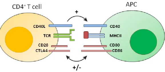
Fig. 1.2 APC activation upon encounter with an antigen-specific CD4

+

T cell. Interactions that control the outcome of CD4

+

T cell-APC encounter are indicated by the receptor-ligand pairs. CD40L, CD40 ligand; TCR, T-cell antigen receptor; CTLA4, cytotoxic T lymphocyte–associated protein 4; MHCII, major histocompatibility complex type II protein (Haanen and Schumacher 2007).

More importantly, CD40 engagement promotes B cell cytokine production, the expression of costimulatory molecules on their surface and facilitates cross-presentation of antigen (Robson et al. 2008, de Wit et al. 2010). Furthermore, CD40 signaling supports survival of germinal center B cells, DCs and endothelial cells (Bishop et al. 2007) and its deregulation has been observed to play a role in multiple autoimmunity diseases (Peters et al. 2009).

The importance of this signaling pathway in acquired immune responses makes it a promising target for pharmacological manipulations for the treatment of immunological diseases. Interference of the CD40-CD40L interaction by use of blocking CD40-antibodies showed positive effects on uncontrolled immune reactions like inflammation, autoimmunity or allo-graft rejection (Larsen et al. 1996, Kirk et al. 1997, Kirk et al. 2001, Pearson et al.

2002, Grammer et al. 2003, Sidiropoulos and Boumpas 2004). Moreover, enhancement of

the CD40-CD40L interaction by use of antagonistic CD40-antibodies was shown to result in

anti-proliferative anti-tumor effects or meditation of immune response against tumors

(Mackey et al. 1998, Diehl et al. 1999, Tong and Stone 2003, Watanabe et al. 2003,

Eliopoulos and Young 2004, Vonderheide et al. 2007). Because of their potential as APC

stimulators, CD40 agonists have been selected as one of the twelve immunotherapy drugs

that could cure cancer (Cheever 2008).

(37)

1.4.2 CD40-Activated B Cells

In 1991, Banchereau et al. discovered that long-term human B-cell lines could be established by stimulation with the CD40L and IL-4 (Banchereau et al. 1991, Banchereau and Rousset 1991). Since then several strategies have been developed to manipulate the CD40-CD40L interaction to activate B cells (Neron et al. 2011). Schultze et al. established a culture system, in which human B cells can be activated and expanded by stimulation with the CD40L and IL-4 (Schultze et al. 1997). Under constant cultivation on CD40L expressing feeder cells, human and murine primary B cells can be expanded from small amount of peripheral blood, upregulate costimulatory, MHC and adhesion molecules and show an increased antigen presenting capacity in vitro (Liebig et al. 2009, Liebig et al. 2010). The expression level of MHC and costimulatory molecules is comparable to those of CD40L/IFN-γ or TNF-α matured DCs (Wiesner et al. 2008). In vitro these CD40-activated B cells present antigen in an MHC class I or II- restricted manner to induce naïve or memory CD8

+

and CD4

+

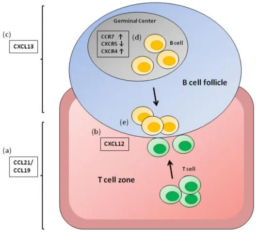
T cells responses, respectively (Schultze et al. 1997, von Bergwelt-Baildon et al. 2002, Lapointe et al. 2003, von Bergwelt-Baildon et al. 2004, Theurich et al. 2011) and overcome T cell tolerance as shown by generating autologous tumor antigen-specific T cells in vitro by help of peptide-pulsed CD40B cells (von Bergwelt-Baildon et al. 2002, Shen et al. 2007, Wu et al. 2010). Moreover, CD40B cells have the capacity to attract T cells and to home to secondary lymphoid organs (von Bergwelt-Baildon et al. 2006, Guo et al. 2009), which is essential for T cell-APC encounter and the induction of immunity. Human and murine CD40B cells express the respective factors and receptors crucial for homing to secondary lymphoid organs including CD62L, C-C-chemokine receptor (CCR) 7/ CXC-Motiv-Chemokinrezeptor (CXCR) 4, and leukocyte function antigen (von Bergwelt-Baildon et al. 2006, Klein-Gonzalez et al. accepted 2015). The receptors CCR7 and CXCR4 are functional and induce chemotaxis toward increasing chemokine concentrations of their ligands CCL21 and CXCL12, respectively (Fig.

1.3).

(38)

Introduction

20

Fig. 1.3. Chemokines involved in lymphocyte positioning in the secondary lymphoid organs.

(a) CCL21/ CCL19, chemokines expressed in the T cell zones of secondary lymphoid organs and the ligand for CCR7; (b) CXCL12, the ligand for CXCR4 mostly involved in T cell zone entry; (c) and CXCL13, a chemokine made in B cell zones of lymphoid organs and the ligand for CXCR5. (d) The chemokine expression pattern of CD40B cells (e) allows them to migrate towards the B/T cell boundary (Klein- Gonzalez et al. accepted 2015).

Nevertheless, very few studies have focused on the in vivo antigen-presenting capacity of CD40B cells and their contribution to anti-tumor immunity (Wennhold et al. 2013). In two preventive vaccination studies, tumor growth was delayed in mice that were immunized with CD40 B cells (Ritchie et al. 2004, Liebig et al. 2011). The group of Mason reported the first study in out-bred large animals using tumor RNA-loaded CD40B cells as cellular adjuvant in privately owned dogs with NHL (Sorenmo et al. 2011). However, the correlation between the immunological response and the clinical outcome was not significant.

Taken together, CD40B cells cannot only serve to identify tumor-antigens or to generate

T cells for adoptive immunotherapy, but show also promise as a versatile platform for

cellular cancer vaccines (Fujiwara et al. 2005, Ivanov et al. 2005, Kondo et al. 2009). Most of

all, the results from preclinical studies of application as antigen presenting cells in

(39)

preventive and therapeutic vaccination appears promising (von Bergwelt-Baildon et al. 2002, Coughlin et al. 2004, Van den Bosch et al. 2005, Yoon et al. 2005, Shen et al. 2008, Wu et al.

2010).

1.4.3 Antigen-Specific B Cells

Antigen-processing and -presentation, and thereby T cells activation, is 1000-10,000 fold more effective in antigen-specific B cells compared to resting or lipopolysaccharide (LPS)- activated polyclonal B cells (Abbas et al. 1985, Lanzavecchia 1990). The BCR has a high affinity for a given antigen and allows B cells to concentrate very small quantities of their specific antigen and present the antigen efficiently. In contrast, presentation after uptake by fluid phase pinocytosis in B cells requires about 5000 higher concentrations.

Specific antigen uptake via the BCR leads to three important changes in the antigen processing machinery that facilitates the traffic of antigen and MHC class II molecules and the generation of peptide-MHCII complexes (Rodriguez-Pinto 2005). First, BCR receptor ligation induces its internalization and the traffic towards MHCII rich compartments (MIIC), the site of peptide-MHCII complex formation. Second, BCR signaling upregulates MHCII expression and trafficking through the MIIC. Finally, BCR crosslinking induces changes in the MIIC to generate the adequate environment for loading of MHCII with peptides. A second characteristic of antigen processing in B cells differs from that in other antigen presenting cells. The expression of HLA-DO, a non-classical MHCII molecule that is expressed only in B cells and thymic epithelium, modulates peptide loading of MHCII molecules by HLA-DM and favoring presentation of peptides derived from antigens internalized through the BCR (Alfonso et al. 1999, Denzin et al. 2005).

1.5 Purpose of this Study

Although polyclonal CD40B cells were shown to efficiently induce anti-tumor immunity in

vivo, the tumor control is still suboptimal when using polyclonal CD40B cells as cellular

adjuvant (Liebig et al. 2011). Based on the fact that antigen-specific B cells present antigen

very efficiently when the antigen concentration is low, this study aimed at investigating

whether the use of tumorantigen-specific CD40B cells instead of polyclonal CD40B cells

improves their antigen-presenting function. For this purpose polyclonal and antigen-specific

(40)

Introduction

22

CD40B cells were compared with regard to their potential to induce antigen-specific T cell

reactions in vitro and in vivo. Moreover, the migratory behavior of tumorantigen-specific

CD40B cells in healthy and tumor bearing mice was investigated with the purpose of

exploiting their potential as drug delivery and imaging vehicle. With regards to the use of

antigen-specific B cells as cellular adjuvant for cancer immunotherapy, their antigen-

presenting function and antibody-secreting capacity were combined for immunotherapy in

tumor bearing mice.

(41)
(42)
(43)

2. Material and Methods 2.1 Material

2.1.1 Chemical

For preparation of buffers and solutions, chemicals of analytical purity were used exclusively.

Bovine Serum Albumin (BSA) Sigma Adrich

CFSE Invitrogen

Active hepatitis b virus Hepatitis B

Surface Antigen full length protein (HBV-antigen) Abcam Albumin from Chicken Egg White (OVA) Sigma Aldrich

Dimethyl sulfoxide (DMSO) Sigma Aldrich

D-Luciferin Regis Technologies

Ethanol Th Geyer

Ethylenediaminetetraacetic acid (EDTA) Sigma Aldrich

Hygromycin B Sigma Aldrich

Isofluran Actavis

Ovalbumin peptide (aa 257-264) Thermo Scientific

Paraformaldehyde (PFA) Sigma Aldrich

Sodium hydrogen carbonate (NaHCO

3

) Sigma Aldrich

Sodium carbonate (Na

2

CO

3

) Sigma Aldrich

Sodium chloride (NaCl) Sigma Aldrich

Trypsin-EDTA Life Technologies

Tween-20 Sigma Aldrich

Sodium Dodecyl Sulfate (SDS) Sigma Aldrich

2.1.2 Consumables

5 ml FACS tubes Sarstedt

6-well culture dish Sarstedt

12-well culture dish Sarstedt

15 ml Falcon tube Sarstedt

26G ½ needle Beckton Dickinson

50 ml Falcon tube Sarstedt

75 cm

2

culture flask (adherent) Sarstedt

75 cm

2

culture flask (suspension) Sarstedt 96-well culture dish, round bottom Sarstedt

96-well high binding plate Greiner

100 µm cell strainer Greiner

Cryo Tubes Sarstedt

Glass object slides Sarstedt

MS Columns Miltenyi Biotech

LD Columns Miltenyi Biotech

LS Columns Miltenyi Biotech

Slide-A-Slizer G2 Dialysis Cassette Thermo Scientific

(44)

Material and Methods

26 2.1.3 Kits

EasySep Mouse T Cell Enrichment Kit Stem Cell Technologies EasySep Mouse Biotin Selection Kit Stem Cell Technologies Pierce Biotin Quantitation Kit Thermo Scientific 2.1.4 Devices

EOOS 350D digital camera Canon

Gallios Flow Cytometer Beckman Coulter

HeraCell 150i CO2 Incubator Thermo Scientific

Heraeus Megafuge 16R Thermo Scientific

HeraSafe hood ThermoScientific

Microplate Reader µQuant BIO-TEK Instruments

Neubauer Counting Chamber OptikLabor

Silk-epil FG1100 Braun

Televal 31 microscope Zeiss

Vernier Caliper Hogotex

Water bath WNB14 Set Memmert

Xenogen IVIS 200 Perkin Elmer

2.1.5 Cell Culture

If not described differently, pipette tips were obtained from BD Falcon.

2.1.5.1 Cell Culture Reagents

10 x Phosphate buffered saline (PBS) Life Technologies

AB-Human serum Biochrom

AIM-V medium Life Technologies

Anti-mouse CD40Ligand (CD40L) antibody Novus Biologicals Anti-mouse IgM(mu-chain), unconjugated Sigma Aldrich

β-Mercaptoethanol (β-ME) Sigma Adrich

CpG ODN 2395 Miltenyi Biotech

Cyclosporin A Sigma Adrich

DMEM medium Life Technologies

Fetal Bovine Serum (FBS) Lonza

G418 Biochrom

GM-CSF Immunotools

HEPES Life Technologies

IMDM medium Life Technologies

Incomplete Freund’s Adjuvant Sigma Aldrich

LPS from Escherichia coli 055:B5 Sigma Aldrich

MEM Life Technologies

Penicillin/ Streptomycin Life Technologies

rh Insulin Novo Nordisk

rh Interleukin-4 Immunotools

rh Transferrin Sigma Aldrich

(45)

rm Interleukin-4 Immunotools

rm Interleukin-21 Immunotools

RPMI 1640 medium Life Technologies

Trypan Blue Stain 0.4 % Life Technologies

VLE-RPMI medium Biochrom

2.1.5.2 Media

DC medium VLE-RPMI medium

+ 5 % FBS + 50 µM β-ME + 10 % Pen/Strep

Freezing medium FBS + 10 % DMSO

HeLa standard medium RPMI 1640

+ 300 µg/ml L-Glutamine + 10 % FBS

+ 10 mM HEPES + 10 % Pen/Strep

HeLa selection medium RPMI 1640

+ 300 µg/ml L-Glutamine + 10 % FBS

+ 10 mM HEPES + 10 % Pen/Strep

+ 0.2 mg/ml Hygromycin B

Human CD40B medium IMDM

+ 584 µg/ml L-Glutamine + 25 mM HEPES

+ 10 % AB-Human serum + 50 µg/ml rh Transferrin + 5 µg/ml rh Inuslin + 10 % Pen/Strep

Murine CD40B medium DMEM

+ 580 µg/ml L-Glutamine + 4.5 mg/ml Glucose + 10 % FBS

+ 10 mM HEPES + 0.1 mM MEM + 10 % Pen/Strep

NIH standard medium DMEM-Ham’s/ F12

+ 365 µg/ml L-Glutamine + 10 % FBS

+ 10 mM HEPES + 10 % Pen/Strep

Tumor medium DMEM

+ 580 µg/ml L-Glutamine + 10 % FBS

+ 10 mM HEPES

+ 10 % Pen/Strep

(46)

Material and Methods

28 2.1.6 Reagents

ABTS Peroxidase Substrate KPL

Biotin anti-mouse IgG1 Biolegend

CD19 MicroBeads human Miltenyi Biotech

CD19 MicroBeads mouse Miltenyi Biotech

Cell wash Beckton Dickinson

EZ-Link NHS-Biotin Reagent Thermo Scientific

HRP Avidin Biolegend

Pancoll Human (density 1.077 g/l) PAN Biotech Pancoll Mouse (density 1.086 g/l) PAN Biotech

Purified anti-chicken Ovalbumin Biolegend

2.1.7 Solutions and Buffers

1x PBS 1 L 10x PBS, 9 L Ampuwa

Blocking solution 1x PBS, 10 % FBS

Coating buffer 8.4 g NaHCO

3

, 3.56 g Na

2

CO

3

, add

H

2

O up to 1.0 L, pH to 9.5

EasySep medium 1x PBS, 2 % FBS, 1 mM EDTA

MACS buffer 1x PBA, 0.5 % BSA, 2 mM EDTA

PBS/Tween 1 L 1x PBS, 0.5 ml Tween-20

2.1.8 Software

FlowJo Software TreeStar

GraphPad Prism GraphPadPrism

Kaluza Software Beckman Coulter

Living Image Software Perkin Elmer

2.2 Methods

2.2.1 Cell Subset Enrichment

2.2.1.1 Purification of Murine Lymphocytes from Spleen

Spleens were removed from 7-12 week old mice. A single suspension was prepared by

squeezing the tissue through a 100 µm cell strainer. For purification of murine lymphocytes

density-gradient centrifugation was performed. Cells of two spleens were resuspended in 8

ml murine CD40B medium and added onto a layer of 5 ml mouse Pancoll separation

medium. Cells were centrifuged at 1080 x g for 15 min without break. Afterwards, the

interphase, containing the splenocytes (lymphocytes from the spleen), was carefully

collected with a 20 G needle and cells were washed with 10 ml PBS.

Abbildung

Fig. 1.2 APC activation upon encounter with an antigen-specific CD4 + T cell. Interactions that control the outcome of CD4 + T cell-APC encounter are indicated by the receptor-ligand pairs
Fig. 1.3. Chemokines involved in lymphocyte positioning in the secondary lymphoid organs.
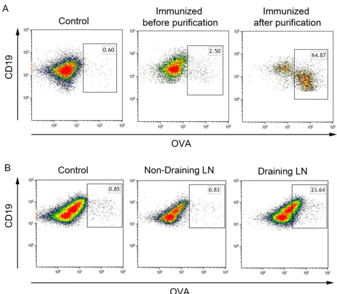
Fig. 3.2 Percentage of OVA-specific B cells. (A) Splenocytes from mice were stained for OVA-specific B cells among the CD19 + B220 + B cell population with OVA-Biotin tetramers
Fig. 3.3 B cell purity of OVA-enriched B cells.
+7

Referenzen

ÄHNLICHE DOKUMENTE

RESULTS: We show for the first time that EC present a quantitatively different peptide repertoire with abundance of certain peptides, compared with leukocytes. The abundance of

Among the CD1 family, CD1e is the only soluble protein, thus supporting a chaperone-like rather than an antigen-presenting function. Furthermore, CD1e is never

and the enormous sensitivity of chromium release Measuring activation of hybridomas started at 10 27 – assays are not ideal prerequisites for monitoring the 10 28 M concentrations

We did not detect secretion of suppressive cytokines (such as IL-4, IL- 10 or TGF-~) from CDS' T cells primed by LSEC that could ex- plain the Joss of specific T-cell

Minami  et  al.  not  only  found  BAG6  associated  with  chemically  induced  DRiPs,  they 

To analyze the influence of BAG6 on MHC class I surface expression, the murine fibroblast cell line B8-D b (H- 2 d + H-2D b ) and HeLa cells were subjected to BAG6

To exclude that the reduced number of virus-specific T cells in Bis VIII–treated mice at day 7 after viral infection was due to inhibition of T cell activation and reduced expansion,

and our phase I trial in cervical cancer patients [25], has demonstrated the efficacy of autologous tumor lysate pulsed DCs (TLDCs), as a next step after optimizing the rhSPAG9