• Keine Ergebnisse gefunden

Molecular and functional studies on the transcript elongation factor FACT and the SAGA-DUBm subunit ENY2 in Arabidopsis

N/A
N/A
Protected

Academic year: 2021

Aktie "Molecular and functional studies on the transcript elongation factor FACT and the SAGA-DUBm subunit ENY2 in Arabidopsis"

Copied!
228
0
0

Wird geladen.... (Jetzt Volltext ansehen)

Volltext

(1)

Molecular and functional studies on the transcript elongation factor FACT and the SAGA-DUBm subunit ENY2 in Arabidopsis

thaliana

DISSERTATION ZUR ERLANGUNG DES DOKTORGRADES DER NATURWISSENSCHAFTEN (DR. RER. NAT.)

DER FAKULTÄT FÜR BIOLOGIE UND VORKLINISCHE MEDIZIN DER UNIVERSITÄT REGENSBURG

vorgelegt von ALEXANDER PFAB

aus

MÜNCHEN

(2)

Der Promotionsgesuch wurde eingereicht am:

17.11.2017

Die Arbeit wurde angeleitet von:

PROF. DR. KLAUS D GRASSER Unterschrift:

ALEXANDER PFAB

ii

(3)

MR.ROBOT

Für Silli, Jari und Loki

(4)
(5)

Contents

Abbreviations 1

1 Introduction 3

1.1 The chromatin structure . . . . 3

1.2 Transcription by RNA polymerase II . . . . 3

1.2.1 Transcript elongation . . . . 4

1.3 The histone chaperone FACT . . . . 6

1.3.1 The FACT architecture . . . . 6

1.3.2 The FACT mechanism . . . . 7

1.3.3 The FACT complex in Arabidopsis . . . . 8

1.4 Co-transcriptional processing of mRNA . . . . 9

1.5 Coupling of the RNAPII and mRNA processing factors . . . . 9

1.6 The coupling factor Sus1/ENY2 . . . . 10

1.7 The transcriptional co-activator SAGA . . . . 11

1.8 Aims of the thesis . . . . 13

1.8.1 The histone chaperone FACT . . . . 13

1.8.2 ENY2, a coupling factor of transcription and mRNA processing . . . . 14

2 Results: Analysis of the HMG-box domain of Arabidopsis SSRP1 15 2.1 Arabidopsis SSRP1 features a conserved C-terminal HMG-box domain . . . . 15

2.2 Truncated SSRP14HMG showed reduced DNA- and nucleosome-binding . . . . 16

2.3 Full-length SSRP1 and truncated SSRP14HMG showed similar protein dynamics 18 2.4 HMG-box-deficient SSRP1 interacted with SPT16 and the transcriptionally active RNAPII . . . . 21

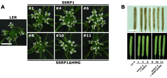
2.5 SSRP1 HMG-box domain is not important for proper plant development . . . . . 22

2.5.1 SSRP1 HMG-box deficiency mutants . . . . 23

2.5.2 Phenotypic analysis of SSRP1 HMG-box deficiency mutants . . . . 25

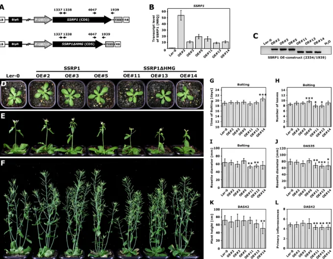
2.6 Overexpression of SSRP1 and SSRP14HMG had no dominant negative effect on plant development . . . . 27

3 Results: The role of FACT in anthocyanin biosynthesis 29 3.1 Transcript profiling of ssrp1-2 and spt16-1 mutants . . . . 29

3.2 GO Analysis revealed that flavonoid biosynthesis genes were downregulated in FACT mutants . . . . 30

3.3 FACT-depleted plants showed defects in the light-induced accumulation of antho- cyanin . . . . 32

3.4 FACT was required for light-induced upregulation of anthocyanin biosynthesis genes . . . . 34

3.5 SSRP1 and SPT16 gene expression was strongly upregulated in response to high light stress . . . . 36

3.6 Proanthocyanin synthesis was not impaired in seeds of FACT mutants . . . . 38

4 Results: The composition of the Arabidopsis transcript elongation complex 41 5 Results: The Arabidopsis SAGA-DUBm component ENY2 43 5.1 ENY2 gene expression in Arabidopsis thaliana . . . . 43

5.2 Localization of eGFP-ENY2 fusion protein in Arabidopsis thaliana . . . . 45

5.2.1 Verification of eGFP-ENY2 fusion protein integrity . . . . 46

5.2.2 In Arabidopsis roots, eGFP-ENY2 is localized in the nuclei . . . . 46

5.2.3 In the nucleoplasm, eGFP-ENY2 is forming speckle-like structures . . . . 47

5.2.4 High mobility of eGFP-ENY2 in the nucleoplasm . . . . 48

(6)

5.3 Affinity purification coupled to mass spectrometry to identify the interaction net-

work of ENY2 in plants . . . . 49

5.4 The SAGA-DUB module in plants . . . . 56

5.4.1 Y2H revealed PPIs between SGF11 and ENY2 as well as SGF11 and UBP22 56 5.4.2 FRET revealed PPIs between SGF11 and ENY2 as well as SGF11 and UBP22 . . . . 57

5.4.3 DUB module components are highly conserved . . . . 59

5.4.4 Homology modelling of Arabidopsis DUB components . . . . 61

5.5 Interaction of ENY2 with the NTC complex and splicing . . . . 64

5.5.1 No direct PPI between ENY2 and NTC/NTR components was detected by Y2H . . . . 65

5.5.2 No direct PPI between ENY2 and NTC/NTR components was detected by FRET . . . . 65

5.5.3 ENY2 and the NTC component MOS4 co-localized in splicing-spleckles in the nucleoplasm . . . . 66

5.6 The SAGA complex and SAGA-DUB module in plants . . . . 67

5.6.1 AP-MS analysis of the Arabidopsis SAGA complex . . . . 67

5.7 Investigation of SAGA and SAGA-DUBm by AP-Superose6-Western Analysis . . 73

5.8 Characterization of plants with altered expression of ENY2 or SGF11 . . . . 76

5.8.1 eny2-1 T-DNA line showed no downregulation of ENY2 . . . . 76

5.8.2 ENY2 knockdown by RNAi showed no obvious phenotype . . . . 76

5.8.3 CRISPR/Cas9-induced mutations to disrupt ENY2 . . . . 79

5.8.4 ENY2 overexpression by the UBI10 promoter . . . . 83

5.8.5 Knockdown of SGF11 showed late flowering phenoytpe . . . . 83

5.8.5.1 The level of FLC is upregulated in sgf11-1 knockout plants . . . 86

5.9 Global H2Bub levels were increased in eny2-RNAi and sgf11-1 mutants . . . . . 87

6 Discussion: Analysis of the HMG-box domain of Arabidopsis SSRP1 89 6.1 The SSRP1 HMG-box domain is important for FACT – nucleosome interaction . 89 6.2 Loss of the HMG-box domain did not alter the nuclear localization of SSRP1 . . 90

6.3 Loss of the HMG-box domain did not alter the SSRP1 – chromatin binding prop- erties in vivo . . . . 90

6.4 HMG-box-deficient SSRP1 was still part of the active transcript elongation com- plex in vivo . . . . 91

6.5 The HMG-box domain of SSRP1 was not important for proper plant development 91 6.6 Other HMGB proteins might compensate the loss of the SSRP1 HMG-box domain in vivo . . . . 94

6.7 Outlook . . . . 94

7 Discussion: The role of the FACT complex in plant anthocyanin biosynthesis 97 7.1 A small subset of genes is differentially expressed in FACT-depleted plants . . . . 97

7.2 Genes are commonly differentially expressed in SSRP1/SPT16-depleted plants . 99 7.3 Iron homeostasis genes are downregulated in FACT mutants . . . . 99

7.4 Circadian clock genes are differentially expressed in FACT mutants . . . 100

7.5 Outlook . . . 101

8 Discussion: The Arabidopsis SAGA-DUBm component ENY2 103 8.1 The expression of ENY2 in Arabidopsis thaliana . . . 103

8.2 The interaction network of ENY2 in Arabidopsis thaliana . . . 103

8.2.1 Old friends: ENY2 and the DUB module of the transcriptional co-activator SAGA . . . 104

vi

(7)

8.2.1.1 Composition and assembly of the putative plant DUB Module is

conserved . . . 104

8.2.1.2 De-ubiquitinating activity of the plant DUB module . . . 105

8.2.2 The composition of the Arabidopsis SAGA complex . . . 105

8.2.3 The evolution of the Arabidopsis SAGA complex . . . 106

8.2.4 The SAGA complex versus the TFIID complex . . . 107

8.2.5 Permanent relationship or temporary liaison? The Arabidopsis SAGA complex and its DUB module . . . 109

8.2.6 The Arabidopsis SAGA complex during transcription by RNAPII . . . 110

8.2.7 No friends anymore: ENY2 and the mRNA export complex TREX-2 . . . 111

8.2.8 New friends: The SAGA – Splicing association . . . 112

8.3 Reverse genetics to learn more about the function of the plant DUB module . . . 115

8.3.1 De-ubiquitination activity of the plant DUB module plays a role in flow- ering time control . . . 116

8.3.2 ENY2, a novel link between histone de-ubiquitination and splicing to con- trol flowering time? . . . 117

8.4 Outlook . . . 117

9 Summary 119 10 Materials 123 10.1 Instruments . . . 123

10.2 Chemicals and enzymes . . . 123

10.3 Oligonucleotides . . . 123

10.4 Plasmids . . . 128

10.5 Organisms . . . 130

10.6 Databases, Online Tools, Software . . . 130

11 Methods 131 11.1 Nucleic acid based methods . . . 131

11.1.1 Isolation of genomic DNA from Arabidopsis leaves . . . 131

11.1.2 Isolation of RNA from Arabidopsis leaves . . . 131

11.1.3 Reverse Transcription (cDNA synthesis) . . . 131

11.1.4 Polymerase chain reaction (PCR) . . . 131

11.1.5 Real time quantitative PCR (qRT-PCR) . . . 132

11.1.6 Restriction-Ligation-based Cloning . . . 132

11.1.6.1 Agarose gel electrophoresis . . . 132

11.1.6.2 DNA extraction from agarose gels and PCR clean up . . . 133

11.1.6.3 Phosphorylation and Annealing of DNA-Oligos . . . 133

11.1.6.4 Restriction digestion, Dephosphorylation and Klenow fragment . 133 11.1.6.5 Ligation . . . 133

11.1.6.6 Isolation of plasmid DNA from E. coli . . . 133

11.1.6.7 Sequencing . . . 134

11.2 Protein based methods . . . 134

11.2.1 Protein Extraction from Arabidopsis flowers . . . 134

11.2.2 Protein purification . . . 134

11.2.2.1 Affinity Purification . . . 134

11.2.2.2 Size-exclusion chromatography . . . 136

11.2.2.3 Ion-Exchange chromatography . . . 136

11.2.2.4 Dialysis . . . 136

11.2.2.5 Acetone precipitation . . . 137

11.2.3 Protein detection (non-specific) . . . 137

(8)

11.2.3.1 Bradford Assay . . . 137

11.2.3.2 SDS-PAGE . . . 137

11.2.3.3 Coomassie Brilliant Blue (CBB) staining . . . 137

11.2.3.4 Ponceau staining . . . 138

11.2.4 Protein detection (specific) . . . 138

11.2.4.1 Western blotting . . . 138

11.2.4.2 Trypsin digestion and Mass spectrometry . . . 138

11.2.5 Protein-DNA interactions by EMSA . . . 139

11.3 Cell based methods . . . 140

11.3.1 Cultivation of bacteria . . . 140

11.3.1.1 Preparation of chemically competent cells . . . 140

11.3.1.2 Transformation by heat shock . . . 140

11.3.2 Protein expression in E. coli . . . 141

11.3.3 Preparation of chemically competent yeast cells . . . 141

11.3.4 Co-transformation of yeast cells by heat shock . . . 141

11.3.5 Yeast-2-Hybrid Assay . . . 142

11.4 Plant based methods . . . 142

11.4.1 Cultivation of Arabidopsis plants . . . 142

11.4.1.1 Transformation of Arabidopsis plants by floral dipping . . . 143

11.4.1.2 Crossing of Arabidopsis plants . . . 143

11.4.1.3 Soil-based phenotypic analysis . . . 143

11.4.1.4 GUS staining and clearing of roots . . . 143

11.4.1.5 Determination of anthocyanin content in Arabidopsis plants . . . 144

11.4.2 Cultivation of Arabidopsis PSB-D cells . . . 144

11.4.3 Cryopreservation and re-initiation of Arabidopsis PSB-D cells . . . 144

11.4.4 Transformation of Arabidopsis PSB-D cells . . . 145

11.4.5 Upscaling of transformed Arabidopsis PSB-D cells . . . 145

11.4.5.1 Tobacco Infiltration . . . 146

11.5 Microscopy . . . 146

11.5.1 Confocal Laser Scanning Microscopy . . . 146

11.5.1.1 Fluorescence recovery after photobleaching (FRAP) . . . 146

11.5.1.2 Förster resonance energy transfer (FRET) . . . 146

11.5.2 Cell stainings . . . 147

11.5.2.1 DAPI . . . 147

11.5.2.2 Propidiumiodid . . . 147

12 Supplements 149 12.1 Phenotypic data . . . 159

12.2 Microarray data . . . 161

12.3 Mass spectrometry . . . 163

13 Publications 175 Manuscript: The Arabidopsis histone chaperone FACT is required for stress-induced expression of anthocyanin biosynthetic genes . . . 176

Acknowledgements 197

List of figures 198

List of tables 202

Bibliography 204

viii

(9)
(10)
(11)

Abbreviations

(v/v) Volume per volume (w/v) Weight per volume

aa Amino acid

AB Antibody

ADA2b Transcriptional adapter 2 b

Ade Adenine

ANS Anthocyanidin synthase

AP-MS Affinity purification coupled to mass spectrometry At Arabidopsis thaliana

bp base pair

CBB Coomassie brilliant blue cDNA complementary DNA CDS Coding sequence CHS Chalcone synthase

CLSM Confacal laser scanning microscopy Col-0 Columbia-0

CTD C-terminal domain Cy3, Cy5 Cyanine dye 3, 5

DAPI 4’,6-Diamidin-2-phenylindol DAS Days after stratification DDO Double drop out medium DFR Dihydroflavonol reductase DNA Deoxyribonucleic acid dNTP Deoxynucleoside triphosphate Ds Drosophila melanogaster DTT Di-thiotreitol

DUBm Deubiquitinating module E. coli Escherichia coli

e. g. For example, latin: exempli gratia EDTA Ethylene diamine tetraacetic acid eGFP enhanced green fluorescent protein EMSA Electrophoretic mobility shift assay ENY2 Enhancer of yellow 2

F3’H Flavonone 3’hydroxylase

FACT Facilitates chromatin transcription FLC Flowering locus C

FPLC Fast performance liquid chromatography FRAP Fluorescence recovery after photobleaching FRET Förster resonance energy transfer

gDNA Genomic DNA

GO Gene ontology

GST Glutathione S-transferase

GUS β-glucuronidase

HAT Histone acetyltransferase His Histidine

HL High light

HMG High-mobility group Hs Homo sapiens

HUB1 Histone monoubiquitination 1

(12)

0 Abbreviations

HYG Hygromycin B

IgG Immunglobulin G

IPTG Isopropyl β-D-1-thiogalactopyranoside

Kan Kanamycin

kDa kilo Dalton

LB Left border OR Luria Bertani

LD Long day

Ler Landsberg erecta

Leu Leucine

LUT Lookup-table

MOS4 Modifier of snc1, 4

mRNA Messenger RNA

mRNP Messenger ribonucleoprotein

MS Murashige-Skoog OR mass spectroscopy MSA Multiple sequence alignment

N. benthamiana Nicotiana benthamiana

NASC Nottingham Arabidopsis stock center NLS Nuclear localization signal

NPC Nuclear pore complex

nptII Neomycin phosphotransferase II (Kanamycin resistance gene)

NTC Nineteen complex

NTR Nineteen complex-related proteins NUPs Nucleoporin proteins

o/n Over night

OD600 Optical density measured at 600 nm PAGE Polyacrylamide gel electrophoresis PCR Polymerase chain reaction PIC Pre-initiation complex

PMSF Phenylmethylsulphonyl fluoride

P-TEFb Positive transcription elongation factor b

QDO Quadruple drop out medium

qRT-PCR Quantitative reverse transcription-polymerase chain reaction

RB Right border

RNAPII RNA polymerase II

RNA Ribonucleic acid

RNAi RNA interference

ROI Region of interest

Rpm Rounds per minute

S. cerevisiae Saccheromyces cerevisiae SAGA Spt-Ada-Gcn5 acetyltransferase SAIL Syngenta arabidopsis insertion library SBP Streptavidin binding peptide

Sc Saccharomyces cerevisiae

SDS-PAGE Sodium dodecyl sulfate polyacrylamide gel electrophoresis Ser2 Serine 2 phosporylated CTD

SG Tag consisting of SBP and 2 x Protein G domains SGF11 SAGA-associated factor 11, DUBm subunit SPT16 Suppressor of Ty 16, FACT subunit

SSRP1 Structure specific recognition protein 1, FACT subunit

TAF10 TATA-box binding protein associated factor 10, SAGA component TAF13 TATA-box binding protein associated factor 13, SAGA component

T-DNA Transfer DNA

TDO Triple drop out medium

TEC Transcript elongation complex TEF Transcription elongation factor

Ter Terminator

TEV Tobacco Etch Virus (TEV) protease

TF Transcription factor

THP1 THO2/HPR1 phenotype 1, TREX-2 subunit TREX-2 Transcription and export complex 2

Trp Tryptophan

UBP22 Ubiquitin carboxyl-terminal hydrolase 22, DUBm subunit

UTR Untranslated region

WT Wild type

Y2H Yeast-two-Hybrid

| 2

(13)

1 Introduction

1.1 The chromatin structure

In the nuclei of eukaryotic cells, the genomic DNA is compacted into chromatin. Nucleo- somes, the building blocks of the chromatin, consist of a histone octamer (two copies of each histones H2A, H2B, H3, H4) with 147 bps of genomic DNA wrapped around. The nucleosomes are connected by the linker DNA into nucleosomal arrays like "beads on a string" that are or- ganized into higher order structures (Luger et al. 1997, Andrews et al. 2011). The chromatin structure does not only package the entire genome into the nucleus, but as repressive barrier it has also profound regulatory implications on all DNA-depending processes including transcrip- tion, replication or repair (Li et al. 2007a, Luger et al. 2012) To make the DNA accessible and to facilitate transcription in the chromatin context, the nucleosomes must be moved, partially disassembled or modified. During transcription, this histone exchange is managed by numer- ous factors including energy-dependent chromatin remodellers, histone chaperones and enzymes that add post-translational modifications (PTMs) to the N-terminal tails of histones (Venkatesh et al. 2015). Histone modifications including acetylation, methylation, phosphorylation, and ubiquitylation do not only change the physical and chemical properties of chromatin, but are also important for recruiting other histone modifiers (Saunders et al. 2006).

1.2 Transcription by RNA polymerase II

DNA templates are transcribed into mRNA molecules by the RNA polymerase II (RNAPII) with the help of several auxiliary factors. The process of transcription is divided into three regulated phases known as initiation, elongation and termination (Figure 1.1). The repetition of these three phases (transcription cycle) over a gene determines its expression levels (Jonkers et al. 2015, Venkatesh et al. 2015). The first step of the transcription cycle starts with the assembly of the pre-initiation complex (PIC) at the promoter of a gene. The PIC consists of the RNAPII and the general transcription factors (GTFs) TFIID, TFIIB, TFIIE, TFIIF and TFIIH. The GTFs recruit and position the RNAPII near the transcription start site and dictate the precise location and direction of the transcription initiation (Sims et al. 2004, Saunders et al.

2006). For regulated transcription, general co-factors like the Mediator or the Spt-Ada-Gcn5

acetyltransferase (SAGA) complex are often required to transmit regulatory signals between

gene-specific activators and the general transcription machinery (Thomas et al. 2006). To ini-

tiate RNA synthesis, the double stranded DNA is melted and the single stranded template is

positioned in the active site of the RNAPII to form the open complex. In the following, the

transition from transcript initiation to elongation is accompanied by structural and functional

changes of the RNAPII including the phosphorylation of the C-terminal domain (CTD) of its

largest subunit NRPB1. The RNAPII loses contact to the GTFs and moves along the template

strand to synthesize the RNA transcript by joining nucleotides that are complement with the

DNA template (Sims et al. 2004, Saunders et al. 2006). In some eukaryotes, the RNAPII can

pause and accumulate at promoter-proximal regions during the initial steps of transcript elon-

gation before productive elongation starts (Jonkers et al. 2015). During the highly regulated

step of transcript elongation, several factors are recruited by the phosphorylated CTD that

are important for efficient mRNA synthesis and co-transcriptional mRNA processing. Finally,

(14)

1 Introduction

Figure 1.1 RNAPII transcription cycle

The transcription by RNAPII is characterized by a cycle of events that starts with the recruitment of the polymerase and the formation of the closed pre-initiation complex (RNAPII with GTFs) at the promoter of a gene. By melting the DNA of the core promoter, an open RNAPII complex is formed to initiate transcription. During the transition from transcript initiation to elongation, the RNAPII undergoes structural and functional changes that are collectively comprised as promoter clearance. Thereby, the RNAPII loses contact to the GTFs and establishes a stable association with the nascent transcript. Approximately 30 nucleotides downstream of the transcription start-site, promoter clearance is complete and the RNAPII becomes engaged in productive elongation. Thereby, a variety of TEFs ensures the efficient mRNA synthesis on chromatin templates. To regulate transcription, the RNAPII can pause and accumulate in some eukaryotes during the initial steps of transcript elongation (usually 20-60 nucleotides downstream of the transcription start-site). The progression of the transcription machinery is accompanied by changes in the phosphorylation pattern of the C-terminal heptapeptide repeats of the largest RNAPII subunit. For instance, the phosphorylation of serine 5 and serine 2 are characteristic for early and later transcript elongation, respectively. Thereby, the phosphorylation of the RNAPII CTD plays an important role in the coordination of the ongoing mRNA synthesis with co-transcriptional mRNA processing events. During termination, the produced transcript is released from the RNAPII. Subsequently, the polymerase dissociates from the DNA and is recycled for the next round of the transcription cycle. The early stages of the transcription cycle (depicted in blue) are well investigated, whereas much less is known about the later stages (dipected in green) that are as well dynamic and highly regulated. The figure is taken from Van Lijsebettens et al.

2014.

transcription is terminated and the transcribed RNA is released from the RNAPII for further processing and export to the cytoplasm. The transcription machinery dissociates from the DNA and is recycled for the next round of mRNA synthesis (Saunders et al. 2006, Selth et al. 2010).

1.2.1 Transcript elongation

Traditionally, the phase of transcript initiation with the recruitment of the RNAPII to the promoter was assumed as the most crucial step in the regulation of mRNA biogenesis. In contrast, during the subsequent phase of transcript elongation, the RNAPII was considered to simply behave like a machine that is quickly "reading the gene". In the recent years, it became more and more apparent that the transcript elongation is as well a dynamic and highly regulated process. Moreover, centrally located in the gene expression pathway, it coordinates the synthesis of mRNA with its maturation and export (Sims et al. 2004, Saunders et al. 2006, Selth et al.

2010, Jonkers et al. 2015).

In the early stage of transcription, the eukaryotic RNAPII exchanges the associated tran- script initiation factors like GTFs with transcript elongation factors (TEFs) to form a transcript

| 4

(15)

Introduction 1

Figure 1.2 A variety of transcript elongation factors (TEFs) enables the efficient transcription of chromatin templates by RNAPII.

TEFs are a heterogenous group of proteins that serve diverse functions during transcription by RNAPII. Factors that directly modulate RNAPII properties, for instance, to allow the polymerase to counteract transcriptional pausing/arrest are depicted in yellow. Histone chaperone (depicted in blue) and ATP-dependent chromatin remodelers (depicted in orange) play important roles to facilitate transcription in the chromatin context. Additional factors like the enzymes depicted in green, can control transcription by modifying histones covalently within transcribed regions. The modifica- tions (methylation, acetylation, and ubiquitination) can be removed by the reverse activity of other enzymes (indicated by double-headed arrows). Some activating marks like H3K4me3, H3K36me3, H3/H4ac, H2Bub are shown. The figure is taken from Van Lijsebettens et al. 2014.

elongation complex (TEC) for processive mRNA synthesis (Ehara et al. 2017). A variety of different TEFs has been identified in the last years (Figure 1.2). These TEFs can be associated with the TEC permanently or just transiently and can therefore affect the global expression or just the expression of a subset of genes. TEFs are a very heterogenous group of proteins that serve diverse functions to facilitate the efficient progression of the transcription machinery through the repressive chromatin. Thereby TEFs can directly modulate the catalytic properties of the processive RNAPII, can modify nucleosomal histones within the transcribed region or can function as histone chaperones and chromatin remodelers (Sims et al. 2004, Selth et al. 2010, Van Lijsebettens et al. 2014). Moreover, the TEC functions as platform for co-transcriptional nuclear transactions including pre-mRNA processing (Perales et al. 2009).

Recently, the structure of the yeast RNAPII transcript elongation complex was elucidated by X-ray crystallography and cryo-EM (Xu et al. 2017a, Ehara et al. 2017). Beside the assembly of the TEC, several studies in yeast revealed genetic/biochemical interactions between various TEFs and tracked the TEC along transcribed regions (Selth et al. 2010, Jonkers et al. 2015). In Arabidopsis and in higher eukaryotes, considerably less is known about the composition of the RNAPII TEC and the interplay of the TEFs among each other and the transcribing RNAPII.

Genetic and biochemical studies in Arabidopsis revealed that TEFs can regulate plant growth

and development. For instance, the histone chaperone FACILITATES CHROMATIN TRAN-

SCRIPTION (FACT) assists the progression of the transcribing RNA polymerase on chromatin

templates by destabilizing nucleosomes (Van Lijsebettens et al. 2014). The Arabidopsis FACT

complex was shown to be necessary for the expression of the floral repressor FLOWERING

LOCUS C (FLC) and thereby for the transition from the vegetative to the reproductive phase

(16)

1 Introduction

(flowering) (Lolas et al. 2010). The POLYMERASE-ASSOCIATED FACTOR 1 (PAF1) COM- PLEX (PAF1-C) has been shown to regulate transcription by promoting specific histone modi- fications including the methylation of lysine residues and the monoubiquitylation of H2B K123 (Tomson et al. 2013). The Arabidopsis PAF1-C controls the flowering time by modifying the chromatin of floral repressors like FLC (He et al. 2004, Oh et al. 2004). The Arabidopsis POSI- TIVE TRANSCRIPTION ELONGATION FACTOR b (P-TEFb) complex can as well regulate the expression level of FLC and affect flowering time. Therefore, P-TEFb that consists of CDKC;2 and CYCT1 influences the phosphorylation status of the Ser2 residue of the RNAPII C-Terminal repeats (CTD) globally (Wang et al. 2014). The TRANSCRIPTION FACTOR S-II (TFIIS) promotes efficient transcription by RNAPII, since it assists in bypassing blocks during mRNA synthesis (Fish et al. 2002). Thereby TFIIS controls the Arabidopsis seed dormancy by regulating the expression of DELAY OF GERMINATION1 (DOG1 ) (Grasser et al. 2009, Mortensen et al. 2014). The SUPPRESSOR OF TY 4/5 (SPT4/5) complex directly interacts with the RNAPII to couple chromatin modification states and RNA processing for processive transcript elongation (Hartzog et al. 2013). In Arabidopsis, SPT4/5 regulates transcript elonga- tion of RNAPII with particular impact on the expression of certain auxin-related genes (Dürr et al. 2014).

1.3 The histone chaperone FACT 1.3.1 The FACT architecture

Figure 1.3 Conserved domain organization and structural alignment of the FACT subunits.

A) Schematic illustration of the conserved domain organization of human, yeast and Arabidopsis FACT subunits. SPT16 is highly conserved from yeast to humans, while the C-terminus of SSRP1/Pob3 shows species-specific characteristics.

CID = C-terminal intrinsically disordered domain, CTD = C-terminal domain, DD = dimerization domain, HMG = high mobility group domain, IDD = intrinsically disordered domain, MD = middle domain, NTD = N-terminal domain.

The figure is taken from Zhou et al. 2015. B) Dimerization of both FACT subunits is accomplished through specific interactions between the centrally and the N-terminally located DDs of Spt16 and SSRP1, respectively. The figure is taken from Winkler et al. 2011a.

The histone chaperone FACT is evolutionarily conserved in eukaryotes including the plant kingdom (Formosa 2008, Lolas et al. 2010). The heterodimer FACT consists of the SUPPRE- SOR OF TY 16 (SPT16) and the STRUCTURE-SPECIFIC RECOGNITION PROTEIN 1 (SSRP1) in metazoans and plants or Spt16 and Pob3 in yeast and fungi (Orphanides et al, 1998). SPT16/Spt16 is highly conserved from yeast to higher eukaryotes, whereas SSRP1/Pob3 shows a more varied degree of sequence conservation and domain organization (Zhou et al. 2015) as shown in Figure 1.3. Most strikingly, the metazoan/plant SSRP1 features a high mobility

| 6

(17)

Introduction 1

group (HMG)-box domain at the C-terminus which is missing in the yeast Pob3. In return, the yeast Spt16/Pob3 complex is loosely associated with the small HMGB protein Nhp6, which pro- vides the HMG-box function and mediates the interaction with DNA and nucleosomes (Brewster et al. 2001, Formosa et al. 2001). Nhp6 seems to have a general role in promoting chromatin dynamics in yeast as it is important for the activities of several other chromatin factors (Mc- Cullough et al. 2011). The genes coding for SPT16 and SSRP1/Pob3 are essential for viability in yeast and metazoans (Winkler et al. 2011a, Formosa 2012). In contrast, the double knockout of the two Nhp6 genes is viable, but results in slow growth of the yeast cells (Costigan et al.

1994, Stillman 2010).

1.3.2 The FACT mechanism

Figure 1.4 Nucleosome reorganization by FACT during transcript elongation.

During transcript elongation, the FACT complex enables the progression of the elongating RNAPII through the chro- matin template by destabilizing nucleosomes in the path of the transcription machinery. Afterwards in the wake of the progressing polymerase, the normal chromatin structure is maintained by the reverse action of the histone chaperone.

The nucleosome reorganization may occur without complete displacement of the histone proteins from the DNA. The figure is taken from Selth et al. 2010.

The FACT complex was first identified as a human factor that facilitated productive tran- script elongation through nucleosomes in vitro (Orphanides et al. 1999). In eukaryotes, FACT plays an important role in many genomic DNA-dependent processes like transcription, replica- tion and repair by both establishing and overcoming the repressive chromatin barrier. Thereby, FACT can temporarily promote the access to DNA by reorganizing nucleosomes. Afterwards FACT can also restrict the access to DNA again by restoring the original chromatin state. Dur- ing transcript elongation (Figure 1.4), FACT binds to H2A-H2B dimers and facilitates the passage of the TEC through the chromatin template by destabilizing the octameric nucleosomes in the path of the transcribing RNAPII (Winkler et al. 2011a, Formosa 2012). After passage of the transcription machinery, FACT most likely restores the normal nucleosome structure by the reverse action (Jamai et al. 2009).

Currently, two main models exist about the mechanism by which FACT performs its func-

tions. According to the "dimer eviction" model, FACT re-organizes nucleosomes and promotes

DNA accessibility by displacing a single H2A-H2B dimer from a nucleosome (Belotserkovskaya

et al. 2003, Orphanides et al. 1999, Reinberg et al. 2006). The "global access" model suggests

that FACT loosens internal contacts of the nucleosome to obtain a more open configuration that

allows access to the DNA. In this model, the loss of the H2A-H2B dimer is not essential, but just

an optional outcome (Xin et al. 2009, Formosa 2008). To what extend the "global access" model

(18)

1 Introduction

and/or "dimer eviction" models apply to FACT function in plants requires further investigations (Zhou et al. 2015). Kemble et al. 2015 revealed that conserved residues in the acidic regions near the C-terminus of both yeast FACT subunits are necessary to bind H2A-H2B dimers. Thereby, the Spt16-Pob3 heterodimer can bind two H2A-H2B dimers simultaneously, because the bind- ing sites of both FACT subunits are overlapping on the histone dimer. All components of the reorganized nucleosome are tethered together by FACT (Figure 1.5). One interesting option is that the interaction of the FACT complex with the H2A-H2B dimer might dependent on the prior association of the C-terminal HMG-box domain with the nucleosomal DNA.

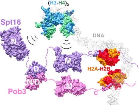
Figure 1.5 C-Terminal peptides of Spt16 and Pob3 are required for H2A-H2B binding and nucleosome reorga- nization.

The model illustrates the binding of FACT to the components of a nucleosome. Two H2A-H2B dimers are bound by C-terminal peptides of Spt16 and Pob3. The H3-H4 histones are bound by the N-terminal and the middle domains of Spt16. The figure is taken from Kemble et al. 2015.

1.3.3 The FACT complex in Arabidopsis

An Arabidopsis FACT complex consisting of SPT16 and SSRP1 has been identified by Duroux et al. 2004. The complete loss of SSRP1 is critical for plant viability, whereas reduced amounts of SSRP1 and SPT16 display various defects in the vegetative and reproductive devel- opment including early flowering, a "bushy" phenotype and a reduced seed set. In accordance to the early transition from the vegetative to the reproductive phase, SSRP1- and SPT16-depleted plants show a reduced expression of the key floral repressor FLC (Lolas et al. 2010). But not much is known about the impact of FACT-depletion on the genome-wide gene expression in Arabidopsis.

In line with its role as TEF, the Arabidopsis FACT complex localizes to the de-condensed euchromatin and associates there with actively transcribed genes like the above mentioned FLC (Duroux et al. 2004). Moreover, FACT was found to interact genetically with HUB1 that catal- yses the mono-ubiquitination of the histone H2B, to regulate various developmental processes

| 8

(19)

Introduction 1

(Lolas et al. 2010, Van Lijsebettens et al. 2014). Additionally, SSRP1 was shown to be required for DNA methylation and genomic imprinting (Ikeda et al. 2011). But interactions of the Ara- bidopsis FACT complex with other TEFs of the RNAPII TEC complex were not investigated yet and would provide new insights into the transcript elongation process of higher eukaryotes.

1.4 Co-transcriptional processing of mRNA

During transcription by RNAPII, the growing pre-mRNA is maturated by processing factors and packaged into an export competent messenger ribonucleoprotein particle (mRNP) that is exported to the cytoplasm for translation (Moore et al. 2009, Saunders et al. 2006, Bentley 2014, Saldi et al. 2016). The processing of pre-mRNA occurs mostly co-transcriptional at the gene. The nascent transcript that is protruding out of the transcribing RNAPII is immediately a substrate for the mRNA processing factors. The maturation of most pre-mRNA molecules includes 5 0 capping, splicing, 3 0 cleavage and polyadenylation as well as RNA editing. The first processing event (5 0 capping) is the attachment of a 7-methylguanosine cap to the 5 0 end of the nascent transcript to convert the 5 0 pppA to a 5 0 7meGpppA end. Next, the spliceosome is assembled on the growing pre-mRNA, to excise the introns and to ligate the exons in a two-step transesterification reaction. Certain exons can be included or excluded from the mature mRNA.

This so-called alternative splicing can result in the production of multiple protein isoforms from a single gene and increases the protein diversity (Jonkers et al. 2015).

Moreover, the transcript elongation rate is highly dynamic and varies between genes (in- tergenic) as well as between regions within a gene (intragenic), what can affect the outcome of co-transcriptional processes like alternative splicing (Bentley 2014). Finally, when the poly- merase transcribes the poly(A) signals, the emerging pre-mRNA sequences are recognized by the polyA complex. This protein machinery catalyses the cleavage of the growing transcript and polyadenylates the 3 0 end by addition of an adenosine monophosphate (Moore et al. 2009, Saunders et al. 2006).

1.5 Coupling of the RNAPII and mRNA processing factors

The transcription and mRNA processing as well as the transport of export competent mRNPs through the nuclear pore complex (NPC) to the cytoplasm depends on the coordinated action of several multiprotein complexes. The functional and physical coupling of the numerous factors of the gene expression machinery and their correct interplay ensures the proper production of proteins in the cell. Several transcription factors were found to influence processing factors and vice versa (Maniatis et al. 2002, Köhler et al. 2007, Komili et al. 2008).

The processing factors are recruited directly by the nascent RNA, the proteins of the TEC or specially by the CTD of the RNAPII that acts as a flexible and versatile "landing pad" for nuclear factors. The phosphorylation patterns on the CTD repeats change during the transcription cycle and determine which factors can bind or not (Phatnani et al. 2006, Bentley 2014).

Beside the linkage of the processing factors to the transcript-elongation machinery, these

factors are also highly connected to each other. This coupling occurs not only between factors

of sequential mRNA processing steps but also between the earliest and latest, which revises the

image of the gene expression pathway as a simple linear assembly line (Maniatis et al. 2002).

(20)

1 Introduction

1.6 The coupling factor Sus1/ENY2

Figure 1.6 The small adaptor protein Sus1/ENY2 links transcript initiation and mRNA export.

In yeast, fruit-fly and humans, Sus1/ENY2 is part of the transcriptional co-activator SAGA and the NPC-associated TREX-2 complex. To gain more clarity, only the protein names of S.cerevisae are indicated in the model. As part of the SAGA-DUB module, Sus1 is important for transcript initiation by de-ubiquitination of the histone H2B. The TREX-2 complex that facilitates the transport of export competent mRNP from the nucleus to the cytoplasm contains two copies of Sus1 that bind to the scaffold protein Sac3. Moreover the yeast TREX-2 complex contains additionally Thp1, Cdc31 and Sem1. Thereby, the conserved Sus1/ENY2 protein bridges the beginning (transcript initiation) and the end (mRNA export) of the gene expression pathway in eukaryotic nuclei (Pascual-García et al. 2009, Kopytova et al. 2010a).

Sus1/ENY2 (yeast/higher eukaryotes) is a small evolutionary conserved protein with ap- proximately 11-kDa in yeast, fruit-flies and humans (Georgieva et al. 2001, Rodríguez-Navarro et al. 2004, Zhao et al. 2008). In agreement with its high sequence conservation, the func- tion of Sus1/ENY2 as an important coupling factor in the gene expression pathway seems to be conserved as well. In yeast and metazoan, Sus1/ENY2 is a shared component of the two multi-protein complexes SAGA and TREX-2 (transcription and export complex 2)(Figure 1.6).

Sus1/ENY2 provides a physical link between the promoter-bound transcriptional co-activator SAGA and the nuclear pore associated mRNA export factor TREX-2. Thereby, Sus1/ENY2 is bridging the ends of the nuclear gene expression pathway, the transcription initiation by RNAPII with the export of the mature mRNA throught the nuclear pore complex (NPC) (Rodríguez- Navarro et al. 2004, Kurshakova et al. 2007, Zhao et al. 2008, Pascual-García et al. 2009, Kopy- tova et al. 2010a). Additionally, Sus1/ENY2 plays a role during transcript elongation that differs between yeast and Drosophila. The Drosophila ENY2 is important for the co-transcriptional recruitment of the THO complex on the nascent mRNA to form export-competent mRNPs, but does not directly associate with the elongating RNAPII (Kopytova et al. 2010b). In addi- tion, ENY2-THO functions independent of SAGA or AMEX (TREX-2). The yeast Sus1 was directly found on coding regions (chromatin) and was associated with the elongating RNAPII in a SAGA and TREX-2 dependent manner (Pascual-García et al. 2008). Under specific circum- stances Sus1/ENY2 (yeast and Drosophila) was observed to a lesser extend in the cytoplasm,

| 10

(21)

Introduction 1

most likely involved in the cytoplasmic mRNA metabolism (Cuenca-Bono et al. 2010, Kopytova et al. 2010a).

1.7 The transcriptional co-activator SAGA

As mentioned earlier (Section 1.2), the transcription is initiated by PIC assembly (RNAPII and GTFs) at the transcription start site (Sims et al. 2004, Saunders et al. 2006). Transcrip- tion factors that are binding gene-specific DNA elements upstream of the core promoter can recruit co-activators like SAGA to the PIC to promote the basal transcription (Thomas et al.

2006). In the last years, the transcriptional co-activator SAGA was the prime example to study gene activation in eukaryotes (Koutelou et al. 2010). The well characterized SAGA com- plex is an approximately 2 MDa multi-protein complex that is structurally and functionally conserved from yeast to humans (Baker et al. 2007, Rodríguez-Navarro 2009, Koutelou et al.

2010, Spedale et al. 2012). In yeast, the SAGA complex regulates the transcription of approx- imately 10 % of the genes genome-wide (Baker et al. 2007, Samara et al. 2011, Weake et al.

2012). The proteins of the SAGA complex are arranged into four functional modules to regulate transcription (Figure 1.7). The HAT (histone H3 acetyltransferase) and the DUB (histone H2B de-ubiquitylation) modules possess enzymatic activities to modify chromatin. Whereas, the TAF (TBP-associated factor) and SPT (suppressor of Ty) modules mediate the interac- tions of SAGA with the activators and the PIC (Rodríguez-Navarro 2009, Koutelou et al. 2010, Samara et al. 2011). In yeast (y), Drosophila (d) and humans (h), the HAT module con- sists of the following four subunits: The acetyltransferase yGcn5/dGCN5/hGCN5 or hPCAF, Ada2/dADA2b/hTADA2b, yAda3/dTADA3/hTADA3 and Sgf29/dSGF29/hSGF29. The HAT module can acetylate the histone H3 at lysine K9 and K14 (Koutelou et al. 2010, Spedale et al. 2012). The DUB module comprises the four following components: The deubiquitinating enzyme yUbp8/dNonstop/hUSP22, the two adaptor proteins ySgf11/dSgf11/hATXN7L3 and ySus1/dENY2/hENY2 as well as the anchoring protein ySgf73/dSgf73/hATXN7 (Köhler et al.

2008, Weake et al. 2008a, Zhao et al. 2008, Lee et al. 2009, Samara et al. 2010, Gurskiy et al.

2012). The crystal structure of the complete DUB module of the yeast SAGA complex revealed that Ubp8, Sus1, Sgf11 and Sgf73 form a highly interconnected complex that is organized into the "catalytic lobe" and the "assembly lobe". Each protein is thereby physically connected the other three (Köhler et al. 2010, Samara et al. 2010). The crystal structure of the yeast DUB module bound to an ubiquitinated nucleosome revealed that the DUB module primarily con- tacts the acidic patch of H2A/H2B with the zinc finger domain (ZnF) of Sgf11 (Morgan et al.

2016). The DUB module catalyses the cleavage of monoubiquitin from K123 (yeast) or K120 (Drosophila and human) of histone H2B (Daniel et al. 2004, Henry et al. 2003, Weake et al.

2008a). All components of the DUB module are required for the activity of de-ubiquitinating enzyme Ubp8 (Köhler et al. 2008).

At the onset of transcription by RNAPII, the co-activator SAGA is recruited to the transcrip-

tional start site. The acetylation of histones by the HAT module leads to de-compaction of the

chromatin structure, which facilitates transcript initiation by the basal transcription machinery

(Sterner et al. 1999, Nagy et al. 2007). The transition from transcript initiation to transcript

elongation is accompanied by changes in the phosphorylation pattern of the RNAPII CTD. Im-

(22)

1 Introduction

Figure 1.7 Composition of the chromatin modifying SAGA complex in Drosophila.

Schematic illustration of the modular structure of the multi-protein SAGA complex. The TAF (depicted in blue) and the SPT module (depicted in yellow) mediate the interactions of the SAGA complex with activators and the RNAPII pre-initiation complex. The HAT module (depicted in green) possesses a histone H3 acetyltransferase activity, while the DUB module (depicted in red) harbours a histone H2B de-ubiquitinase activity. The figure is taken from Mohan et al. 2014.

mediately after transcript initiation, the heptapeptides repeats of the CTD are phosphorylated at serine 5 and 7 to recruit factors for the early steps of transcript elongation. Subsequent these CTD repeats are phosphorylated at serine 2 to recruit factors for the following productive elon- gation phase (Buratowski 2009, Workman 2016). The monoubiquitin at the N-terminal tail of H2B blocks the recruitment of the kinase Ctk1, which phosphorylates the serine 2 of the RNAPII CTD. Therefore, the de-ubiquitylation of H2B by the SAGA DUB module promotes transcript elongation by removing the repressive histone mark (Wyce et al. 2007, Workman 2016).

Little is known about the SAGA complex in plants. The potential Arabidopsis counterparts of the yeast and human SAGA components were predicted bioinformatically (Moraga et al.

2015, Srivastava et al. 2015). Mutant studies on Arabidopsis GCN5 and ADA2b, the homologs of the yeast HAT components Gcn5 and Ada2, revealed that these proteins are critical for plant growth and development (Vlachonasios et al. 2003). The interaction of Arabidopsis ADA2b with GCN5 was shown in vitro by pulldown and yeast two-hybrid assays (Stockinger et al. 2001, Mao et al. 2006). ADA2b can stimulate the HAT activity of GCN5 on nucleosomal histones in vitro (Mao et al. 2006). In ADA2b or GCN5 depleted plants, the expression of 5 % of the 8200 investigated genes was affected (Vlachonasios et al. 2003). Another study showed that 40 % of the investigated promoters are associated with the Arabidopsis GCN5 protein (Benhamed et al.

2008). Furthermore, the Arabidopsis GCN5 was shown to be required for the homeostasis of the recently discovered histone modification H3K36ac, which is conserved in plants and highly enriched in euchromatin (Mahrez et al. 2016a).

| 12

(23)

Introduction 1

1.8 Aims of the thesis

1.8.1 The histone chaperone FACT

FACT, a conserved heterodimer of SSRP1 and SPT16, facilitates the progression of the tran- scription machinery through the chromatin template by destabilizing nucleosomes in the path of the elongating RNAPII. The histone chaperone FACT reorganizes nucleosomes and promotes access to the genomic DNA by binding to H2A-H2B dimers. One attractive possibility is that the association of FACT with chromatin depends, in the first place, on the association of the SSRP1 HMG-box domain with the nucleosomal DNA. To gain more knowledge about the SSRP1 HMG-box domain in higher eukaryotes, the proposed DNA- and nucleosome-binding properties of the Arabidopsis SSRP1 HMG-box domain will be analysed by Electrophoretic Mobility Shift Assays (EMSA) in vitro. In the following, FRAP experiments will reveal if removing of the C-terminal HMG-box domain alters the kinetics of SSRP1 in living Arabidopsis cells, which is a good indication if the HMG-box domain is required for the binding of FACT to chromatin. Ad- ditionally, immunoblotting analysis will reveal if the plant SSRP1 lacking the HMG-box domain still associates with the transcriptionally active RNAPII in vivo. Moreover, the generation and phenotypic analysis of Arabidopsis HMG-box-deficiency mutants will reveal if the DNA-binding domain of SSRP1 is important for proper plant growth and development. Collectively, we will gain new insights about the role of the SSRP1 HMG-box domain in Arabidopsis and whether the DNA-binding domain is indispensable for the association of FACT with nucleosomes.

During transcript elongation, a heterogenous group of TEFs including the histone chaperone FACT enables efficient mRNA synthesis. A proteomic approach using reciprocal tagging in combination with affinity purification and mass spectrometry will reveal the composition of the Arabidopsis transcript elongation complex. Furthermore, the effect of TEF-depletion in Arabidopsis plants is ranging from mild phenotypes (e.g. TFIIS) to severe and lethal phenotypes (e.g. SPT5). The phenotypic analysis of Arabidopsis double mutants that are defective in different combinations of TEFs will reveal if there are genetic interactions between the genes encoding subunits of FACT, TFIIS and PAF1-C.

The reduced expression of either SSRP1 or SPT16 results in similar pleiotropic phenotypes

affecting plant growth and development like early flowering and a reduced seed set. SSRP1-

and SPT16-depleted plants will be analysed by genome-wide transcript profiling compared to

wild-type, to identify differentially expressed genes.

(24)

1 Introduction

1.8.2 ENY2, a coupling factor of transcription and mRNA processing

ENY2, an evolutionary conserved adaptor protein, bridges both ends of the nuclear gene expression pathway as part of the transcriptional co-activator SAGA and the mRNA export complex TREX-2 in yeast and metazoa. The small protein with its important function in con- trolling eukaryotic gene expression is well described in yeast, fruit-fly and humans, but nothing is known about ENY2 in plants. As well little is known about the chromatin-modifying SAGA complex in plants, although its counterparts in other organism play critical roles during tran- scription by RNAPII. To identify the interaction network of Arabidopsis ENY2 and its putative interactors, the SAGA complex and the TREX-2 complex, a proteomic approach using recip- rocal tagging in combination with affinity purification and mass spectrometry will be applied.

Additionally, to gain more knowledge about the function of ENY2 in plants, the following ex- periments will be conducted. The ENY2 promoter activity will be analysed in Arabidopsis by GUS staining, to characterize the spatio-temporal ENY2 gene expression throughout the entire life cycle of a plant. The subcellular localization and the dynamics of ENY2 in Arabidopsis root nuclei will be investigated in transgenic plants expressing GFP-tagged ENY2 by in-detail CLSM analysis. Moreover, a reverse genetics approach using RNAi and CRISPR/Cas9 and the phenotypic characterization of the generated knockdown/knockout mutants will give insights about the function of ENY2 during plant growth and development.

| 14

(25)

2 Results: Analysis of the HMG-box domain of Arabidopsis SSRP1

2.1 Arabidopsis SSRP1 features a conserved C-terminal HMG-box domain

Among eukaryotes, the high mobility group (HMG)-box is a common features of various chromosomal proteins that are important for the regulation of DNA-dependent processes. To bind DNA, the 75 aa HMG-box domain forms a L-shaped molecule consisting of three alpha- helices (Thomas et al. 2001, Štros et al. 2007, Malarkey et al. 2012). In humans and Arabidopsis, the essential histone chaperone FACT features a conserved HMG-box domain at the C-terminus of SSRP1 (Figure 2.1) (Duroux et al. 2004). This HMG-box domain may help FACT to recognize, bind and reorganize chromatin by binding to nucleosomal DNA (Winkler et al. 2011b).

The yeast ortholog Pob3 lacks the the HMG-box domain. However, a third protein called Nhp6A/B is loosely associated with the yeast Pob3/Spt16 heterodimer. This small HMGB protein binds to DNA and provides the HMG-box function. Whereas SSRP1/Pob3 is an essential protein, Nhp6 is not required for viability, but important for normal cell growth (Stillman 2010, Formosa 2012).

Figure 2.1 The amino acid sequence of Arabidopsis SSRP1 is highly conserved among different species.

The SSRP1 protein sequence was aligned to its orthologs from the following other species: Homo sapiens (Hs), Mus

musculus (Mm), Drosophila melanogaster (Dm), Saccharomyces cerevisiae (Sc), Zea mays (Zm) and Physcomitrella

patens (Pp) as representatives for mammals (Ms, Mm), invertebrates (Dm), fungi (Sc), dicotyledons (At), mono-

cotyledons (Zm) and mosses (Pp). The yeast ortholog of AtSSRP1 is Pob3 that is lacking the C-terminal HMG-box

domain. Nhp6 provides the HMG-box function for the yeast FACT complex. The multiple sequence alignment was

generated using Clustal Omega (Sievers et al. 2011) and the conservation of aa residues was highlighted with blue

coloration using JalView (Waterhouse et al. 2009).

(26)

2 Results: Analysis of the HMG-box domain of Arabidopsis SSRP1

2.2 Truncated SSRP14HMG showed reduced DNA- and nucleosome-binding

Figure 2.2 Two-step purification of the recombinant GST-SSRP1 and GST-SSRP14HMG proteins.

A) Overview of the two-step chromatography with adjacent dialysis of the recombinant proteins. The GST affinity purification was followed by an anion exchange chromatography and dialysis. B) First, the GST-tagged SSRP1 proteins were expressed in E. coli BL21 cells and affinity purified using Glutathione Sepharose. All steps were monitored by SDS-PAGE and Coomassie staining (CBB). A representative gel for the expression/purification of the full length SSRP1 was depicted. The whole cell extracts, before (-) and two hours after (+) IPTG induction are shown. Additionally, the samples of the supernatant (S), the pellet (P), the flow-through (FT) and the eluate (E) were analyzed. Following affinity purification, the recombinant SSRP1 proteins were subjected to an anion exchange chromatography and dialysis.

The two-step purified proteins were first analyzed by SDS-PAGE and Coomassie staining (C) and secondly by SDS-PAGE and immunoblot analysis using SSRP1 antisera (D).

In yeast/human it has been shown that the HMG-box domain of Nhp6/SSRP1 binds nucle- osomal DNA (Winkler et al. 2011b). This was also demonstrated in plants by investigations of the maize SSRP1 HMG-box domain (Röttgers et al. 2000, Lichota et al. 2001). To get more information about the HMG-box domain of Arabidopsis SSRP1 (AT3G28730), electrophoretic mobility shifts assays (EMSA) were performed. Therefore, recombinant full length and truncated SSRP1 proteins were expressed in E. coli with an N-terminal GST (Glutathione S-Transferase) tag and purified by two-step chromatography (Figure 2.2 A). Thereby, the affinity purification of the GST-fusion proteins was followed by an anion exchange chromatography to obtain recom- binant SSRP1 and SSRP14HMG proteins in higher purity. Subsequently, the purified proteins were desalted by dialysis and subjected to the EMSA experiments. All steps were monitored by SDS-PAGE and Coomassie staining (Figure 2.2 B-C) as well as by Western Blot analysis with SSRP1 specific (Duroux et al. 2004) antiserum (Figure 2.2 D). This verified the high purity of the recombinant SSRP1 fusion proteins. In the following, the interaction of SSRP1 and SSRP14HMG with mononucleosomes and DNA was examined. The mononucleosomes that were either lacking or possessing linker DNA were kindly provided by Prof. Dr. Gernot Längst.

Recombinant Drosophila histones were reconstituted into nucleosomes by salt dialysis with DNA fragments of 147 bps (without linker DNA) or 198 bps (with linker DNA) that were containing the 601 nucleosome position sequence. The mononucleosomes or the free DNA were incubated with increasing amounts of recombinant SSRP1 and SSRP14HMG proteins, respectively. The formation of complexes was analysed by electrophoretic mobility shift assays using agarose gels.

In contrast to the truncated SSRP14HMG, the full length SSRP1 showed a high affinity for the Cy5-labeled free DNA (Figure 2.3 A-B). The smear indicated that multiple different DNA-

| 16

(27)

Results: Analysis of the HMG-box domain of Arabidopsis SSRP1 2

Figure 2.3 EMSA analysis revealed that the HMG-box domain of SSRP1 was crucial for the binding to DNA and mononucleosomes with linker DNA.

Increasing amounts of the recombinant GST-SSRP1(M1-N646) or GST-SSRP1(M1-K557) proteins were incubated

with A-B) free DNA, C-D) mononucleosomes with linker DNA (198 bps) E-F) or mononucleosomes with only core

nucleosomal DNA (147 bps). The formation of the SSRP1/DNA or SSRP1/mononucleosome complexes was analyzed

by agarose gel electrophoresis. The fluorescent Cy5-labeled DNA was detected by scanning the gels with the Typhoon

FLA9500.

(28)

2 Results: Analysis of the HMG-box domain of Arabidopsis SSRP1

SSRP1 complexes were formed and SSRP1 bound DNA a non sequence-specific manner. In addition, full length SSRP1 formed a specific complex with linker-DNA containing nucleosomes (Figure 2.3 C). Half of the nucleosomes were shifted with 50 nM of SSRP1 protein. In contrast, SSRP1 without HMG-box domain showed no binding at these concentrations and approximately twice the amount of SSRP14HMG protein was needed to shift the nucleosomes (Figure 2.3 D). On the other hand, similar concentrations of full length SSRP1 (∼ 120 nM) and truncated SSRP14HMG (∼ 140 nM) were needed to start shifting core nucleosomes without linker DNA (Figure 2.3 E-F). Compared to nucleosomes with linker DNA, the affinity of SSRP1 was lower and 160 nM recombinant SSRP1 protein was needed to shift half of the nucleosomes. The affinity of truncated SSRP14HMG was similar for nucleosomes with and without linker DNA.

Taken together, the ability of SSRP1 to bind DNA is mediated by the HMG-box domain.

Full length SSRP1 had a high affinity for DNA and nucleosomes with linker DNA, whereas the affinity for nucleosomes with core DNA was reduced. In contrast, SSRP1 lacking the HMG- box domain did not bind to DNA and showed a reduced binding affinity to nucleosomes with linker-DNA in comparison to SSRP1.

2.3 Full-length SSRP1 and truncated SSRP14HMG showed similar protein dynam- ics

Figure 2.4 Generation of transgenic Arabidopsis cell lines -expressing eGFP-SSRP1 and eGFP-SSRP14HMG.

A) Schematic illustration of the eGFP-SSRP1, eGFP-SSRP14HMG and eGFP-NLS transgenes. The genomic sequences of the full length or the truncated SSRP1 were expressed as N-terminal translational fusions with eGFP under the native SSRP1 promoter. Free eGFP-NLS was expressed under control of the CaMV 35S promoter. The primers for the genotyping PCR are indicated as arrows (Black bars = exons, dark grey bars = promoter, dotted lines = introns, green bar = eGFP coding sequence, light grey bars = UTR, KAN = Kanamycin resistance marker, RB/LB = Right Border/Left Border). B) The genotyping PCR of wild type and transgenic cell lines. The following primer pairs were used to detect the transgenes: 3193/1840 (eGFP-SSRP1 fusions) and 3555/1840 (eGFP-NLS).

The previously described EMSA experiments showed that the HMG-box domain of SSRP1 was crucial for the binding of SSRP1 to nucleosomes in vitro. In a next step, the role of the SSRP1 HMG-box domain in mediating SSRP1 – chromatin interactions was examined by fluo- rescence recovery after photo bleaching (FRAP) experiments. To characterize the dynamics of SSRP1 and SSRP14HMG in living cells, transgenic Arabidopsis suspension cell lines-expressing eGFP-SSRP1 fusion proteins were generated. Therefore, transgenes were created to drive the expression of either genomic SSRP1 or SSRP14HMG with eGFP as N-terminal translational fusion under the SSRP1 promoter, a 614 bps region upstream of the translational start site (Figure 2.4). A plasmid driving the expression of eGFP-NLS under the CaMV 35S promoter was generated as control. The constructs were introduced into the genome of cultured Ara-

| 18

(29)

Results: Analysis of the HMG-box domain of Arabidopsis SSRP1 2

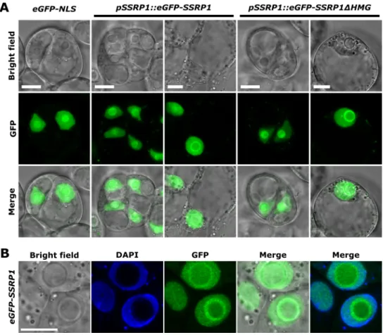
Figure 2.5 Full length and truncated SSRP1 showed the same nuclear localization in living cells.

A) Transgenic Arabidopsis cell lines -expressing either eGFP-SSRP1, eGFP-SSRP14HMG or eGFP-NLS were generated by co-cultivation of PSB-D cells with transgenic Agrobacterium. Three days old cells were investigated by confocal microscopy. One or two representative images were selected for cell line -expressing eGFP-NLS or eGFP-SSRP1 fusion proteins, respectively. The bar indicates 10 µm. B) Cells -expressing eGFP-SSRP1 were fixed using 1 % formaldehyde and stained with DAPI. The bar indicates 10 µm.

bidopsis cells (PSB-D) by co-cultivation with transgenic Agrobacterium (Van Leene et al. 2011, Pfab et al. 2017) and the generated cell lines were analyzed by confocal microscopy. The control cell line showed a homogenous distribution of eGFP-NLS in the nucleus. The signal in the nucleolus was more intense (Figure 2.5 A). The GFP signals of both, the full length and the truncated SSRP1 were detected in the nucleoplasm and nucleolus. In approximately 5 % of the cells-expressing either full length or truncated eGFP-SSRP1, a ring-like structure surrounding the nucleolus was observable. DAPI staining of fixed transgenic cells showed that the eGFP fusion proteins accumulated at the periphery of the nucleolus (Figure 2.5 B). Taken together, the removal of the HMG-box domain did not change the subcellular localization of SSRP1.

In a following step, the transgenic cell lines were subjected to fluorescence recovery after

photobleaching (FRAP) experiments using time-lapse confocal microscopy. The GFP signal

was bleached in a defined region of interest (ROI) in the nucleoplasm and the fluorescence

recovery was monitored over time (Figure 2.6 A-C). The time of the fluorescence recovery

after the bleaching was not significantly different for the full length (t 1/2 = 1.93 s) and truncated

SSRP1 (t 1/2 = 1.79 s) (Figure 2.6 D,F). Both versions of SSRP1 showed a similar high

mobility. But both proteins recovered significantly slower than free eGFP-NLS (t 1/2 = 0.5

s). This indicated that the full length and the truncated SSRP1 were not freely diffusing in

(30)

2 Results: Analysis of the HMG-box domain of Arabidopsis SSRP1

Figure 2.6 Full length and truncated SSRP1 showed the same kinetics in living cells.

A) The FRAP of transgenic eGFP-SSRP1, eGFP-SSRP14HMG and eGFP-NLS -expressing cells is shown. The circular 3 µm ROIs (indicated by doted circles) were photobleached and the recovery of the fluorescence intensity was measured over time in these areas. The Pre-Bleach indicates the first timepoint of the series (t = 0 s), the Post-Bleach the first timepoint after the bleaching (t = 4.56 s) and the Post-Bleach 2 the last time point of the series (t = 43.3 s).

Pseudo-coloured images (modified fire LUT) with respective colour calibration bar are shown. B) The full scale- and C) double- normalized mean fluorescence recovery curves for eGFP-SSRP1 (n = 16), eGFP-SSRP14HMG (n = 16) and eGFP-NLS (n = 12) are shown. This plot shows the normalized GFP intensities at 50x pre-bleach and 90x post-bleach time points. The white dots indicate eGFP-NLS, the blue dots indicate eGFP-SSRP1, and the yellow dots indicate eGFP-SSRP14HMG. D) The Half-time (t

1/2

) of eGFP-NLS (0.5 s), eGFP-SSRP1 (1.93 s) and eGFP-SSRP14HMG (1.79 s). E) The mobile fraction of eGFP-NLS (95.6 %), eGFP-SSRP1 (90.6 %) and eGFP-SSRP14HMG (88.2 %).

F) The exact half-time and mobile fraction values of the indicated cell lines. P-Value (P) of (1) eGFP-SSRP1 or eGFP-SSRP14HMG versus eGFP-NLS and (2) eGFP-SSRP1 versus eGFP-SSRP14HMG. All data are means ± SD.

The significance was tested by the Student

0

s T-Test.

| 20

(31)

Results: Analysis of the HMG-box domain of Arabidopsis SSRP1 2

the nucleoplasm, but were transiently interacting with other proteins/structures to a similar extend. Additionally, the mobile fractions of SSRP1 (90.6%) and SSRP14HMG (88.2%) were comparable, but both were significantly lower than the mobile fraction of free eGFP (95.6%) (Figure 2.6 E,F). This indicated that a small percentage of SSRP1 proteins was immobilized, for instance, at large structure like chromatin. In summary, SSRP1 lacking its HMG-box domain showed similar kinetics as wild type SSRP1. But the mobility of both, SSRP1 and SSRP14HMG was significantly different to the free diffusion of eGFP-NLS. This suggested that the loss of the SSRP1 HMG-box domain, that mediates the binding to the nucleosomal DNA, did not impair the interaction of SSRP1 and chromatin in living cells.

2.4 HMG-box-deficient SSRP1 interacted with SPT16 and the transcriptionally ac- tive RNAPII

Figure 2.7 SSRP1 lacking the C-terminal HMG-box domain interacted with SPT16 and the elongating RNAPII.

Immunoprecipitation of eGFP-SSRP1 and eGFP-SSRP14HMG proteins from transgenic Arabidopsis cell lines using GFP-Trap

TM

. Whole cell extracts were used for the affinity purifications. Co-purified proteins were analysed by SDS- PAGE and CBB staining (A). Endogenous SSRP1 is always indicated by red asterics and eGFP-SSRP1 fusions by green asterics. The bait proteins showed the electrophoretic mobility that is consistent with theirs predicted masses of 98.6 kDa (eGFP-SSRP1) and 88.4 kDa (eGFP-SSRP14HMG). Immunoblot analysis of co-purified proteins (IP) versus input is shown. Different antibodies were used as following: (B) a-SSRP1 (Duroux et al. 2004) and a-GFP (Chromotek, 3H9), (C) a-SPT16 (Duroux et al. 2004), (D) a-RNAPII (CTD) (Abcam, ab817) specifically binding the non-phosphorylated C-terminal domain (CTD) of the largest subunit of RNAPII (NRPB1) and a-RNAPII (Ser2) (Abcam, ab5095) binding specifically the Ser2 phosphorylated CTD of NRPB1. Two bands appeared in the input sample by using the a-RNAPII (Ser2) antibody (Indicated by red arrows). The band migrating at a lower molecular weight represents the hypophosphorylated (NRPB1A) form of the RNAPII, the upper band the hyperphosphorylated (NRPB1O) form.

The FRAP experiments revealed that the loss of the HMG-box domain did not change the kinetics of SSRP1 and its putative interaction with chromatin. In Arabidopsis, the FACT com- plex was detected over the entire transcribed region of actively transcribed genes (Duroux et al.

2004). This raised the question if SSRP14HMG is still part of FACT and is associated with

the elongating RNAPII? Therefore, the proteins co-purifying with either the full length SSRP1

or the truncated SSRP1 lacking the HMG domain were analyzed by Western Blotting. For

this purpose, the transgenic Arabidopsis suspension cell lines-expressing either eGFP-SSRP1 or

Abbildung

Figure 1.2 A variety of transcript elongation factors (TEFs) enables the efficient transcription of chromatin templates by RNAPII.
Figure 1.5 C-Terminal peptides of Spt16 and Pob3 are required for H2A-H2B binding and nucleosome reorga- reorga-nization.
Figure 1.6 The small adaptor protein Sus1/ENY2 links transcript initiation and mRNA export.
Figure 1.7 Composition of the chromatin modifying SAGA complex in Drosophila.
+7

Referenzen

ÄHNLICHE DOKUMENTE

Vrtti and abhinaya are classified on the basis of the innate human

As we approached Walvis Ridge at the beginning of the week the petrologists attempted unsuccessfully to sample an isolated seamount flanking Walvis Ridge to the south.. Later in

First, RNAPII is recruited to the promoter, and then the general transcription factors bind RNAPII and initiate transcription (pre-initiation complex assembly,

When comparing chromatin of SPT4-R3 and Col-0 plants, increased levels of SPT5 and transcribing RNAPII (Ser5P, Ser2P) were found along the transcribed region of the long At3g02260

A rat glioma cell line (RuGli) adhered via α1ß1 integrin to collagen XVI (Eble et al., 2006), whereas human fibrosarcoma cells showed mainly interaction via predominantly

Feeding experiments with specifically 13 C-labeled glucose disclosed that the diterpenoid part of the striatals/striatins is formed via the mevalonate pathway, whereas the

fragment. Thus, these complexes react predominantly end-on by using the phosphorus lone pair. 4 In complexes of type B the lone pair is already used by coordination to Lewis

WITH THE POWER CONTROL MODULE ORIENTED AS SHOWN IN FIGURE 2, CAREFULLY ROUTE THE MODULE CONNECTORS THROUGH THE OPENING BETWEEN THE FAN HOUSING AND THE POWER SUPPLY BOARD.. THE