in male C57BL/6 mice – impact of the central oxytocinergic system
DISSERTATION ZUR ERLANGUNG DES DOKTORGRADES DER
NATURWISSENSCHAFTEN (DR. RER. NAT.) DER FAKULTÄT FÜR BIOLOGIE UND VORKLINISCHE MEDIZIN DER UNIVERSITÄT REGENSBURG
vorgelegt von Sebastian Thomas Peters
aus Landshut im Jahr 2014
Das Promotionsgesuch wurde eingereicht am: 18.02.2014
Die Arbeit wurde angeleitet von: Prof. Dr. rer. nat. Inga D. Neumann
Unterschrift:
Dissertation
Durchgeführt am Institut für Zoologie der Universität Regensburg unter Anleitung von
Prof. Dr. rer. nat. Inga D. Neumann
Gewidmet meiner Familie
Content
Chapter 1
General Introduction 11
1 Introduction 12
2 Stress system 13
2.1 The acute stress response 14
2.1.1 The sympathetic nervous system (SNS) 14
2.1.2 The hypothalamo-pituitary-adrenal (HPA) axis 16
2.2 Acute vs. chronic stress 20
2.3 Psychosocial stress 23
3 Stress and carcinogenesis 25
3.1 Inflammation-mediated colon carcinogenesis 25
3.1.1 Clinical relevance 25
3.1.2 Immunological and cellular issues 27
3.1.3 The azoxymethane (AOM) – dextran sulphate sodium (DSS) – model of chemically-induced inflammation-related
colon carcinogenesis 29
3.2 Stress and colon carcinogenesis 32
4 Stress and drug addiction 38
4.1 Alcohol addiction 40
4.2 Stress and addiction in general 42
4.3 Stress and alcohol addiction 46
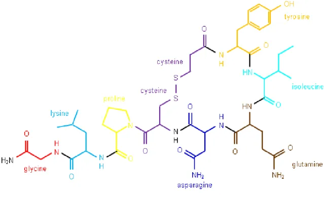
5 Stress and oxytocin 47
5.1 The oxytocinergic system 47
5.2 Oxytocin, stress and anxiety 50
5.3 Oxytocin and addiction 52
6 Effects of CSC on physiological, immunological and behavioural parameters 53
7 Aims of the present thesis 57 Chapter 2
Effects of chronic psychosocial stress on inflammation-related colon
carcinogenesis 61
Chapter 3
Effects of baclofen and oxytocin on ethanol consumption following chronic
psychosocial stress 88
Chapter 4
Effects of chronic oxytocin on behaviour, physiology and the oxytocin system 112 Chapter 5
General Discussion 140
Summary in German 160
References 164
Abbreviations 192
CV and publications 198
Acknowledgements 202
Author´s declaration 205
Chapter 1
General Introduction
1 Introduction
In the modern, fast moving society stress is an omnipresent phenomenon and a concept everybody can identify with. The elevated activity of the hypothalamo-pituitary-adrenal (HPA) axis and the sympathetic nervous system (SNS) upon stressor exposure mediates a broad variety of functions within an organism by the secretion of hormones and neurotransmitters (Dhabhar and McEwen 1996). Although stress is often perceived as negative, an acute exposure to stressors has profound positive effects on an organism, since it results in enhanced attention and allocation of additional energy to increase the physical capability.
From an evolutionary point of view, those stress-induced alterations are important, because they increase the fitness of an organism and, therefore, its chances to survive. In contrast, chronic stress exposure and in particular chronic psychosocial stress is a major burden of modern societies and an accepted risk factor for the development of a variety of disorders including somatic disorders like inflammatory bowel disease (IBD) (Duffy et al. 1991), cancer (Reiche et al. 2004), cardiovascular diseases (Dimsdale 2008), as well as affective disorders like anxiety and major depression (Reiche et al. 2004, McEwen et al. 2012), but also substance abuse disorders (Koob 2008). Due to the omnipresence of psychosocial stressors and a rising number of people suffering from stress-related diseases, a deeper understanding of stress-related symptoms at physiological, behavioural and cellular levels is needed for a sufficient treatment of those pathologies. Concerning the lack of sufficient medications to treat stress-induced disorders, the development of new therapeutics is urgently required. The chronic subordinate colony housing (CSC) paradigm represents an established and clinically relevant animal model of chronic psychosocial stress in male mice (Reber et al. 2007). Mice exposed to CSC develop various symptoms including increased anxiety-related behaviour and colonic inflammation, which reflect the human situation following psychosocial stressor exposure nicely. During the last years neuropeptides got in the focus of interest to serve as possible new treatment options for stress-induced pathologies (Griebel and Holsboer 2012) with the nonapeptide oxytocin (OXT) being one of those candidates. Animal as well as human studies demonstrate that OXT exerts anxiolytic
and stress-protective effects following acute administration (Meyer-Lindenberg et al. 2011, Neumann and Landgraf 2012). In addition, the oxytocinergic system is often discussed to be a possible target for treating substance abuse disorders (McGregor and Bowen 2012).
2 Stress systems
The French physiologist Claude Bernard (1813-1878) postulated the importance of a relative constancy of the internal environment for the functional integrity and the independent existence of an organism. The internal environment defined Bernard as the “internal milieu”, which we call “homeostasis” today (Bartolomucci 2007).
Our understanding of the stress response and the physiological and behavioural reactions of an organism to maintain the homeostasis began during the Great American Depression. In the year 1935, the physiologist Walter Cannon described the extraordinary flexibility of an organism and its ability to respond to stressful situations or “accidents of existence” (Sorrells et al. 2009). The focus of his work was on exploring the sympathetic adrenal response to an immediate threat, and he found that an organism prepares itself by the release of the adrenal hormones, which subsequently activate the body´s energy reserves and, therefore, primes the organism for a fight-or-flight (Bartolomucci 2007, Thiel and Dretsch 2011) or freeze response (Bracha et al. 2004, Thiel and Dretsch 2011). In the year 1936, Hans Selye gave a more precise definition of the terminus stress. According to him, stress is the “non-specific reaction of the body to any demand for change”. In his eyes, stress is a typical defensive reaction of an organism exposed to stressors, and he further described this stereotypic reaction to different stressors as the “General Adaption Syndrome” comprising three different phases: (1) the alarm stage (i.e., physiological activation of the HPA axis and the SNS in preparation to deal with the stressor), (2) a resistance stage (i.e., the period following the initial reaction to the threat whereby the body mediates ongoing stress and attempts to return to steady-state levels), and (3) an exhaustion stage (i.e., when a prolonged stress response overexerts the body´s defence system, thus draining it of its reserve sources and leading to
illness) (Selye 1956, Thiel and Dretsch 2011). A more recent definition explains stressors as internal or external threats that disturb the homeostasis of an organism, leading to specific reactions (stress response) of the body to restore the homeostasis. However, the stress response has a degree of specificity depending on the particular stressor, the organism´s perception of the stressor and the ability to cope with it (McEwen and Stellar 1993, Goldstein and Kopin 2007).
2.1 The acute stress response
Stressor exposure results in an acute stress response which involves a variety of physiological as well as behavioural alterations including increased attention, cardiovascular activity, respiration and allocation of additional energy on the one hand and decreased digestion, growth and sexual activity on the other, to restore the homeostasis (Tsigos and Chrousos 2002, Carrasco and Van de Kar 2003). The HPA axis and the SNS act in concert to enable those alterations, although the two systems act with different velocity. The nerve fibres of the SNS allow a rapid neuronal regulation of vital functions compared to the relatively slow hormonal responses of the HPA axis, whose products have to be transported to their respective target tissues via the blood stream (Kyrou and Tsigos 2009).
2.1.1 The sympathetic nervous system (SNS)
Exposure to stressors results in the disruption of the homeostatic balance of an organism, which, in turn, activates the SNS and the HPA axis. The autonomic nervous system comprising the SNS, the parasympathetic nervous system and the enteric nervous system responds rapidly to stressor exposure and controls a broad range of functions. The cardiovascular, respiratory, gastrointestinal, renal, endocrine and other systems are under the control of the SNS and the parasympathetic nervous system with the latter assisting the SNS functions either by withdrawing or antagonizing and therefore increasing its activity
(Chrousos 1998). The major regulatory region of the SNS is the locus coeruleus (LC), an area located in the brain stem comprising a cluster of noradrenalin-containing neurons (Berridge and Waterhouse 2003). The LC represents the major noradrenergic nucleus within the brain, innervating almost the entire neuroaxis and getting activated by many different stressors, including hypotension, forced swim, shock, immune challenge, but also social stressors (Valentino and Van Bockstaele 2008). Interestingly, this stressor-induced activation of the LC might be due to the monosynaptical innervations of catecholamine-containing dendrites by afferents of the paraventricular nucleus (PVN) (Reyes et al. 2005) whereas LC neurons were found to project to the PVN (Aston-Jones et al. 1986). That PVN-LC circuitry represents an important mechanism to restore the homeostasis upon stressor exposure.
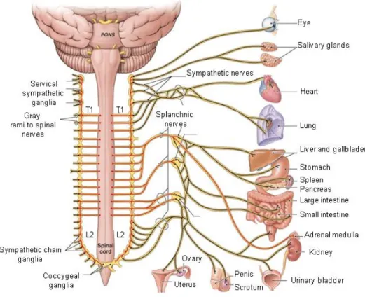
Moreover, the LC directly projects to the sympathetic preganglionergic neurons (Valentino and Van Bockstaele 2008) and regulates the activity of the SNS by the secretion of noradrenalin that binds to α1-adrenoreceptors (Lewis and Coote 1990). The cell bodies of the preganglionergic neurons originate in the intermediolateral column (lateral horn) at the thoracic and lumbar region (“thoracolumbar system”) of the spinal cord. Preganglionergic neurons of the sympathetic nervous system synapse with the postganglionergic neurons within the sympathetic trunk, representing an area of ganglia which are located on each side of the spinal cord. Postganglionergic neurons innervate the peripheral organs (Abboud 2010). However, sole exception represents the adrenal gland, with the adrenal medulla being directly innervated by the great splanchnic nerve, triggering the release of catecholamines, mainly adrenalin from chromaffin cells, into the blood stream (Holgert et al. 1998).
Acteylcholine (ACh) represents the major neurotransmitter of the preganglionergic neurons whereas for the postsynaptic neurons the main neurotransmitter is noradrenalin to activate the target organs (Holgert et al. 1998). Noradrenalin as well as adrenalin binds to their respective α- and β-adrenoreceptors (α1, α2, β1, β2, β3) that belong to a family of seven- transmembrane domain receptors, also known as G-protein coupled receptors (GPCR), which stimulate or inhibit intracellular pathways (Molinoff 1984). Due to different expression
patterns of the adrenoreceptors, the catecholamine-mediated effects are tissue- and organ- specific.
Figure 1: Schematic illustration of the autonomic nervous system. Preganglionergic neurons originating from the first thoracic segment (T1) and extending to the second lumbar segment (L2) of the spinal cord synapse with postganglionic neurons lying in small sympathetic ganglia, named the sympathetic trunk and innervating the peripheral organs. As an exception, the greater splanchnic nerve, originating from the thoracic spinal cord, directly innervates chromaffin cells of the adrenal medulla, without synaptic contact in the sympathetic ganglia. [taken and adapted from:
http://www.highlands.edu/academics/divisions/scipe/biology/faculty/harnden/2121/images/sympathetic .jpg]
2.1.2 The hypothalamo-pituitary-adrenal (HPA) axis
The HPA axis (Fig. 2) plays a major role in mediating the body`s response to a physical or emotional stressor (Papadimitriou and Priftis 2009). As the name implies, the HPA axis consists of several parts including the hypothalamus, the adenohypophysis (pituitary) and the adrenal gland. The hypothalamus as part of the diencephalon located next to the third
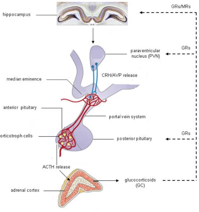
ventricle consists of several nuclei (hypophyseotropic areas) with neurons producing neuropeptides that regulate various functions of the pituitary (Moyes 2008). When the homoeostasis of an organism is disrupted, the PVN gets stimulated. Each PVN comprises both parvicellular and magnocellular subdivisions. Parvocellular neurons of the medial part of the PVN mostly produces corticotropin-releasing hormone (CRH), a 41-amino-acid peptide, upon stimulation, the intermediate part synthesizes the nonapeptide arginine-vasopressin (AVP), and neurons of the lateral area primarily produce CRH and innervate noradrenergic and other neurons of the stress system in the brain stem (Saper et al. 1976, Chrousos 1992, Chrousos and Gold 1992). Axons of parvocellular PVN neurons project to the Eminentia mediana of the hypophyseal stalk and secrete CRH (and AVP) into the thalamic-hypophyseal portal vein system. Via these portal blood vessels CRH obtains the anterior lobe of the pituitary and binds to its respective receptors located in the plasma membrane of the corticotroph cells (Aguilera 2011) that produce proopiomelanocortin (POMC), a precursor of adrenocorticotropic hormone (ACTH).
Figure 2: Schematic illustration of HPA axis activation. Upon stimulation, the paraventricular nucleus (PVN) of the hypothalamus releases corticotropin-releasing hormone (CRH) and arginine- vasopressin (AVP). Neurons from the PVN reach the Eminentia mediana and trigger the release of CRH and AVP into the portal vein circulation. Both peptides bind to their receptors within the anterior pituitary and corticotroph cells secrete adrenocorticotropic hormone (ACTH) into the blood stream.
After binding to its receptors ACTH triggers the release of glucocorticoids into the blood stream (GC, cortisol in humans and corticosterone in rodents) which inhibit their own release via negative feedback loops to the pituitary, the PVN and the hippocampus. [adapted from (Lightman and Conway-Campbell 2010)]
AVP is the second regulator of the HPA axis, especially during chronic stressor exposure. It is transported to the anterior pituitary on the identical route like CRH and stimulates the production and secretion of ACTH through binding to vasopressin type 1b (V1b) receptors (Aguilera 2011). CRH was originally thought to be the major stimulus for ACTH secretion, and AVP was thought to only stimulate ACTH functions as a co-activator with additive effects (Kyrou and Tsigos 2009). However, recent studies assume that the two peptides act in concert and that AVP is a strong synergistic partner of CRH to stimulate ACTH-secretion (Aguilera et al. 2008). After its secretion into the general circulation, the major target of ACTH is the adrenal cortex. Here, ACTH binds to melanocortin-2-receptors (Mc2r) and thereby stimulates the synthesis and secretion of glucocorticoids (GCs; cortisol in humans and corticosterone (CORT) in rodents) (Gorrigan et al. 2011). GCs play a major role in the allocation of energy by enhancing blood glucose and lipid levels, the modulation of the immune response and the cardiovascular functions, but they are also known to affect emotional responses and cognitive processes (Sapolsky et al. 2000). Before binding to their respective receptors, GCs are regulated at a variety of different stages. In blood, about 90%
of GCs are bound to corticosterone-binding globulin, but only unbound GCs are able to cross the blood-brain barrier and cell membranes due to their lipophilic character as steroids. Once in the cytoplasm, GCs bind to two different types of receptors, the mineralocorticoid receptor (MR) and the glucocorticoid receptor (GR), which are bound to heat shock proteins (HSPs) while being unoccupied. After activation by GC-binding the receptors homodimerize, shed their HSP chaperones and translocate to the nucleus, where they regulate gene transcription (Sorrells et al. 2009). Those receptors play a major role in mediating the effects of GCs on basal HPA axis activity, including also the coordination of circadian rhythms, food intake and the sleep/wake cycle. Further, MRs and GRs play an important role in the termination of the stress response. The two receptors display different primary structures, localisations, and affinities to bind GCs. The major expression sides of the MRs are limbic structures like the hippocampus, the lateral septum (LS) and, to a lower extend, the amygdaloid nuclei, the hypothalamus and the pituitary (Moguilewsky and Raynaud 1980, Reul and de Kloet
1985).The GR is expressed all over the brain, including the LS, hippocampus, nucleus tractus solitarii, amygdala and the PVN (Reul and de Kloet 1985). Binding studies were able to demonstrate that the MR has a 10-fold higher affinity (Kd ≈ 0.5) to bind GCs compared to the GR (Kd ≈ 5.0) resulting in an occupation of the MR already at low levels of circulating GCs (Reul et al. 2000). In contrast, the GRs become activated at higher GC levels, e.g. at the circadian peak and during stressor exposure (Reul and de Kloet 1985). Those facts suppose the MR to be involved in the tonic inhibition of the HPA axis under basal conditions and the GR is thought to play a major role in the termination of the stress response, during recovery from stressor exposure and the preparation to following stressors. Termination of the HPA axis activity is mediated via negative feedback loops of the activated MRs and GRs at various levels axis like the pituitary, the hypothalamus and also the hippocampus (see Fig.
2) (De Kloet et al. 1998, Ladd et al. 2004). Altogether, the activation of the SNS and the release of the catecholamines lead to the “fight or flight” response with the main effects representing an enhanced heart rate, respiration, blood pressure, and allocation of additional energy to increase the biological fitness of an organism.
2.2 Acute vs. chronic stress
As already stated above, “stress” consists of a chain of physiological and behavioural alterations that reinstate homeostasis after disruption of the internal milieu of an organism.
Thereby, stressful life events called stressors can be defined as “good”, “tolerable”, or “toxic”, depending of the ability of an organism to control the stressor and whether there are enough resources left to cope with the stressor (McEwen and Gianaros 2011). The brain is the main organ in stress processing. It determines, what is threatening during stressor perception and therefore stressful for an individual and regulates all physiological, behavioural, emotional and immunological responses an organism needs for coping with the stressor. Independent of the nature of the stressor, the orchestrated interplay of all of those responses leads to the activation of the cardiovascular, respiratory, neuroendocrine, but also the immune system
facilitating the “fight or flight” response (Dhabhar 2009, McEwen and Gianaros 2011). The sum of all active processes of the body to return to homeostasis was defined as allostasis by Sterling and Eyer in 1988, literally meaning “maintaining stability through change”. The stress hormones, mainly catecholamines and GCs secreted by the adrenal medulla and adrenal cortex, respectively, act as key players for the achievement of the allostasis (McEwen and Seeman 1999). Acutely, these alterations are beneficial, since they enhance the biological fitness of an individual and therefore its chances to survive. Acute stress is also often referred to as “eustress” (Dhabhar 2000). On the contrary, whether an individual gets exposed to immoderate stressors or for a prolonged period, referred to as “distress”
(Dhabhar 2000), those beneficial adaptations exert deleterious effects and are the origin of a variety of different affective as well as somatic pathologies (Chrousos 2009). For this circumstances McEwen and Stellar introduced the term “allostatic load” describing the wear and tear of the body and brain that results from chronic dysregulation, either overactivity or inactivity, of mediators of the stress response (McEwen and Stellar 1993). Therefore, important characteristics for the discrimination of stressors are its duration, but also its intensity. An acute stressor exposure lasts minutes to hours, whereas chronic stressor exposure continues for days or months. The strength or magnitude of a given stressor can be estimated by peak levels of stress hormones or neurotransmitters, but also by the determination of the heart rate and the blood pressure (Dhabhar 2000). Rodent, but also human studies demonstrated a collapse in the regulation of the circadian CORT rhythm as one significant indicator for the deleterious effects of chronic stressor exposure (Dhabhar 2000, Sephton et al. 2000). Accordingly, blood GC levels are shown to peak after 15 to 30 min following acute stress and to decline to baseline levels after 60 to 120 min depending on the nature and magnitude of the stimulus (de Kloet et al. 2005). However, whether a chronic exposure to stressors is accompanied by sustained exaggerated plasma CORT levels is controversially discussed within the literature and strongly depends on the nature of the stressor, the endurance of stressor exposure but also individual characteristics like genetic predisposition, education and personal experiences. Animal studies demonstrated enhanced
basal plasma CORT levels in male rats after a 14-day exposure to the visible burrow system paradigm, representing an animal model of social stress (Albeck et al. 1997), but also an exposure to restraint stress (1.5h/day) for 7 consecutive days was shown to result in enhanced basal plasma CORT levels in rats (Zelena et al. 1999). Accordingly, male mice exposed to repeated social defeat for 3 consecutive weeks displayed similar sustained elevated plasma CORT levels compared to respective unstressed animals (Keeney et al.
2006). As stated above, persistent high levels of circulating GCs may exert deleterious effects on an individual leading to several pathologies (Sorrells et al. 2009). On the other hand, there are also animal studies displaying an unaffected level of circulating CORT upon chronic stressor exposure or a decline to basal levels after an originally rise (Armario et al.
1986, Djordjevic et al. 2012), representing a possible mechanism of adaption enabling an organism to acclimatise to consistent stressors and preventing the negative effects of hypercorticism. It has to be emphasized that for habituation processes, the stressor must be of homotopic nature, since individuals are unable to adapt to heterotopic stressors (Bartolomucci 2007). Those adaptive mechanisms might be due to changes in the secretion of hypothalamic regulators like CRH and AVP, alterations of the CRH/AVP receptor density, but also an altered GC feedback signalling during chronic stressor exposure (Aguilera 1994).
As stated above, the effects of chronic stressor exposure not only depend on the duration and the nature of the stressor, but also on individual characteristics, like the genetic predisposition and life experiences including trauma or abuse (McEwen and Gianaros 2011).
In animal studies, Dhabhar and colleagues demonstrated that hairless SKH1 mice with an increased anxiogenic phenotype display an increased stress burden and are more prone to develop UVB-induced skin cancer compared to mice with normal anxiety-related behaviour (Dhabhar et al. 2012). In line, mice selectively bred for different levels of anxiety show a diverse susceptibility for developing affective and somatic consequences induced by chronic psychosocial stressor exposure. Individuals with a high anxious phenotype were shown to be more vulnerable to chronic psychosocial stress compared to animals with a low anxiety- related behaviour, giving evidence for the anxiety-related phenotype to mediate resilience to
stress-induced pathologies (Liebsch et al. 1998, Liebsch et al. 1998, Füchsl et al. 2013).
Regarding early life experiences, Levine was the first to demonstrate a reduction in emotional as well as neuroendocrine responses upon stressor exposure in adulthood in rats after maternal separation (Levine 1957). In contrast, various other studies showed an increased vulnerability for developing stress-related pathologies after repeated daily maternal separation (3h/day) during postnatal day 2 to 14 in rats (Ladd et al. 2004) and mice (Veenema et al. 2008).
2.3 Psychosocial stress
The General Adaption Syndrome introduced by Hans Selye and mentioned in chapter 1.2 describes the occurring adaptations of an organism during stressor exposure, but is only referred to physical stressor like exposure to cold, surgical injury and intoxication. More recent studies demonstrate different physiological and behavioural effects of stressful events, depending on the type of the stressor. For example, water deprivation produced a duration- dependent anxiolytic effect in rats tested on the elevated plus maze (EPM) whereas 1 h of restraint stress induced an anxiogenic phenotype in the same behavioural paradigm, although both stressors triggered increased circulating plasma CORT levels (McBlane and Handley 1994). In addition, social defeat was shown to decrease various cardiac parameters compared to non-social stressors such as restraint, shock and forced swimming, with the latter leading to an increased or unaffected variability (Sgoifo et al. 1999). Further studies demonstrated a reactivation of a latent herpes simplex virus type 1 in mice after disrupting the social hierarchy, but not following restraint stress (Padgett et al. 1998). Given the diverse responses to different stressors, ongoing research is supposed to focus on stressful stimuli with varying qualities that serve as stressors across mammalian species, including humans (Blanchard et al. 2001), for the development of adequate strategies to treat stress-induced pathologies. For the human situation, social or psychological stressors are the most important and naturalistic threats with psychosocial stress combining those two important
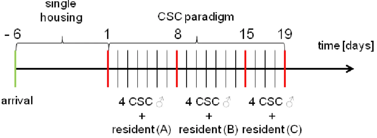
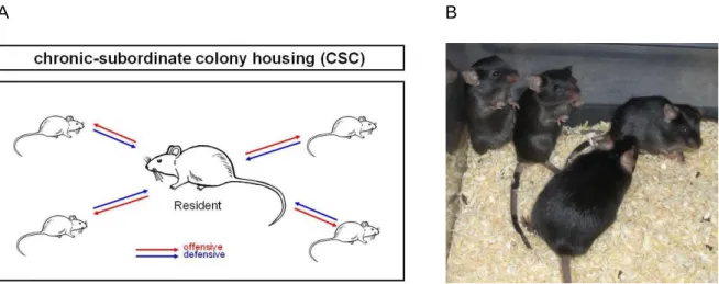
aspects of stress. Psychological stressors are a product of cognitive assessment of environmental claims that are thought to exceed the individual´s capability to deal with (Cohen et al. 2007). These stressors reflect a learned response to previously experienced adverse stimuli, whereas social stressors originate from disturbed interactions among different individuals (Pacak and Palkovits 2001). Based on the literature chronic exposure to psychosocial stressors is a well described risk factor for the development of stress-related disorders (Reiche et al. 2004, Reber 2010). Therefore, animal models of chronic psychosocial stress are clinically relevant, since they reflect powerful paradigms to investigate the underlying mechanisms of stress-derived pathologies in humans. According to that, the CSC paradigm (for details see chapter 1.6) represents a pre-clinically relevant and well-established murine model to induce chronic psychosocial stress by housing four small male mice together with a larger dominant male mouse for 19 consecutive days. CSC was shown to induce a broad variety of physiological, neuroendocrine, immunological and behavioural changes. Exposure to CSC has been repeatedly shown to induce thymus atrophy as well as adrenal hypertrophy (Reber et al. 2007, Slattery et al. 2012, Uschold- Schmidt et al. 2012, Füchsl et al. 2013, Uschold-Schmidt et al. 2013). Further important alterations seen after 19 days of CSC are the induction of a spontaneous colonic inflammation (Reber et al. 2007), aggravation of a chemically induced colitis (Reber et al.
2008) and the enhancement of anxiety-related behaviour (Reber and Neumann 2008, Slattery et al. 2012). Therefore, the CSC paradigm seems to provide a powerful and relevant tool for investigating the the underlying mechanisms of the development and maintenance of stress-related pathologies in humans.
3 Stress and carcinogenesis
3.1 Inflammation-mediated colon carcinogenesis
3.1.1 Clinical relevance
The term tumour or neoplasia describes a disease with an excessive or uncontrolled growth of structurally and biological abnormally cells that can originate from any tissue of the body.
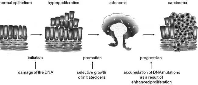
Thereby, benign and malign neoplasm can be differentiated with the benign neoplasm showing close morphological similarity to the tissue of origin, displaying slow growth patterns and forming circumscribed and encapsulated masses. Further, benign tumours do not infiltrate their surrounding tissue and do not metastasize. In contrast, malign neoplasms resemble their adjacent tissue less closely and are characterized by more abnormal cells regarding their cellular form and function. The majority of malign tumours grow rapidly and extend gradually throughout their surrounding tissue and metastasize to different sites within the organism (Tanaka 2009). The progression of colon carcinogenesis can be subdivided into three sections namely the initiation, promotion, and progression phase, which are known as adenoma-carcinoma-sequence, which represents the progress from the first mutation within the epithelium of the colon to the invasive growing colon carcinoma (Fearon and Vogelstein 1990).
Figure 3: Schematic illustration of the adenoma-carcinoma sequence. [taken and adapted from:
http://ediss.sub.uni-hamburg.de/volltexte/1999/161/html/1_%20Einleitung.htm
During the last century there was an increase of the cancer-related death rate, which is the most frequent cause of death within the Western countries (Karim-Kos et al. 2008, Jemal et al. 2009). Epidemiological studies demonstrate rather personal life-style and dietary habits as the cause of the increased colorectal cancer (CRC) rate than the genetic predisposition (Hollman 2000). This aspect became evident in the Japanese population developing more distal CRC after adapting to a more Western high fat diet (Tajima and Tominaga 1985).
Another important factor supporting tumour development within an organism is inflammation.
In 1863, Rudolf Virchow noted leukocytes in neoplastic tissue and postulated a connection between inflammation and cancer. He further suggested the “lymphoreticular infiltrate”
reflecting the origin of cancer at the sides of inflammation with certain cytokines and chemokines in combination with tissue damage and the resulting inflammatory processes leading to an increased proliferation (Balkwill and Mantovani 2001). Although, an enhanced proliferation of cells is not the only reason to cause cancer, a sustained proliferation in an environment rich of inflammatory cells, growth factors and DNA-damage promoting agents, certainly potentiates and/or promotes neoplastic risk (Coussens and Werb 2002). For example, patients who longlastingly suffer from Crohn´s disease (CD) or ulcerative colitis (UC) have an increased risk to develop pre-cancerogenic dysplastic alterations or colon
cancer. For the latter 8-10 years after diagnosis the risk for CRC increases by 0.5-1% per year (Munkholm 2003, Herszenyi et al. 2007). Studies reporting the risk for developing CRC for patients suffering from UC to be at 2% after 10 years, 8% after 20 years and 18% after 30 years of disease (Eaden et al. 2001). Compared to sporadic colorectal carcinomas, CRC developing in IBD patients affects individuals at younger age more often than the general population and progresses to invasive adenocarcinoma from flat and nonpolypoid dysplasia.
3.1.2 Immunological and cellular issues
The hallmarks of inflammation -related cancer include the presence of inflammatory cells and mediators like chemokines, cytokines and prostaglandins in tumour tissues. These factors are also involved in tissue remodelling and angiogenesis similar to that seen in chronic inflammation (Mantovani et al. 2008). They have also been detected in breast cancer without any ongoing inflammation (Balkwill et al. 2005). In addition, cancer cells are able to produce a variety of inflammation-related mediators by their own leading to the recruitment and infiltration of leukocytes that are known to secrete cytokines and cytotoxic substances like reactive oxygen and nitrogen compounds, but also interleukins (IL) and interferons (IFN) (Wahl and Kleinman 1998, Kuper et al. 2000). Enhanced levels of circulating reactive oxygen and nitrogen compounds as a result of infectious stimuli can increase the amount of DNA damages in tissues with an increased proliferation ratio (Maeda and Akaike 1998).
Peroxinitrite for example, acts as a mutagen by interacting with the DNA on sides of an enhanced proliferation resulting in permanent genomic alterations like point mutations, deletions or reorganisations (Coussens and Werb 2002). According to this, mutations of the gene encoding for the protein p53, which is known to exert tumour suppressive properties, are almost as frequent in tumours as in chronic inflammation (Yamanishi et al. 2002). In addition, there are various essential factors being involved in the processes of inflammation- mediated colon carcinogenesis, resembling transcription factors like nuclear factor kappa- light-chain-enhancer of activated B cells (NF-KB), activators of signal transducer and activator
of transcription 3 (STAT3) as well as proinflammatory cytokines like tumour necrosis factor (TNF) and IL-6 (Marazziti and Catena Dell'osso 2008, Colotta et al. 2009). NF-KB resembles a key regulator of inflammatory processes mediated by the innate immune system.
Unactivted, NF-KB forms inactive dimers within the cytoplasm and gets activated by signal- transduction-cascades, initiated by toll-like-receptors or TNF and IL-1β, after binding to their respective receptors. Upon activation NF-KB affects the expression of genes, which can be arranged into three different groups: anti-apoptotic genes, genes regulating immune response and proliferation, and genes encoding for their own negative regulation (Naugler and Karin 2008). Therefore, activation of this transcription factor is closely linked to different types of cancer including breast cancer, prostate cancer, Hodgkin´s lymphoma and colon cancer (Li et al. 2005). Lee and colleagues demonstrated that for a consistent NF-KB-activity within carcinomas STAT3 is essentially required (Lee et al. 2009). STAT3 itself also affects the cell cycle and apoptosis by regulating the expression of different genes e.g. encoding for the antiapoptotic B cell lymphoma (Bcl-2). One major effector molecule downstream of NF-
KB-activation represents the cytokine IL-6, which is secreted by activated T-lymphocytes, epithelial cells but also by a variety of different tumour cells, and again activates STAT3. The transcription of IL-6 is regulated by bacterial endotoxines like lipopolysacharide, proinflammatoriy cytokines as TNF, but also IL-6 itself (Holländer 2006). In addition, recent studies were able to demonstrate that a process called Il-6-trans-signalling is essentially involved in proliferation processes. Thereby, the membrane bound IL-6 receptor transfers by a metalloprotease-mediated process into a soluble form, achieves its target cells and stimulates tumour growth (Becker et al. 2004). Becker and colleagues demonstrated a decelerated adenoma development in late stages of colon carcinogenesis by inhibition of the IL-6 mediated signal cascade (Becker et al. 2004). After injection of hyper-IL-6, a designer cytocine consisting of IL-6 and a covalently linked soluble IL-6 receptor, for 1 week, mice displayed an enhanced proliferation of dysplastic epithelial cells and also an increased amount of phosphorylated STAT3. In addition, a reduced inflammation-induced colon carcinogenesis was observed in mice after blocking the Il-6-trans-signalling (Becker et al.
2004). Grivennikov and colleagues were able to confirm these results by using IL-6-/- knockout mice with the wild-type animals displaying an enhanced carcinogenesis after the AOM-DSS-model compared to the knockout mice (Grivennikov et al. 2009). In addition to IL- 6, TNF - another proinflammatory cytokine - is mainly involved in the development of tumours. TNF, originally thought to hold neoplasia-destroying properties, was found to increase carcinogenesis instead. After administration of dimethylbenzanthrancen, a strong carcinogen, TNF-deficient mice displayed resistance for developing skin cancer compared to wild-type animals (Moore et al. 1999). A possible mechanism for TNF to promote tumour development as well as progression is the activation of cyclooxigenase II (COXII), an enzyme known to be involved in processes like apoptosis, angiogenesis and invasion of tumour cells into the surrounding tissue. In detail, human colonic cancer cells, overexpressing COX II were shown to be resistant to apoptosis (Sun et al. 2002). Regarding cancer progression, COX II is known to increase the invasiveness of colon cancer cells by activating metalloproteinase-2 (Li et al. 2002), whereas COX II suppression resulted in reduced levels of metalloproteinase-2 and -9 in human prostate cancer (Attiga et al. 2000).
Further, in relation to angiogenesis, expression levels of COX II in colon cancer cells correlate with high levels of vascular endothelial growth factor, basic fibroblast growth factor and endothelin-1, which stimulate endothelial migration and endothelial tube formation (Tsujii et al. 1998). Finally, an enhanced concentration of circulating TNF leads to the inhibition of tyrosinephosphatase, resulting in an attenuated expression of MHC-I-antigens on the cell surface. Therefore, it is impossible for the immune system to detect degenerated cells facilitating their development into solid tumours by continuous replication (Reiche et al.
2004).
3.1.3 The azoxymethane (AOM) - dextran sulphate sodium (DSS) model of chemically-induced inflammation-related CRC
The AOM-DSS- model established by Tanaka and colleagues is based on the enhanced risk to develop CRC following inflammatory processes (Tanaka et al. 2003). The principle of this animal model consists of one single ip injection of the carcinogen AOM followed by repeated chemical inductions of colitis via several cycles of DSS administrations in mice. Here, the order of administering the chemicals is important for a successful induction of colon carcinogenesis with the best result occurring with a single AOM injection followed by one DSS administration for seven days via the drinking water (Tanaka et al. 2003). The genetic background of the animals is the second important factor, since there are mouse strain differences in the susceptibility to AOM-DSS treatment (Suzuki et al. 2006, Rosenberg et al.
2008). Suzuki and colleagues were able to demonstrate different tumour ratios in diverse mouse strains, beginning with the most fragile: Balb/c > C3H/HeN > C57BL6/N > DBA/2N (Suzuki et al. 2006).
Azoxymethane (AOM)
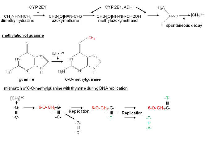
AOM is a metabolite of the colon carcinogen 1, 2-dimethylhydrazine (DMH) with both being precursors of methylazoxymethanol (MAM). This carcinogen was identified by a study about the inhabitants of Guam (largest and southernmost of the Mariana Islands) consuming cycad and developing colorectal cancer at a high percentage. The carcinogen cycasine being a metabolic precursor of MAM reflects the carcinogen localized within the cycad (Laqueur 1964). Compared to DMH and MAM, AOM is known to exert a higher effectiveness and stability (Papanikolaou et al. 1998, Neufert et al. 2007). Both, DMH and AOM are pre- carcinogens that require a metabolic activation to produce reactive products which are able to alter the DNA. One key player for the production of the reactive MAM is the cytochrome – isoform CYP2E1. Within the colon MAM decays into an alkyl radical and due to its strong electrophilic character, is able to alkylate the DNA and forms 6-O-methulguanine. If not being eliminated by the 6-O-methylguanine-DNA-methyltransferase it establishes a mismatch pair
with thymine during the process of replication resulting in adenine-thymine pair instead of a guanine-cytosine pair (Fiala 1977, Fiala et al. 1984, Pegg 1984, Sohn et al. 2001).
Figure 5: Schematic illustration of the GC-AT transition induced by azoxymethane (AOM). The carcinogens dimethylhydrazine (DMH) and azoxymethane (AOM) build free radicals within the colon that methylate guanine. The methylated guanine (6-O-methylguanine) pairs with a thymine instead of a cytosine resulting in a GC-AT transition during the cell cycle [taken and adapted from (Fähndrich 2005)]
Dextran-sulphate-sodium (DSS)
The risk for developing colorectal cancer is closely linked to IBDs like CD and UC. As stated above, the risk for developing CRC increases with severity and duration of the colonic inflammation which can be reliably induced by DSS administration (Okayasu et al. 1990, Eaden et al. 2001, van Hogezand et al. 2002). Animals treated with DSS display marked loss of body weight, rectal bleeding, reduction of colonic length and destruction of the epithelial
layer and glandular architecture of the large intestine (Okayasu et al. 1990, Cooper et al.
1993, Kitajima et al. 1999) and due to the restriction to the large intestine DSS colitis is considered to be a useful and relevant animal model for UC (Okayasu et al. 1990, Kitajima et al. 1999).
Figure 6: Chemical structure of dextran sulphate sodium (DSS). [adapted from http://www.sigmaaldrich.com/content/dam/sigmaaldrich/docs/Sigma/Product_Information_Sheet/d890 6pis.pdf]
3.2 Stress and carcinogenesis
The hypothesis that psychological states affect the development of diseases is an old one.
About 200 AD the Greek physiologist Galen postulated “melancholic” women being more susceptible to develop “swellings” of the breast than “sanguine” woman (Reiche et al. 2004).
Stressors can affect the proliferation, the growth of tumours, development of metastases as well as the immune response via the endproducts of the stress response, the catecholamines and GCs. Animal studies demonstrated that the catecholamine-mediated
effects on carcinogenesis are mediated via β-adrenergic receptors. Thaker and colleagues demonstrated that mice receiving an injection of a HeyA8-ovarian-tumour cell line followed by a daily administration of isoproterenol (nonspecific β-receptor agonist), terbatuline (specific β2-receptor agonist), xamoterol (specific β1-receptor agonist), or isoproterenol (nonspecific β-receptor antagonist) differed in the amount of solid tumours, but also the weight of the tumours varied. In more detail, administration of unspecific β-receptor agonists and specific β2-receptor agonists led to an increase in the number of tumours and also their weight, whereas unspecific β-receptor antagonists resulted in an opposite outcome (Thaker et al. 2006). The catecholamine-mediated stimulation of β-adrenergic receptors activates the proteinkinase C-/-Erk1/2-/-cyclooxygenase II cascade, which is known to be involved in cell proliferation (Thaker and Sood 2008, Armaiz-Pena et al. 2009). Interestingly, it has to be mentioned that acute and chronic stress exerts different effects on cancerogenisis. Acute stressor exposure induced a redistribution of immune cells and increased the concentration of leukocytes at sides of wounds and an activated immune system and, thus, strengthens the innate and adaptive immune response (Viswanathan and Dhabhar 2005). In line daily acute restraint stress (2.5h, 2 weeks) activated the immune system resulting in a reduced development of skin cancer in mice. In contrast, chronic stress suppressed the innate as well as the adaptive immune response by reducing the levels of circulating immune cells, but also activated immune-suppressive mechanisms like the activation of regulatory T-cells (Dhabhar 2009). Therefore, stressors affect also the production and secretion of cytokines derived by those immune cells (Reiche et al. 2004). Dhabhar and colleagues demonstrated an increased susceptibility of mice, exposed to chronic restraint stress (6h, 2 weeks) to develop UV-induced skin cancer (Saul et al. 2005). Similar results namely an enhanced tumour development and accelerated growth, was demonstrated in studies with mice receiving an injection of a T-cell lymphoma in combination with chronic restraint stress (6h, 3 weeks) (Frick et al. 2009). It has to be emphasized that most of the studies are not about cancer development but rather investigate the effects of stressor exposure on injected tumour cells and therefore on the progression of cancer. In addition, there are only a few studies about
the development or the progression of colonic cancer during or after stressor exposure, although colon cancer is one of the most common cancers worldwide and therefore possesses a high clinical and social relevance.
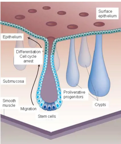
Morphologically, the intestinal tract comprises three different compartments. The small intestine, including duodenum, jejunum, and ileum, the caecum, and the large intestine comprised of the colon and rectum (Moyes 2008). The colonic surface contains invaginations/crypts with continuously dividing stem cells on their base, which are responsible for the regeneration and renewal of the colonic epithelial layer. The new cells migrate from the base of the crypt to the tip and compensate via apoptosis the continuous proliferation (D’Errico and Moschetta 2008) (see Fig. 7).
Figure 7: Consistently dividing stem cells on the base of the crypts form proliferative progenitor cells that migrate from the base of the crypt to the tip, regenerate and renew the colonic surface epithelial layer, and compensate via apoptosis the continuous proliferation. [taken and adapted from (D’Errico and Moschetta 2008)
One major control element of this colonic cell renewal is the WNT/β-catenin cascade with the main effector molecule β-catenin. At inactivated WNT receptors, β-catenin is bound to the two cytoplasmatic proteins of the “destruction” complex, the adenomatous polyposis coli- protein and axin, and inactive. Mutations in the genes encoding for those two proteins can result in a continuous activity of β-catenin and an enhanced proliferation (Reya and Clevers 2005). Recent studies demonstrate the nuclear receptor liver-receptor-homolog-1 (LRH-1) that is mainly expressed in omnipotent embryonic stem cells and is involved in early developmental and differentiation stages, to be also involved in processes of cell renewal within the intestinal tract (Botrugno et al. 2004). In the adult organism LRH-1 is predominantly produced within the liver, the exocrine pancreas and the intestinal crypts and
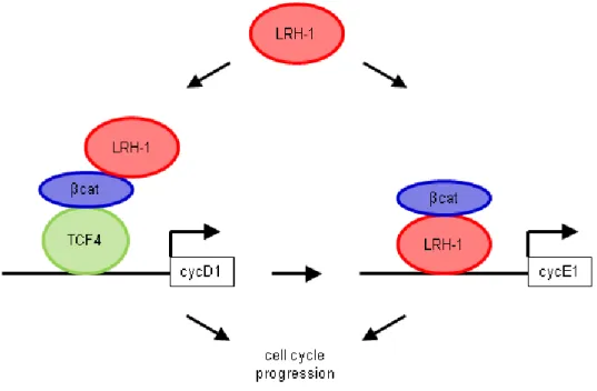
controls cascades regulating the homeostatsis of cholesterol and bile acid (D’Errico and Moschetta 2008). With respect to cell renewal processes, LRH-1 is able to affect the cell cycle via two different cascades. First, LRH-1 acts as a co-activator of the β-catenin/Tcf4- cascade with stimulating the expression of cyclin D1 and c-Myc. Cyclin D1serves as a cell cycle regulatory switch in actively proliferating cells with regulating the continuation of the cell cycle. Thereby, cycline D1 is required in G1 phase for the initiation of DNA synthesis and for the entry into S phase (Stacey 2003). With respect to the cell cycle, c-Myc is known to regulate cell cycle duration with high levels of c-Myc leading to a more rapid cycle (Karn et al.
1989). For the second possibility a conserved LRH-1 element binds to the promotorregion of cyclin E1 with an excess cyclin E1 activity causing cells to progress through G 1 phase more quickly (Hwang and Clurman 2005). Therefore, combination of those two cascades, result in an enhanced and accelerated cell cycle.
Figure 8: Schematic illustration of the dual mechanism of LRH-1 mediated cell proliferation.
LRH-1 promotes proliferation via two mechanisms. First, inducing cyclinD1 expression by co- activating the β-catenin/Tcf4 cascade and second by directly binding to the promoter of cyclinE1 with β-catenin co-activating LRH-1 transcription. The combination of both DNA-dependent and – independent transcriptional events leads to an accelerated cell cycle progression. [taken and adapted from (Botrugno et al. 2004)]
In addition, LRH-1 is also involved in mediating inflammatory processes within the intestine.
Studies with heterocygote LRH-1 knockout mice demonstrate a more severe inflammation and a reduced capability to re-built the colonic epithelium of the knockout mice compared to wild type animals. In addition, LRH-1 regulates the enterocyte-dependent local production of GCs which is decreased for the haploinsufficient LRH-1 mice (Coste et al. 2001). In combination, those results reveal an enhanced risk for developing CRC due to an increased LRH-1 expression. Accordingly, the LRH-1 haploinsufficient mice are less susceptible for developing AOM-induced CRC (Schoonjans et al. 2005).
4 Stress and drug addiction
Addiction to drugs of abuse reflects a chronically relapsing disorder of the central nervous system (CNS) including the hallmarks of compulsive drug use and loss of control over drug intake. Addictive behaviour is described to consist of three different phases: (1) preoccupation and anticipation, (2) binge consumption and intoxication and (3) withdrawal with its negative effects. The characteristics of those phases are impulsive behaviour, which often dominates the early stages of addiction, whereas compulsive behaviour is more prominent for the later phases. The shift from impulsivity to compulsivity induces the changes from positive reinforcement driven motivated behaviour into negative reinforcement driven motivated behaviour. Those three stages are thought to interact with each other, thus becoming more and more intense and finally resulting in the pathological state known as addiction (Koob and Volkow 2010). A more clinical definition states the term addiction as a consumption of high doses of psychoactive substance for a prolonged period, craving for the drug but also unsuccessful attempts to discontinue drug intake, accompanied by consumption of the substance of abuse despite negative effects on the social and professional life, and increasing tolerance and withdrawal symptoms and therefore an elevated intake to reduce or alleviate those symptoms (Filip and Frankowska 2008). Cursory examinations display only a few similarities among all drugs of abuse with some exerting sedative effects like barbiturates, alkohol (EtOH), opiates and benzodiazepines, whereas nicotine, amphetamine, and cocaine display more stimulant effects. Further, EtOH and opiates are known to produce striking degrees of physical dependence while others like cocaine produce little or any signs of physical dependence. There are some similarities of central actions of all kind of drugs of abuse. They are pleasurable, reinforcing, and all of them activate the mesolimbic dopaminergic reward system (Gardner 2011). These effects are due to the fact that all of them resemble functional direct, indirect or transsynaptic dopamine (DA) agonists (Gardner 2011). The mesolimbic dopaminergic reward system was first discovered by Olds and Milner in 1950 by electrical brain stimulation (Olds and Milner 1954). The reward system is formed by dopaminergic cell bodies within the ventral tegmental
area (VTA) and their projections systems to the nucleus accumbens-olfactory tubercle complex. First, the posteriomedial VTA selectively project to the ventromedial striatum, which includes the medial olfactory tubercle and the medial NAc shell. Second, the lateral VTA projects largely to the ventrolateral striatum, which includes the NAc core, the medial NAc shell and the lateral olfactory tubercle. (George et al. 2012). A population of GABAergic neurons located within the VTA supplies inhibitory effects on dopaminergice neurons, but also affect other structures like the pedunculopontine tegmental nucleus and glutamatergic neurons (Dobi et al. 2010). The VTA receives the major excitatory glutamatergic and cholinergic inputs from the ventromedial prefrontal cortex, ventral subiculum, subthalamic nucleus, pedunculopontine tegmental nucleus, laterodorsal tegmental nucleus (Kalivas 1993), but also prominent inputs from the nucleus accumbens (NAc) shell and the ventromedial ventral pallidum (Oades and Halliday 1987). Following acute administration, drugs of abuse trigger the release of DA within the NAc. As a result, DA receptors, comprising two receptor families, become activated and trigger different intracellular signal cascades. The DA D1-like receptors, comprising D1 and D5 receptors, were shown to enhance the activity of the adenylyl cyclise, whereas the DA D2-like receptors including D2 and D4 receptor exert inhibitory effects (Spanagel 2009). Due to the brain reward enhancing properties of drugs of abuse, Blum and colleagues hypothesized a chronic basal brain reward deficiency to be causally involved in addiction (Blum et al. 1996). In line, animal studies with Lewis and Fisher 344 rats, a strain selectively bred for different vulnerabilities to drug addiction, demonstrate differences in the dopaminergic reward circuitries (Guitart et al.
1992). Nestler and colleagues found that Lewis rats, which display a behavioural phenotype of huge addictive drug acceptance and an escalated drug seeking behaviour, have a pathological reduction in the neurofilamentous transport system for the dopamine- synthesizing enzyme tyrosine hydroxylase compared to Fisher 344 animals not displaying such excessive drug-seeking behaviour. Further, Lewis rats reveal a deficiency of tyrosine hydroxylase in the dopaminergic axon terminals of the NAc and reduced extracellular DA
levels within the NAc leading to pathological aberrations in the postreceptor signal transduction mechanisms (Beitner-Johnson et al. 1991, Guitart et al. 1992).
Figure 9: Schematic illustration of dopamine synthesis. Dopamine synthesis starts with the tyrosine hydroxylase mediated conversion of tyrosine to dihydroxyphenylalanine (DOPA), which represents the rate-limited step in the production of dopamine. Next, dopamine is formed by the DOPA decarboxylase. [taken and adapted from http://www.vivo.colostate.edu/hbooks/pathphys/endocrine/
adrenal/medhormones.html]
For the human situation, family, adoption and twin studies were able to demonstrate a genetic influence on the development of drug addiction disorders. It has to be emphasized that also individual characteristics including, sensation and novelty seeking, trait impulsivity, antisocial conduct disorder, depression but also attention deficit/hyperactivity disorder are known to enhance the susceptibility to develop substance abuse disorders. (Uhl 2004, Uhl et al. 2008)
4.1 Alcohol addiction
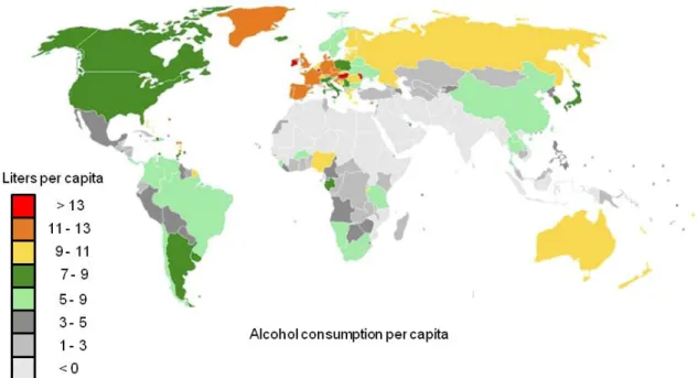
The consumption of alcoholic beverages is a major global burden and an extremely common feature of social gatherings throughout many cultures. Therefore, EtOH is one of the most frequent substances of abuse worldwide with recent statistics revealing that EtOH consumption increases the risk for developing adverse health and social consequences that are related to its noxious and dependence producing properties (World Health Organization
2012). In fact, EtOH abuse is related to more than 60 different pathologies, including cancer, cardiovascular disorders, injuries but also neuropsychiatric diseases (Spanagel et al. 2010).
Figure 10: Alcohol consumption per capita in the year 2006. [taken and adapted from (Spanagel 2009)
Despite the enormous health and socioeconomic problems of the use and abuse of alcoholic beverages, EtOH also exerts some positive effects, at least in light to moderate doses. EtOH is known to improve mood, to attenuate stress and to reduce the risk for developing coronary heart disease, type-2 diabetes mellitus as well as cancer (Spanagel 2009). In addition to the social component mentioned above, developing EtOH abuse disorders is also promoted by a number of risk factors including personal history of alcohol usage, genetic factors like alterations in EtOH-pharmacokinetics, -pharmacodynamics and genes modulating neurophysiological responses (Buscemi and Turchi 2011) as well as mood and anxiety disorders (Kessler et al. 1996, Hines et al. 2005, Barrenha et al. 2011). Furthermore, chronic stress, and in particular chronic psychosocial stress, which is very common in modern societies, has been shown to represent a high risk for developing substance abuse and addiction in general, alcoholism in particular, as well as causing relapse in abstinent
individuals (Brown et al. 1990, Litt et al. 1990, Brown et al. 1995, Cooney et al. 1997, Sinha 2008) (see chapter 4.2 and 4.3). During the last decades different theories about the possible binding sides within the CNS were discussed. Until the 1990s different lipid theories hypothesized that EtOH acts via some perturbation of the membrane lipids of CNS neurons.
Recent studies mostly describe membrane proteins like receptors and ion channels as the major side of action of EtOH. Several studies demonstrate EtOH to inhibit NMDA receptor activity (Weight et al. 1991), to potentiate GABAA receptor functioning and to directly affect glycine receptors (Mihic et al. 1997). Furthermore, EtOH potentiates neuronal ACh (Narahashi et al. 1999) and serotonin receptor functioning (Lovinger and White 1991, Machu and Harris 1994) and directly interacts with non-ligand ion channels by either inhibition (Wang et al. 1994) or activation (Kobayashi et al. 1999). In addition, it has to be mentioned that the inhibitory or excitatory effects of EtOH on receptors depend on a variety of variables including EtOH concentration and the subunit composition of the respective channel or receptor (Spanagel 2009).
4.2 Stress and addiction in general
Exposure to stressors is often related to the development of substance abuse disorders with a variety of different hypothesises trying to explain the connection of stressor exposure and addiction. The stress-coping model of addiction described by Shiffman postulates that consumption of drugs of abuse reduces the negative effects of stressor exposure and additionally enhances the positive effects. Thus, this theory predicts drug taking as a maladaptive, but effective strategy for better stress coping (Shiffman 1982). In line, Marlatt and Gordon described individuals with poor coping resources in addition to other risk factors including parental drug use and peer pressure to be more prone to use addictive substances (Marlatt GA and JR 1985). Finally, the reinforcement theory (Conger 1956), the tension reduction theory (Sher and Levenson 1982) and self-medication hypothesis (Khantzian 1985) predict that stressed individuals being in a drug naive state are more prone to consume
substances of abuse due to their mood-enhancing and emotional distress-reducing properties (Sinha 2001). Affective pathologies like anxiety-related disorders are induced by chronic exposure to stressors and are associated with dys-regulation of several brain circuitries (Arborelius et al. 1999), which might lead to an enhanced sensitivity to the reinforcing properties of substances of abuse and, therefore, to an increase in the amount of drugs consumed in those patients (Sinha 2001). In line with these hypothesises several animal studies were able to demonstrate a stress-induced increase in the self-administration of drugs of abuse. Chronic immobilization stress (15 min/day; 50 days) exaggerated the self- administration of morphine or fentanyl in rats compared to unstressed animals (Shaham 1993). Additionally, Long Evans rats displayed an enhanced cocaine self-administration following exposure to repeated restraint stress (Covington and Miczek 2001) or repeated social defeat (25 min on days 1, 4, 7, 10) (Cruz et al. 2011). Further, human studies were able to reveal an increased consumption of drugs of abuse including nicotine, EtOH and cocaine (Maddahian et al. 1988) after exposure to chronic stressful life events like physical and sexual abuse (Harrison et al. 1997) with the susceptibility for developing substance abuse disorders correlating with the exposure to stressors (i.e. amount/duration of abuse during childhood) (Lo and Cheng 2007).
As already stated above, all drugs of abuse exert their rewarding properties via activation of the mesolimbic dopaminergic reward pathways, which were shown to be also responsive to exposure to stressors (Cleck and Blendy 2008). For example, microdialysis studies demonstrated an increased DA release within the shell of the NAc after an acute stressor exposure (mild footshock) (Kalivas and Duffy 1995) or (10-minute tail pinch) (Rouge-Pont et al. 1998) in rats. Further, drugs of abuse including cocaine, EtOH and nicotine as well as acute stressor exposure (cold forced swim; 6°C, 4-6 min) lead to an increased excitatory synaptic transmission in dopaminergic neurons within the VTA, as evidenced by an increase in glutamate receptor activation (Saal et al. 2003) in mice. Finally, repeated injections of cocaine and amphetamine increase the number dendritic branches and the density of dendritic spines within the NAc and prefrontal cortex (PFC) (Robinson and Kolb 1999),
whereas repeated injections of morphine were shown to reduce dendritic branching within the NAc and the neocortex in rats (Robinson and Kolb 1999), an effect, which is also described for the medial PFC of rats exposed to repeated restraint stress (6 h/day, 21 days) (Liston et al. 2006). These findings give evidence for stressors as well as substances of abuse to affect the neurochemistry, electrophysiology and morphology of neurons involved in the rewarding pathways in a similar manner.
The mechanisms that increase the intake of drugs of abuse due to stressor exposure include dysregulation of the activity of the HPA axis and the sympathoadrenomedullary system and their products interacting with the mesolimbic dopaminergic reward system and its glutamatergic and GABAergic modulation (Kalivas and Volkow 2005, Hyman et al. 2006, Koob and Kreek 2007). CORT plays a crucial role in the acquisition of drug intake. Goeders and colleagues demonstrated that the inhibition of CORT release by adrenalectomy attenuates cocaine self-administration in rats (Goeders and Guerin 1996). In line, rats displaying an enhanced locomotor activity and increased circulating CORT levels (high responders) upon exposure to a novel environment displayed an exaggerated cocaine intake compared to animals with low CORT levels and a low locomotor activity (low responders) (Piazza et al. 1991). Moreover, a daily CORT administration switched the low-responding phenotype into the high-responding phenotype indicated by an induction and maintenance of comparable levels of amphetamine self-administration (Piazza et al. 1991). The CORT- mediated effects of drug intake are mediated via GRs located on neurons of the mesolimbic dopaminergic reward system (Harfstrand et al. 1986). Adrenalectomy results in a reduced DA release from the NAc shell in response to a subcutaneous (sc) morphine or an intraperitoneal (ip) cocaine injection (Barrot et al. 2000), and also after acute stressor exposure (10 min tail pinch) (Rouge-Pont et al. 1998) in rats, which can be prevented by CORT replacement (Barrot et al. 2000).
Another prominent candidate linking stress and addictive behaviour is the neuropeptide CRH. In addition to its HPA axis activating properties, CRH is known to mediate neurotransmission within the CNS. The expression patterns of CRH and its respective
receptors, namely CRH receptor 1 (CRHR1) and CRHR2, throughout the brain suggest a fundamental role in affective disorders and addiction (Sarnyai et al. 2001). The CRH system is known to be involved in mediating hormonal effects of drugs of abuse, including the drug- induced activation of the HPA axis, but also adaptation to chronic administration. Further, CRH mediates conditioned as well as unconditioned behavioural effects of substances of abuse and is involved in drug self-administration and rewarding processes. In addition, exposure to drugs of abuse and also drug withdrawal is known to alter the central CRH mRNA and protein levels (Sarnyai et al. 2001). Studies using CRHR antagonists demonstrate the involvement of the CRH system in the initial behavioural and biochemical effects of cocaine. Non-specific (α-helical CRH) as well as specific CRHR1 antagonists (CP- 154,526) but not CRHR2 antagonists (anti-sauvagine-30) decreased cocaine-induced DA release from the NAc and VTA and reduced the rewarding and locomotor activating properties of cocaine (Lu et al. 2003). Further, animal studies indicate the CRH system as an attractive target for addiction pharmacotherapy. Blocking CRH activity by CRHR1 antagonism was shown to attenuate stress-induced relapse to drug consumption (Cleck and Blendy 2008).
Recent studies indicated another peptidergic system to be involved in the stress response as well as addiction-related behaviours (Schank et al. 2012). Besides its potent anxiolytic properties (Jungling et al. 2008, Leonard et al. 2008, Rizzi et al. 2008, Vitale et al. 2008), neuropeptide S (NPS) is known to induce arousal and stress-responsive mechanisms (Smith et al. 2006, Schank et al. 2012). Regarding its involvement in addiction processes, neurochemical studies demonstrated a facilitated corticomesolimbic DA neurotransmission in rats following central NPS infusion (Mochizuki et al. 2010, Si et al. 2010). In addition, NPS given intracerebroventricular (icv) potentiated relapse to EtOH in rats (Cannella et al. 2009) and also reinstated extinguished lever pressing for cocaine in mice (Paneda et al. 2009).
These behavioural effects are likely to be mediated due to a downstream activation of the central CRH system, since they were prevented by administration of a CRHR1 antagonist and completely absent in CRHR1 knockout mice (Paneda et al. 2009).