HPA axis functionality in male C57BL/6 mice and the impact of trait anxiety on the individual
stress vulnerability
DISSERTATION ZUR ERLANGUNG DES
DOKTORGRADES DER NATURWISSENSCHAFTEN (DR. RER. NAT.) DER FAKULTÄT FÜR BIOLOGIE UND VORKLINISCHE MEDIZIN
DER UNIVERSITÄT REGENSBURG
vorgelegt von Andrea Monika Füchsl
aus Straubing im Jahr 2013
Das Promotionsgesuch wurde eingereicht am: 04.10.2013
Die Arbeit wurde angeleitet von: Prof. Dr. rer. nat. Inga D. Neumann Unterschrift:
D ISSERTATION
Durchgeführt am Institut für Zoologie der Universität Regensburg
Table of Contents
Chapter 1 – Introduction
1 Stress ... 1
1.1 The Stress System ... 1
1.1.2 Sympathetic nervous system (SNS) ... 2
1.2.2 Hypothalamic-Pituitary-Adrenal (HPA) axis ... 4
1.2 Acute vs. chronic/repeated stress ... 13
1.3 Psychosocial stress ... 18
2 GC Signalling ... 20
2.1 Corticosteroid availability ... 20
2.2 Corticosteroid receptor types in the brain ... 21
2.3 Transcriptional Regulation ... 25
2.4 Non-genomic pathways ... 28
2.5 Negative feedback effects of GC ... 28
3 Chronic stress and GC signalling ... 36
4 Risk factors influencing the individual stress vulnerability... 39
5 Effects of chronic subordinate colony housing (CSC) on physiological, neuroendocrine, immunological and behavioural parameters ... 44
6 Aim and outline of the present thesis ... 50
Chapter 2- Material and Methods
1 Material ... 55
1.1 Antibodies for protein, immunohistochemical and immunological analysis ...55
1.1.1 Primary Antibodies ...55
1.1.2 Secondary antibodies ...56
1.2 Oligonucleotides ...56
1.3 RNA isolation and quantitative real-time PCR (qPCR) ...57
1.4 Protein Extraction and Western Blotting ...57
1.5 Immunohistochemical analysis ...58
1.6 Neuroendocrine analysis ...58
1.7 Immunological analysis ...58
1.8 In vitro studies ...59
1.9 Chemicals, Enzymes, Reagents and Equipment ...60
1.10 Software ...60
2 Methods ... 61
2.1 Animal models ...61
2.1.1 Mice ...61
2.1.2 Chronic subordinate colony housing (CSC) ...62
2.1.3 Acute heterotypic stressor exposure - Forced swim (FS) ...62
2.2 Elevated plus-maze (EPM) ...63
2.3 Trunk blood sampling and analysis of plasma ACTH and CORT ...63
2.4 Dexamethasone suppression test (DST) ...64
2.5 Preparation of organ tissue ...64
2.5.1 Removal of different brain regions ...64
2.5.2 Determination of pituitary weight and pituitary cell number ...65
2.5.3 Determination of body weight gain, adrenal and spleen weight...65
2.6 Molecular Techniques - quantitative real-time PCR (qPCR) using Taqman
technology ... 66
2.7 Protein Techniques ... 68
2.7.1 Preparation of total cell lysates ... 68
2.7.2 Preparation of fractionated cell lysates ... 68
2.7.3 Quantitative Determination of protein concentrations ... 69
2.7.4 SDS Polyacrylamide Gel Electrophoresis (SDS-PAGE) ... 70
2.7.5 Western Blottting ... 71
2.8 Immunohistochemistry ... 74
2.8.1 ACTH immunostaining in pituitary cryo-sections ... 74
2.8.2 AVP and OXT immunostaining in brain cryo-sections ... 75
2.9 In vitro Techniques ... 77
2.9.1 ACTH stimulation of adrenal explants in vitro ... 77
2.9.2 Hippocampal cell isolation und stimulation ... 77
2.10 Analysis of immunological parameters ... 79
2.10.1 Determination of the histological damage score of the colon ... 79
2.10.2 Isolation and incubation of mesenteric lymph node cells ... 80
2.11 Statistics ... 81
Chapter 3 – Results
1 Effects of 19 days of CSC on different parameters of the pituitary
... 85
1.1 Basal and acute heterotypic stress-induced plasma ACTH levels ...85
1.2 Absolute pituitary weight and total pituitary cell number ...86
1.3 ACTH staining in the pituitary ...87
1.4 POMC mRNA and protein expression ...88
1.5 CRH-R1 and AVPR-1b mRNA and protein expression ...89
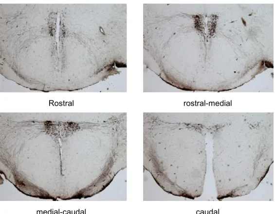
2 Effects of 19 days of CSC on AVP and OXT expression in the PVN ... 91
2.1 Number of AVP positive neurons in the PVN ...91
2.2 Number of OXT positive neurons in the PVN ...93
3 Effects of 19 days of CSC on the negative feedback regulation at different levels ... 94
3.1 Analysis of the negative feedback at the level of the pituitary ...94
3.1.1 GR and MR mRNA and protein expression ...94
3.1.2 Dexamethasone suppression test (DST) ...96
3.1.3 FKBP51 protein expression ...97
3.1.4 GR protein expression in the nuclear fraction ...97
3.2 Analysis of the negative feedback at the level of the hippocampus ...99
3.2.1 Absolute hippocampus weight and number of hippocampal cells ...99
3.2.2 GR and MR mRNA and protein expression ...99
3.2.3 FKBP51 protein expression ... 101
3.2.4 Per1 hnRNA expression in hippocampal cells in vitro ... 101
3.2.5 Hippocampal cell viability in response to corticosterone (CORT) stimulation ... 102
3.3 Analysis of the negative feedback at the level of the PVN ... 104
3.4 Analysis of the negative feedback at the level of the PFC ... 105
3.4.1 GR and MR protein expression ... 105
3.4.2 FKBP51 protein expression ... 106
4 Influence of trait anxiety on the consequences of 19 days of CSC ... 107
4.1 Anxiety-related behaviour on the EPM ... 107
4.2 Assessement of physiological parameters ... 110
4.3 Assessement of neuroendocrine parameters ... 112
4.3.1 Plasma CORT and ACTH concentrations ... 112
4.3.2 Adrenal in vitro ACTH responsiveness ... 114
4.4 Assessement of immunological parameters ... 115
4.5 GR, MR and FKBP51 protein expression in the pituitary ... 117
Chapter 4 - Discussion
1 Effects of CSC on HPA axis functionality – a closer look at the level of the pituitary and PVN ... 121
1.1 Do adaptations at the level of the pituitary contribute to changes in HPA axis functionality following CSC? ... 121 1.2 Are increased plasma ACTH levels mediated by the pituitary? ... 123 1.3 Is there an influence of the PVN on the increased plasma ACTH secretion?
... 126 2 Effects of CSC on the negative feedback inhibition at different
levels ... 129 2.1. Effects of CSC on MR, GR and FKBP51 expression in different brain regions ... 129 2.2 Effects of CSC on the negative feedback at the level of the pituitary ... 136 2.3. Summary of the negative feedback analysis ... 138 3 CSC and the influence of trait anxiety on the individual stress
vulnerability ... 140 3.1 Effects of CSC on behavioural, physiological, neuroendocrine and
immunological parameters ... 141 3.2 Why are mice of the low-anxious phenotype resilient to the consequences of CSC? ... 149 4. Summary and concluding remarks ... 152
Addendum
Summary in German - Deutsche Zusammenfassung ... 157
References ... 165
Abbreviations ... 199
CV and publications ... 203
Acknowledgements - Danksagung ... 205
Author`s declaration – Eidesstattliche Erklärung ... 207
CHAPTER 1
G ENERAL I NTRODUCTION
1 Stress
In 1865 the French physiologist Claude Bernard (1813 – 1878) was the first one describing the importance of a stable internal equilibrium of an organism compared to the fluctuating external environment. This idea was rendered more precisely over half a century later by Walter Cannon (1871 – 1945) defining the internal equilibrium and its maintenance within an adequate range as
“homeostasis” (Cannon, 1929). Disruption of this homeostasis, e.g. through exposure to cold, traumatic pain or emotional distress, activates the adrenal medulla and the sympathetic nervous system (SNS), coined together the
“sympathoadrenal” system. This promotes the “fight or flight” response which restores the internal milieu. It was in 1936 when Hans Selye, with his groundbreaking paper “A syndrome produced by diverse nocuous agents”, defined “stress” as “the non-specific response of the body to any physical demand” (Selye, 1936a). He also established the term “stressor”. Stressors, which can be physical or chemical in nature, cause the physiological stress response (Selye, 1975).
The modern concept of stress research entitles stressors as consciously or unconsciously sensed threat to homeostasis (McEwen and Stellar, 1993;
Goldstein and Kopin, 2007). The specificity of the stress response is dependent on the particular type of stressor, the way in which the organism perceives the stressor, and the personal ability to cope with it (Goldstein and Kopin, 2007).
1.1 The Stress System
Following stressor exposure the two main stress response systems, the SNS and the hypothalamic-pituitary-adrenal (HPA) axis, get activated. Both systems, differing in their time course and in their effector hormones, coordinate the restoration of the homeostasis of the organism following stressor exposure, a processed called allostasis (Sterling et al., 1988). Moreover, also behavioural responses to a stressful experience take place, like increased arousal, alertness, heightened attention, anxiety or aggression, while feeding or sexual behaviour
are suppressed (Stratakis and Chrousos, 1995; Herman and Cullinan, 1997;
McEwen and Wingfield, 2003). In the following sections the SNS and the HPA axis are discussed in detail, with the main focus directed on the latter one.
1.1.2 Sympathetic nervous system (SNS)
In response to stressful stimuli, the SNS, together with the parasympathetic and enteric nervous system forming the autonomic nervous system, gets rapidly activated and controls cardiovascular, gastrointestinal, respiratory and other somatic systems (Chrousos, 1998). The control station of the SNS is the locus coeruleus (LC), located in the brain stem and containing a cluster of norepinephrine (NE)-containing neurons. The LC-NE system innervates nearly the entire central nervous system (CNS) (Berridge and Waterhouse, 2003) and gets activated by diverse stressors, e.g. shock, hypotension, swim and social stressors (Valentino and Van Bockstaele, 2008), probably mediated by the afferents of the paraventricular nucleus (PVN) which innervate dendrites in the LC (Reyes et al., 2005). The LC also projects to sympathetic preganglionic neurons, emphasizing its role in regulating the SNS (Valentino and Van Bockstaele, 2008).
The cell bodies of the preganglionic neurons originate in the intermediolateral column (lateral horn) at the thoracic and lumbar region (“thoracolumbar system”) of the spinal cord. These sympathetic preganglionic neurons synapse with postganglionic neurons located in ganglia which lie on each side of the spinal cord, the sympathetic trunk. The postganglionic neurons innervate the peripheral organs, like e.g. heart and blood vessels leading amongst others to vasoconstriction and increase in the heart rate (Abboud, 2010) (see Fig. 1).
Sympathetic preganglionic neurons also can directly innervate the adrenal medulla, for stimulating the release of catecholamines from chromaffin cells which are embryologically and anatomically homologous to the sympathetic ganglia. As the adrenal medulla consists of postganglionic neurons which originate from the SNS and release their neurotransmitters directly into the blood stream, the adrenal functions as an endocrine gland (Elenkov et al., 2000). While
the transmitter of the preganglionic neurons is acetylcholine, the end organs get stimulated by catecholamines secreted by the postsynaptic sympathetic fibers, thereby, NE/noradrenaline represents the major neurotransmitter (Holgert et al., 1998). In contrast, the adrenal gland secretes mainly epinephrine/adrenaline and only to a lesser extent NE following sympathetic stimulation (Holgert et al., 1998).
The catecholamines bind to α- and β-adrenoreceptors (α1, α2, β1, β2, β3) which belong to the family of seven-transmembrane domain receptors, also known as G-protein coupled receptors (GPCR) (Minneman et al., 1981). The catecholamines released in response to activation of the SNS prepare the organism for the “fight or flight” response. The major effects are increase in heart and breathing rate, blood pressure, attention and mobilization of energy while e.g. digestion is reduced.
Figure 1: Schematic representation of the sympathetic nervous system (SNS). The preganglionic neurons of the SNS start from the first thoracic segment (T1) and extend to the second lumbar segment (L2) of the spinal cord. Their synapses with the postsynaptic neurons lie in small sympathetic ganglia, forming a chain near the spinal cord, called the sympathetic trunk. The postganglionic neurons run from the ganglia to the target organs.
[taken and adapted from: wps.aw.com/bc_martini_ fap_9_oa/185/47590/12183 208.cw/- /12183 232/ index.html]
Eye Salivary glands Heart
Lung Liver
Stomach Spleen
Intestine Adrenal medulla Kidney Urinary bladder Penis
Scrotum Uterus
Ovary Sympathetic
Chain ganglia Preganglionic
neurons Postganglionic
neurons
Spinal cord T1
L2 Pons
1.2.2 Hypothalamic-Pituitary-Adrenal (HPA) axis
Along with the SNS, the HPA axis gets, albeit in a delayed manner, activated in response to stressor exposure. In the following section the components of the HPA axis, i.e. hypothalamus, pituitary, and adrenal, are discussed in detail (see Fig. 2).
Figure 2: Schematic illustration of the HPA axis. Corticotropin-releasing hormone (CRH) and arginine vasopressin (AVP) are released from parvocellular neurons of the paraventricular nucleus (PVN) of the hypothalamus. Neurons of the PVN reach the median eminence and release CRH and AVP into the portal blood system. They trigger the release of adrenocorticotropic hormone (ACTH) from corticotroph cells of the anterior pituitary into the blood stream. ACTH in turn binds to its receptor in the adrenal cortex, stimulating the synthesis and release of glucocorticoids (GC, cortisol in humans, corticosterone in rats and mice) which negatively signal back to pituitary and PVN. [taken and adapted from (Lightman and Conway-Campbell, 2010)]
Glucocorticoids (GC)
The Paraventricular nucleus of the Hypothalamus
The activation of the HPA axis starts in a distinct set of neurons located in the parvocellular PVN, one nucleus of the hypothalamus (Whitnall, 1993). These neurons produce the main adrenocorticotropic hormone (ACTH) secretagogues, corticotropin releasing hormone (CRH) and arginine vasopressin (AVP) (Sawchenko et al., 1984; Whitnall et al., 1985; Aguilera, 1994; Herman and Cullinan, 1997). The PVN gets excitatory input from ascending brainstem systems, circumventricular organs and hypothalamic-basal forebrain pathways. In detail, catecholaminergic neurons from the lower brainstem, mainly activated in response to systemic stress, activate the PVN (Sawchenko and Swanson, 1982;
Ziegler and Herman, 2002). Innervation from the circumventricular organs, activated during osmotic stress, is angiotensinergic and stimulates release and synthesis of CRH (Kovacs and Sawchenko, 1993; Ziegler and Herman, 2002).
The majority of projections to the PVN originates from the dorsomedial nucleus of the hypothalamus, medial preoptic area, anterioventral third ventricular region and ventral medial, posterior medial and intermediate divisions of the bed nucleus of the stria terminalis (BNST) (Ziegler and Herman, 2002). These local projections can be either excitatory or inhibitory in nature whereby most of them are gamma-aminobutyric acid (GABA)ergic and, therefore, inhibit the PVN and in turn HPA axis activity (see also Sec. 3.5). Limbic activation of the HPA axis is associated with the amygdala, from where the information is relayed via catecholaminergic pathways in the brainsteam or via the lateral BNST to the PVN (Herman and Cullinan, 1997; Ziegler and Herman, 2002).
The PVN is comprised of magnocellular and parvocellular neurons, whereupon HPA axis activation is supposed to be mainly regulated by the latter one (see Fig.
3). Both cell types can be differentiated according to their localization, their cell size, and their synthesized peptides (Swanson and Kuypers, 1980; van den Pol, 1982; Ma et al., 1999). The CRH-containing axons of the parvocellular neurons, whereby about 50% of the neurosecretory cells coexpress AVP (Sawchenko et al., 1984; Whitnall et al., 1985; Aguilera, 1994) project to the external zone of the median eminence, located at the bottom of the brain and release CRH and AVP
in the pituitary portal circulation from where they can directly target the anterior pituitary (Gillies et al., 1982; Rivier and Vale, 1983). While AVP alone is a weak ACTH secretagogue (Vale et al., 1983) it plays a role in regulating the HPA axis during acute and chronic stress (see also Sec. 4).
Magnocellular neurons of the PVN and also of the supraoptic nucleus (SON) express the neuropeptides Oxytocin (OXT) and AVP (Brownstein et al., 1980) and either project to the posterior pituitary from where the neurohormones are directly released into the blood stream or they can also project to limbic regions via their axon collaterals (Neumann and Landgraf, 2012). OXT is mainly associated with parturition and lactation, but it also exhibits anxiolytic properties and is implicated in the stress response (Cook, 1997; Hashiguchi et al., 1997;
Neumann et al., 2000; Landgraf and Neumann, 2004; Waldherr and Neumann, 2007; Neumann and Landgraf, 2012). OXT release was demonstrated in response to various acute stressors, like shaker stress (Nishioka et al., 1998), forced swim (FS) (Wotjak et al., 1998) or Morris water maze testing (Engelmann et al., 2006). However social defeat (SD) elicited no increased OXT release in the PVN and SON (Wotjak et al., 1996), indicating stressor-dependend differences in the OXT release. HPA axis activation in response to an acute stressor was shown to be suppressed by OXT (Neumann et al., 2000; Windle et al., 2004).
Magnocellular AVP, also known as antidiuretic hormone, controls water conservation in the kidney and is released upon osmotic stimulation (Leng et al., 1999; Stricker and Sved, 2002) and also regulates vasoconstriction (Altura and Altura, 1984).
Autonomic parvocellular neurons which express CRH and to a lesser extent other neuropeptides project to the brainstem and spinal cord (Swanson and Kuypers, 1980) and are involved in the regulation of the sympathoadrenal system (Aguilera and Liu, 2012). CRH is also present in other brain regions, mainly in limbic structures, like the BNST, the central amygdala (CeA), LC, cerebral cortex, cerebellum and the dorsal root neurons of the spinal cord (Sawchenko and Swanson, 1985). CRH in these brain regions acts as a neurotransmitter and is implicated in the behavioural response to stress.
Figure 3: Schematic illustration of the different subregions of the PVN. The parvocellular division (pc) contains neurons expressing corticotrophin releasing hormone (CRH) and arginine vasopressin (AVP). Both are released in the pituitary portal circulation, stimulating the release and synthesis of adrenocorticotropic hormone (ACTH) in the anterior pituitary. The magnocellular division (mc) of the PVN and of the supraoptic nucleus (SON) contain AVP and oxytocin (OXT) expressing neurons which project to the posterior pituitary and mediate the release of AVP and OXT in the peripheral circulation.
The autonomic parvocellular neurons (ap) contain mainly CRH and to a lesser extent other neuropeptides and project to the brainstem and spinal cord. [taken and adapted from (Aguilera and Liu, 2012)]
The Pituitary
The pituitary gland, situated at the basis of the brain, is comprised of the anterior pituitary, consisting of the anterior and intermediate lobes and the neuropituitary also known as posterior lobe or posterior pituitary. While anterior and intermediate lobes consist of oral ectoderm, the posterior pituitary originates from neural tissue, i.e. from the infundibulum (Kelberman et al., 2009) (see Fig. 4).
pc mc
CRH AVP
AVP OXT
AVP OXT
ACTH
AVP OXT posterior pituitary
anterior pituitary 3rd
ventricle
ap
SON
Figure 4: Rat pituitary section stained with antibody against γ-melanocyte stimulating hormone. The staining reveals the three regions of the pituitary; the anterior lobe/anterior pituitary, the intermediate lobe and the posterior lobe/posterior pituitary or neuropituitary. PL, posterior lobe, AL, anterior lobe; IL, intermediate lobe [taken and adapted from (Bicknell, 2008)]
The anterior pituitary contains five major cell types: the corticotrophs, thyrotropes, gonadotropes, somatotropes and lactotropes, producing different types of hormones which regulate adrenal function, thyroid function, reproduction, growth, and lactation, respectively. Corticotroph cells, comprising about 10 – 20 % of the total cell population of the anterior pituitary, produce ACTH and constitute the prinicipal site for HPA axis regulation (Yeung et al., 2006). The posterior lobe receives input from the axon terminals of the magnocelluar neurons of the PVN and also of the SON (Kelberman et al., 2009). In contrast, the anterior pituitary receives the endocrine information from parvocellular neurons via the portal blood (see also Fig. 3).
CRH released from these parvocellular neurons binds to the membrane-bound CRH receptor 1 (CRH-R1), the main subtype in pituitary corticotroph cells (Chalmers et al., 1996). CRH receptors, the CRH-R1 and the CRH receptor 2 (CRH-R2), are members of the GPCR and are coupled to adenylatcyclase which stimulates cyclic adenosine monophosphate (cAMP) synthesis and proteinkinase A (PKA) activity. CRH can bind to both receptor types but shows a higher affinity for the CRH-R1. The CRH-R1 is not only expressed in the pituitary but also widely distributed in the brain, especially in the cerebral cortex, cerebellum, amygdala, hippocampus, LC, and in the olfactory bulb (Grigoriadis et al., 1996).
In the periphery it is amongst others localized in the adrenal, heart, spleen, colon, skin, testis, and placenta (Hillhouse and Grammatopoulos, 2006). In contrast, the CRH-R2 is widely expressed in the peripheral blood vessels, skeletal, smooth, and cardiac muscles but only in distinct areas of the brain, like lateral septal
IL
AL
AL
PL
nuclei, ventromedial nucleus of the hypothalamus, PVN, and BNST (Grigoriadis et al., 1996; Hillhouse and Grammatopoulos, 2006).
A binding protein for CRH (CRH-BP), located at different sites of the CNS, modulates CRH actions and probably acts as a negative regulator of CRH signalling (Thomson, 1998; Seasholtz et al., 2001). Moreover, some CRH-like peptides, the urocortins (urocortin 1, urocortin 2, urocortin 3), are distributed throughout stress-sensitive central and peripheral structures like the pituitary, gastrointestinal tract, testis, immune tissue, kidney, heart, adrenal, peripheral blood cells, muscle, and skin (Aguilera et al., 2004). While urocortin 1 binds to CRH-R1 and CRH-R2, Urocortin 2 and 3 selectively activate CRH-R2 (Dautzenberg and Hauger, 2002). The distribution pattern of the urocortins suggests a role in coordinating the stress response (Aguilera et al., 2004;
Jamieson et al., 2006).
CRH administration was shown to increase gene transcription of pro- opiomelanocortin (POMC), the precursor of ACTH, in the anterior and intermediate lobe in in vivo as well as in in vitro studies (Affolter and Reisine, 1985; Lundblad and Roberts, 1988; Autelitano et al., 1990). After CRH binding to its receptor, cAMP and PKA activate via both calcium (Ca2+)- dependent and independent transduction signals the mitogen-activated protein kinase (MAPK) pathway which in turn activates Nur77 and Nurr1, two transcription factors (TF) involved in regulation of POMC expression (Murphy and Conneely, 1997;
Kovalovsky et al., 2002).
As already mentioned before, POMC is only a precursor protein, from which different end products can be generated by endoproteolytic cleavage through proproteins and prohormone converting enzymes in a tissue specific manner (Marcinkiewicz et al., 1993). POMC is produced in corticotroph cells of the anterior pituitary and in melanotroph cells of the intermediate lobe (Bicknell, 2008). In the anterior pituitary, POMC, in a first step, is processed to pro-µ- melanocyte stimulating hormone, ACTH, and β-Lipotrophin. A main proportion of β-Lipotrophin is then further cleaved to β-endorphin. In the intermediate lobe POMC is cleaved to corticotrophin-like intermediate peptide, α- melanocyte
stimulating hormone, µ-Lipotrophin, and β–Endorphin whereby these products can be further processed (Bicknell, 2008).
In addition to its role in regulating POMC expression, CRH binding to CRH-R1 leads, as described above, to the activation of the PKA, which in turn - via a so far unknown pathway - opens voltage-dependent Ca2+ channels (VDCC), leading to increased influx of extracellular Ca2+ and eliciting the release of ACTH vesicles into the blood stream (Won et al., 1990; Kuryshev et al., 1996). Moreover, extracellular Ca2+ influx, via binding to a ryanodine receptor, enhances intracellular Ca2+ release which further promotes ACTH release (Yamamori et al., 2004) (see Fig. 5).
AVP receptors also belong to the family of GPCR and are divided into three major subtypes: AVP receptor 1a (AVPR-1a), AVP receptor 1b (AVPR-1b or V3), and V2. While the V2 receptor, which regulates the water resorption in the kidney, is coupled to Gs signalling via adenylate cyclase, the AVPR-1a, localized in smooth muscle cells and in the liver, and the AVPR-1b, localized in corticotroph cells, are coupled to Gq and signal via phospholipase C (PLC) (Aguilera and Rabadan-Diehl, 2000a).
While AVP alone is a weak stimulus for ACTH secretion, together with CRH it acts in a synergistic way on the corticotroph cells (Gillies et al., 1982; Antoni, 1993; Aguilera, 1994; Lightman, 2008). Activated PLC cleaves phosphatidylinositol 4,5-bisphosphate (PIP2) into diacylglycerol (DAG) and inositol 1,4,5-trisphosphate (IP3). IP3 promotes the release of Ca2+ from IP3 – sensitive intracellular stores (Tse and Lee, 1998) while DAG, in addition, activates proteinkinase C (PKC) which stimulates influx of extracellular Ca2+ via L-type Ca2+ channels (Won et al., 1990) (see Fig. 5). The synergistic effect of AVP and CRH on ACTH release seems to be mediated by the activation of PKC which contributes to the CRH-stimulated cAMP production by inhibiting the activity of phosphodiesterases leading in turn to a reduced degradation of cAMP (Abou-Samra et al., 1987). Interestingly, there are also studies describing heterodimers of CRH-R1 and AVPR-1b, probably contributing to the synergistic effect of CRH and AVP (Young et al., 2007) (see Fig. 5).
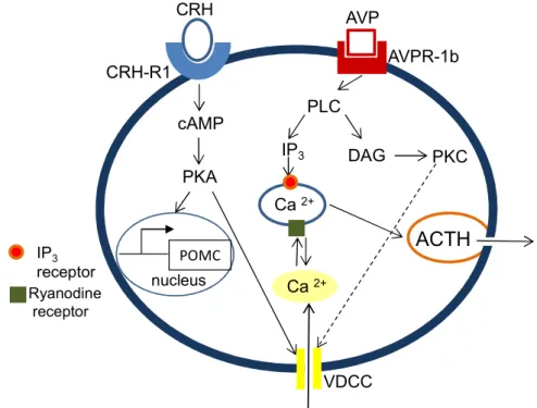
Figure 5: Schematic illustration of the intracellular pathways involved in corticotropin releasing hormone (CRH)- and arginine vasopressin (AVP)-mediated ACTH release in pituitary corticotroph cells. CRH binds to the CRH receptor 1 (CRH- R1), activates proteinkinase A (PKA) via cyclic AMP (cAMP) and stimulates pro- opiomelanocortin (POMC) gene transcription or triggers the influx of Ca2+ via voltage- gated channels (VDCC). Ca2+ that entered the cell via the VDCC can in turn stimulate the release of intracellular Ca2+ via binding to the ryanodine receptor. AVP, by binding to the AVP receptor 1b (AVPR-1b) activates phospholipase C (PLC) and via inositol 1,4,5- trisphosphate (IP3) promotes intracellular Ca2+ release. Diacylglycerol (DAG) can activate proteinkinase C (PKC) which can act synergistically with CRH to increase intracellular Ca2+. Ca2+ is of importance for the fusion of the ACTH vesicles with the plasma membrane of the cell. [adapted from (Yamamori et al., 2004)]
Adrenal gland
ACTH released from the pituitary into the blood stream can bind to the melanocortin-2-receptor (Mc2r) at the adrenal cortex and triggers the synthesis and release of glucocorticoids (GC, cortisol in humans, corticosterone (CORT) in rats and mice) (Elias and Clark, 2000). The adrenal is composed of the adrenal cortex, producing primarily steroid hormones, and the adrenal medulla,
CRH-R1 AVPR-1b
cAMP PKA
nucleus POMC
PLC
IP3 DAG PKC Ca2+
VDCC Ca2+
ACTH
Ryanodine receptor
IP3 receptor
CRH AVP
composed of mainly chromaffin cells, modified postganglionic sympathetic neurons, secreting primarily the catecholamines ephinephrine/adrenaline and NE/noradrenaline upon stimulation by the sympathetic preganglionic neurons (Diaz-Flores et al., 2008) (see also Sec. 2.1.2). In detail, the adrenal cortex consists of three layers. The innermost layer, the zona reticularis produces GC and in some species like in humans also androgens, estrogens and progestins.
The outermost layer, the zona glomerulosa, synthesizes and secretes mineralocorticoids (aldosterone) for controlling the salt and water balance in the kidney. The medial layer, the zona fasciculata, synthesizes the main amount of GC (Rosol et al., 2001). GC secretion is mainly stimulated by ACTH, but also other hormones produced by the medulla, like adrenaline, have a stimulating effect (Bremner et al., 1996).
The Mc2r belongs to the family of melanocortin receptors (Mc1r to Mc5r), members of the GPCR, and stimulates the activation of adenylatcyclase and the production of cAMP. Within minutes, the protein expression of the steroid acute regulatory protein - expressed in all steroid hormone-producing cells (adrenal, gonads) - is increased which stimulates steroidogenesis (Cherradi and Capponi, 1998; Miller, 2008). Mc2r mRNA expression was also shown to be increased following stimulation with ACTH in human as well as in mouse adrenocortical cells in vitro (Mountjoy et al., 1994).
GC, mainly released from the zona fasciculata and zona reticularis, belong to the family of steroid hormones. Because of their common origin and their similar effectiveness with the mineralocorticoids they are denotet corticosteroids. While in humans the cortisol is the main corticosteroid (Nishida et al., 1977), in mice and rats it is CORT. GC can exert diverse effects on the whole body. For instance they play a major role in energy mobilization by increasing the blood glucose levels, they are implicated in modulation of the immune systems as well as in the regulation of cardiovascular functions (Sapolsky et al., 2000). Moreover, GC play a central role in the basal HPA axis activity as well as in the termination of the stress response by exerting a negative feedback at different levels of the HPA axis (see also Sec. 3.5).
1.2 Acute vs. chronic/repeated stress
The term “stress“ is mostly used in a negative sense, whereby it is often buried in oblivion, that the acute stress response constitutes one of the major survival mechanisms of an organism. The synchronal interplay between all physiological systems, resulting in coordinated activation of cardiovascular, locomotor, neuroendocrine and also immune system facilitates the “fight or flight” reaction (Dhabhar, 2009). Sterling and Eyer (1988), therefore, coined the term “allostasis”, meaning that the body returns to homeostasis by active processes. To achieve this allostasis, the stress hormones, mainly catecholamines and GC from the adrenal medulla and the adrenal cortex, respectively, are released (McEwen, 1998b).
Stress is composed of a series of events, starting with a stimulus (stressor) that is perceived by the brain (stress perception), disrupts the homeostasis and in turn activates physiological systems in the body (stress response) to restore the physiological balance (Dhabhar, 2009). The acute stress response is beneficial and adaptive for the body, contributes to the well-being of an organism, promotes survival, and increases its biological fitness. In contrast, repeated or chronic stressor exposure can have deleterious effects, promoting the development of somatic as well as affective disorders (McEwen, 1998a; Chrousos, 2009).
McEwen and Stellar in 1993 introduced the term “allostatic load“ describing the long lasting burden and negative consequences of prolonged activation of the stress system (McEwen and Stellar, 1993).
Important criteria for distinguishing between acute and chronic stress are duration and intensity of the stressor. While acute stress lasts from minutes to hours, chronic stress occurs from days to month (Dhabhar, 2000). The intensity may be estimated by measurement of stress hormone levels, GC and catecholamines, and of heart rate and blood pressure (Dhabhar, 2000).
An acute stressor elicits a peak in blood GC levels within 15 – 30 min and a decline to basal levels 60 – 120 min later (Paskitti et al., 2000; de Kloet et al., 2005). In contrast, mice exposed to chronic SD over a period of 3 weeks (Keeney et al., 2006; Hartmann et al., 2012a) or exposed to chronic subordination in the
visible burrow system for 14 days (Albeck et al., 1997) showed sustained elevated plasma CORT levels compared with control mice. This prolonged release of GC can have deleterious effects on the organism, e.g. the damage of the hippocampus (McEwen, 1998a) and inhibition of the immune function (Dhabhar, 2000). Nevertheless, there are also chronic stress paradigms resulting in unchanged or even decreased plasma GC levels. For instance, in rats, repeated exposure to noise (4 h/day for 3 weeks) did not affect basal plasma CORT levels (Armario et al., 1986), whereby chronic social isolation for 3 weeks resulted in reduced basal plasma CORT levels compared with controls (Adzic et al., 2009; Djordjevic et al., 2012).
Chronic stress paradigms can be further divided in homotypic (same stimulus) or heterotypic (different stimuli) stressors whereby homotypic stressor exposure commonly results in adaptation of the stress response (Bartolomucci, 2007;
Wood et al., 2010). As the homeostasis of an organism is important to guarantee survival, such adaptational mechanisms are indispensable to cope with repeated internal as well as external stimuli or stressors which are not life threatening.
However, despite adapting to repeated innocuous stimuli, it is important for an organism to respond to a novel and perhaps dangerous challenge, in an adequate manner. Nevertheless the mechanisms underlying this adaptation and sensitization process are only poorly understood (Aguilera, 1994). The adaptation of the ACTH response, in-depth described in a review of Aguilera (1994), during chronic stress involves a bulk of mechanisms, with the regulation of the ACTH secretagogues CRH and AVP as well as their receptors on the pituitary, and the GC feedback playing a major role (Aguilera, 1994).
Three different patterns, characterized by the degree of adaptation to the persistent stressor and the magnitude of response to a novel, heterotypic superimposed stressor, are described. The first one is characterized by a desensitization of the ACTH response to a repeated/chronic stressor (physical or psychological stressor like repeated immobilization) and a hyperresponsiveness to the novel stressor (see Fig. 6 top). In the second pattern, no desensitization of the ACTH release (repeated painful stimuli like ip hypertonic saline injection) but
a hyperresponsiveness to the novel stimulus is observed (see Fig. 6 bottom). The third pattern is characterized of a small and transient increase in plasma ACTH (e.g. prolonged osmotic stimulation during water deprivation) and a hyporesponsivenss to the novel stressor (Aguilera, 1994).
Figure 6: Plasma adrenocorticotropic hormone (ACTH) release during adaptation to repeated/chronic stressor exposure. Shown are plasma ACTH levels in controls and rats subjected to repeated immobilization (2 h/day, upper panel) or ip injection of hypertonic saline (5 ml, lower panel) for 14 days under basal conditions and following exposure to an acute novel stressor (ip hypertonic saline injection in the upper group and 30 min immobilization in the lower group). [Taken and adapted from (Aguilera, 1994)]
According to Aguilera and colleagues, AVP, under basal conditions only a weak stimulator of the ACTH release, becomes more and more attention as an important factor in the process of adaptation to repeated/chronic stressors and in the hyperresponsiveness to a novel heterotypic stressor, mediated by its synergistic effect on CRH-mediated ACTH secretion (Aguilera, 1994; Aguilera and Rabadan-Diehl, 2000a). Interestingly, following repeated immobilization, a significant increase in CRH neurons coexpressing AVP (from 50 % in controls to 90 % in chronically stressed animals) (Bartanusz et al., 1993) and also an increase in AVP mRNA (Ma and Lightman, 1998; Ma and Aguilera, 1999) has been described. This indicates that stress paradigms associated with a hyperresponsiveness to a novel heterotypic stressor (repeated immobilization,
repeated hypertonic saline injection) increase AVP expression while prolonged osmotic stress elicited no enhanced AVP activation in parvocellular neurons (Aguilera, 1994).
Also regulation of CRH mRNA expression depends on the type and duration of the homotypic stressor (Aguilera, 1998; Aguilera and Rabadan-Diehl, 2000a). In stress paradigms associated with sustained ACTH responses, like foot shock (Imaki et al., 1991; Sawchenko et al., 1993) or ip hypertonic saline injection (Ma and Aguilera, 1999), CRH mRNA levels are elevated. In contrast, following repeated restraint stress (Ma and Lightman, 1998; Ma et al., 1999) or colony- housing (De Goeij et al., 1992), associated with desensitization of the ACTH response, CRH levels are unchanged.
As indicated above, the expression of AVP increases in chronic stress paradigms associated with hyperresponsiveness to a novel heterotypic stressor but not during osmotic stimulation (Aguilera, 1994; Aguilera and Rabadan-Diehl, 2000a).
Besides changes in PVN AVP expression, thereby, regulation of pituitary AVPR- 1b expression seems to play a major role, as there is a good correlation between receptor content in the pituitary and corticotroph ACTH secretion in response to a superimposed heterotypic stressor (Aguilera, 1994; Aguilera et al., 1994; Aguilera and Rabadan-Diehl, 2000b). Under chronic stress conditions associated with reduced ACTH release in response to a novel stressor (chronic osmotic stimulation), AVP receptors decrease, while stress paradigms with enhanced ACTH response to a novel stressor (repeated immobilization, repeated hypertonic saline injection) result in receptor up-regulation (Aguilera and Rabadan-Diehl, 2000b), whereby mRNA not always correlates with AVP binding (Rabadan-Diehl et al., 1995; Rabadan-Diehl et al., 1997). The increase in AVPR- 1b expression seems to be mediated by AVP as well as by other neuropeptides in the pituitary portal blood and also by GC. While increased AVP mRNA expression in the PVN positively correlated with the number of AVP-R1b receptors and ACTH response to a novel stressor following chronic stress (Aguilera, 1994), GC were found to reduce AVPR-1b binding despite increasing its mRNA levels as well as the coupling of the receptor to PLC. The increased
coupling of the AVPR-1b to PLC facilitates the corticotroph response to AVP in spite of eleveated GC levels (Rabadan-Diehl and Aguilera, 1998; Aguilera and Rabadan-Diehl, 2000b).
Given that in response to many chronic stress paradigms AVP, in contrast to CRH expression, is increased, this suggested for a long time that AVP plays a primary role in adaptation of the HPA axis response to chronic stimulation (de Goeij et al., 1991; Ma et al., 1997). However, studies conducted in AVP deficient Brattleboro rats or following pharmacological blockade of AVP receptors refuted the hypothesis of AVP becoming the main regulator during chronic stressor exposure. Administration of an AVPR-1b antagonist significantly reduced the response to acute restraint stress or lipopolysaccharide stimulation in rats (Spiga et al., 2009). AVP deficient Brattleboro rats showed a normal response to most acute stressors and only a slight reduction in ACTH release during repeated restraint stress (Zelena et al., 2004). Moreover, administration of a non-selective V1 receptor antagonist was not able to block the hyperresponsiveness of repeatedly restraint rats to an acute stressor, indicating that AVPR-1b up- regulation, described for this stress paradigm, is not required for the sensitization of the ACTH response (Chen et al., 2008). Overall, these studies suggest that AVP is important for the full ACTH response in some acute stress paradigms, but it is not necessary for the ACTH hyperresponsiveness to a superimposed acute heterotypic stressor, indicating that AVP might play an alternative role in the process of stress adaptation (Aguilera et al., 2008). A role for AVP in stimulating proliferation of corticotroph cells has been shown in the murine corticotroph tumor cell line AtT20 (van Wijk et al., 1995), during GC deficiency and chronic stress (Subburaju and Aguilera, 2007).
In chronic stress paradigms associated with ACTH hyperresponsivenss to a heterotypic stressor and also in chronic stress models associated with maintained ACTH release (repeated hypertonic saline injection) CRH-R1 gets down-regulated and desensitized, indicated by a reduced cAMP and ACTH response to CRH (Kiss and Aguilera, 1993; Aguilera, 1994). However, for the CRH-R1 it could be demonstrated that the receptor number in the pituitary does
not always correlate with the pituitary responsiveness. Thus, a decreased CRH binding following chronic homotypic stressor exposure (e.g. repeated restraint) resulted in increased POMC mRNA expression (Aguilera et al., 2001). In addition, there is no correlation between CRH-R1 binding and CRH-R1 mRNA levels, suggesting that CRH-R1 down-regulation seems to be mediated by changes in the translation efficiency and/or receptor internalization or desensitization (Aguilera et al., 2001). Down-regulation of CRH-R1 binding can be ascribed to CRH and was shown to be facilitated by concomitant administration of AVP (Hauger and Aguilera, 1993). In addition, administration of GC decrease CRH binding in vivo and in vitro (Childs et al., 1986; Schwartz et al., 1986; Hauger et al., 1987) while CRH-R1 mRNA levels decreased only transiently (Rabadan-Diehl et al., 1996; Iredale and Duman, 1997), suggesting an inhibition of the CRH-R1 by GC at the post-transcriptional level. Overall the studies indicate, that i) the number of CRH-R1 is not indicative for the corticotroph responsiveness, as a small number of receptors is sufficient for a full ACTH response and that ii) post-transcriptional mechanisms play the major role in the regulation of its expression (Aguilera et al., 2004).
1.3 Psychosocial stress
Selye`s proposed concept of the general adaptation syndrome only referred to physical stressors like cold exposure or surgical injury (Selye, 1936b).
Nevertheless, it became more and more evident, that the type of stressor, e.g.
psychological, social or physical in nature, determines the consequences on behaviour and physiology. While water deprivation produced a duration- dependent anxiolytic effect on the elevated plus-maze (EPM), restraint stress (1 h) had an opposite effect even though plasma CORT levels were increased in both groups (McBlane and Handley, 1994). These differences in stress responsiveness clearly indicate that stress research should focus on those types of stressors that reflect the natural condition a mammalian species, especially humans, are normally exposed to, in order to find treatment strategies for e.g.
stress-related disorders. In humans, the most naturalistic type of stressor every
individual is exposed to is of social and/or psychological nature (Brown and Prudo, 1981; Bartolomucci et al., 2005). Psychological stress can be caused by environmental demands that are thought to exceed the individual’s capability to deal with, implying the cognitive appraisal of the stimulus (Sapolsky, 2005;
Cohen et al., 2007). Social stress is mainly the result of alterations in the social life of an individual, concerning the relationship and interaction between individuals including e.g. disputes over resources or the social rank (Blanchard et al., 2001). Given that chronic psychosocial stressors are acknowledged risk factors for the development of stress-related disorders in humans (Hemingway and Marmot, 1999; Wahrendorf et al., 2012), animal models mimicking this chronic psychosocial burden are important and powerful tools for studying the physiological, neuroendocrine and immunological mechanisms that underlie the development of these diseases.
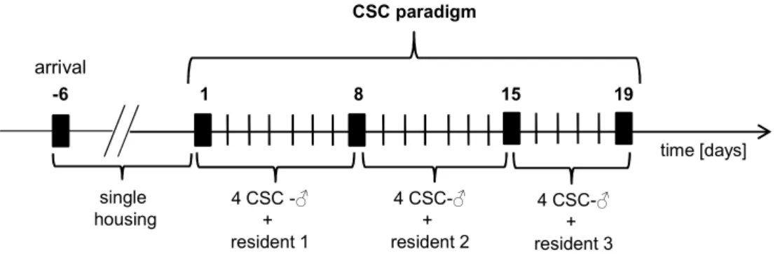
The chronic subordinate colony housing (CSC, 19 days) paradigm constitutes one of these appropriate animal models as it induces chronic psychosocial stress in male mice. Thereby four experimental mice are housed together with a larger dominant male mouse for 19 consecutive days, resulting in a number of physiological, neuroendocrine, immunological and also behavioural alterations.
CSC mice develop somatic as well as affective disorders (Reber et al., 2007;
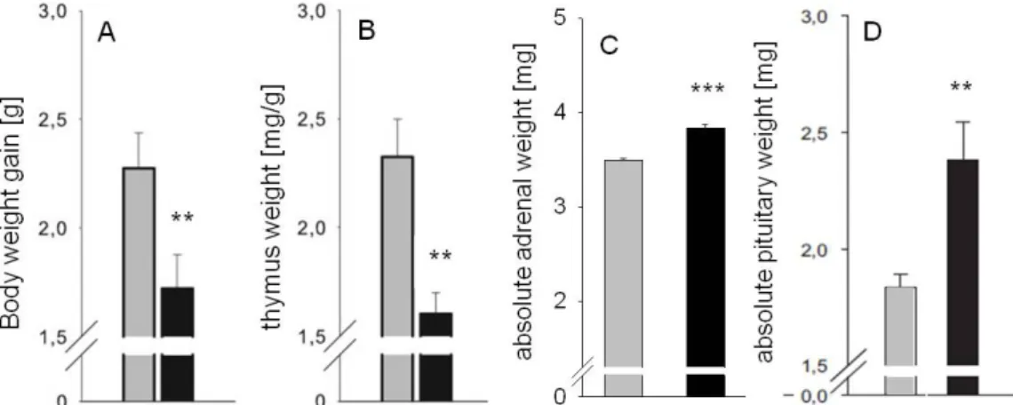
Slattery et al., 2012; Uschold-Schmidt et al., 2012) and display typical signs of chronic stress, like adrenal hypertrophy and thymus involution (Reber et al., 2007; Uschold-Schmidt et al., 2012). Moreover, following CSC exposure, mice exhibit basal hypocorticism in the evening and show a GC resistance in different cells in the periphery (Reber et al., 2007). But, interestingly, in response to an acute heterotypic stressor exposure (5 min elevated platform (EPF)), they show an exaggerated CORT release (Uschold-Schmidt et al., 2012). This indicates an adaptation to the chronic stressor exposure and a concomitant sensitization of the HPA axis in response to an acute challenge, at least at the level of the adrenal glands. Whether or not changes at higher HPA axis levels, for instance at the pituitary, are also contributing to these adaptation/sensitization processes have not been analysed in detail yet.
2 GC Signalling
Upon binding of ACTH to the adrenal cortex GCs are released into the blood stream to exert a variety of effects, which are determined by a number of factors, like the availability of the corticosteroids in the cell as well as the type of receptor.
2.1 Corticosteroid availability
The activity of the HPA axis, i.e. ACTH and GC release, follows a circadian rhythm. Peak cortisol levels are secreted at the end of the resting period, i.e. in the morning. In contrast, nocturnal animals like rodents show the CORT peak at the end of the afternoon immediately before the start of the dark phase (Young et al., 2004; Lightman et al., 2008). Coordination of this rhythmic release pattern is orchestrated by the suprachiasmatic nucleus of the hypothalamus. This nucleus controls on the one hand CRH and AVP release (Reppert and Weaver, 2002) and on the other hand, via the autonomic nervous system, the adrenal gland`s sensitivity to ACTH (Ulrich-Lai et al., 2006), preparing the organism for the increased metabolic demand during the activity phase.
Not only the release of GC from the adrenal glands is of importance but also the availability in the target tissue is regulated by multiple factors. Corticosteroids can be bound to specific carrier proteins, e.g. the corticosteroid-binding globulin (CBG) or albumin and they are also subjected to enzymatic conversions, both influencing the availability and, thus, bioactivity of these hormones.
CBG is a glycoprotein, synthesized in the liver, to which under normal conditions about 80 – 90 % of circulating corticosteroids are bound. 10 – 15 % are bound to albumin, resulting in only about 5 - 10 % of free corticosteroids (Cizza and Rother, 2012). As only free corticosteroids are able to enter the target cells, the binding proteins constitute an important mechanism to protect, particularly the brain, from the dangerous effects of high corticosteroid levels. Regulation of CBG levels enables a fast mechanism of adaptation of the corticosteroid levels to different situations, e.g. ultradian or circadian variations, stress (Qian et al., 2011) or disease. Levels of circulating CBG rise in response to an acute stressor in
order to restrain the CORT rise (Qian et al., 2011) while chronic stressor exposure evoques a decrease in CBG leading to an increase in the free corticosteroid levels (Stefanski, 2000; Henley and Lightman, 2011)
Entering the brain involves the passage of the blood-brain barrier, composed of specialized endothelial cells, protecting the brain from probably damaging compounds and maintaining the neuronal homeostasis. While the small and highly lipophilic CORT can pass the blood-brain barrier via diffusion, the penetration of cortisol as well as of synthetic GC, e.g. dexamethasone (Dex), is limited as they are substrates for the multidrug resistance (mdr) 1a P- glycoprotein, located in the membrane of endothelial but also of other peripheral cells (Meijer et al., 1998; Karssen et al., 2001). In the cell, tissue specific enzymes, the most common one is the 11ß-hydroxysteroid dehydrogenase (11ß- HSD), regulate the interconversion of active GC (cortisol, CORT) and their inert 11-keto forms (cortisone, 11-dehydrocorticosterone). 11ß-HSD type 1 is expressed in the liver, adipose tissue, bone, lung and pituitary and in the CNS, especially in the hippocampus, cerebellum and neocortex, catalizing the conversion of the 11-keto in its active form (Harris et al., 2001; Cooper and Stewart, 2009). The type 1 form of this enzyme, therefore, plays a major role in regulating GC levels in the brain under basal and stress conditions (Harris et al., 2001). 11ß-HSD type 2 is mainly expressed in mineralocorticoid target tissue, like kidney, colon, and salivary glands, converting GC in their inactive form, rendering them unable to bind to the mineralocorticoid receptor (MR) and, therefore, enabling aldosterone to exert its effects by binding to the MR (Cooper and Stewart, 2009).
2.2 Corticosteroid receptor types in the brain
The actions of the corticosteroids are mediated by two types of receptors; namely the MR and the glucocorticoid receptor (GR). Their names already implicate the processes they are involved in, mineral balance (Yang and Young, 2009) and gluconeogenesis (Revollo and Cidlowski, 2009), respectively. Binding studies revealed, that the MR binds GC with a 10-fold higher affinity (KD ~ 0.5 nM) than
GR (KD ~ 5 nM) (Reul and de Kloet, 1985; de Kloet et al., 1998). MR are mainly distributed in limbic brain structures with the highest density in the hippocampus and the lateral septum, while low expression levels are found in the amygdala, hypothalamus and also outside of the brain in the pituitary (Moguilewsky and Raynaud, 1980; Reul and de Kloet, 1985). GR are distributed all over the brain, e.g. in the lateral septum, hippocampus, nucleus tractus solitarii, LC, amygdala, and PVN (Reul and de Kloet, 1985). According to the binding studies, low freely circulating GC levels in the brain during the diurnal through (0.5 – 1 nM) already occupy most of the MR (Reul et al., 2000), whereas GR become additionally occupied at higher CORT levels, i.e. at the circadian peak and during stress (Reul and de Kloet, 1985). As under most circumstances the MR is already occupied, it is hypothesized that for the MR its protein expression while for the GR the ligand concentration constitute the most important regulators (de Kloet et al., 2000). For the GR, two main isoforms, the GRα and the shorter form, the GRß, are described which differ in their carboxy termini. While GRα binds GC and mediates a variety of GC effects, the GRβ is unable to bind GC and is, therefore, transcriptionally inactive and, in addition, can inhibit the transcriptional activity of GRα (Oakley et al., 1999; Revollo and Cidlowski, 2009). For the MR four isoforms exist, with the MRα and MRß representing the two main types which show different tissue-specific expression patterns (Zennaro et al., 1995;
Zennaro et al., 1997).
GR and MR are members of an evolutionary conserved family of nuclear receptors which are composed of three functional domains. The N-terminal transactivation domain with an activation function motif (AF-1) for the interaction with the transcriptional machinery and/or TF, the central DNA-binding domain (DBD) containing two zinc fingers for interaction with the DNA and receptor dimerization, and the C-terminal ligand-binding domain, containing a ligand- binding motif, a nuclear localization signal (NLS) and a further activation motif (AF-2) for interaction with other TF (Giguere et al., 1986; Bamberger et al., 1996;
Revollo and Cidlowski, 2009) (see Fig. 7).
Figure 7: Schematic illustration of the protein structure of the human GR. The gene is composed of a N-Terminal region, containing an activation function (AF-1) motif, a DNA binding domain (DBD), a C-Terminal region, containing a nuclear localization signal (NLS) and another activation motif (AF-2). [adapted from (Revollo and Cidlowski, 2009)]
The properties of the different domains already give insight into the functions of these receptors. The unactivated receptor is bound to a multiprotein complex consisting of molecular chaperones, the heat shock proteins (hsp90 and hsp70), which serve the proper folding of peptides and proteins in the cytoplasm. Other co-chaperones, like the acidic protein p23 and immunophilins that bind via their tetratricopeptide repeat (TPR) domain to a TPR domain on the hsp90 complement the complex (Tai et al., 1992; Hutchison et al., 1993; Pratt and Toft, 1997). Immunophilins, disposing peptidylproly-isomerase activity, bind immunosuppressive drugs, like FK506, rapamycin and cyclosporine A. Some of these participate in the corticosteroid receptor-hsp90 complex, e.g. FK506- binding protein 51 (FKBP51) and FK506-binding protein 52 (FKBP52). Given that both bind to the same site on the hsp90 independent heterocomplexes can be created (Pratt and Toft, 1997). The whole complex around the GR regulates the receptor folding, maturation, activation, and also trafficking into the nucleus (Grad and Picard, 2007). Upon ligand binding primarily a conformational change of the receptor takes place, exposing two NLS which promote the translocation into the nucleus (Picard and Yamamoto, 1987). The receptors then either form dimers which bind to the DNA on so called GC-responsive elements (GRE) enabling the transcription of different target genes or they can also act as monomers. The general opinion was that the GR has to dissociate from the hsp90 chaperone complex in order to promote translocation (de Kloet et al., 1998). Nevertheless, pharmacological inhibition of hsp90 has been shown to lead to a delay in nuclear translocation of the GR-ligand complex (Czar et al., 1997). Therefore, the translocation concept gets renewed by conveying the immunophilins FKBP51
AF-1 DBD NLSAAF-2 AF-2
N-Terminal C-Terminal
DNA Binding
and FKBP52 an important role in hormone-bound receptor trafficking (Galigniana et al., 2010a). It suggests that FKBP51, bound to hsp90, has to be exchanged by FKBP52 which binds to dynein, enabling the translocation to the nucleus via microtubules (Davies et al., 2002; Galigniana et al., 2010b) (see Fig. 8).
Figure 8: Schematic illustration of the concept of glucocorticoid receptor (GR) nuclear translocation. In the cytoplasm, the GR is associated with heatshock proteins (hsp 90 and hsp70), two acidic proteins (p23) and one immunophilin, the FK506-binding protein 51 (FKBP51). Once CORT binds to the GR receptor complex FKBP51 is exchanged against FKBP52. In turn, dynein can bind and the receptor complex translocates into the nucleus through the nuclear pore where it can bind to the DNA. To simplify the illustration only a monomer of the receptor complex is depicted. [adapted from (Binder, 2009)]
FKBP51 and FKBP52
Since their discovery, the FK506 binding proteins gained more and more attention as they represent important regulators of GR (Denny et al., 2000) and also MR (Gallo et al., 2007). As already mentioned before, FKBP51 binds to hsp90 via a TPR-domain and lowers the receptors affinity for its ligand (Wochnik et al., 2005). The significance for FKBP51 was discovered in New World Monkey, a monkey line characterized by extremely high cortisol levels compared with other monkeys or humans. But, surprisingly, they show no behavioural or
GR
hsp90
hsp70
FKBP51 p23 p23
FKBP52 dynein
CORT
physiological signs known to be linked with hypercorticism, as the target organs exhibit GC resistance mediated by an overexpression of FKBP51 (Denny et al., 2000). In addition, in vitro studies demonstrated that increased FKBP51 levels not only reduce the affinity of the receptor for its ligand but also reduce subsequent nuclear translocation (Wochnik et al., 2005). Interestingly, GC induce FKBP51 transcription via GR which in turn restrains GR functionality, forming part of a short negative feedback loop regulating GR activity (Vermeer et al., 2003).
Scharf and colleagues (2011) could show that FKBP51 is not only expressed in peripheral tissues but also in GR-expressing brain regions, like hippocampus, amygdala, and PVN. Stressor exposure was shown to induce FKBP51 expression whereby the basal expression pattern influenced the extent to which its expression was induced. Regions containing low levels of FKBP51 under basal conditions showed a higher induction compared with regions showing higher basal expression levels. An evidence for FKBP51 regulating GR sensitivity and that high levels result in lower GR responsiveness which suggests the use of baseline FKBP51 levels as marker for GR sensitivity (Binder, 2009; Scharf et al., 2011). Genetic polymorphisms in the gene encoding for FKBP51, associated with an increased FKBP51 expression, lead to decreased negative feedback regulation of the stress hormone system and, therefore, increase the risk for development of psychiatric disorders, like major depression, bipolar disorder, and post-traumatic stress disorder (Binder, 2009).
In opposite to FKBP51, FKBP52 is a positive regulator for trafficking of GR and MR into the nucleus via interaction with dynein (Silverstein et al., 1999;
Galigniana et al., 2010b). FKBP52 also stimulates GR-mediated gene transcription (Ning and Sanchez, 1993) but not the transcriptional activity of the MR (Gallo et al., 2007).
2.3 Transcriptional Regulation
Upon translocation into the nucleus, MR and GR can modulate gene transcription by binding to a GRE or by interaction with other TF (Beato and Sanchez- Pacheco, 1996). GR and MR have a nearly identical DBD recognizing the GRE
on the DNA in the proximity of gene promoters where they can bind either as homo- (Karst et al., 2000) or heterodimers (Liu et al., 1995; Ou et al., 2001). It is hypothesized that GR are more potent transcription activators than MR and that heterodimers have different functions compared to homodimers (de Kloet et al., 2000). Hence, according to the GC concentrations, three different dimer conformations are possible, that can bind to different DNA targets and regulate a variety of target genes in a distinct manner (Kellendonk et al., 2002).
Heterodimers are observered predominantely at higher concentrations, i.e. under conditions of stressful events (Nishi and Kawata, 2007). Nevertheless not much is known about the exact differences between the binding of hetero- or homodimers to the promoter sequence of different genes. Upon binding to the GRE, a number of co-factors are recruited to the receptor complex and interaction with the general transcription machinery takes place. Transcription of target genes is then either enhanced, by binding to positive GRE, a process called transactivation, or repressed, by binding to negative GRE. Negative GRE are less commonly observed, but some are part of the negative feedback process of the HPA axis (see also Sec. 3.5) or of the immune system (Zhang et al., 1997; Datson et al., 2008).
GR can also interact as monomer with other TF, e.g. activator protein 1, cAMP response element binding protein, or nuclear factor-kappa B, that are activated by other signalling pathways in response to inflammation or immune activation.
Normally, GR repress the effects of these TF on gene transcription (transrepression), accounting for the inhibitory effects of GR on the immune system (de Kloet et al., 1998; Datson et al., 2008) (see Fig. 9). The transcriptional response is further regulated by phosphorylations of the receptor by cell-specific kinases (e.g. MAPK, cyclin-dependent kinase CDK, glycogen synthase kinase GSK-3) which can modulate the receptor activity (Galliher- Beckley and Cidlowski, 2009).
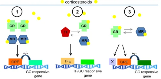
Figure 9: Molecular mechanisms of glucocorticoid actions on gene expression. (1) Homodimerization: Upon binding of corticosteroids, mineralocorticoid receptor (MR) or glucocorticoid receptor (GR) can translocate into the nucleus, form homodimers and bind to specific elements on the DNA, the GC-responsive elements (GRE), in the promoter region of GC-responsive genes and increase (transactivation) or decrease their transcription. (2) Transrepression: Activated GR can interact with transcription factors (TF) by direct protein-protein interaction, inhibiting the binding of the TF to the TF elements (TFE), resulting in a down-regulation of TF and/or GC-responsive genes. (3) Heterodimerization: Activated MR and GR can build heterodimers and bind to GRE or yet unknown DNA elements (indicated by X), affecting expression of GC-responsive genes.
[adapted from (de Kloet et al., 1998)]
The transcriptional response mediated by the GR is highly dynamic, displaying different expression waves over the time. In hippocampal slices, administration of 100 nM CORT down-regulated a majority of genes after 1 h, up-regulated some genes after 3 h and following 5 h of stimulation, gene transcription was nearly back to baseline (Morsink et al., 2006).
GC regulate a wide number of genes involved in cellular processes like energy metabolism (e.g. glycolysis and gluconeognesis), signal transduction, regulation of neurotransmitters and their receptors, e.g. AVP-R1a, OXT receptor and in neuronal plasticity, e.g. the expression of the clock gene period 1 (Per1).
Importantly, they are also implicated in the regulation of the HPA axis and in corticosteroid signalling itself (Datson et al., 2008; Lightman and Conway- Campbell, 2010).
GR
MR GR
MR
GR
MR
TF GR MR
GRE
GC responsive gene +/-
TFE
TF/GC responsive gene
GRE
GC responsive gene +/- X
corticosteroids
1 2 3