Synthesis of compound libraries for the study of glucose uptake and splicing inhibition
For the achievement of the academic degree of the
Faculty of Chemistry and Chemica Technical University Dortmund
Javier de Ceballos Cerrajería
ynthesis of compound libraries for the study of glucose uptake and splicing inhibition
Dissertation
For the achievement of the academic degree of the Doctor in Natural Sciences
(Dr. rer. nat.)
Submitted to
Faculty of Chemistry and Chemical Biology Technical University Dortmund
by
Javier de Ceballos Cerrajería
From Santander, Spain
Dortmund 2017
ynthesis of compound libraries for the study of
glucose uptake and splicing inhibition
The following work was performed from September 2012 until February 2017 under the supervision of Prof. Dr. Herbert Waldmann at the Max Planck Institute for Molecular Physiology in Dortmund.
1st Examiner: Prof. Dr. Herbert Waldmann
2nd Examiner: Prof. Dr. Daniel Summerer
Some of the work described in this doctoral thesis can be found in the scientific publication:
Identification of a small molecule inhibitor that stalls splicing at an early step of spliceosome activation
Anzhalika Sidarovich, Cindy L. Will, Maria M. Anokhina, Javier Ceballos, Sonja Sievers, Dmitry E. Agafonov, Timur Samatov, Penghui Bao, Berthold Kastner, Henning Urlaub, Herbert Waldmann, Reinhard Lührmann, eLIFE, just accepted.
Dedicated to Helena and my family for their continuous support.
1. Abstract/Zusammenfassung
Despite having the same "purpose", the uncontrollable cellular growth to the detriment of the organism, cancer presents itself in humans in great variety of forms. This variety makes developing effective treatments a highly challenging task. Nevertheless, cancer has a handful of hallmarks that are present almost ubiquitously in all types of tumours. Among them is the upregulated glucose uptake, known as the Warburg effect. However, there has yet to be a drug or treatment that makes use of this aberrant cancer metabolism. This is due to the lack of potent glucose uptake inhibitors. The identification of small molecules that efficiently target the glucose uptake is hence of high value. Through an in-house established glucose uptake assay, new scaffolds were identified as glucose uptake inhibitors, such as the Glupins. This novel identified scaffold presented a natural product-like structure and a high potency. In the present work, a synthetic route towards the Glupin core was established. The structure-activity relationships of the Glupin class was extensively studied by the synthesis of a library of analogues. The analogue, (+)-Glupin-1, was discovered as one of the most potent glucose uptake and was therefore selected for further biological studies.
The splicing process, the process by which premature messenger RNA is transformed into a mature form ready to be translated, is performed by a highly complex machinery, the spliceosome. The splicing process occurs in a series of steps, each of them catalysed by a different spliceosome complex constituted by many proteins and ribozymes. Decades of research on the spliceosome complex have provided an understanding of the process but much remains to be discovered. The in-house identification of a novel splicing inhibitor, cp028, enabled the isolation of a previously unknown spliceosome stage. In this work, the cp028 compound was studied with the synthesis of a library of analogues around its core. The library of analogues was assayed as splicing inhibitors yielding a SAR analysis of this novel class.
Furthermore, efforts towards the identification of the cp028 target were made. Cp028 joins a short list of powerful splicing inhibitor that have allowed for a deeper study of the splicing process and opens the door for the identification of novel key protein players of this highly complex process.
Krebs ist das unkontrollierte Wachstum von Zellen zum Nachteil des Gesamtorganismus. Auf zellulärem Niveau sind die Eigenschaften von Krebszellen jedoch sehr unterschiedlich, was eine effektive Behandlung von Tumorerkrankungen erschwert. Es gibt jedoch einige Merkmale, die von fast allen malignen Tumorzellen geteilt werden, unter anderem ein veränderter zellulärer Metabolismus. Dieser äußert sich häufig in erhöhter Aufnahme von Glukose und einer erhöhten Glykolyserate, auch Warburgeffekt genannt. Bisher fehlen jedoch effektive Wirkstoffe, die dieses Charakteristikum von Krebszellen nutzen, um selektiv Krebszellen zu adressieren. Die Identifikation von wirksamen Hemmern der Glukoseaufnahmen ist deshalb von großem Interesse. Durch einen intern etablierten zellulären Glukoseaufnahmetest im Hochdruchsatzverfahren konnten verschiedene kleine Moleküle mit gemeinsamen Grundstrukturen, die die Glukoseaufnahme von kultivierten Krebszellen effizient inhibieren identifiziert werden. Eine dieser Substanzklassen, die Glupine, basiert auf einem naturstoffähnlichen Grundgerüst, dessen Synthese im Rahmen dieser Arbeit etabliert wurde und auf dessen Grundlage eine Substanzbibliothek hergestellt wurde um eine Struktur- Wirksamkeitsbeziehung zu erstellen. Die Substanz (+)-Glupin-1 wurde als wirksamstes Mitglied dieser Molekülklasse identifiziert und für weitere biologische Studien genutzt.
Das Spleißen von prä-mRNA wird von einer fein regulierten molekularen Maschinerie aus Enzymen und Ribozymen gesteuert, dem Spleißosom. Obwohl seit vielen Jahrzehnten untersucht, sind viele Details des komplexen Assemblierungsprozess des Spleißosoms bis heute nicht komplett verstanden. Mit Hilfe eines neu identifizierten Inhibitors des Spleißens, cp028, konnte ein bisher unbekannter Spleißosom-Komplex entdeckt werden. Auf Grundlage dieses Inhibitors wurde im Rahmen dieser Arbeit eine Substanzbibliothek hergestellt und auf ihre Aktivität in Bezug auf die Inhibierung des Spleißens getestet. Cp028 erweitert die bisher kurze Liste von Inhibitoren des Spleißings und eröffnet die Möglichkeit neue Schlüsselproteine dieses hoch komplexen Prozesses zu identifizieren.
Table of Contents
1. Abstract/Zusammenfassung 9
Glucose Uptake Inhibition
2. Introduction 15
2.1. Glucose Uptake 15
2.1.1. The GLUT transporter family 15
2.1.2. GLUT structure and glucose uptake mechanism 16
2.1.3. Regulation of glucose uptake 17
2.1.4. Glucose uptake as apart of glucose metabolism 19
2.2. Glucose Uptake in Cancer: the Warburg Effect 20
2.3. Targeting Glucose Metabolism in Cancer 23
2.3.1. Targeting hexokinase 24
2.3.2. Targeting pyruvate kinase 25
2.3.3. Targeting lactate dehydrogenase 25
2.3.4. Targeting pyruvate dehydrogenase 25
2.4. Targeting the Facilitative Glucose Transporters 26
3. Aims of the Project 30
4. Results and Discussion: 31
4.1. Screening Assay and Compound Class Identification 31 4.2. Validation of the Selected Compound Classes 34
4.2.1. Compound class of triazole 5 34
4.2.2. Glupins 36
4.3. Synthesis of Glupins 39
4.3.1. One-pot synthesis of the tryptamine-morphan core scaffold 40
4.3.2. O-Methylation of alcohol 44 42
4.3.3. Indole N-alkylation 45
4.3.4. Synthesis (+/-)-Glupin-1 47
4.4. Determination of a Structure-Activity Relationship for the Glupin class 49 4.4.1. Validation of the tryptamine-morphan core scaffold of the glupin compound class 50
4.4.2. SAR analysis for position R1 51
4.4.3. SAR analysis for position R2 54
4.4.4. SAR analysis for position R3. 56
4.4.5. SAR analysis of position R4 58
4.5. (+) and (-) Glupin-1 62
4.6. Absolute Configuration of (+)-Glupin-1 63
4.7. Synthesis of the Chemical Probes Based on Glupin-1 67
4.8. Biological Characterization of Glupin-1 70
4.8.1. [3H]-Glucose uptake 70
4.8.2. Glucose uptake across tissues 70
4.8.3. Cell proliferation of MDA-MB 231 71
4.8.4. Changes in metabolites upon glupin-1 treatment 72
4.8.5. Inhibition of aminoacid starvation-induced autophagy 73
4.8.6. (+)-Glupin-1 reduces lipid droplet formation 73
Splicing Inhibition
5. Introduction 75
5.1. The Splicing Process 75
5.2. The Spliceosome 76
5.2.1. The spliceosome ribonucleoproteins: the snRNPs 76
5.2.2. The spliceosome cycle 77
5.2.3. The spliceosome structure 78
5.3. Splicing Inhibition 79
6. Aims of the Project 82
7. Results and Discussion 83
7.1. Assay and Compound Class Identification 83
7.2. Synthetic Route for Cp028 and Analogues 86
7.3. 1st SAR Round of Cp028 Analogues 87
7.3.1. Potential covalent inhibitor 88
7.3.2. SAR analysis for the synthesised analogues 89
7.3.3. SAR analysis for the commercial set of analogues 92
7.4. 2nd SAR Round of Cp028 Analogues 94
7.5. Target Identification of Cp028 96
7.5.1. Synthesis of chemical probes based on cp028 96
7.5.2. Affinity-based proteomics 100
7.5.3. Targeting kinases involved in the splicing process 100 7.6. Characteriazation of the stalled B028 spliceosome complex. 103
8. Summary of the Thesis 104
9. Experimental Part: Glucose Uptake Inhibition 108
10. Experimental Part: Splicing Inhibition 159
11. List of Abbreviations 174
12. References 177
13. Curriculum Vitae 185
14. Acknowledgements 187
Glucose Uptake Inhibition
2. Introduction
2.1. Glucose Uptake
The monosaccharide D-glucose is the principal source of energy in humans as it fuels the tricarboxylic acid cycle (TCA). Furthermore, it is a main source for anabolic pathways that feed from the TCA cycle and is involved in glycosylation processes. Hence, glucose is a key player in metabolism and cell homeostasis. It is the hydrolysis productof more complex sugars such as starch, sucrose or lactose, obtained from the diet. Although potentially synthesised by the liver, the levels of glucose in blood and tissues are mainly modulated by the synthesis and hydrolysis of the polysaccharide glycogen, which is used as a glucose reserve.The uptake of glucose and other hexoses into cells can be promoted by two different types of transporters: the sodium- glucose transporters (SGLTs) and the facilitative glucose transporters (GLUTs).1-2 The SGLTs are a family of symporters that use the Na+-K+ gradient to actively transport glucose into the cells. Given this mode of transport, these symporters are situated where glucose needs to be absorbed or recovered against the glucose gradient at the membrane, such as the intestines or the renal tubules.The main two transporters of this family are SGLT 1 and 2.The first has a high affinity but low capacity of glucose transport and is present at the small intestine membrane and at the proximal tubules(kidneys). The SGLT2, on the other hand, hasa low affinity but a high capacity and ispresentmostly at the proximal tubules (kidneys). The second family of transporters responsible for glucose uptake is the GLUT family.
2.1.1. The GLUT transporter family
The family of GLUTs is a group of transporters that promote glucose diffusion across the membrane in a bidirectional manner according to the concentration gradient. This family is responsible for glucose uptake in the vast majority of tissues and presents a wide variety of affinities, substrate specificities and different cellular localisations.2 This array of characteristics ensures a tight control of glucose uptake into the different tissues according to cellular requirements and to glucose availability. The GLUT family comprises 14 members which are classified into three subfamilies according to sequence similarities (Figure 1). The most studied subfamily is the well-characterizedclass 1, that comprises the GLUT1 to 4 plus the recently identified GLUT14.3 The main difference within this subgroup is the distribution across tissues:
GLUT1, ubiquitously expressed, is responsible for the basal supply of glucose to all tissues but
exhibits higher expression on erythrocytes and the brain, whereit representsthe main transporter at the blood brain barrier.4-5 GLUT1 has a Km of 3-7 mM for glucose uptake.Since the average glucose concentration in blood is around 5mM, GLUT1 has the ability to transport glucose at a high rate.6The second member, GLUT2, displays a comparatively low affinity for glucose (Km
of 15-20 mM) and other sugars but has a high transport capacity.6 It is mostly expressed on the liver, pancreas and kidney, and its main function is believed to be glucose-sensing.7-8 GLUT3 presents the highest affinity for glucose (Km of ca. 2 mM) and is highly expressed in the neurons at the brain, where glucose is in high demand and of high priority.2 GLUT14 is almost identical to GLUT3 but is specifically expressed in the testis.3 The last GLUT transporter of the class 1 is GLUT 4, whose main characteristic is its insulin-dependent regulation. GLUT4 is stored in vesicles and is recruited to the membranes upon insulin stimulation.It is mostly expressed in adipocytes, and heart and muscle tissue.9
Figure 1. Dendrogram of all members of the GLUT transporter family. The numbers represent the percentages of sequence identity. Adapted from Scheepers et al.2
2.1.2. GLUT structure and glucose uptake mechanism
The GLUT transporters share a common core structure of two symmetrical six transmembrane α-helix domains, where both termini face the intracellular side (Figure 2a). The transmembrane domains form a cavity that allows glucose to diffuse by the rocker-switch alternative-access mechanism, in which the substrate-binding site is available from both sides of the membranevia conformational changes (Figure 2b).10 The recent solution of the crystal structures of several GLUT transporters has enabled a deeper understanding of the mechanism and functions of the different members.11-13 The GLUT1 structure was reported in 2014 by Deng and co-workers in an inward-open conformation bound to nonyl-glucopyranoside.11 This structure complements,
65%
55%
35-42%
42%
59%
29%
35%
45%
41%
GLUT1 GLUT4 GLUT3 GLUT14 GLUT2 GLUT5 GLUT7 GLUT9 GLUT11 GLUT6 GLUT8 GLUT10 GLUT12 HMIT
Class 1
Class 2
Class 3 95%
and was resolved with the aid of, the previously reported bacterial homologue of glut1-4, crystallized in an outward-facing conformation bound to glucose.14
Figure 2. a) Illustration of the human GLUT1 transporter crystal structure (PDB: 4PYP).11 Adapted from Kapoor et al.15 The drawing shows the N-terminal (yellow) and the C-terminal (orange) domains and the intracellular (IC) domains (green). Cytochalasin B is represented by the red-stick structure at its binding site. The α-helix domains are represented as rods.b) Rocker- switch predicted alternative access transport for the GLUT1transporter. Adapted from Denget al.11
2.1.3. Regulation of glucose uptake
Glucose uptake can be considered controlled, at its simplest stage, by the presence or lack of glucose in the extracellular media. This is particularly true for unicellular organisms in which up-regulation or down-regulation of the glucose metabolism is mostly driven by an extracellular nutrient-sensing mechanism. Nevertheless, in higher organisms such as humans, the extracellular media has no shortage of nutrient supply. Consequently, the glucose metabolism needs to be regulated by external signalling factors (e.g. growth factors) to prevent, for example, aberrant cell proliferation. The GLUT transporters are regulated by a series of transcriptional pathways and direct protein or compound interactions. When a greater glucose metabolism is needed, for example in proliferating cells, signalling factorspromote the up-regulation of the glucose uptake capacity. This occurs by modulating the GLUT transporters ability, as for the growth factor IL-3 (interleukin-3),16 or by over-expressing the GLUT transporters at the membrane, as for the hormone insulin.17 The main mechanism of glucose uptake regulation is insulin secretion by the pancreas, which transiently recruits GLUT4 to cell membranes of
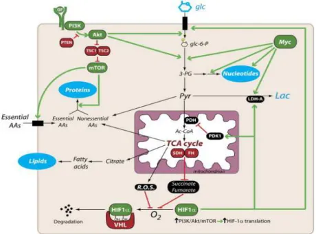
insulin-responsive tissues. The PI3K/Akt/mTOR pathway is another important regulator of glucose metabolism by, among other mechanisms, increasing the presence of GLUT1 at the cell surface (Figure 3).18-19 Furthermore, the PI3K/Akt/mTOR pathway also regulates the activation of HIF-1(hypoxia inducible factor-1 alpha),which promotes the over-expression of the GLUT transporters.20 HIF-1 activation can also be triggered by alterations in ROS levels or in hypoxic conditions (Figure 3). The transcription factor MYC has also been shown to control the expression of the GLUT1 transporter as well as other glycolytic enzymes (Figure 3).21
Figure 3. Transcriptional activation of GLUT1 expression regulated by the PI3K/Akt/mTOR signaling pathway, the transcription factors HIF-1 and Myc. Adapted from DeBerardinis et al.19
Another mechanism for the regulation of glucose uptake is the direct interaction with natural compounds such as methylxanthines and ATP, or proteins such as Stomatin. Methylxanthines bind to the external face of the transporter in a non-competitive mode of inhibition promoting a conformational change that down-regulates glucose uptake.22 Glucose uptake is also regulated by glycolysis and the oxidative phosphorylation product ATP. Hence, at high ATP/ADP ratios, the triphosphate analogue binds to GLUT1 promoting conformational changes that decrease the transport activity.23-24 The transmembrane protein Stomatin has also been shown to interact and modulate the function of GLUT1.25
Some post-translational modifications can also influence GLUT activity. For example, phosphorylation of GLUT1 by protein kinase C (PKC) is reported to increase its cell surface localisation and hence glucose uptake.26 This phosphorylation is promoted by growth factors
such as VEGF or by aberrant mutations. N-glycosylation of the glucose transporter-1 is required for maintaining a high affinity for glucose and, as a consequence, a high glucose uptake.27
2.1.4. Glucose uptake as apart of glucose metabolism
The GLUT transporters represent the first proteins involved in the glycolytic route to convert glucose to pyruvate, which generatestwo molecules of ATP in the process. The glycolytic route is an anaerobic energy-producing process which is highly conserved from the early unicellular organisms, where it represents the main energy source, to high organisms that make further catabolic use of pyruvate to increase the free energy release of the overall process. The glycolytic enzymes include key glucose metabolism regulators like hexokinase (HK),28 or phosphofructokinase (PFK).29-30 The GLUT transporters have no affinity for glucose-6- phosphate and since the transport of glucose goes according to the concentration gradient, HK directly regulates the glucose uptake rate. Furthermore, TCA intermediates such ascitrate or ATP are known inhibitors of PFK, and hence of the glycolytic route (Figure 4).
Aerobic organisms, in terms of energy production, metabolize glucose down to carbon dioxide and water. The energy production of this process is18 times higher than anaerobic fermentation (the glycolytic route). The resulting pyruvate from glycolysis is transferred to the mitochondria where it is transformed into acetyl-CoA. Acetyl-CoA then feeds the TCA cycle generating the H+ and reduced cofactors (NADH and FADH2) necessary for oxidative phosphorylation(OXPHOS) (Figure 4). This pathway is highly conserved in complex organisms because of its high efficiency in ATP production. The mitochondrial metabolic activity is connected to the GLUT transporters through a series of complex signalling pathways, thus enabling "feedback" from energy production at the organelle to regulate glucose uptake. For example, elevation of ROS, generated in the mitochondria, plays an important part in activation of the HIF-1 transcription factor and hence promotes GLUT1 expression (see section 2.1.3.).20,
31 Increased glucose uptake results in the generation of higher quantities of NAD(P)H (mainly through the pentose phosphate pathway), that can revert the elevated ROS levels.32 The reverse process has also been reported. When GLUT1 is inhibited or mutated, the ROS levels increase.33
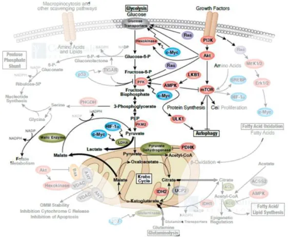
Figure 4. Signalling network around the glucose metabolic pathways. The glycolytic route occurs in the cytosol and is regulated by many transcriptional pathways and the allosteric inhibition of the glycolytic enzymes. Pyruvate can then enter the mitochondria to feed the TCA cycle. Illustration reproduced courtesy of Cell Signaling Technology, Inc.
(www.cellsignal.com).
In conclusion, the uptake of glucose, the main energy source and a major feed source to the anabolic routes, is highly regulated by the enzymes and signalling pathways involved in the glucose metabolic routes, glycolysis and the TCA cycle. Hence, variations in glucose uptake have an extended effect throughout cell metabolism.
2.2. Glucose Uptake in Cancer: the Warburg Effect
In 1923, Otto Heinrich Warburg (1883-1970, Nobel laureate in medicine in 1931)observed that, in the presence of sugar, cancerous tissues displayed a much higher (over 70-fold) lactic acid production than healthy tissues (Figure 5).34-35 Furthermore, he observed that the Pasteur effect (i.e. decrease or complete stop of the fermentation process in the presence of oxygen) did not
seem to apply to cancerous tissues. In normoxia (normal oxygen concentrations in the media), the tumours continued to produce high quantities of lactic acid.36 Warburg hypothesised that the presence of lactic acid fermentation indicated an impaired respiration in the tumours and that this impaired respiration was the cause of carcinogenesis. The presence of such aerobic glycolysis in cancer cells was later named the Warburg effect.
Figure 5. Changesin pressure caused by the carbon dioxide generated from the acidification of extracellular media in the presence of a bicarbonate solution. Picture taken from Warburg and Minami (1923).34 Columns (from left to right): Type of tissue. Tumour, liver, kidney and heart.
Weight of the tissue slice, without sugar. Change in pressure after 60 minutes, without sugar.
Weight of the tissue slice, with sugar. Change in pressure after 60 minutes, with sugar. Lactic acid generated per mg and h.
This hypothesis was soon to be refuted when studies revealed a functioning respiration mechanism across many types of cancers.37 Nevertheless, his observations have not been disputed and remain a hallmark in cancer metabolism. Contrary to healthy cells, excluding highly proliferative cells such as embryos, cancer cells engage, almost ubiquitously, in aerobic glycolysis, regardless of the extent to which respiration is performed. A high fermentation rate means a higher glucose demand which is met through the over-expression of the GLUT transporters, particularly the GLUT1 and GLUT3 members.38-39 The rationale behind this behaviour in cancer is still under debate, but it has become clear that the Warburg effect
provides an evolutionary advantage to the cancer cells compared to the surrounding tissues.40-42 In the past decade, the scientific community has accepted the hypothesis that cancerous cells engage in aerobic glycolysis to boost the anabolic routes with glycolytic intermediates.40, 43 This allows for fast cell division and tumour proliferation. This hypothesis is partly supported by the observed aerobic glycolytic phenotype in fast proliferating healthy cells such as lymphocytes.44 Nevertheless, aerobic glycolysis could appear as a hypoxia defense mechanism but also confers a phenotypic advantage to the tumour,which would further promote aerobic glycolysis regardless of the oxygen levels. For example, the acidification of the extracellular media has been hypothesised to confer a proliferation advantage to the tumour, which has adapted to it, in contrast to the healthy surrounding tissues to whichit becomes toxic, thus facilitating the tumour invasiveness.42 Overall, there is still not clear understanding of the original causes of aerobic glycolysis.
The mechanisms by which the aerobic glycolysis phenotype is achieved are not fully clear.
Many signalling pathways have been discovered to promote the necessary metabolic changes to acquire the aerobic glycolytic phenotype, but the molecular mechanism that triggers those pathways has yet to be fully understood. Since glycolysis is highly up-regulated, the pathways and transcription factors that regulate key glycolytic enzymes (see section 2.1.4.), such as glucose transporters or hexokinase, have been extensively studied.20, 45-46
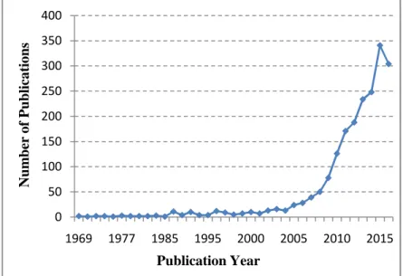
Figure 6. Number of publications per year since 1969. Source: Web of Science, under the theme search of "Warburg Effect".
0 50 100 150 200 250 300 350 400
1969 1977 1985 1995 2000 2005 2010 2015
Numberof Publications
Publication Year
In the last decade, the interest of the scientific community in the Warburg effect has increased exponentially (Figure 6). Being one of the hallmarks of cancer, the Warburg effect has lately been viewed as a potential window in the fight against cancer.47 Based on the high dependence of most cancers on glucose, deprivation of it would sensitize tumours towards cancer therapies.
This idea has sparked, at both the academic and industrial levels, the search for inhibitors of the glycolytic route, including glucose uptake.
2.3. Targeting Glucose Metabolism in Cancer
For many decades, the selectivity towards cancer cells in medicaltreatments has mainly been based on their higher proliferative character. Chemotherapeutic agents that target characteristic cell proliferation processes, such as cell division, have a stronger toxic effect on the cells with higher proliferation rates. Famous examples are Cisplatin, a chemotherapeutic drug that targets DNA replication, or the cytoskeletal natural product Taxol, which interferes with mitotic spindle assembly. Nevertheless, the recent characterization of alterations in tumour metabolism, such as the highly up-regulated glycolysis (see section 2.2.), have opened a new window for a more selective targeting of cancer.48 Consequently, a variety of drugs targeting tumour metabolism are currently under study, including in clinical trials.49
A very common metabolic alteration found in cancer is aerobic glycolysis. The increased glycolytic flux indicates a strong dependence on glucose metabolism, which if inhibited or dysregulated should impair cancer cell growth or viability. Consequently, several compounds have been developed targeting known upstream oncogene modulators of the glycolytic route, such asHIF, PI3K, AKT or mTOR.49 Furthermore, much research has also been dedicated to developing inhibitors of the enzymes involved in glucose metabolism (Figure 7).50-51
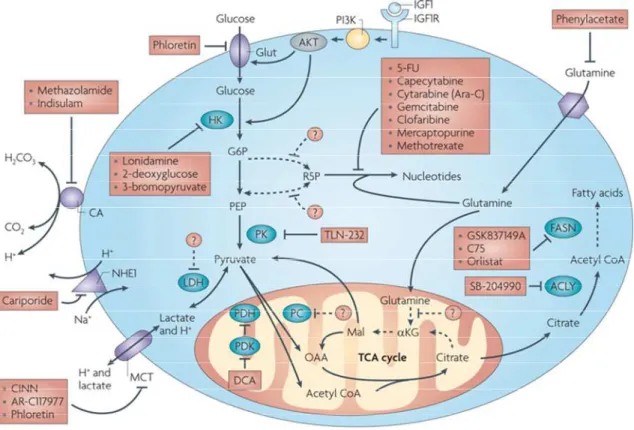
Figure 7.Metabolic pathways and enzymes with inhibitors that have entered clinical trials.
Figure from Tennant et al.49
Examples below targeting enzymes involved in glucose metabolism will be discussed. A selection was made for representative examples of compounds that have entered clinical trials, further validating modulation of the Warburg effect as a promising anti-cancer therapy. The GLUT inhibitors, which bear more importance for the work of this doctoral thesis, will be discussed in more detail (see section 2.4.).
2.3.1. Targeting hexokinase
The glycolytic enzyme hexokinase, particularly the isoform 2 (HK2), has been found up- regulated in many cancers and has thus received much attention as a potential cancer target, The glucose analogue 2-deoxyglucose (2-DG), which cannot be metabolised in glycolysis, acts as a competitive inhibitor of hexokinase, obstructing glycolytic flux (Figure 7). 2-DG has been subjected to several clinical trials against different solid tumours, alone or in combination with other chemotherapeutic drugs. 2-DG alone, however, has displayed very discouraging results in early clinical trials due partly to the fact that the maximal tolerated dose of 2-DG is approximately one order of magnitude below the average glucose concentrations in the blood.52
Nevertheless, 2-DG remains in clinical trials in combination with other drugs.53-54 Other hexokinase inhibitors that made it into clinical trialsare, 3-bromopyruvate (3-BrPA) and lonidamine. 3-BrPA, displayed good results against liver cancer when administered locally in animals but has yet to conclude successfully a clinical trial.55
2.3.2. Targeting pyruvate kinase
The final step of glycolysis, which is catalysed by pyruvate kinase (PK), consists on transferring the phosphate group from phosphoenolpyruvate to ADP, generating ATP and pyruvate (Figure 7). PK is considered a very promising target because in proliferative tissues, including tumours, an alternative spliced form (PKM2) is expressed rather than the normal tissue splice variant, PKM1.56 This selectivity window was addressed by Vander Heiden et al. by developing PKM2 inhibitors.57 It has also lead to compounds in clinical trials such as the cyclopeptide TLN-232.
This PKM2 inhibitor showed promising results in phase II of clinical trials against metastatic melanoma and renal cell carcinoma. Nevertheless, the PK is currently not considered as a useful target in cancer.
2.3.3. Targeting lactate dehydrogenase
Another important glycolytic enzyme targeted is lactate dehydrogenase (LDH) (Figure 7). LDH is the enzyme responsible for the conversion of pyruvate into lactate and has been found over- expressed in many cancers. The natural product gossypol was shown to inhibit LDH and, consequently, the synthesis of pyruvate. After lead optimization studies, the gossypol analogue AT-101 was developed and submitted to clinical trials. Nevertheless, positive results are scarce and cannot be directly linked to the inhibition of LDH.53
2.3.4. Targeting pyruvate dehydrogenase
Down-regulationof the aerobic glycolytic flux has also been achieved by “diverting” the glycolytic intermediate pyruvate to the mitochondrial respiration process rather than allowing it to be converted into lactate (as in aerobic glycolysis). Pyruvate dehydrogenase(PDH, the enzyme responsible for converting pyruvate into acetyl-CoA) can be activated by inhibiting a known PDH positive regulator, pyruvate dehydrogenase kinase (PDHK) (Figure 7). The result is a down-regulated aerobic glycolytic flux that can impair tumour growth. The small molecule dichloroacetate (DCA), was found to inhibit PDHK, thereby activating PDH and promotingtumour size reductions in mice. DCA was hence subjectedto several clinical trials against differenttumours, but no clear clinical benefits have yet been discovered.53
2.4. Targeting the Facilitative Glucose Transporters
As for the above-mentioned glycolytic enzymes, small molecule inhibition of the GLUT transporters hasalso been reported to impair cancer growth and viability. For example, the glucose inhibitor compound 30 (Table 1) displayed a cytotoxic effect against prostate adenocarcinoma cells while not affecting healthy cells.58 Compound 30 was, however, devalidated. Another GLUT inhibitor, WZB117 (Table 1) was shown to impair cell proliferation of A549 lung cancer cells more efficiently than that of NL20 healthy lung cells.59 The natural product Phloretin was reported to induce apoptotic cell death of B16 melanoma cells by inhibition of glucose transport.60 The biological effects of these glucose uptake inhibitors in cancer cells further validate the idea of selectively targeting cancer by depriving it from glucose. However, due to the striking lack of selective and potent GLUT inhibitors, a glucose transport inhibitor has yet to enter a clinical trial.61 Furthermore,the GLUT inhibitor Cytochalasin B, despite being a toxic compound andpreventing actin polymerization, continues to be used as the standard tool compound.62
The first low nanomolar GLUT inhibitor, the natural product Glucopiericidin A (GPA), was discovered in 2009 by Kitagawa and co-workers(Table 1). GPA was found to inhibit protrusion growth in combination with the known metabolic respiration inhibitor piericidin A (PA).63 The authors could identify the GLUT transporters as the targets of GPA through a chemical genomic screening of small molecules with identified targets that would emulate GPA´s activity in combination with PA. Interestingly, GPA contains in its structure a glucose molecule (Figure 8), which may suggest a "glucose-directed" GLUT interaction. GPA was identified from a microbial broth and was not chemically synthesised.
The identification of novel glucose uptake inhibitors was enabled by the development of a pairwise chemical genetic screen by Ulanovskaya and co-workers.64 In this screen, the authors suppressed the oxidative phosphorylation process and measured the ATP decrease upon compound treatment. If oxidative phosphorylation is inhibited, ATP production comes solely from the glycolytic path, hence a decrease in ATP concentration would identify a glycolytic inhibitor, hence potentially a GLUT inhibitor. The authors, however, could only report compounds of low micromolar activity like pyrrolidinone 12 (Table 1). Nevertheless, based on this assay, Bayer Pharmaceuticals established a high throughput screening(HTS)assay and developed the first series of highly potent glucose uptake inhibitors. Compounds Bay-GLUT1,
BAY-876, GLUT-i1 and GLUT-i2 (Table 1, Figure 8) were reported in 2016 while this doctoral thesis doctoral thesis work was performed.
Table 1. Potency, mode of action and GLUT selectivity of some reported glucose uptake inhibitors.
Compound
Repo rted in
IC50
(µM) Assay Cell line
Mode of action[b]
GLUT 1-4 selectivity Ref
BAY-GLUT1[a] 2016
0.311 3H-2DG[c]
DLD-1 (CHO-K1 HeLaMaTu)
Competitive 1 65
0.025 ATP
detection[d]
WZB117[a] 2012 ~0.5 3H-2DG A549 - - 66,
59
Phloretin 1964 1-50 3H-2DG SW620 - - 67,
68
Cytochalasin B 1972 ~0.5 3H-2DG Various Non
competitive
Non selective
15, 62
Cmpd. 30[a] 2012 2.0 3H-2DG LNCaP Non
competitive
Non
selective 58
Pyrrolidinone 12[a]
2011 2.0 3H-2DG
A549, CHO and ghosts
Non
competitive - 64
2011 10.0 ATP
detection[c]
Indinavir 2002 50-100 3H-2DG GLUT4
oocytes
Non
competitive 4 69
Glucopiericidin
A 2010 0.005 3H-2DG A431, 3T3-
L1 adipocytes - unselective to 1 and 4 70
GLUT-i1 2016 0.267 ATP
detection[c] DLD1 - 1 and 4
15
GLUT-i2 2016 0.140 ATP
detection[c] DLD1 - 1 and 4
BAY-876[a] 2016 0.002 ATP
detection[c] DLD1 Competitive 1 71
PUG-1 2016 0.450 3H-2DG CHO (GLUT1
transfected) - - 72
IOmed-341[a] 2014 < 1.0 3H-2DG HEK-293 - unselective 73 [a] Compound is a representative example of a reported library. [b] Compared to the natural substrate glucose. [c] 3H- 2DG: [3H]-2-deoxy-D-glucose. [d] In the presence of a mitochondrial respiration inhibitor.
Figure 8. Chemical structures of the glucose uptake inhibitors of Table 1.
Bay-GLUT1 was reported as the first potent GLUT1 selective inhibitor. The following reported GLUT inhibitor, BAY-876 displayed a much higher potency coupled with a very high selectivity towards GLUT1. The identification of these inhibitors has, however, not led to any clinical trials. This may suggest that inhibiting GLUT1 alone may not be sufficient to kill or
sensitize cancer cells, and other receptor family members over-expressed in cancers such as GLUT3 may have to be targeted as well.38-39, 74 It is possible that cancer cells overexpress GLUT3 or another transporter as a survival mechanism if GLUT1 is targeted. If so, a high selectivity towards any transporter migth be a disadvantage in cancer treatment.
In conclusion, it is my belief that the current trend of targeting the altered mechanism of cancer, particularly the GLUT transporters, has to be accompanied by the development of novel glucose uptake inhibitors and that further effort in this direction is necessary and will eventually lead to a powerful and selective cancer-targeting drug.
3. Aims of the Project
Despite the extensive variety of unique characteristic mutations of each type of cancer, tumors present a series of general trends or hallmarks. The Warburg effect is an example of a cancer hallmark. As such, the scientific community has accepted the potential of targeting the dysregulated metabolism of cancer, in general, and the Warburg effect in particular. The scarcity of potent and selective compounds that target the glucose transport mechanism is, therefore, the inspiration behind this project.
The general aim of this project is the identification of potent glucose uptake inhibitors.
Identification of such compounds would meet a growing demand of GLUT inhibitors in the drug development area and provide the scientific community with more reliable tool compounds to inhibit the glucose uptake.
The general aim of this project was envisioned to be met by first; to identify a chemical core structure with glucose uptake-inhibiting properties. Second, to establish a suitable synthetic route for the compound class. Third, to synthesise a library of analogues of this class that can provide a structure-activity-relationship analysis. Once met, these objectives will allow for a biological characterization of a selected glucose uptake inhibitor.
This chemical biology project isa cooperative effort with the dissertation project of Melanie Schwalfenberg, in which the biological part is addressed.
4. Results and Discussion:
4.1. Screening Assay and Compound Class Identification
Based on the lack of compounds targeting the glucose facilitative transporters (GLUTs), there is a strong need for identification of compounds with novel scaffolds that can efficiently inhibit glucose uptake. There are several reported assays to study glucose uptake; so far, the use of radio-labelled [3H]-glucose has been the gold standard. However, due to the use of a radioactive compound, this method is not suitable for a medium/high-throughput assay. Our group has developed an assay monitor glucose uptake in cells, by adaptating the reported procedure by Yamamoto et al.75-76 This method uses a non-hydrolysable analogue of glucose, 2-deoxy-D- glucose (2DG), which is also taken up by glucose transporters (Figure 9). 2DG is taken up by the GLUT transporters into the cell where it is directly phosphorylated by hexokinases to yield 2-deoxy-D-glucose-6-phosphate (2DG6P). Unlike its glucose analogue, 2DG6P cannot be further metabolised because it lacks the 2-hydroxyl group and consequently accumulates in cells. At this point the cells are lysed and the lysate is depleted from NADH. Subsequent NAD+ and glucose-6-dehydrogenase addition prompts the oxidation of 2DG6P to 6-phospho-2- deoxyglucoronic acid (2DGA6P). During this reaction, the nicotinamide cofactor NAD+is reduced to NADH which can be used to reduce the non-fluorescent resazurin to the fluorescent resorufin, a process catalysed by the enzyme diaphorase. By measuring resorufin fluorescence, the amount of glucose taken up can be quantified. The reported assay by Yamamoto et al. was adapted to the COMAS screening facilities. The cell line of choice, HCT116, had given the best signal to noise ratios. The glucose uptake assay was then run on a library of over 150,000 compounds with an initial hit rate of 0.44%.
Figure 9. Assay scheme for the detection of glucose uptake. Assay established by Melanie Schwalfenberg and the COMAS screening facility in Dortmund.
All compounds that induced at least a 70% inhibition of glucose uptake, were further tested by different orthogonal assays: against diaphorase inhibition or interaction with the fluorescent readout. Furthermore, their ability to inhibit hexokinase and their cytotoxicity were assayed.
After the validation assays, the active compounds were subjected to IC50 determination. A total of 121 compounds were found to inhibit glucose uptake in a dose-dependent manner with IC50
values ≤ 10 µM.
The active hits were clustered into different families according to their core scaffolds. Based on the relatively manageable number of hits, the clustering was performed by simple observation.
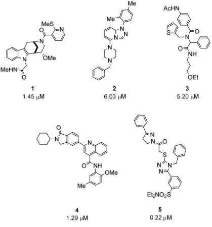
For this purpose, the substituents of the different heterocycles were removed to obtain the core scaffolds but the presence of different heteroatoms in the core structure was allowed to vary within members of the same class when the similarities were readily observed. In this fashion, most of the active hits could be grouped into 9 families (Figure 10). Recently, members of the compound classes represented by analogues 2, 3 and 4 were reported in the literature as glucose uptake inhibitors by Bayer Pharma and collaborators.15, 65, 71
These reports further validate our assay and the identified hits as starting points for the synthesis of libraries around them.
Figure 10. Representative analogues of some of the identified classes.
Compound 5 (Figure 10) had no common core scaffold with any other active hit from our assay.
Other singletons (ungrouped active hits) were found in the assay but a clear common core scaffold with compound 5 could not be identified (Figure 11a). A similarity search in our complete library showed several compounds, such as 10 and 11 (Figure 11b), with the same core scaffold as compound 5 albeit with no activity as glucose uptake inhibitors. Nevertheless, compound 5 presented a very high potency with an IC50 around 200 nM. At the time, this compound represented, to the best of our knowledge, the most potent glucose-uptake-inhibiting synthetic compound. Consequently, despite not having identified any other active member of this class, I decided to further study the structure of triazole 5 as a potential glucose-uptake- inhibiting class of compounds.
Figure 11.a) Other active singletons. b) Inactive analogues from the same compound class represented by compound 5.
The core-scaffold of the family represented by compound 1 (Figure 10), from here on referred to as the Glupin family, was identified in two other active compounds (Figure 12a). This common scaffold consists of a fused tryptamine-morphan hybrid moiety (Figure 12b). Besides the compounds 1, 12 and 13, I could identify additional analogues in the library that shared the same scaffold and presented a wide range of potencies (IC50s ˃ 10 µM)(see Experimental Part).
Figure 12. a) Active hits of the Glupin family identified in the assay and their IC50 values as glucose uptake inhibitors. b) Tryptamine-morphan Glupin core scaffold.
Both the tryptamine and the morphan scaffolds can be found across natural products, especially in alkaloids derived from tryptophan or the opioid morphine from which morphan takes its name. Although a similar core scaffold can be found in the Strychnos family of alkaloids, e.g.
Strychnine and Brucine (Figure 13a), the Glupin core is not present in nature. Furthermore, since its first appearance in the literature as a potential opioid, this novel scaffold has remained virtually chemically unexplored (Figure 13b).77-80 Its strong potency, natural product-like structure and the synthetic challenge of exploring this scaffold, led me to select the Glupin compound class for further study.
Figure 13. a) Tryptamine-morphan examples in nature. b) Examples of synthetic compounds with a tryptamine-morphan scaffold.77, 81-82
4.2. Validation of the Selected Compound Classes
4.2.1. Compound class of triazole 5
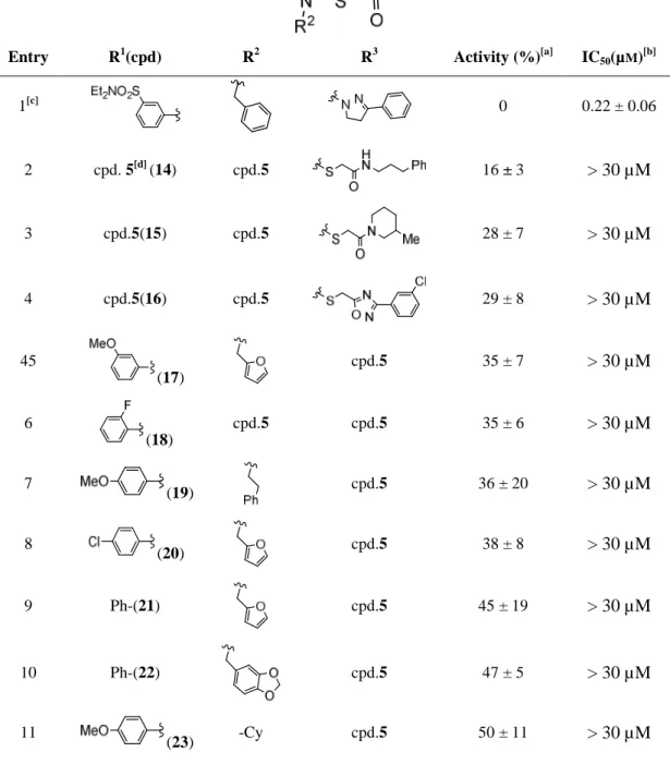
In order to validate the class represented by triazole 5 (Figure 10) as a glucose uptake inhibiting scaffold, a selection of 21 close analogues was purchased from Enamine (Table 2). All the purchased analogues showed significantly lower activities in single point measurements than compound 5. The potencies were within the 60 to 70% range of residual glucose uptake compared to the parent compound (set as 0%). Furthermore, when the dose-response effect was
measured, all compounds except for the parent compound showed an IC50 ˃ 30 µM. These results were not very promising, since regardless of the substitutions around the core, the analogues displayed a constant inactivity and only triazole 5 showed a big potency leap. Given that no active analogues were obtained to delineate an initial SAR, this class of compounds was not further explored as glucose uptake inhibitors.
Table 2. Selected examples of the commercial library of analogues of compound 5.
Entry R1(cpd) R2 R3 Activity (%)[a] IC50(µM)[b]
1[c] 0 0.22 ± 0.06
2 cpd. 5[d] (14) cpd.5 16 ± 3 > 30 µM
3 cpd.5(15) cpd.5 28 ± 7 > 30 µM
4 cpd.5(16) cpd.5 29 ± 8 > 30 µM
45
(17)
cpd.5 35 ± 7 > 30 µM
6
(18)
cpd.5 cpd.5 35 ± 6 > 30 µM
7 (19) cpd.5 36 ± 20 > 30 µM
8 (20) cpd.5 38 ± 8 > 30 µM
9 Ph-(21) cpd.5 45 ± 19 > 30 µM
10 Ph-(22) cpd.5 47 ± 5 > 30 µM
11 (23) -Cy cpd.5 50 ± 11 > 30 µM
12 (24) cpd.5 cpd.5 53 ± 10 > 30 µM
Assay performed in HCT cells, n=3; [a] Glucose uptake % residual activities compared to compound 5 assigned arbitrarily as 0% of glucose uptake with a compound concentration of 30 µM;[b] Activities given as IC50(µM) ± SD (n ≥ 3). Compounds with IC50> 30 µM were considered inactive; [c] Entry 1 corresponds to compound 5; [d]
cpd.5 = same structure as compound 5.
4.2.2. Glupins
In order to validate the Glupin class, a set of 19 close analogues was purchased from Edelris (Table 3). These analogues were selected based on variations of compounds 1, 12, and 13, while maintaining the tryptamine-morphan core scaffold. It should be noted that all of the commercial compounds (including the ones in our in-house library) were racemic mixtures. The introduction of an ethyl ester at the indole (R3, Table 3) led to a 25-fold potency increase (entry 2, (+/-)- Glupin-1). The same potency leap, arising from the introduction of the ester moiety on position R3, was observed when either a 2-pyrazine or a morpholinecarboxy group was present on the morphan nitrogen (R1) (entries 6,7 and 3, 9 resp.). In combination with the pyrazine group, the free amide at the R3 position did not yield any activity, suggesting that the free amide was detrimental for activity (entry 8). On the other hand, on position R1, heteroaromatics such as furan or thiophene led to active compounds, but both showed only low micromolar potencies (entries 4 and 5). Some ureas on R1 delivered inactive compounds (entries 9, 10 and 18). In contrast to compound 13 (Figure 4a), the rest of the compounds with no N-substitution at the indole (R3=H) were inactive (entries 11 to 16, 19 and 20). Comparing 13 (Figure 12) with 36 (entry 15), revealed that introduction of a bigger group on R2, such as 2-pyridine, was deleterious to the activity. In the rest of the compounds where the indole was not N-substituted, the lack of activity could then be attributed to an unsuitable R1 substitution (entries 11 to 13), to an introduction of the bulkier 2-pyridine group on R2 (entries 14 to 16, 19 and 20) or by a combination of both.


![Table 3. Commercial library of analogues of the Glupin compound class. Entry R 1 (cpd) R 2 R 3 IC 50 (µM) [a] 1 (1) -Me -CH 2 CONHMe 1.45 ± 0.6 2 ((+/-)-Glupin-1) -Me -CH 2 CO 2 Et 0.051 ± 0.025 3 (12) -Me -CH 2 CO 2 Et 3.16 ± 0.6 4 (25)](https://thumb-eu.123doks.com/thumbv2/1library_info/3632493.1502271/37.892.163.735.180.1114/table-commercial-library-analogues-glupin-compound-conhme-glupin.webp)
