and
Selective ABC Transporter Modulators
Dissertation
Zur Erlangung des Doktorgrades der Naturwissenschaften (Dr. rer. nat.)
der Fakultät für Chemie und Pharmazie der Universität Regensburg
vorgelegt von Qiu Sun
aus
Chengdu (China) 2015
The experimental part of this work was carried out between October 2011 and December 2014 under the supervision of Prof. Dr. Burkhard König at the Institute of Organic Chemistry, University of Regensburg.
The thesis was submitted on: 2nd, March, 2015 Date of the colloquium: 27th, March, 2015 Board of examiners: Prof. Dr. Achim Göpferich (Chairman) Prof. Dr. Burkhard König (1st Referee) Prof. Dr. Jörg Heilmann (2nd Referee) PD Dr. Sabine Amslinger (Examiner)
Dedicated To
My family
路漫漫其修远兮,吾将上下而求索。
The journey is long; I'll search up and down.
屈 原
Qu Yuan
Table of Contents
Chapter 1 ... 1
Natural phenolic metabolites with anti-angiogenic properties – a review from the chemical point of view ... 1
Abstract ... 1
Introduction ... 3
4-Hydroxybenzyl alcohol... 8
Curcumin ... 9
Ellagic acid ... 12
Resveratrol ... 13
Quinoline substituted phenols ... 14
4-Amino-2-sulfanylphenol derivatives ... 16
Acylphloroglucinol derivatives ... 17
(-)-Epigallocatechin-3-O-gallate (EGCG) ... 20
Xanthohumol ... 22
Genistein ... 24
Fisetin and Quercetin ... 24
(2S)-7,2’,4’-Trihydroxy-5-methoxy-8-dimethylallyl flavanone ... 27
Conclusions ... 27
References ... 28
Chapter 2 ... 33
Synthesis of natural and natural-like acylphloroglucinols with anti-proliferative, anti-oxidative and tube- formation inhibitory activity ... 33
Abstract ... 33
Introduction ... 35
Results and discussion ... 36
Conclusions ... 42
Experimental ... 43
References ... 53
1H and 13C NMR spectra of selected final compounds ... 55
Chapter 3 ... 58
Flavonoid derivatives as selective ABCC1 modulators: synthesis and functional characterization ... 58
Abstract ... 58
Introduction ... 59
Results and discussion ... 60
Conclusions ... 72
Experimental ... 72
References ... 94
1H and 13C NMR spectra of selected final compounds ... 99
Chapter 4 ... 102
Quinoline carboxamide-type ABCG2 modulators: quinoline moiety as anilide replacement ... 102
... 102
Abstract ... 102
Introduction ... 103
Results and Discussion ... 104
Conclusions ... 112
Experimental ... 112
References ... 130
1H and 13C NMR spectra of selected final compounds ... 132
Chapter 5 ... 135
Triphenylphosphine mediated photo-rearrangement and methanol addition of aryl chalcones to 1- propanones ... 135
Abstract ... 135
Introduction ... 136
Results and Discussion ... 137
Conclusions ... 143
Experimental ... 143
References ... 151
1H and 13C NMR spectra of selected compounds ... 154
Abbreviation ... 157
Summary ... 159
Zusammenfassung ... 161
Curriculum Vitae ... 163
Acknowledgement ... 167
This chapter has been published:
Q. Sun, J. Heilmannand B. König.Beilstein J. Org. Chem. 2015, 11, 249-264.
Author contributions:
Q. Sun wrote the manuscript.
Chapter 1
Natural phenolic metabolites with anti-angiogenic properties – a review from the chemical point of view
Abstract
Within the secondary natural metabolites from plants, phenolic compounds have a special impact on human health as they occur in significant amounts in several fruits, vegetables and medicinal plants. In this review natural phenolic compounds of plant origin with significant anti-angiogenic properties are summarized. Thirteen representatives of eight different natural or natural like
phenolic subclasses are presented with a particular emphasis on their synthesis and the methods to modify the parent compounds. Whenever available, the consequence of structural variation on the pharmacological activity of the molecules is described.
Keywords
natural phenolic compounds, angiogenesis, synthesis, structure-activity relationship
Introduction
The term “angiogenesis” is commonly used to describe the biological process of blood vessel growth. Nevertheless, it should be more precisely defined as formation of new blood vessels from pre-existing ones. Under physiological conditions angiogenesis is vital for foetal development, tissue regeneration and wound healing. Patho-physiologically, massive vascular growth or abnormal shape formation promotes many diseases including cancer, inflammation, and eye illness. However, inadequate vessel preservation or growth leads to ischemia causing myocardial infarction, stroke, and neurodegenerative or obesity-associated conditions.[1]
The generation of new blood vessels is based on a strictly controlled balance between various soluble and membrane-bound factors showing either anti- or pro-angiogenic activity and thus embedded together with enzymes and signalling molecules (Table 1) into a very complex network of signal pathways.[2] In case of cancer development the growing tumor disturbs the angiogenic balance in a tissue and induces the secretion of pro-angiogenic factors either by the tumor cells or by cells of the tumor microenvironment. When the tumor grows to the diameter of 1-2 mm, the tumor cells located far away from blood vessels undergo apoptosis or necrosis resulting from the lack of oxygen and nutrients. At this moment, tumor cells express pro- angiogenic factors including growth factors such as the vascular endothelial growth factor (VEGF) and fibroblast growth factor (FGF) and enzymes like cyclo-oxygenase 2 (COX-2) and protein kinase A (PKA) as well as signalling molecules like the integrins. The evoked cascades induce the formation of new blood vessels quickly connected with the pre-existing blood vessels providing sufficient supplies for tumor survival.[3] In addition, the new blood vessels allow cancer cells to transfer from a parent location to other new locations causing metastases.
Nevertheless, the morphology and pathophysiology of these blood vessels differs significantly from physiological ones as they work less effective and show a lower state of organization and control.[2a] After discovering the mechanism of angiogenesis and its crucial role in the tumor development, different therapies targeting to interfere with this process were investigated.[4]
Preferred clinical target are the VEGF receptors leading to the development and approval of monoclonal antibodies against VEGF and VEGF receptor tyrosine kinase inhibitors.[2a]
Nevertheless, the existing therapy options with antibodies and VEGF receptor inhibitors showed,
from the clinical point of view, several limitations making the search for further clinically relevant targets and other drugs mandatory to combat tumor related angiogenesis.
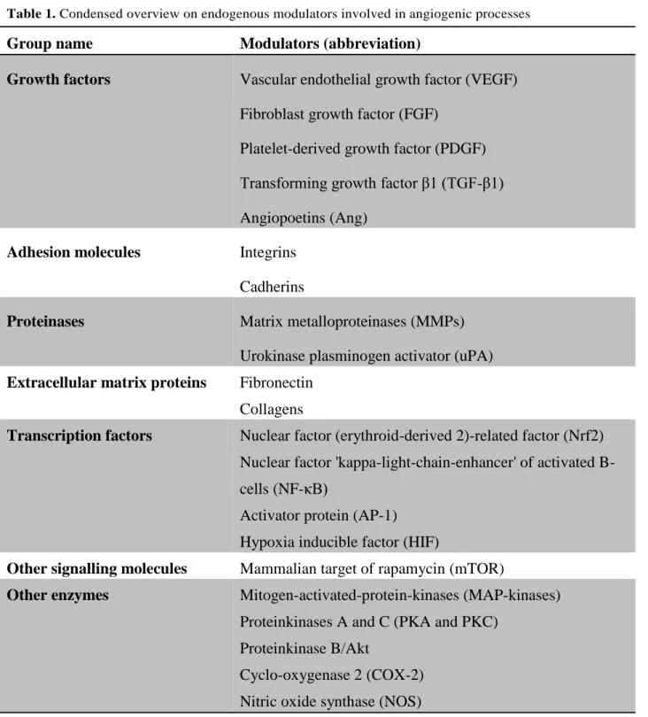
Table 1. Condensed overview on endogenous modulators involved in angiogenic processes
Group name Modulators (abbreviation)
Growth factors Vascular endothelial growth factor (VEGF) Fibroblast growth factor (FGF)
Platelet-derived growth factor (PDGF) Transforming growth factor β1 (TGF-β1) Angiopoetins (Ang)
Adhesion molecules Integrins Cadherins
Proteinases Matrix metalloproteinases (MMPs) Urokinase plasminogen activator (uPA) Extracellular matrix proteins Fibronectin
Collagens
Transcription factors Nuclear factor (erythroid-derived 2)-related factor (Nrf2) Nuclear factor 'kappa-light-chain-enhancer' of activated B- cells (NF-κB)
Activator protein (AP-1) Hypoxia inducible factor (HIF)
Other signalling molecules Mammalian target of rapamycin (mTOR)
Other enzymes Mitogen-activated-protein-kinases (MAP-kinases) Proteinkinases A and C (PKA and PKC)
Proteinkinase B/Akt
Cyclo-oxygenase 2 (COX-2) Nitric oxide synthase (NOS)
Accumulating research also showed that secondary natural metabolites are attractive candidates for the therapy of pathologically induced angiogenesis.[5] Among such natural products phenolic or polyphenolic compounds with anti-angiogenic properties have been investigated and the opinion on their pharmacological impact has changed over the last years. While the pharmacological activity of polyphenols was previously considered as unspecific, more recently observations of a specific interference with biological mechanisms at the molecular level are exponentially growing. Especially in the field of anti-inflammatory activity, chemoprevention and cytoprotection natural phenolic metabolites like flavonoids, caffeic acid derivatives and diarylheptanoids showed pleiotropic influence on cellular signalling e. g. by the inhibition of transcription factors like NF-B or Nrf2,[6] or anti-oxidative effects.[6b, 7] Furthermore, polyphenols are abundant in many plants used as fruits and vegetables in high concentrations, resulting in a continuous and long-term intake of such plant phenols. Consequently, their beneficial and protective impact on unbalanced angiogenic processes has been intensively discussed.[5]
In the last decade, many excellent review articles summarized the biological and pharmacological aspects of anti-angiogenic compounds including natural compounds with a phenolic substructure.[8] Complementary to the previously discussed pharmacological point of view this review focuses on recent reports of anti-angiogenic natural phenolic compounds specifically addressing their chemistry, synthesis and possible structure modifications.
Nevertheless, it should be mentioned that the selection of compounds for this review is based on the reports on their pharmacological activity. As the term “anti-angiogenic compound” is not unequivocally defined and somewhat inflationarily used, appropriate inclusion criteria for the review had to be defined. Compounds included in our survey have shown anti-angiogenic activity not only in convenient (and often descriptive) cellular in vitro assays (Table 2), but also in molecular in vitro test systems related to the signalling cascades of pathological angiogenesis.
Further inclusion criteria were the existence of anti-agiogenic activity obtained in ex vivo and in vivo assays (Table 2). Additionally, the observed in vitro activity should be in the lower µM range (or better) and thus high enough to realistically speculate on an anti-angiogenic activity in vivo. As endothelial cells (ECs) have an extraordinary significance in angiogenesis, results from cellular assays using primary or immortalized ECs like human umbilical vein endothelial cells (HUCEC) or human microvascular endothelial cells (HMEC-1) have been given special attention.
In contrast, compounds showing in vitro anti-angiogenic activity, but also most likely signs of strong unspecific cytotoxic effects in vitro have been excluded. The discussed secondary metabolites include six flavonoids from different subclasses namely quercetin, fisetin, epigallocatechin-3-O-gallate, xanthohumol, (2S)-7,2’,4’-triihydroxy-5-methoxy-8-dimethylallyl flavanone and genistein. Other compounds belong to the groups of simple phenols (4- hydroxybenzyl alcohol), hydrolysable tannins (ellagic acid), stilbenoids (resveratrol) and diarylheptanoids (curcumin). In addition, acylphloroglucinols, quinoline substituted phenols and 4-amino-2-sulfanylphenol derivatives were discussed. Some important aspects of the described pharmacological activities of the compounds are summarized in Table 3.
Table 2. In vitro, ex vivo and in vivo assays to characterize anti-angiogenic activity*
In vitro assays Assay principles / detection, read out Endothelial cell proliferation
assays
Cell counting / Increase of cell number Crystal violet / Increase of cell number
MTT / Activity of dehydrogenase activity (positively correlated to cell number)
Incorporation of [3H]thymidine, 5-bromodeoxyuridine into DNA / DNA synthesis (positively correlated to cell number) Endothelial cell migration
assays
Scratch assay / Migration into a denuded area (wound healing)
Endothelial cell differentiation assays
Tube formation e.g. in Matrigel/ Formation of capillary like tubules
Endothelial-Mural cell co- culture assays
Interaction between two cell types (endothelial/mural) / Influence on cell differentiation and proliferation
Ex vivo assays
Aortic ring assay Aorta of rodents cultured in biological matrices / Outgrowth of branching microvessels
In vivo assays
-Table 2 continued- Chick chorioallantoic membrane
assay (CAM)
Extra-embryonic membrane (in ovo, ex ovo) / growth and branching of blood vessels
Hen's egg test on chorioallantoic membrane (HETCAM)
CAM modification / Growth and branching of blood vessels Zebrafish Zebrafish embryos or transgenic zebrafish embryos /
Visualization of vascularisation (e.g. with confocal microscopy)
Corneal angiogenesis assay Corneal injury or implantation of pellets / Vascular response of the cornea
Dorsal air sac model Ring (filled with tumor cell suspension) implantation (dorsal skin) / Tumor induced angiogenesis
Mouse models Genetic engineered mouse models; xenografts
*Molecular or enzyme assays not included
Table 3. Natural phenolic compounds with anti-angiogenic activity and their evaluated molecular mechanisms of anti-angiogenesis
Compound name Mechanisms of anti-angiogenic action
4-Hydroxybenzyl alcohol Down-regulation of VEGF and MMP9 protein expression Curcumin Reduction of VEGF expression, inhibition of transcription
factors, mTOR pathway and MMP9 protein expression Ellagic acid Inhibition of VEGF and PDGF receptor phosphorylation Resveratrol Abrogation of VEGF-mediated tyrosine phosphorylation of
vascular endothelial (VE)-cadherin, inhibition of VEGF- induced and FGF-2 neovascularization
Quinoline substituted phenols Inhibition of VEGF and transforming growth factor-β1 (TGF- β1) expression
-Table 3 continued- 4-Amino-2-sulfanylphenol
derivatives
Inhibition of protein kinase B/Akt and ABL tyrosine kinase
Nature-like acylphloroglucinol derivatives
Under investigation
(-)-Epigallocatechin gallate (EGCG)
Inhibition of estrogen-stimulated VEGF expression, HIF-1α and Nf-κB, inhibition of MMP-2 and MMP-9, inhibition of urokinase plasminogen activator
Xanthohumol Inhibition of Nf-κB and Akt pathways
Genistein Inhibition of VEGF and HIF-1α protein expression
Fisetin Down-regulation of VEGF and eNOS expression, inhibition of MMPs activities
Quercetin Inhibition of the expression of VEGF-2, inhibition of COX-2 and arachidonate 5-lipoxygenase (LOX-5), inhibition of Nf-
B, In some cell types it activates angiogenesis.
(2S)-7,2’,4’-Triihydroxy-5- methoxy-8-dimethylallyl flavanone
Down-regulation of reactive oxygen speics (ROS) levels and VEGF expression
4-Hydroxybenzyl alcohol
4-Hydroxybenzyl alcohol (HBA, 1) (Figure 1) is a well-known phenolic compound from plants and has been for example found in flowers of carrot (Daucus carrota L., Apiaceae). In 2007, Park and co-workers [9] found in the chick chorioallantoic membrane (CAM) assay no change of the vascular density in the presence of HBA, indicating that HBA has no influence on the growth of blood vessels. In contrast, the branching pattern of blood vessels was reduced dose- dependently in the same assay, making an inhibition of angiogenesis likely. Later, Laschke et al.
(2011) [10] performed experiments in vitro with an aortic ring assay and in vivo in an
endometriosis model as well as systematic analysis of the mechanism underlying the anti- angiogenic activity of HBA. They found that HBA is capable of inhibiting several steps of the angiogenic mechanism. Western blot analysis showed the down-regulation of VEGF and MMP9 protein expression. The effect of HBA was confirmed [11] by mouse dorsal skinfold chamber experiments. Incubation of CT26.WT colon carcinoma cells with HBA showed a dose dependent decrease of their viability and integrity. In addition, the cells expression of the apoptosis marker cleaved caspase-3 increased significantly and the expression of vascular endothelial growth factor (VEGF) and matrix metalloproteinase (MMP)-9 decreased compared to controls. No influence on the normal behaviour of the animals was observed. In general, HBA represents an interesting anti-angiogenic agent for the treatment of angiogenic diseases.
Figure 1. Structure of 4-hydroxybenzyl alcohol (HBA).
Curcumin
Curcumin (3) is a natural product isolated from different Curcuma species (Zingiberaceae) some of them used as raw material of the spice turmeric. It has been evaluated as a chemopreventive agent since the early nineties and in 1998, Arbiser and his co-workers found that the compound showed also anti-angiogenic properties in vitro and in vivo.[12] In the following years, many studies on the anti-angiogenic properties in different tumor cell lines or in animal models were reported.[13] They include interactions with the transcription factor Nf-B, mTOR pathway, and reduction of VEGFA and MMP9 expression. Despite of its promising pharmacological properties, curcumin suffers from a low in vivo bioavailability as a consequence of its low aqueous solubility, poor chemical stability and low adsorption. Therefore many analogues (Figure 2) were synthesised in order to overcome these drawbacks and enhance the activity. In addition, their structure-activity relationships were studied to gain better insight into the mode of action. The general synthesis of curcumin itself (Scheme 1) requires masking of the reactive
methylene group of acetylacetone by formation of a complex with boric oxide, followed by reaction with vanillin. Instead of boric oxide, alkyl borate esters and boric acid can be used.[14]
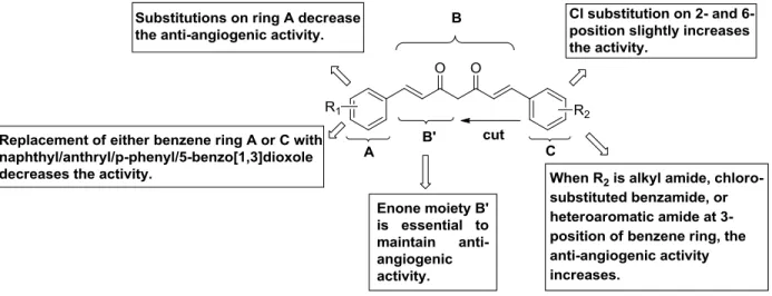
The first attempt of modification was to truncate the general structure to either a single enone or dienone system. The latter group showed a diarylpentanoid instead of the natural diarylheptanoid backbone, in some cases amended by a central ring system, and was labelled as monocarbonyl analogues of curcumin (MACs, Figure 3). Bowen et al.(2003) [15] used Claisen–Schmidt reaction for synthesis of these analogues. The C7- chain between the two aromatic rings was shortened and a series of compounds (Scheme 2) with different substitutions on the aromatic rings was synthesised to explore stereoelectronic effects. It was demonstrated that those analogues of curcumin were excellent anti-angiogenic compounds, having inhibition patterns equivalent or better than the parent natural product. This work was continued by more comprehensive bioactive studies on aromatic enones utilizing the substituted chalcone backbone.[16] The study showed that the presence of the enone moiety played an important role in maintaining the activity in the curcumin analogues. Ahn et al. (2005) [17] left the enone part unchanged to the previous principle and prepared various curcumin mimics with asymmetric units with bearing alkyl amide, chloro-substituted benzamide, or heteroaromatic amide moieties. Those analogues showed stronger anti-angiogenic activity than curcumin against HUVEC. Up to now the number of synthesised single enones and MACs clearly broke the 1000 mark.
Scheme 1: Synthesis of Curcumin 3. Reagents and conditions: (a) vanillin, 1,2,3,4-tetrahydroquinolin, HOAc, H3BO3, DMF, Δ 4 h.
Figure 2. Structure-activity relationship of curcumin analogues.
Figure 3. Backbone and substitution of monocarbonyl analogues of curcumin (MACs) showing their structural diversity.
Scheme 2. Exemplary synthesis of monocarbonyl analogues of curcumin (MACs). Reagents and conditions: (a) 40% KOH, EtOH, 5°C, stir 10 h, rt. X=C, N. R=OH, OMe, Cl, F.
Ellagic acid
Ellagic acid (7) is a naturally existing phenol antioxidant widely found in numerous fruits and vegetables like raspberries (Rubus idaeus L., Rosaceae), strawberries (Fragaria spec. L., Rosaceae) and pomegranates (Punica granatum L., Lythraceae). It shows potent antioxidant effects by radical scavenging and the inhibition of lipid peroxidation.[18] Ellagic acid is also capable of interfering with some angiogenesis-dependent pathways. It possesses anti- carcinogenic activity through inhibiting tumor cell proliferation, migration and induction of apoptosis. In addition, it is a dual inhibitor of the phosphorylation of VEGF and PDGF receptors, intercepting the angiogenesis processes required for tumor growth.[19] Recently, it was reported that its anti-angiogenesis mechanism affects the VEGFR-2 signalling pathway by forming hydrogen bonds and aromatic interactions within the ATP-binding region of the VEGFR-2 kinase unit.[20] Shankar and Srivastava et al. (2013) [21] treated PANC-1 xenografted mice with the ellagic acid and measured the expression of Akt, Shh and Notch. The results suggested that ellagic acid effectively inhibited human pancreatic cancer growth by suppressing protein kinase B (Akt), sonic hedgehog (Shh) and Notch pat hways. The preparation of ellagic acid (7) can be achieved by oxidative coupling of gallic acid (Scheme 3).[22] In the presence of H2SO4, gallic acid (4) was esterified to methyl gallate (5). The gallate (5) was oxidized by o-chloranil followed by reduction with Na2S2O4 to obtain the hexahydroxy biphenyl (6). Subsequent lactonization afforded the final product ellagic acid (7) in high yield.
Scheme 3. Synthesis of ellagic acid 7. Reagents and conditions: (a) H2SO4, CH3OH; (b) (1) o-chloranil, Et2O, -40°C; -40°C → r.t, 3 h; (2) Na2S2O4, r.t, 30 min; (c) MeOH:H2O=1:1, reflux.
Resveratrol
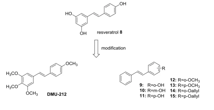
Resveratrol (8) is a natural polyphenol belonging to the stilbenoids and widely existing in a number of plants. It was primarily extracted from grape (Vitis vinifera L., Vitaceae), and mulberry (Morus L., Moraceae) (Figure 4).[23] It has anti-oxidant effects, anti-estrogenic activities and the ability to reduce the synthesis of hepatic lipids and eicosanoids. It inhibits platelet aggregation and protects vessels from arteriosclerosis.[23-24] In recent years, it has been reported that resveratrol is sufficiently potent to inhibit VEGF-induced and FGF-2 neovascularization in vivo.[25] It was also found that resveratrol showed direct inhibition to bovine aorta endothelial cell proliferation, migration and tube formation in vitro.[26] Resveratrol has also been found to effectively abrogate VEGF-mediated tyrosine phosphorylation of vascular endothelial (VE)-cadherin and its complex partner, β-catenin.[27] But unfortunately, resveratrol has dual effects on cells depending on the situation and cell type, meaning it can either induce or suppress angiogenic effects.[28] The low oral bioavailability and metabolic stability of resveratrol also limited its application.[29] Therefore, in an attempt to increase its bioavailability and stability,
the structure of resveratrol was modified by methylation of the phenol group [30] and introduction of other groups on the phenyl ring (see compound 9-15).[31] Trans-3,4,5,4’-tetramethoxystilbene (DMU-212) has pharmacokinetic properties that are better compared to resveratrol and shows anti-proliferative activities in different cancer cells.[32] The further investigation of its role in angiogenesis by Dai and Zhang et al.(2013) [33] showed that DMU-212, a potential anti- angiogenic agent, inhibits VEGFR2 phosphorylation and thereby acts as a suppressor of signalling pathways mediated by VEGFR2 inducing apoptosis in endothelial cells.
Figure 4. Structure of resveratrol 8 and its analogues.
Quinoline substituted phenols
Up to now, quinoline substituted phenols (Qsps) have not been reported as natural secondary metabolites, but the individual substructures quinoline and alkyl phenol are common structural elements of secondary plant products. The quinoline skeleton is present in alkaloids derived from tryptophane, like quinine or camptothecine, whereas alkyl phenols with a varying length of the alkyl side chain are common metabolites from the shikimate pathway. Qsps were reported in a recent patent to be effective for the treatment of angiogenesis-related diseases or disorder.
Among other assays a transgenic line of zebrafish that express a fluorescent reporter (EGFP) in vasculature was used in their study to identify anti-angiogenic compounds.[34] They particularly
looked at the integrity of vessels developing in the eyes and in the trunk. Quinoline-substituted phenols were identified being active based on a significant inhibition of the hyaloid vessel formation in the zebrafish model. The synthesis of two representative compounds 20 and 23 of this class are shown in Scheme 4 and Scheme 5.[34]
Scheme 4. Synthesis of quinoline substituted phenol 20. Reagents and conditions: (a) Ac2O, 2- hydroxybenzaldehyde, 130°C; (b) (1) Br2, AcOH; (2) Ac2O; (c) (1) DBU, THF; (2) Ac2O; (d) Na2CO3, MeOH/THF.
Scheme 5. Synthesis of quinoline substituted phenol 23. Reagents and conditions: (a) Ac2O, 2- hydroxybenzaldehyde; (b) (1) NaOH, EtOH/H2O, 100°C; (2) HCl.
After condensation of 16 with an aldehyde, product 17 was subsequently brominated, followed by twofold elimination of HBr forming a triple bond. Finally, ester hydrolysis provides
compound 20. Compound 23 was obtained from an analogous condensation product 22 hydrolysis under basic condition.
4-Amino-2-sulfanylphenol derivatives
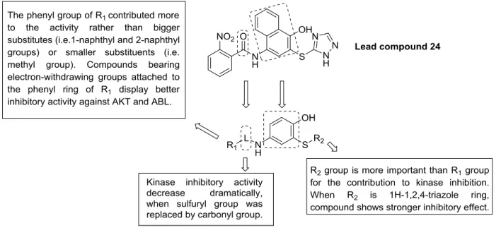
The group of 4-amino-2-sulfanylphenols is obviously an outlier among the reviewed compounds as the 2-sulfanylphenol structure is not a structural element in natural products. The vast majority of thiol groups in secondary metabolites derive from the amino acid from cysteine, producing aliphatic secondary metabolites with thiol functionality instead of phenolic ones. Nevertheless, this group contains thiol-analogues of the naturally occurring o-catechol substructure and thus they have been included to this review. Zhang and Xu et al. (2013) [35] have reported that 4- amino-2-sulfanylphenol compounds display high specific protein kinase and angiogenesis inhibitory activities. Based on their previous findings, the structure of compound 24 was optimized by replacing the naphthalene ring by a phenolic skeleton and a sulfonamide fragment.[36] These compounds show in vitro anti-angiogenic activities compared to Pazopanib in both human umbilical vein endothelial cell (HUVEC) tube formation assay and the rat thoracic aorta rings test. They inhibited protein kinase B/Akt and ABL tyrosine kinase in the micro-molar range. The preliminary structure-activity relationship is summarized in Figure 5.
Figure 5. Design of 4-amino-2-sulfanylphenol derivatives and their structure-activity relationship.
The synthesis of 4-amino-2-sulfanylphenol compounds (Scheme 6) [37] starts from the 4- aminophenol hydrochloride salt 25, which was dissolved in pyridine and reacted with various substituted sulfonyl chlorides yielding compounds 26 that differ in the substituent R1. Oxidation by NaIO4/SiO2 gives the quinone-type structures 27, which were reacted without further purification with thiols R2SH. The addition to the unsaturated system yielded the target compounds 28 with different arylthiol groups under rearomatization.
Scheme 6. Synthesis of 4-amino-2-sulfanylphenol deriatives. Reagents and conditions: (a) R1SO2Cl, Pyridine, 0°C; (b) NaIO4/SiO2, DCM; (c) DMF, R2SH.
Acylphloroglucinol derivatives
Acylphloroglucinols are typical secondary metabolites biosynthetically derived from the polyketide pathway and mainly accumulating in Hypericaceae [38] and Clusiaceae. Hyperforin, likely the most prominent acylphloroglucinol derivative and present in higher amounts in St.
John’s wort (Hypericum perforatum L., Hypericaceae), has been recently reported to exhibit strong anti-proliferative effects [39] and strongly inhibited angiogenesis in vitro and in vivo models. Mechanistically, it interferes with MMP-2 and an urokinase plasminogen activator
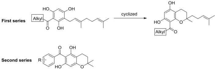
(uPA),[40] but due to its instability in aqueous solution, complex structure and limited availability, hyperforin is neither a drug candidate nor a good model compound. The finding that structurally simpler natural acylphloroglucinol derivatives showed also anti-proliferative effects against endothelial cells with inhibitory effects in a tube-formation assay on Matrigel catalysed the search for simple acylphloroglucinols with anti-angiogenic activity.[41] Within this group some geranylated monocyclic and bicyclic acylphloroglucinol derivatives have been found, which are structurally much simpler than hyperforin, but exhibiting potent anti-proliferative activity for a human microvascular endothelial cell line (HMEC-1) at low micromolar concentration.[41] Two series of natural-like acylphloroglucinols were synthesised (Figure 6) for the systematic investigations of their anti-angiogenic properties.[42] Compound 47 (R1=H, R2=OH, R3=OH) showed anti-proliferative activity with an IC50 of 0.88 ± 0.08 µM in vitro. Compound 38 (alkyl = CH(CH3)CH2CH3) exhibited moderate anti-proliferative effects (IC50 =11.0 ± 1 µM) and inhibited the capillary-like tube formation of HMEC-1 in vitro, whereas 47 is inactive.
Furthermore some of the compounds showed significant anti-oxidative activity. The most active compound in the ORAC assay was 47, which exhibited an anti-oxidative effect of 6.6 ± 1.0 TE.
However, this compound showed only weak activity during the proliferation assay (IC50 = 53.8 ± 0.3 µM) and did not inhibit tube-formation.
Figure 6. Structures of two series of nature-like acylphloroglucinols.
A basic structure-activity relationship of the aliphatic mono- and bicyclic acylphloroglucinol derivatives with short acyl side chains indicates that the in vitro anti-proliferative activity of these acylphloroglucinols in HMEC-1 increases with increasing logP. Increasing the number of carbon atoms in the acyl group provides higher lipophilicity, which allows the compound a better penetration across cellular membranes in vitro assay. In contrast, the activity of the derivatives with aromatic acylside chain depends on other properties. The molecular aspects of the observed cellular effects are currently under investigation.
Compounds 30-34 were synthesised via Friedel-Crafts acylation (Scheme 7) with 55 to 81%
yield. The alkylation of 30-34 and geranyl bromide gave products 35-39 with moderate yields from 55 to 60%. Finally, a para-toluenesulfonic acid (pTSA) catalysed cyclisation afforded the target compounds 40 and 41 in 53 and 65% yield, respectively.
Scheme 7. Synthesis of acylphloroglucinol derivatives 35-41. Reagents and conditions: (a) acyl chloride, AlCl3, CS2-PhNO2, 55°C, 2 h; (b) geranyl bromide, K2CO3, acetone, reflux overnight; (c) pTSA, benzene, reflux, 2 h.
Amberlyst 15 efficiently catalysed the condensation of 1,3,5-trihydroxybenzene 29 with isoprene in 53% yield in the synthesis of the second series of compounds (Scheme 8). The following
Friedel-Crafts acylation gave intermediates 43-46, which were subsequently demethylated using BBr3 to give 47-51 with 48 to 78% yield.
Scheme 8. Synthesis of acylphloroglucinol derivatives 43-51. Reagents and conditions: (a) isoprene, Amberlyst 15, THF-Hexane; (b) benzoyl chloride, AlCl3, DCM, -5°C to r.t., overnight; (c) BBr3, DCM, - 78°C to r.t., overnight.
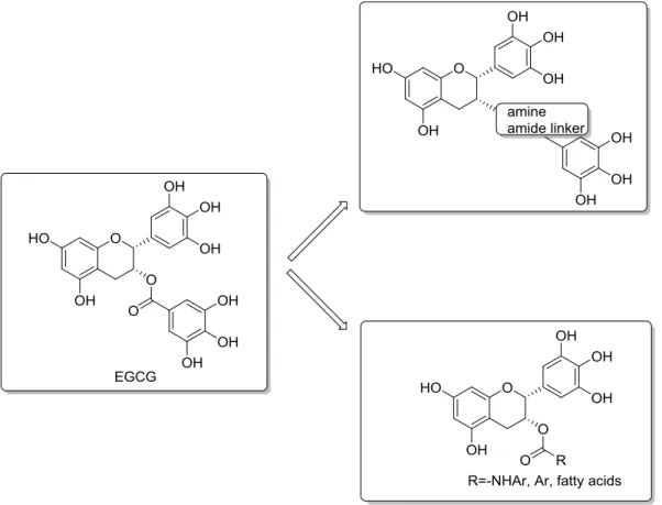
(-)-Epigallocatechin-3-O-gallate (EGCG)
(-)-Epigallocatechin-3-O-gallate (EGCG, 52) is the most abundant catechin in green tee (Camellia sinensis L. KUNZE, Theaceae). It is the esterification product of epigallocatechin and gallic acid. Many studies provide evidence that EGCG modulate multiples signal transduction pathways controlling the unwanted proliferation of cells. It inhibits the activation of HIF-1α, NF- κB and VEGF expression, thereby suppressing tumor angiogenesis and cancer progression.[43]
Furthermore, EGCG inhibited MMP-2 and MMP-9 (in different cell type), which seem to play
an important role in tumor invasion and metastasis. Also, the inhibition of uPA by EGCG has been observed.[44] uPA has the ability to prevent apoptosis, stimulate angiogenesis, mitogenesis, cell migration, and to modulate cell adhesion. The presence of the 3-galloyl moiety in catechins led to higher biological activities,[45] but an increasing number of aromatic hydroxyl groups results in low stability and the inability of the compound to cross cellular membranes.[46] To prevent oxidation and improve its bioavailability, modifications of EGCG focus on synthesising more stable analogues (Figure 7). Anderson et al.(2005) [47] replaced the hydrolytically labile ester bond with a more stable amine and amide bond and evaluated their efficacy as modulators for β-lactam resistance in S. aureus. Landis-Piwowar.and Chi-chui Wang et al (2005) [48]
protected the hydroxy groups by peracetate. These analogues behave as prodrugs and the acetyl group is removed by cellular cytosolic esterases. Liao (2002) and Huang (2010) et al. [49]
acylated the phenol group at 3-position, introduced fatty acids of different size and extensively explored their structure-activity relationship to 5α-reductase. Park and coworkers (2010) [50] also synthesised 3-O-alkyl analogues of epicatechin. They found that the introduction of alkyl groups instead of acyl groups enhanced antimicrobial activities and stability at pH 7.4.
Figure 7. Analogues of (-)-EGCG to prevent oxidation and improve bioavailability of the compounds.
Xanthohumol
Xanthohumol (XN, 58), a naturally occurring prenylated chalcone in hop plants (Humulus lupulus L., Cannabaceae), has been suggested to have potential to halt the development and progression of cancer and is therefore also a compound with a chemopreventive potential.[51] For example, XN shows proliferative inhibition of human breast (MCF-7), colon (HT-29), and ovarian cancer (A-2780) cells in vitro with IC50 ranging from 0.52 to 13.3 μM. Because most cancer chemopreventive agents have also anti-angiogenic properties in vitro and in vivo, further investigations [52] showed that XN repressed both the NF-κB and Akt pathways in endothelial cells, inhibited VEGF-A expression in a wound-healing assay and exhibited interference in the angiogenic process. The first total synthesis of xanthohumol was accomplished by Erhardt et al.
in six steps with an overall yield of 10% in 2007 (Scheme 9).[53] The method was improved by Vogel and Heilmann et al. (2008) [54] to yield also several xanthohumol derivatives occurring as
minor compounds in hop cones or as in vivo metabolites after xanthohumol intake.[54a] Up to now xanthohumol in vivo metabolites are not investigated regarding their anti-agiogenic activity.
Scheme 9. Scheme 9. Synthesis of xanthohumol 58. Reagents and conditions: (a) MOMCl, diisopropyl ethyl amine, CH2Cl2; (b) 3-methyl-2-butene-1-ol, diethylazodicarboxylate, PPh3, toluene/THF; (c) N,N- dimethylaniline, reflux; (d) (CH3O)SO2, K2CO3, acetone, reflux; (e) 4-methoxymethylbenzaldehyde aqueous NaOH, MeOH, reflux; (f) concentrated HCl (pH=1), MeOH/H2O, rt.
Genistein
Genistein (60) is an isoflavone extracted from soybeans (Glycine max (L.) MERR, Fabaceae). It is present as the 7-O-glycoside genistin in the plant, but during the processing of soya products a significant amount of the aglycone genistein is liberated. Genistein was originally described as an exclusive inhibitor of tyrosine-specific protein kinases.[55] These kinases are responsible for the tyrosine-specific protein phosphorylation, which is required for the regulation of cell functions, including cell proliferation and cell transformation. Later on genistein was also found to act as oestrogen receptor agonist.[56] The anti-angiogenic potential of genistein was first reported by Fotsis et al in 1993.[57] Then further studies showed that genistein inhibited angiogenic processes in various in vitro and in vivo models.[58] The typical synthesis of genistein starts from m-trihydroxybenzene 29 (Scheme 10).[59] After Houben–Hoesch reaction or Friedel- crafts acylation, cyclization of the resulting hydroxyketone (59) in the presence of BF3-Et2O gave genistein (60) in good yield.
Scheme 10. Synthesis of genistein 60. Reagents and conditions: (a) 4-hydroxyphenyl acetonitrile, anhydrous HCl, ZnCl2-Et2O, then aq HCl, heat or 4-hydroxyphenylacetic acid, BF3-Et2O, 120°C; (b) BF3- Et2O, DMF, MeSO2Cl, 100°C, 2 h.
In the Friedel-crafts acylation, BF3-Et2O was used as the catalyst and solvent. The following formation of the pyrone was also catalysed by BF3-Et2O, a convenient one-pot synthesis of 60 was achieved without isolation of 59.[60]
Fisetin and Quercetin
The flavonoids fisetin (67) and quercetin (68) belong to the flavonol subgroup exhibiting a double bond between C-2/C-3 and a hydroxyl group at C-3. Flavonols are the most abundant
flavonoid subtype in plants which mainly occur as glycosides. Nevertheless, pharmacological testing concentrated (historically based) on the investigation of the aglycones. This has been often criticised, but it is most likely an important aspect with regard to flavonoid metabolism. It has been shown that the flavonoid glycosides are not absorbed after oral intake but are cleaved by lactase-phlorizin hydrolase and absorbed as the corresponding aglycone. The aglycone passes the cell membrane by passive diffusion and undergoes phase-II metabolism in enterocytes and the liver leading to glucuronides as the main metabolites. At least, a release of the aglycones from the glucuronides in tissues or cells with -glucuronidase activity is possible.[61]
Fisetin can be found in many fruits like strawberries and apples (Malus spec. MILL., Rosaceae) as well as in vegetables like onions (Allium cepa L., Amaryllidaceae, subfamily Allioideae former family Alliaceae).It possesses anticancer activities in various cancer models, for example, it can inhibit androgen receptor signalling and tumor growth in athymic nude mice,[62] it can cause apoptosis and cell cycle arrest in human prostate cancer LNCaP cells,[63] in HCT-116 human colon cancer cells and it can induce apoptosis associated with an increased level of p53.[64]
P.Singh and A.Bhat et al.(2012) [65] tested its anti-angiogenic activity for the first time. Their study revealed that fisetin (10–50 µM) strongly inhibited the growth, proliferation and cell cycle progression in HUVEC by down-regulating the expression of VEGF and eNOS in endothelial cells. Another recent study also demonstrated that fisetin inhibits MMPs and reduces tumor cell invasiveness and endothelial cell tube formation.[66]
Quercetin is besides kaempferol the most abundant aglycone in flavonol glycosides. Quercetin glycosides occur in higher concentrations in onions, red wines, and green tea or in various medicinal plants.[67] In a number of early studies, quercetin showed a strong ability to inhibit tumor growth in vivo.[68] Quercetin, inhibits angiogenesis through multiple mechanisms such as inhibition of COX-2 and lipoxygenase (LOX)-5, interference with the EGF receptor, the HER-2 intracellular signaling pathway, and the NF-κB nuclear transcription protein. Chen et al.(2008)
[69] reported that quercetin inhibited the proliferation of choroids-retina endothelial cells and the migration and tube formation of RA/6A cells were also significantly inhibited by quercetin in a dose-dependent manner, but in some cell types quercetin is also able to activate the angiogenic pathway by inhibiting HIF-prolyl hydroxylase.[70] Zhao et al.(2014) [71] investigated that the anti- angiogenic activity of quercetin in zebrafish embryos and in human umbilical vein endothelial
cells (HUVECs). The formation of intersegmental vessels was disrupted in transgenic zebrafish embryos. In HUVECs, quercetin inhibited cell viability, the expression of VEGF-2 and tube formation dose-dependently.
The synthesis of fisetin and quercetin can be achieved by two different methods. The first choice for synthesising fisetin and quecetin is by the Allan-Robinson reaction.[72] However, this reaction has some drawbacks as very harsh experimental conditions and the necessity of selective protection and deprotection of the free hydroxyl groups with benzyl and/or benzoyl groups. An alternative method is the Algar-Flynn-Oyamada (AFO) reaction which gives flavone-3-ols directly, but the yield varies depending on different substrates.[73] Simpson and co-workers (1955)
[74] improved the reaction conditions of AFO in order to synthesise the flavonol rhamnocitrin by using bismuth carbonate and acetic acid, which increased the yields to 71% and the overall yields to 52% over two steps. According to this, a synthesis of fisetin and quercetin with methyl protected chalcones as starting material was proposed (Scheme 11).
Scheme 11. Synthesis of fisetin 67 and quercetin 68. Reagents and conditions: (a) 3,4- dimethoxybenzaldehyde, KOH, DMF, 0°C; (b) BiCO3, AcOH, 2-ethoxyethanol, Δ; (c) BBr3, DCM, -78°C
→r.t.
(2S)-7,2’,4’-Trihydroxy-5-methoxy-8-dimethylallyl flavanone
(2S)-7,2’,4’-Triihydroxy-5-methoxy-8-dimethylallyl flavanone (69, Figure 8) is a prenylated flavanone isolated from Sophora flavescens by Wang and Yuan et al in 2013.[75] It displays inhibitory effects on cell proliferation, cell migration, cell adhesion and tube formation with the human umbilical vein endothelial cell line ECV304, which are the four important steps in angiogenesis process. The mechanistic study showed that compound 69 is able to regulate ROS levels and VEGF expression in a dose-depended manner down, and induce cell cycle arrested in G0/G1 phase.
Figure 8. Structure of (2S)-7,2’,4’-triihydroxy-5-methoxy-8-dimethylallyl flavanone 69.
Conclusions
As pathological angiogenic processes are supposed to contribute to several diseases, compounds with anti-angiogenic activity have been intensively investigated. Besides antibodies also several low-molecular weight compounds have been chemically and pharmacologically characterized among them several secondary metabolites of natural origin. In anti-angiogenic strategies natural products with phenolic substructures or belonging to the polyphenols are of special importance as they occur in several food and medicinal plants important for human diet and health. Despite the fact that phenolic compounds often show specific interactions in biological systems most of them are pleiotropic substances with an effect on different cellular networks or targets. Several natural phenolic angiogenic inhibitors like curcumin (3), epigallocatechin-3-O-gallate (52) and xanthohumol (58) also showed remarkable chemopreventive activity. However, the stability, availability from natural sources and bioactivity of the natural compounds are typically limited with so far no example of very strong anti-angiogenic activity in the nano-molar range. Thus,
synthetic approaches for the production, diversification and optimization are mandatory. The modification of the structures of polyphenols improving stability and bioactivity, and further enhancing their anti-angiogenic activity, is the main goal of current research in the field.
Analogues of the natural phenols with improved drug properties may be promising candidates for future oncology treatment.
References
[1] a) P. Carmeliet, Nat Med 2003, 9, 653-660; b) J. Folkman, M. Klagsbrun, Science 1987, 235, 442-447.
[2] a) M. Potente, H. Gerhardt, P. Carmeliet, Cell 2011, 146, 873-887; b) Z. K. Otrock, R. A.
Mahfouz, J. A. Makarem, A. I. Shamseddine, Blood Cells, Mol., Dis. 2007, 39, 212-220.
[3] V. Baeriswyl, G. Christofori, Semin Cancer Biol 2009, 19, 329-337.
[4] F. A. Scappaticci, J Clin Oncol 2002, 20, 3906-3927.
[5] a) S. M. Sagar, D. Yance, R. K. Wong, Curr Oncol 2006, 13, 14-26; b) E. J. Seo, V. Kuete, O.
Kadioglu, B. Krusche, S. Schroder, H. J. Greten, J. Arend, I. S. Lee, T. Efferth, J. Evidence- Based Complementary Altern. Med. 2013, 2013, 131306.
[6] a) S. Prasad, K. Phromnoi, V. R. Yadav, M. M. Chaturvedi, B. B. Aggarwal, Planta medica 2010, 76, 1044-1063; b) I. Rahman, S. K. Biswas, P. A. Kirkham, Biochem Pharmacol 2006, 72, 1439- 1452.
[7] P. G. Pietta, J Nat Prod 2000, 63, 1035-1042.
[8] a) S. P. Ivy, J. Y. Wick, B. M. Kaufman, Nat Rev Clin Oncol 2009, 6, 569-579; b) K. M. Cook, W. D. Figg, CA: a cancer journal for clinicians 2010, 60, 222-243; c) O. Wahl, M. Oswald, L.
Tretzel, E. Herres, J. Arend, T. Efferth, Curr Med Chem 2011, 18, 3136-3155.
[9] E. J. Lim, H. J. Kang, H. J. Jung, E. H. Park, J Pharm Pharmacol 2007, 59, 1235-1240.
[10] M. W. Laschke, A. E. V. van Oijen, C. Scheuer, M. D. Menger, Brit J Pharmacol 2011, 163, 835- 844.
[11] M. W. Laschke, A. E. V. van Oijen, C. Korbel, C. Scheuer, M. D. Menger, Life Sci 2013, 93, 44- 50.
[12] J. L. Arbiser, J. D. Fine, D. Murrell, A. Paller, S. Connors, K. Keough, E. Marsh, J. Folkman, Mol Med 1998, 4, 191-195.
[13] a) S. Bimonte, A. Barbieri, G. Palma, A. Luciano, D. Rea, C. Arra, Biomed Res Int 2013, Article ID 810423, 8 pages; b) F. Zhang, Z. L. Zhang, L. Chen, D. S. Kong, X. P. Zhang, C. F. Lu, Y.
Lu, S. Z. Zheng, J Cell Mol Med 2014, 18, 1392-1406; c) T. Kalinski, S. Sel, H. Hutten, M.
Ropke, A. Roessner, N. Nass, Plos One 2014, 9, e99296; d) P. Yoysungnoen-Chintana, P.
Bhattarakosol, S. Patumraj, Biomed Res Int 2014, Article ID 817972, 12 pages.
[14] a) H.J.J.Pabon, Recueil des Travaux Chimiques des Pays-Bas 1964, 83, 379-386; b) K. V. D.
Babu, K. N. Rajasekharan, Org Prep Proced Int 1994, 26, 674-677.
[15] T. P. Robinson, T. Ehlers, R. B. Hubbard, X. H. Bai, J. L. Arbiser, D. J. Goldsmith, J. P. Bowen, Bioorg Med Chem Lett 2003, 13, 115-117.
[16] T. P. Robinson, R. B. Hubbard, T. J. Ehlers, J. L. Arbiser, D. J. Goldsmith, J. P. Bowen, Bioorgan Med Chem 2005, 13, 4007-4013.
[17] H. B. Woo, W. S. Shin, S. Lee, C. M. Ahn, Bioorg Med Chem Lett 2005, 15, 3782-3786.
[18] a) D. H. Han, M. J. Lee, J. H. Kim, Anticancer Res 2006, 26, 3601-3606; b) K. I. Priyadarsini, S.
M. Khopde, S. S. Kumar, H. Mohan, J Agr Food Chem 2002, 50, 2200-2206.
[19] L. Labrecque, S. Lamy, A. Chapus, S. Mihoubi, Y. Durocher, B. Cass, M. W. Bojanowski, D.
Gingras, R. Beliveau, Carcinogenesis 2005, 26, 821-826.
[20] N. Wang, Z. Y. Wang, S. L. Mo, T. Y. Loo, D. M. Wang, H. B. Luo, D. P. Yang, Y. L. Chen, J.
G. Shen, J. P. Chen, Breast Cancer Res Tr 2012, 134, 943-955.
[21] M. Zhao, S. N. Tang, J. L. Marsh, S. Shankar, R. K. Srivastava, Cancer Lett 2013, 337, 210-217.
[22] S. Quideau, K. S. Feldman, J Org Chem 1997, 62, 8809-8813.
[23] G. J. Soleas, E. P. Diamandis, D. M. Goldberg, Clin Biochem 1997, 30, 91-113.
[24] a) H. Arichi, Y. Kimura, H. Okuda, K. Baba, M. Kozawa, S. Arichi, Chem Pharm Bull 1982, 30, 1766-1770; b) L. Belguendouz, L. Fremont, A. Linard, Biochem Pharmacol 1997, 53, 1347-1355;
c) E. N. Frankel, A. L. Waterhouse, J. E. Kinsella, Lancet 1993, 341, 1103-1104; d) R. Q. Lu, G.
Serrero, J Cell Physiol 1999, 179, 297-304.
[25] E. Brakenhielm, R. Cao, Y. Cao, Faseb J 2001, 15, 1798-1800.
[26] K. Igura, T. Ohta, Y. Kuroda, K. Kaji, Cancer Lett 2001, 171, 11-16.
[27] M. T. Lin, M. L. Yen, C. Y. Lin, M. L. Kuo, Mol Pharmacol 2003, 64, 1029-1036.
[28] Y. Chen, S. H. Tseng, In Vivo 2007, 21, 365-370.
[29] T. Walle, F. Hsieh, M. H. DeLegge, J. E. Oatis, U. K. Walle, Drug Metab Dispos 2004, 32, 1377- 1382.
[30] A. Gosslau, M. Chen, C. T. Ho, K. Y. Chen, Brit J Cancer 2005, 92, 513-521.
[31] R. Marti-Centelles, R. Cejudo-Marin, E. Falomir, J. Murga, M. Carda, J. A. Marco, Bioorgan Med Chem 2013, 21, 3010-3015.
[32] a) Z. S. Ma, O. Molavi, A. Haddadi, R. Lai, R. A. Gossage, A. Lavasanifar, Cancer Chemoth Pharm 2008, 63, 27-35; b) S. Sale, R. D. Verschoyle, D. Boocock, D. J. L. Jones, N. Wilsher, K.
C. Ruparelia, G. A. Potter, P. B. Farmer, W. P. Steward, A. J. Gescher, Brit J Cancer 2004, 90, 736-744; c) S. Sale, R. G. Tunstall, K. C. Ruparelia, G. A. Potter, W. P. Steward, A. J. Gescher, Int J Cancer 2005, 115, 194-201.
[33] L. K. Chen, P. F. Qiang, Q. P. Xu, Y. H. Zhao, F. Dai, L. Zhang, Acta Pharmacol Sin 2013, 34, 1174-1182.
[34] B. O. S. Kennedy, Jacintha; Reynolds, Alison; Kilty, Claire; Baxter, Andrew Douglas, WO2014012889A1, 2014.
[35] F. M. Xu, L. Zhang, Y. P. Jia, X. J. Wang, X. G. Li, Q. L. Wen, Y. J. Zhang, W. F. Xu, Eur J Med Chem 2013, 69, 191-200.
[36] F. M. Xu, Y. P. Jia, Q. L. Wen, X. J. Wang, L. Zhang, Y. J. Zhang, K. H. Yang, W. F. Xu, Eur J Med Chem 2013, 64, 377-388.
[37] F. M. Xu, H. Xu, X. J. Wang, L. Zhang, Q. L. Wen, Y. J. Zhang, W. F. Xu, Bioorgan Med Chem 2014, 22, 1487-1495.
[38] E. W. Ades, F. J. Candal, R. A. Swerlick, V. G. George, S. Summers, D. C. Bosse, T. J. Lawley, J Invest Dermatol 1992, 99, 683-690.
[39] B. Kraus, H. Wolff, E. F. Elstner, J. Heilmann, N-S Arch Pharmacol 2010, 381, 541-553.
[40] B. Martinez-Poveda, A. R. Quesada, M. A. Medina, Int J Cancer 2005, 117, 775-780.
[41] a) S. Schmidt, G. Jurgenliemk, H. Skaltsa, J. Heilmann, Phytochemistry 2012, 77, 218-225; b) S.
Schmidt, G. Jurgenliemk, T. J. Schmidt, H. Skaltsa, J. Heilmann, J Nat Prod 2012, 75, 1697- 1705.
[42] Q. Sun, S. Schmidt, M. Tremmel, J. Heilmann, B. Konig, Eur J Med Chem 2014, 85C, 621-628.
[43] a) X. Y. Li, Y. Feng, J. H. Liu, X. W. Feng, K. Y. Zhou, X. D. Tang, J Nutrigenet Nutrige 2013, 6, 169-178; b) Y. Sakamoto, N. Terashita, T. Muraguchi, T. Fukusato, S. Kubota, Biosci Biotech Bioch 2013, 77, 1799-1803.
[44] Y. C. Ho, S. F. Yang, C. Y. Peng, M. Y. Chou, Y. C. Chang, J Oral Pathol Med 2007, 36, 588- 593.
[45] a) M. Z. Fang, Y. M. Wang, N. Ai, Z. Hou, Y. Sun, H. Lu, W. Welsh, C. S. Yang, Cancer Res 2003, 63, 7563-7570; b) S. Nam, D. M. Smith, Q. P. Dou, J Biol Chem 2001, 276, 13322-13330;
c) E. Navarro-Peran, J. Cabezas-Herrera, F. Garcia-Canovas, M. C. Durrant, R. N. F. Thorneley, J. N. Rodriguez-Lopez, Cancer Res 2005, 65, 2059-2064.
[46] J. Hong, H. Lu, X. F. Meng, J. H. Ryu, Y. Hara, C. S. Yang, Cancer Res 2002, 62, 7241-7246.
[47] J. C. Anderson, C. Headley, P. D. Stapleton, P. W. Taylor, Bioorg Med Chem Lett 2005, 15, 2633-2635.
[48] a) L. C. M. Chiu, C. K. L. Kong, V. E. C. Ooi, Int J Mol Med 2005, 16, 735-740; b) C. C. Wang, H. Xu, G. C. W. Man, T. Zhang, K. O. Chu, C. Y. Chu, J. T. Y. Cheng, G. Li, Y. X. He, L. Qin, T. S. Lau, J. Kwong, T. H. Chan, Angiogenesis 2013, 16, 59-69.
[49] a) R. A. Hiipakka, H. Z. Zhang, W. Dai, Q. Dai, S. T. Liao, Biochem Pharmacol 2002, 63, 1165- 1176; b) S. F. Lin, Y. H. Lin, M. J. Lin, Y. F. Kao, R. W. Wang, L. W. Teng, S. H. Chuang, J. M.
Chang, T. T. Yuan, K. C. Fu, K. P. Huang, Y. S. Lee, C. C. Chiang, S. C. Yang, C. L. Lai, C. B.
Liao, P. N. Chen, Y. S. Lin, K. T. Lai, H. J. Huang, J. Y. Yang, C. W. Liu, W. Y. Wei, C. K.
Chen, R. A. Hiipakka, S. S. Liao, J. J. Huang, Eur J Med Chem 2010, 45, 6068-6076.
[50] K. D. Park, S. J. Cho, Eur J Med Chem 2010, 45, 1028-1033.
[51] a) C. Gerhauser, A. Alt, E. Heiss, A. Gamal-Eldeen, K. Klimo, J. Knauft, I. Neumann, H. R.
Scherf, N. Frank, H. Bartsch, H. Becker, Mol Cancer Ther 2002, 1, 959-969; b) C. L. Miranda, J.
F. Stevens, A. Helmrich, M. C. Henderson, R. J. Rodriguez, Y. H. Yang, M. L. Deinzer, D. W.
Barnes, D. R. Buhler, Food Chem Toxicol 1999, 37, 271-285.
[52] A. Albini, R. Dell'Eva, R. Vene, N. Ferrari, D. R. Buhler, D. M. Noonan, G. Fassina, Faseb J 2005, 19, 527-529.
[53] R. S. Khupse, P. W. Erhardt, J Nat Prod 2007, 70, 1507-1509.
[54] a) S. Vogel, S. Ohmayer, G. Brunner, J. Heilmann, Bioorg Med Chem 2008, 16, 4286-4293; b) S.
Vogel, J. Heilmann, J Nat Prod 2008, 71, 1237-1241.
[55] T. Akiyama, J. Ishida, S. Nakagawa, H. Ogawara, S. Watanabe, N. Itoh, M. Shibuya, Y. Fukami, J Biol Chem 1987, 262, 5592-5595.
[56] L. Markiewicz, J. Garey, H. Adlercreutz, E. Gurpide, The Journal of steroid biochemistry and molecular biology 1993, 45, 399-405.
[57] T. Fotsis, M. Pepper, H. Adlercreutz, G. Fleischmann, T. Hase, R. Montesano, L. Schweigerer, P Natl Acad Sci USA 1993, 90, 2690-2694.
[58] a) B. Wang, Y. Zou, H. Li, H. Yan, J. S. Pan, Z. L. Yuan, J Ocul Pharmacol Th 2005, 21, 107- 113; b) S. Kiriakidis, O. Hogemeier, S. Starcke, F. Dombrowski, J. C. Hahne, M. Pepper, H. C.
Jha, N. Wernert, Brit J Nutr 2005, 93, 317-323.
[59] Y. C. Chang, M. G. Nair, R. C. Santell, W. G. Helferich, J Agr Food Chem 1994, 42, 1869-1871.
[60] K. Wahala, T. A. Hase, J Chem Soc Perk T 1 1991, 3005-3008.
[61] A. Ishisaka, K. Kawabata, S. Miki, Y. Shiba, S. Minekawa, T. Nishikawa, R. Mukai, J. Terao, Y.
Kawai, Plos One 2013, 8, e80843.
[62] N. Khan, M. Asim, F. Afaq, M. Abu Zaid, H. Mukhtar, Cancer Res 2008, 68, 8555-8563.
[63] N. Khan, F. Afaq, D. N. Syed, H. Mukhtar, Carcinogenesis 2008, 29, 1049-1056.
[64] D. Y. Lim, J. H. Y. Park, Am J Physiol-Gastr L 2009, 296, 1060-1068.
[65] T. A. Bhat, D. Nambiar, A. Pal, R. Agarwal, R. P. Singh, Carcinogenesis 2012, 33, 385-393.
[66] J. H. Park, Y. J. Jang, Y. J. Choi, J. W. Jang, J. H. Kim, Y. K. Rho, I. J. Kim, H. J. Kim, M. J.
Leem, S. T. Lee, Nutr Cancer 2013, 65, 1192-1199.
[67] C. A. RiceEvans, N. J. Miller, G. Paganga, Free Radical Bio Med 1996, 20, 933-956.
[68] a) M. H. Castillo, E. Perkins, J. H. Campbell, R. Doerr, J. M. Hassett, C. Kandaswami, E.
Middleton, Am J Surg 1989, 158, 351-355; b) J. V. Formica, W. Regelson, Food Chem Toxicol 1995, 33, 1061-1080.
[69] Y. Chen, X. X. Li, N. Z. Xing, X. G. Cao, Graef Arch Clin Exp 2008, 246, 373-378.
[70] H. Jeon, H. Kim, D. Choi, D. Kim, S. Y. Park, Y. J. Kim, Y. M. Kim, Y. J. Jung, Mol Pharmacol 2007, 71, 1676-1684.
[71] D. X. Zhao, C. J. Qin, X. H. Fan, Y. C. Li, B. H. Gu, Eur J Pharmacol 2014, 723, 360-367.
[72] J. Allan, R. Robinson, J. Chem. Soc. 1926, 2334-2336.
[73] T. Oyamada, H. Baba, B Chem Soc Jpn 1966, 39, 507-511.
[74] T. H. S. J. M. Guider, D. B. Thomas, J. Chem. Soc. 1955, 170-173.
[75] X. L. Zhang, M. A. Cao, L. P. Pu, S. S. Huang, Q. X. Gao, C. S. Yuan, C. M. Wang, Pharmazie 2013, 68, 369-375.
This chapter has been published:
Q. Sun, S. Schmidt, M. Tremmel, J. Heilmann, B. König. Eur J Med Chem. 2014, 85, 621-628.
Author contributions:
Q. Sun synthesized all the compounds and wrote the manuscript.
Chapter 2
Synthesis of natural and natural-like acylphloroglucinols with anti- proliferative, anti-oxidative and tube-formation inhibitory activity
Abstract
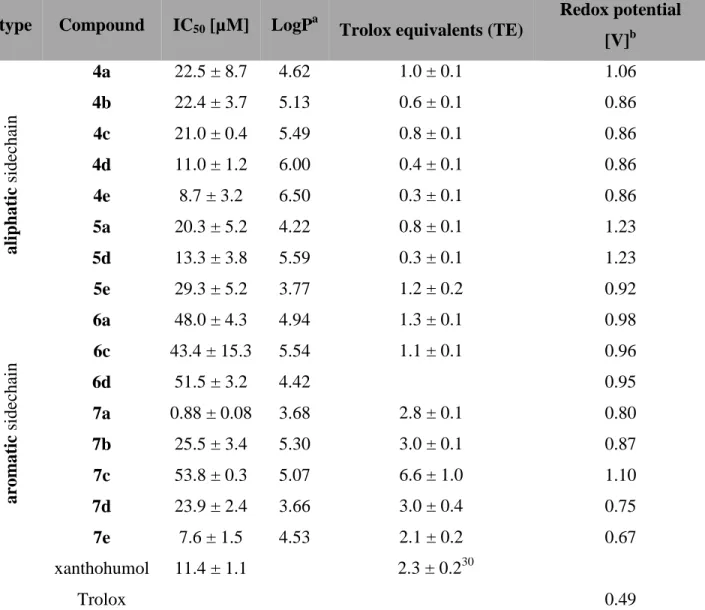
Two series of natural and natural-like mono- and bicyclic acylphloroglucinols derived from secondary metabolites in the genus Hypericum (Hypericaceae) were synthesised and tested in vitro for anti-proliferative and tube-formation inhibitory activity in human microvascular endothelial cells (HMEC-1). In addition, their anti-oxidative activity was determined via an ORAC-assay. The first series of compounds (4a-e) consisted of geranylated monocyclic acylphloroglucinols with varying aliphatic acyl substitution patterns, which were subsequently cyclised to the corresponding 2-methyl-2-prenylchromane derivatives (5a and 5d). The second series involved compounds containing a 2,2-dimethylchromane skeleton with differing aromatic acyl substitution (6a-d and 7a-e). Compound 7a, (5,7-dihydroxy-2,2-dimethylchroman-6-yl)- (3,4-dihydroxyphenyl)methanone), showed the highest in vitro anti-proliferative activity with an
IC50 of 0.88 ± 0.08 µM and a remarkable anti-oxidative activity of 2.8 ± 0.1 TE from the ORAC test. Interestingly, the high anti-proliferative activity of these acylphloroglucinols was not associated with tube-formation inhibition. Compounds (E)-1-(3-(3,7-dimethylocta-2,6-dien-1- yl)-2,4,6-trihydroxyphenyl)-2-methylbutan-1-one (4d) and (5,7-dihydroxy-2,2-dimethyl- chroman-6-yl)(3,4-dimethoxyphenyl)methanone (6a) exhibited moderate to weak anti- proliferative effects (IC50 11.0 ± 1 µM and 48.0 ± 4.3 µM, respectively) and inhibited the capillary-like tube formation of HMEC-1 in vitro, whereas 7a was inactive. The most active compound in the ORAC assay was 7c, which exhibited an anti-oxidative effect of 6.6 ± 1.0 TE.
However, this compound showed only weak activity during the proliferation assay (IC50 53.8 ± 0.3) and did not inhibit tube-formation.
Keywords
acylphloroglucinol; chromane; HMEC-1; anti-proliferative activity; tube formation; ORAC