Molecular Genetics of Dupuytren´s Disease
Inaugural-Dissertation
zur
Erlangung des Doktorgrades
der Mathematisch-Naturwissenschaftlichen Fakultät der Universität zu Köln
vorgelegt von
Kerstin Becker aus Köln
2012
Berichterstatter: Prof. Dr. Peter Nürnberg
(Gutachter) Prof. Dr. Angelika A. Noegel
Tag der mündlichen Prüfung: 22.01.2013
- iii -
Abstract
Dupuytren’s disease (DD) is a fibromatosis of connective tissue within the palm of the hands. It is characterised by progressive collagen deposition that leads to hardening and thickening of the connective tissue and results in permanent contraction of affected fingers.
The aetiology of Dupuytren’s disease is so far unknown and the pathogenesis is favoured by ageing, genetic predisposition, mechanic trauma and possibly other risk factors. Blood and tissue samples of over 850 German and Swiss DD patients were collected in order to analyse the genetics, gene expression patterns and the in vitro behaviour of disease tissue derived fibroblast in 2-D and 3-D models.
A genome wide association study (GWAS) with 565 unrelated DD patients and 1,219 controls was performed. Data for 5,204,451 single-nucleotide polymorphisms (SNPs; 186 cases genotyped with Affymetrix Genome-Wide Human SNP Array 6.0 and 379 cases, 1219 controls genotyped with Axiom CEU 1 Array; data imputed with HapMap CEU reference panel) were analyzed for association with DD. SNP rs2290221 on chromosome 7p14, showed the strongest association signal with a p-value of 2.2x10
-10and odds ratio of 2.13.
SNP rs2290221 is located intronic of the genes secreted frizzled-related protein 4 (SFRP4) and ependymin related protein 1 (zebrafish) (EPDR1).
In addition an integrative replication study with 2,325 Dutch, English and German DD cases and 11,562 controls was performed. It identified nine different susceptibility loci that showed genome-wide significance. Six of these loci contain genes known to be involved in the Wnt/β-catenin signalling pathway. Again the strongest association was seen for the 7p14 locus.
Consistent with GWAS findings, a whole genome expression analysis with 12 DD primary
disease tissue samples and 12 normal fascia controls revealed upregulation of the Wnt/β-
catenin signalling pathway and also changes in mitochondrial function and oxidative stress
response in disease tissue. The Wnt signalling pathway is therefore likely to be a key player
in the fibromatosis process observed in DD. Primary disease tissue derived fibroblasts at
least in part retained their disease associated characteristics in vitro, they exhibited higher
proliferation rates and generated strong contraction forces in 3-D collagen gels, and thus
present an excellent model for investigating the mechanisms of DD in the context of aging
and aging associated diseases.
- iv -
Zusammenfassung
Bei der Dupuytren´schen Erkrankung (DD) handelt es sich um eine Fibromatose des Bindegewebes in der Handinnenfläche und der Innenseite der Finger. Im Verlauf der Krankheit verhärtet und verdickt sich das Bindegewebe aufgrund progressiver Kollagenablagerungen. Dies führt zu einer dauerhaften Kontraktion von betroffenen Fingern.
Die Ursache des Morbus Dupuytren ist bisher unbekannt. Der Krankheitsverlauf wird durch Alter, genetische Veranlagung, mechanische Traumata und möglicherweise weitere Risikofaktoren begünstigt. Von über 850 deutschen und Schweizer DD-Patienten wurden Blut und Gewebeproben gesammelt, um die Genetik, Genexpression und das In-vitro- Verhalten von Fibroblasten in 2-D und 3-D-Modelle zu analysieren.
In einer genomweiten Assoziations-Studie (GWAS) wurden 565 DD-Patienten und 1.219 Kontrollen miteinander verglichen. Dabei wurden 5.204.451 genetische Marker (single- nucleotide polymorphisms (SNPs)) untersucht. SNP rs2290221 auf Chromosom 7p14, zeigte das stärkste Assoziationssignal mit einem P-Wert von 2,2x10
-10und eine Odds Ratio von 2,13. SNP rs2290221 befindet sich im Intron der beiden Gene secreted frizzled-related protein 4 (SFRP4) und ependymin related protein 1 (zebrafish) (EPDR1).
Parallel dazu wurde eine integrative Replikations-Studie mit 2325 Niederländisch, Deutsch und Englisch DD Patienten und 11.562 Kontrollen durgeführt. Diese identifizierte neun verschiedene Loci, die mit genomweiter Signifikanz mit DD assoziierten. Sechs dieser Loci enthalten Gene, die im Wnt/β-catenin-Signalweg eine Rolle spielen.
In Übereinstimmung mit den GWAS Erkenntnisse ergab eine genomeweite Expressions-
Analyse, bei der 12 DD- und 12 Kontroll-Gewebeproben miteinander verglichen wurden,
eine Hochregulation des Wnt/β-Catenin-Signalweges in den DD Proben. Außerdem wurden
Veränderungen in der oxydative Stressreaktion festgestellt. Der Wnt-Signalweg stellte daher
wahrscheinlich eine wichtige Komponente im Krankheitsprozess dar. Primäre Fibroblasten,
die aus DD Gewebe gewonnen wurden, behielten zum Teil ihre mit der Krankheit
verbundenen Eigenschaften in vitro. Sie zeigten eine höhere Proliferationsrate und erzeugt
starke Kontraktionskräfte in 3-D Kollagengelen. Sie stellen somit ein hervorragendes Modell
dar für die Untersuchung der Krankheitsmechanismen im Kontext des Alterns und alters-
assoziierter Krankheiten.
- v -
Table of Content
1 Introduction ... 9
1.1 Aging and Aging Associated Diseases ... 9
1.1.1 Free Radical Theory of Aging ... 10
1.1.2 Insulin/IGF Signalling and Aging ... 11
1.2 Dupuytren´s Disease ... 12
1.2.1 The Myofibroblast ... 15
1.2.2 History and Prevalence ... 16
1.2.3 Treatment ... 16
1.2.4 Genetics and other Risk Factors ... 18
1.2.5 Social Relevance and Civic Impact ... 19
1.2.6 Other related Diseases ... 19
1.3 Mitochondrial Dysfunction and Oxidative Stress ... 20
1.3.1 Mitochondrial Dysfunction ... 20
1.3.2 ROS Production in Mitochondria ... 21
1.3.3 ROS Production at the Plasma Membrane ... 21
1.3.4 Mitochondrial Dysfunction without enhanced ROS Production ... 21
1.4 WNT/β-catenin Signalling Pathway ... 22
1.4.1 Wnt/β-catenin Signalling ... 22
1.4.2 Wnt Signalling: Modifications, Antagonists and Activators ... 24
1.4.3 Wnt/β-catenin Signalling and Aging ... 25
1.5 Objectives and Hypotheses ... 27
2 Materials and Methods ... 28
2.1 Sample Collection, Storage and Handling ... 28
2.1.1 Subjects ... 28
2.1.2 Questionnaire ... 28
- vi -
2.2 DNA Extraction ... 29
2.2.1 DNA Extraction from Blood ... 29
2.2.2 DNA Extraction from primary Fibroblasts ... 30
2.2.3 DNA Quality control and Quantification ... 31
2.3 Whole Genome Association Study ... 31
2.3.1 DNA Sample Preparation for Affymetrix Chips ... 32
2.3.2 Affymetrix 6.0 Chip ... 32
2.3.3 Axiom CEU 1 Chip ... 32
2.3.4 Imputation ... 38
2.3.5 SNPstream Genotyping ... 39
2.3.6 PCR and Sequencing reaction of Candidate Genes ... 41
2.4 RNA Extraction, Quality Control and Quantification ... 42
2.4.1 RNA Extraction from primary Tissue ... 43
2.4.2 RNA Extraction from Primary Fibroblasts ... 44
2.4.3 RNA Quantification with NanoDrop Spectrophotometer ... 45
2.4.4 RNA Quality Control with Bioanalyzer Instrument ... 45
2.5 Whole Genome Gene Expression Analysis ... 46
2.5.1 RNA Amplification for Array Analysis ... 48
2.5.2 Whole Genome Gene Expression Direct Hybridisation Assay ... 50
2.5.3 Expression Data Analysis with Illumina Genome-Studio ... 51
2.5.4 Expression Data Analysis with Ingenuity Pathway Analysis ... 51
2.6 Quantitative Real-Time PCR ... 51
2.7 Cell Culture of Human Fibroblast Cell Lines ... 54
2.7.1 Isolation of Human Fibroblasts from primary Tissue ... 54
2.7.2 Cultivation of Human Fibroblasts ... 55
2.7.3 Cultivation of Fibroblasts on Coverslips ... 55
2.7.4 Collagen Matrix Contraction Assay ... 57
2.7.5 Functional Assays: Oxidative Stress Enzyme Function ... 57
- vii -
2.8 Functional Assays: RNA interference ... 58
2.9 Histology of primary Tissue Samples ... 59
2.9.1 Fixation, Paraffin Embedding and Sectioning ... 59
2.9.2 Haematoxylin-Eosin (HE) Staining ... 60
2.9.3 Immunohistochemical (IHC) Staining ... 60
2.10 Statistical Tests ... 62
3 Results ... 64
3.1 Epidemiology ... 64
3.1.1 Age at first Surgery ... 64
3.1.2 Affected Hands ... 65
3.1.3 Family Predisposition ... 66
3.1.4 Ectopic Manifestations and other Diseases ... 68
3.1.5 Diabetes Mellitus ... 69
3.1.6 Alcohol and Smoking ... 70
3.1.7 Early Menopause ... 71
3.1.8 Trauma and Sudeck-Dystrophy ... 72
3.1.9 Occupational Exposure ... 72
3.2 Genome wide Association Study ... 74
3.2.1 GWAS results: Axiom Genome-Wide CEU 1 Array Data ... 75
3.2.2 GWAS Results: Imputed Data ... 86
3.2.3 SNPstream Genotyping ... 89
3.2.4 Sequencing of selected Candidate Genes ... 100
3.3 Expression analysis ... 102
3.3.1 Whole Genome Gene Expression Analysis... 102
3.3.2 Quantitative Real-Time PCR ... 113
3.4 Cell culture and Immunohistological Staining ... 117
3.4.1 Functional Assays: Collagen Matrix Contraction Assay ... 117
3.4.2 Functional Assays: Oxidative Stress Enzyme Function ... 118
- viii -
3.4.3 Functional Assays: RNA interference ... 120
3.4.4 Immunohistological Staining of Primary Tissue and Fibroblasts ... 122
4 Discussion ... 126
4.1 Epidemiology ... 126
4.1.1 Heredity ... 126
4.1.2 Diabetes Mellitus ... 127
4.1.3 Other Risk Factors (Smoking, Alcoholism, Epilepsy) ... 127
4.1.4 Occupational Exposure ... 128
4.1.5 Inter-Centre Variance ... 129
4.1.6 Age at Menopause in Women ... 130
4.2 Genetic Association ... 130
4.3 Expression and Function ... 133
4.3.1 Extra-Cellular Matrix Proteins overexpressed in DD ... 133
4.3.2 Genes involved in Wnt/β-catenin Signalling ... 135
4.3.3 Mitochondrial Dysfunction and Oxidative Stress ... 141
4.3.4 Other Genes Deregulated in DD ... 146
5 Conclusions ... 149
6 References ... 150
7 Appendix ... 180
7.1 Questionnaires and Informed Consent Forms ... 180
7.2 Primer Sequences ... 184
7.3 GWAS detailed Results ... 187
7.4 Whole Genome Expression Analysis detailed Results ... 191
8 Erklärung ... 194
1 Introduction
1.1 Aging and Aging Associated Diseases
Aging is a progressive process that leads to death. It is characterised by decline in body functions and loss of fertility. Aging occurs in many organisms although bacteria and some multicellular organisms e.g. Hydra do not age. In general all organisms with clear separation of soma and germline are thought to undergo aging. On the cellular level the loss of replicative capacity is called senescence.
There are several theories that try to explain aging from an evolutionary point of view.
Kirkwood and Austad (2000) argued that animals in the wild mostly die early in life of extrinsic hazards, e.g. infection, predation or starvation and only very few animals live long enough to die of old age. Consequently evolutionary aging theories explain the aging process not as a trait under natural selection but as a by-product of life. In the “mutation accumulation” theory, first developed by P.B. Medawar (1952), delirious mutations that affect late stages in life but have no impact on the earlier phases (e.g. reproduction) accumulate over generations because there is no selection against them (Martin et al. 1996, Kirkwood and Austad 2000, Charlesworth 2001). One human example for this may be Huntington’s disease, which is caused by a dominant mutation that was not selected against because the disease manifests only after the onset of reproduction (Partridge and Gems 2002). The “pleiotropy” or “trade-off” theory (Williams 1957) states that a mutation with positive effect early in life is favoured by selection regardless of possible deleterious effects the same mutation has late in life if the probability of surviving to be affected is small.
Support for this theory is thought to come from experiments with Drosophila melanogaster where early reproducing flies were short lived compared to late reproducing ones (Partrigde and Gems 2002). One consistent observation in humans was that women who lived to the age of 100 years were four times more likely to have had children while in their forties than women who survived to age 73 (Perls et al. 1997). The “disposable soma” theory (Kirkwood 1977), is based on optimal allocation of limited metabolic resources between somatic maintenance and reproduction. Because metabolic resources are limited investment in e.g.
reproduction at the expense of repair allows damage to accumulate with age (Kirkwood and Austad 2000).
All these theories are based on the observation that death in wild living animals is primarily
caused by extrinsic factors at a young age. But looking at survival curves (Kirkwood and
Austad 2000) may only reveal part of the age-structure of populations. Another point of view
is offered by mortality curves. Especially in larger animals (mammals) mortality by extrinsic hazards is high in newborn animals and may peak again when offspring leave their parental care and e.g. try to establish a territory. Only individuals that live beyond this point may reproduce. At the same time these individuals also have a higher chance to reach old age.
Therefore the aging process may have a larger effect on the population subgroup that reproduces than on the whole population itself.
Although aging is not an ordered process under natural selection like development, it is still a common process that is regulated through common mechanisms in different (model) animals (Partrigde and Gems 2002). That is because aging is affected by common genetically controlled processes, reproduction and damage repair that are conserved in animals. Several studies from Drosophila melanogaster and Caenorhabditis elegans show that fecundity and lifespan are inversely connected (Sgrò and Partridge 1999, Friedman and Johnson 1988). In humans extrinsic hazards have been greatly reduced in developed countries resulting in proportionately more and older old people. Aging associated diseases are therefore more common forming serious health and resource (cost) issues. The arising interest in aging and aging associated diseases results mainly from the questions how we age, and how healthy aging can be achieved. To answer these questions one has to study the mechanisms of aging on cellular and molecular level downstream of its evolutionary origin.
1.1.1 Free Radical Theory of Aging
Again several theories were developed that try to explain the molecular mechanisms that
drive the aging process. Most prominent among these is the Free Radical Theory of Aging
(also termed Mitochondrial Free Radical Theory of Aging (MFRTA) or Oxidative Stress
Theory of Aging (OSTA)). It states that the damage of reactive oxygen species (ROS)
accumulates over time and that this damage to macromolecules and cells underlies the aging
process. Since it was first proposed (Harman 1956) this theory was much discussed because
it remains difficult to prove. Long lived species show reduced ROS production compared to
short lived species (Sanz et al. 2010, Lambert et al. 2007, Sohal et al. 1995, Ku and Sohal
1993), with exceptions (Andziak et al. 2005). But experimental manipulations to reduce or
increase ROS production rather did not result in increased or reduced lifespan, respectively
(Sanz et al. 2010, Pérez et al. 2009, and Doonan et al. 2008). Nevertheless the production of
ROS is a complex process whose disruption in disease and aging leads to oxidative damage
(de Magalhães and Church 2006). Other theories were developed highlighting other
important processes that get deregulated with age and that may be associated with
(disrupted) ROS production. The immune system may play an important role in the aging process. The Immune oxidation-inflammation theory of aging tries to link oxidative damage in cells of the immune system to aging (De la Fuente 2008). Most age-related diseases are associated with a low level of chronic inflammation (De la Fuente and Miquel 2009) and the immune system is a source of ROS. Neutrophils, macrophages, dendritic cells and monocytes release ROS (oxidative burst), as part of their innate immune function towards different pathogens (Cannizzo et al. 2011, Cathcart 2004, Park 2003). Another line of argument highlights that protein aggregates accumulated in aging cells. These are removed by autophagy and lysosomal degradation (Terman et al. 2006). Lysosoms are especially sensitive to ROS and may therefore also play an important part in the aging process. The mechanisms of ROS function in conjunction with oxidative stress shall be discussed in greater detail in chapter 1.3 of this introduction.
1.1.2 Insulin/IGF Signalling and Aging
The best studied signalling pathway involved in aging is the Insulin signalling pathway. C.
elegans strains with mutations in components of this pathway have extended lifespans (Ogg et al. 1997, Kimura et al. 1997, Kenyon et al. 1993, Friedman et al. 1988). In the worm food stimulates the secretion of insulin-like hormones by chemosensory neurons. These bind to the neuronal receptor Daf-2. Upon receptor binding intracellular signalling leads to the inhibition of Daf-16, a transcription factor that regulates e.g. the transcription of genes involved in ROS reduction (superoxide dismutase (SOD2, SOD3) and catalase) (Honda and Honda 1999, Murphy et al. 2003, Lee et al. 2003). In mammals there are three related receptors: the insulin receptor (Insr), the insulin-like growth factor 1 (Igf1) receptor and the insulin receptor-related receptor (Insrr). A decrease in insulin/Igf1 signalling has been shown to extend longevity in mice (Brown-Borg et al. 1996, Coschigano et al. 2000, Flurkey et al.
2001). Mutations in these mice affect the activities of the multiple components of these signalling pathways, i.e. phosphatidylinositol-3-phosphate (PI3K), Akt and Forkhead proteins (FOXO) (Blüher et al. 2003; Holzenberger et al. 2003) which are also targets of the ROS-mediated redox pathways (Papaconstantinou 2009).
These last findings come from research on model organisms (yeast, worm, fly, and mouse).
Another approach to access aging mechanisms is the study of aging associated diseases. In
the developed countries aging was proposed to be the major risk factor for diseases and
death in humans after the age of 28 (Harman 2006). Therefore studying aging associated
diseases is one important approach to access healthy aging in humans.
1.2 Dupuytren´s Disease
Dupuytren’s disease (DD) (OMIM 126900) is one of the most common genetic disorders of connective tissue (CT). Patients develop non-malignant (benign) CT tumours in the palm of the hand and in the fingers (fibromatosis). The fibromatosis is characterised histological by the proliferation of fibroblasts and the massive deposition of collagen. The tumour shows an aggressive clinical behaviour with frequent local recurrence.
Connective Tissue (CT) is one of the four traditional classes of tissues (the others being epithelial, muscle, and nervous tissue). It is found throughout the body and consists mostly of extracellular matrix (ECM) that can be either loosely or densely packed. Collagen is the main component of connective tissue in animals and also the most abundant protein in mammals, making up about 25% of the total protein content. There are also specialised CT subtypes which include cartilage, bone, blood, adipose, haematopoietic and lymphatic tissue.
A prominent CT structure in the palm of the hand that is affected by DD is the superficial palmar fascia (palmar aponeurosis), which forms a thin triangular layer of connective tissue underlying the dermis in the palm of the hand and extending into the fingers (see figure 1 and 2). It is attached to the undersurface of the skin above it and the bones and muscle coverings below it. The superficial palmar fascia acts as scaffolding which anchors palm skin to the bones in the hand and maintains the shape of the skin. The fascia is a normally unnoticeable layer, with few resident cells. In DD it thickens and contracts causing permanent bending of affected fingers.
Figure 1. Schematic representation of three main structures of the palmar fascia.
a. normal anatomy, b. Dupuytren’s contracture.
Figure 2. Cross-section of the hand. Highlighted in yellow is the palmar aponeurosis that partly extends into the overlying dermis and underlying muscle layer.
In the early stages DD affects the bands of aponeurotic fascial fibres that run longitudinally in the palm (Luck 1959) (figure 1). In the course of DD first one or more small tender lumps form in the palm of the hand, these are called nodules. These nodules are characterised by high content of proliferating fibroblasts and relatively low amounts of collagen. The skin can become attached to the nodules and invaginated (figure 4). In the progresses of the disease hard cords do form along the fascial fibres. These cords consist mostly of collagen fibres and few proliferating fibroblasts. As cords form or extent into fingers, these get contracted bending permanently towards the palm. Once contracture develops, the fingers get in the way affecting simple everyday tasks such as face washing (poking the eye with affected digit), combing hair, putting the hand in a pocket or glove, driving or playing sports.
Dupuytren's disease often develops in both hands and most commonly affects the ring
fingers and the little fingers, followed by the index, the middle fingers and the thumbs
(Bayat and McGrouther 2006). Contractures often span several adjacent joints. For affected
joints, if bending one joint allows the adjacent joint to be fully straightened and vice versa,
the contracture is referred to as a "composite contracture". If an affected joint cannot be fully
straightened in any hand position, the result is called a "fixed contracture". The disease may
cause deformity of the affected hand, limiting hand function and diminishing the patient’s
quality of life (figures 3 and 4).
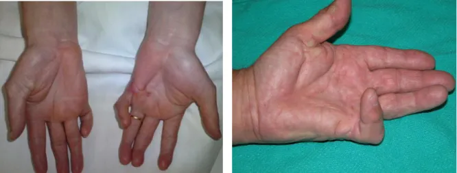
Figure 3. Clinical photograph showing the hands of two patients with advanced stage DD showing severe deformity. First picture (white background) from: Couto-Gonzalez et al. 2010. Second picture (green background) from: Howard et al. 2003.
Figure 4. Photograph showing a patients hand with Dupuytren’s disease in digit 4. Photo: Frank C. Müller.
Three different stages of disease tissue can be defined: proliferative, involutional and residual (Luck 1959). The first stage in characterised by proliferating fibroblasts in the nodules. Cells, rather than collagen, make up a large portion of the tissue, and the nodules are likely to be vascular. In the involutional stage, fibroblasts within the nodules align along the major lines of stress, predominately in the longitudinal axes of the hand (Luck 1959). As the contracture progresses, the nodules become smaller and increasingly ill-defined. In the residual stage, the nodules disappear, leaving a hypocellular and tendon-like fibrous cord (Luck 1959). The progression through these stages varies greatly in different individuals (Shih and Bayat 2010) from an aggressive course were the disease progresses to severe contraction in a few years to mild progression with no apparent change over many years.
Nodules and cords can be found simultaneously in affected tissue from one patient.
1.2.1 The Myofibroblast
The myofibroblast is the celltype thought to be responsible for the contraction on cellular level. Myofibroblasts differentiate from fibroblasts. Myofibroblasts differentiate after tissue injury. They are involved in normal wound healing and organ development (Tomasek et al.
2002). They synthesise ECM components, remodel the ECM and exhibit great contraction forces responsible for wound contraction leading to wound closure (Hinz et al. 2007).
Figure 5. Immunohistological staining of a myofibroblast.
The actin filaments arrayed in parallel sheets (stress fibers) are stained in red (phalloidin), the nucleus is stained in blue (DAPI) and αSMA is stained in green. 40x magnification.
Unchallenged fibroblasts exhibit few or no actin-associated cell-cell and cell-matrix contacts
and little ECM production (Tomasek et al. 2002). After tissue injury, they become activated
to migrate into the damaged tissue by cytokines locally released from inflammatory cells
(Werner and Grose 2003). Macrophages and T lymphocytes have been observed in DD
tissue (Baird et al. 1993). Fibroblasts are normally shielded from mechanical stress through
the ECM. Disruption of the ECM in tissue injury results in mechanic stress, which also
activates fibroblasts. In response to mechanical challenge, fibroblasts produce contractile
stress fibres. These are composed of cytoplasmic actins (Tomasek et al. 2002). Stress fibres
are connected to fibrous ECM proteins at sites of integrin-containing cell-matrix junctions
(Hinz 2006) and between cells via de novo established N-cadherin-type adherents’ junctions
(Hinz et al. 2004). Additionally differentiated myofibroblasts are defined by their de novo
expression of alpha smooth muscle actin (αSMA) (figure 5), which localises to stress fibres.
In culture fibroblasts generate stress fibres when grown on normal plastic or glass surface.
To stimulate αSMA expression fibroblast must be additionally treated with transforming growth factor beta 1 (TGFβ1) (Hinz 2006, Hinz et al. 2007). In vitro αSMA positive fibroblasts show twofold stronger contraction forces than αSMA negative fibroblasts (Hinz et al. 2001). At the end of tissue repair, the reconstructed ECM again takes over the mechanical load and myofibroblasts are released from stress and disappear by massive apoptosis (Tomasek et al. 2002).
1.2.2 History and Prevalence
DD was first described by the Swiss physician Felix Plater in 1614 and was later named after Baron Guillaume Dupuytren, a French physician, who in 1831 extensively lectured on the subject (Brenner and Rayan 2003). DD is common in the north of Europe. It is present in Germany (Brenner et al. 2001, Loos et al. 2006), Scandinavia (Bergenudd et al. 1993), Iceland (Guğmundsson et al. 2000), the UK (Gerber et al. 2011), and Ireland and has also been described in white populations in Northern America and Australia and in parts of Japan. It is rare in other populations, e. g. from Africa were only sporadic cases without family history are reported (Mitra and Goldstein 1994), or Asia (except Japan) (Slattery 2010). DD is significantly more frequent in the north than in the south of France (Maravic et al. 2005).
DD is a common disease. Estimative more than 2.3% of the German population are affected (Brenner et al. 2001). In UK the prevalence is about 4% in men and 2% in women (Early 1962). The prevalence of DD increases with age (Hindocha et al. 2009). In Iceland it increased from 7.2% in men aged 46-49 years to 39.5% in men >70 years old (Guğmundsson et al. 2002). Patients treated surgically for DD have an increased risk for cancer and cancer associated death (Wilbrand et al. 2000, Guğmundsson et al. 2002).
1.2.3 Treatment
There are a variety of classification systems (Rayan 1998, Woodruff et al. 1998, Falter et al.
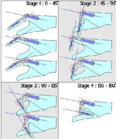
1991, Tubiana 1986) to group disease severity. Tubiana’s system (figure 6) grades the
contracture into one of four stages based on the combined angles of contracture of the joints
of the finger (metacarpal phalangeal (MCP) and proximal interphalangeal (PIP)). The
contraction degree at each finder joint is measured and classified. Classifications from each
finger can be added for one hand resulting in a maximal value of twenty.
Figure 6 Tubiana's classification. It grades the contracture into one of four stages based on the combined angles of contracture of the MCP and PIP joints, and may be applied to both composite and fixed contractures: This angle is illustrated by the blue lines in this diagram. MCP: metacarpal phalangeal;
PIP: proximal interphalangeal. From www.handcenter.org.
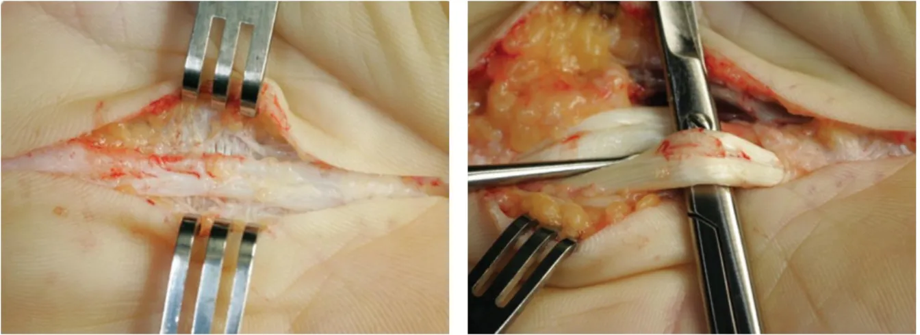
There is no cure for DD so far. When disease hampers finger movement corrective surgical treatment becomes necessary. Roughly two different approaches are frequently applied. The more non-invasive method involves the cutting across tight bands of diseased fascia, letting the edges gape apart and heal back at or closer to their natural length to restore the area's original flexibility (fasciotomy). This can be done without opening the affected area, by inserting a small-gauge needle through the skin along the length of the cord (needle fasciotomy). The cord is incised, using sweeping motions. Thus the cord is weakened and allows an extension force over the finger to rupture it (Desai and Hentz 2011). Most common is the surgical removal of affected tissue (limited fasciectomy) (figure 7). This procedure involves careful dissection and excision of the involved diseased fascia. After surgery, the hand is splinted, with the MCP and PIP joints in an extended position. Short- and long-term effectiveness of splinting following surgery was only poorly evaluated (Larson and Jerosch-Herold 2008). A resent non-surgical approach is based on the injection of collagenases into the affected tissue in order to resolve the collagen deposits (Hurst et al.
2009). Collagenase is injected directly into a Dupuytren cord, leading to lysis of the collagen
found within the diseased tissue. The patient returns the following day for joint manipulation in an attempt to rupture the cord (Desai and Hentz 2011).
Postoperative complications include haematoma, skin necrosis, infection, nerve injury, vascular injury, prolonged oedema, reflex sympathetic dystrophy and, rarely, finger loss. The most common complication is postoperative joint stiffness and loss of pre-operative flexion.
Regardless of the technique, there is a high rate of recurrence following surgical correction (Bayat and McGrouther 2006, Denkler 2010). For needle fasciotomy it was in the range of 65% after 33 months (van Rijssen et al. 2006).
Figure 7. A. Exposure of Dupuytren’s cord in the palm of the hand. From Rozen et al. 2012.
1.2.4 Genetics and other Risk Factors
DD is clearly an aging associated disease with onset from the fourth and fifth decade of life.
A strong risk factor is male gender. Women are less severe affected and develop the disease later in live. In men the time of first surgical treatment peaks around the fifth decade of live while women mostly present for surgery approximately one decade later.
Frequent familial occurrence of DD indicates a genetic basis for the disease. Studies have
determined a family predisposition in 12.5% (Brenner et al. 2001) and 27% of cases (Coert
et al. 2006). Several extended pedigrees were identified particularly in Scandinavia, pointing
to an autosomal dominant inheritance with reduced penetrance (Burge 1999). There is one
report so far describing a whole genome approach to identifying an underlying gene in an
extended Swedish family (Hu et al. 2005). They found linkage to an interval on chromosome
16 with a lod score value of 3.2, assuming 90% penetrance in family members over 45 years
of age. Another small genome wide association study (GWAS) has been conducted with 40
cases and 40 controls (Ojwang et al. 2010). But no candidate genes have been identified so
far. Reduced penetrance in several pedigrees and the large number of apparently sporadic cases indicate that Dupuytren disease is a complex disease in which genetic and environmental risk factors are involved. Several environmental factors have been proposed to contribute to DD development. Smoking and alcohol consumption have been associated with DD (e.g. Burge et al. 1997, Godtfredsen et al. 2004, Guğmundsson et al. 2000).
Elevated blood glucose levels, low body weight, and low BMI (body mass index) have also been associated with DD (Guğmundsson et al. 2000). Heavy manual labour and exposure to vibrations probably contribute to the disease (Liss and Stock 1996, Descatha et al. 2011).
DD is common among diabetes mellitus type 2 patients, but they may be in general less severe affected (Noble et al. 1984). In a study with epilepsy patients 56% had DD (Critchley et al. 1976). The authors proposed that this association is probably due to epileptic drug intake and subsequent stimulation of tissue growth factors. DD is common in patients with frozen shoulder, another fibrotic disorder (Smith et al. 2001). DD is rare among patients with rheumatoid arthritis (Arafa et al. 1984) and DD patients had less frequently stiff joints and rheumatic disorders (Guğmundsson et al. 1999).
1.2.5 Social Relevance and Civic Impact
DD is a common complex disorder. High prevalence rates result in considerable economic burden for treatment of this disease (Maravic et al. 2005, Gerber et al. 2011). Although DD is not life threatening it affects a constantly used tool, the hand. Finger contraction hampers already small tasks in the daily routine e.g. handshake. And patients may additionally experience pain in the affected hand. There is no cure for DD. High reoccurrence rates are observed after surgical treatment and the risk of complications is even higher at repeated surgery (Denkler 2010).
1.2.6 Other related Diseases
There are a number of clinically related conditions that occur less frequent and manifest in different parts of the body. Patients that present with one of these conditions often also have DD. In some DD patients connective tissue depots form at the dorsum of the proximal interphalangeal finger joints, the so called knuckle pads. In plantar fibromatosis (Ledderhose´s disease, LD) the sole of the feet is affected. Often the fibromatosis manifests in the part of the sole that is not in contact with the ground and thus does not hamper walking but in extreme cases the toes can contract.
Penile contracture (Peyronie's disease, PD) is characterized by the formation of thickened
fibrous plaques on the dorsum of the penis. Comparison of gene expression profiles indicates
that Dupuytren´s disease and Peyronie's disease share a common pathophysiology (Qian et al. 2004). PD was described as a genetic disorder with autosomal dominant inheritance (Bias et al. 1982). It was also linked to trauma (Carrieri et al. 1998). In this study it will be evaluated how many DD patients additionally suffer from these related conditions.
1.3 Mitochondrial Dysfunction and Oxidative Stress
In the following section recent knowledge of mitochondrial dysfunction and oxidative stress in the context of aging is discussed briefly. Mitochondrial dysfunction was implicated in the aetiologies of type 2 diabetes (Patti et al. 2003, Petersen et al. 2004, Lowell and Shulman 2005) and age related neurodegenerative disorders (Bowling and Beal 1995), e.g.
Alzheimer's disease (Swerdlow and Khan 2004), Parkinson's disease (Langston et al. 1983, Schapira et al. 1989, Bindoff et al. 1989), and Huntington's disease (e.g. Kuwert et al. 1990, Kim et al. 2010).
1.3.1 Mitochondrial Dysfunction
The main function of mitochondria is ATP production, which occurs during mitochondrial oxidative phosphorylation. During oxidative phosphorylation, electrons from reduced substrates are transferred to O
2through a chain of respiratory electron transporters including the complex I, III, and IV proton (H
+) pumps, which in turn generate a proton gradient across the mitochondrial inner membrane. The electrochemical energy of this gradient is then used by the ATP synthesis (complex V) which couples H
+reuptake with ADP phosphorylation in the matrix to generate ATP (Feissner et al. 2009). Proteins of the electron transport chain are downregulated with aging (Ghosh et al. 2011). Electrons leak from reduced sites in the respiratory chain and react with oxygen to form reactive oxygen species (ROS) which play an important role in cell signalling, but are better known for creating oxidative stress (Brookes et al. 2002). In mitochondria ROS cause mutations of mitochondrial DNA (mtDNA) which affect the electron transport chain function triggering increased ROS generation and accumulation of mtDNA damage over time (Mammucari and Rizzuto 2010).
During aging, giant non-functional mitochondria, defective in ATP production together with
aberrant macromolecules, accumulate especially in post-mitotic organs, such as the nervous
system and the cardiac and skeletal muscle. These are removed by autophagy, which also
declines with aging (Bergamini et al. 2007) accelerating the accumulation of these large
aggregates with age (Mammucari and Rizzuto 2010).
1.3.2 ROS Production in Mitochondria
ROS is implied in the aetiology of a number of diseases such as diabetes (Newsholme et al.
2007) and hypertension (Paravicini and Touyz 2006). In the mitochondrial respiratory chain, the transport of electrons is coupled with the formation of ROS. In particular, the redox reactions at respiratory complexes I and III generate superoxide (O
2•−) (Rigoulet et al. 2011).
Under physiologic conditions, the superoxide production is around 0.1% of the respiratory rate (Tahara et al. 2009). O
2•−in solution is short lived and rapidly converted into hydrogenperoxid (H
2O
2) e.g. by the mitochondrial superoxide dismutase (SOD2). H
2O
2is not a free radical (one or two unpaired electrons in the outer electron orbital) and is a more- stable molecule that is able to diffuse across biologic membranes. By the so called Fenton reaction H
2O
2generates the highly reactive hydroxyl radicals
•OH and H
•in the presence of metals such as Fe, which is a co-factor of many proteins. Other proteins in mitochondria are able to produce ROS, e.g. dehydrogenases, a-ketoglutarate dehydrogenase complex (aKGDHC) and glycerol-3-phosphate dehydrogenase (GPDH) and uncoupling proteins (UCPs) (Tahara et al. 2009) and monoamine oxidase (MAOA) in the outer mitochondrial membrane (Peña-Silva et al. 2009).
1.3.3 ROS Production at the Plasma Membrane
Mitochondria are the main side of ROS production. Another source of ROS are cells of the immune system. Neutrophils, macrophages, dendritic cells and monocytes release ROS in response to pathogens (Cannizzo et al. 2011). ROS in these cells is generated by NADPH oxidases (NOXs) located in the plasma membrane. They catalyze the production of superoxide by the one-electron reduction of oxygen, using NADPH as the electron donor
(Babior 1999): .
O
2•−is then converted into H
2O
2by the cytosolic superoxide dismutase (SOD1) (Cannizzo et al. 2011). The NOX gene family includes seven members. They are expressed in a variety of different tissues (Krause 2004). NOX4 is most widely expressed and was associated with transforming growth factor β (TGFβ1) induced fibroblast to myofibroblast differentiation (Cucoranu et al. 2005).
1.3.4 Mitochondrial Dysfunction without enhanced ROS Production
The Mitochondrial Free Radical Theory of Aging predicts that a normal metabolism causes
ROS production in mitochondria. ROS cause damage to lipids, proteins, and mtDNA. ROS-
induced mtDNA mutations lead to the synthesis of functionally impaired respiratory chain
subunits, causing respiratory chain dysfunction which in turn enhances ROS production.
This cycle over time promotes aging (Harman 2006) and involves an exponential increase of mtDNA mutations over time. But the formation of ROS in the course of normal metabolism does not necessarily increase significantly with age (Barja 1999). Trifunovic et al. (2005) used a mouse model that accumulates mtDNA mutations over time because of a defect mitochondrial polymerase lacking proofreading 3’-exonuclease activity (Trifunovic et al.
2004) and found no increase in ROS and oxidative damage and at the same time severely impaired respiratory chain function. The authors concluded from these findings that mtDNA mutations rather than ROS initially drive the aging process. But absolute mutation levels (deletions) have been argued to be too low to drive premature aging in these mice (Kraytsberg et al. 2009).
1.4 WNT/β-catenin Signalling Pathway
Wnts are secreted glycoproteins that act as short range signals in multiple processes. They are expressed in spatially restricted and dynamic patterns during development (Cadigan and Nusse 1997) and in the adult organism (Moon et al. 2004). Wnt signalling has been implicated in the pathogenesis of cancer (van de Wetering et al. 2002, Giles et al. 2003, Uematsu et al. 2003), cardiovascular disease (van Gijn et al. 2002), and Alzheimer's disease (De Ferrari and Inestrosa 2000) and upregulation of Wnt signalling was associated with aging (Marchand et al. 2011, Brack et al. 2007, Liu et al. 2007). The Wnt protein family comprises 19 members in mammals which can trigger different pathways in the signal receiving cell (Veeman et al. 2003, Sugimura and Li 2010). One extensively studied pathway involves the stabilisation of β-catenin which then translocates into the nucleus and acts on the expression of target genes. Wnt signalling is activated during fibroblast proliferation (Cheon et al. 2004) and elevated levels of β-catenin were found in pulmonary fibrosis (Chilosi et al. 2003), aggressive fibromatosis (Cheon et al. 2002), and also in DD (Varallo et al. 2003). Expression profile of human lung fibroblasts treated with Wnt3a revealed that among genes differentially expressed in response to Wnt stimulation, where genes that play a role in differentiation of fibroblasts to a myofibroblast or smooth muscle phenotype (Klapholz-Brown et al. 2007).
1.4.1 Wnt/β-catenin Signalling
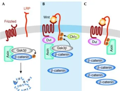
In the absence of Wnt, β-catenin is targeted to a multimeric protein complex called the
destruction complex, which includes the scaffolding protein axin and the tumour suppressor
protein adenomatous polyposis coli (Apc). β-catenin is then phosphorylated by casein kinase
1 (Ck1, CSNK1A1) and subsequent glycogen synthase kinase 3β (Gsk3β) (Liu et al. 2002).
This phosphorylation targets β-catenin for ubiquitination and subsequent degradation by the proteasome (figure 8). In the nucleus prospective target genes of the pathway are kept in a repressed state by T-cell factor (Tcf) and lymphoid enhancer-binding protein (Lef) transcription factors and associated co-repressors (Gordon and Nusse 2006, Logan and Nusse 2004).
Figure 8 Model for the activation of the Wnt/β-catenin pathway. (A) In the absence of a Wnt signal, β- catenin is phosphorylated and targeted for proteasome-mediated degradation by a destruction complex that contains axin and Gsk3β among other proteins. (B) On binding of Wnt to the receptors Fzd and LRP, Dvl binds to Fzd and recruits the destruction complex through interaction with axin. Subsequently, Gsk3β phosphorylates critical sites on LRP, which, together with residues phosphorylated by CkIγ, act as docking sites for axin. (C) Binding of axin to LRP leads to inhibition of the destruction complex and stabilization of β-catenin. CkIγ, casein kinase Iγ; Dvl, dishevelled; Fz, Frizzled; Gsk3β, glycogen synthase kinase 3β. From Fuerer et al. Wnt signalling in development and disease. Max Delbrück Center for Molecular Medicine meeting on Wnt signalling in Development and Disease. EMBO Rep. 2008 Feb;9(2):134-8.
Two cell-surface receptors cooperate to transmit Wnt signals across the plasma membrane to
activate β-catenin signalling. Wnts bind to the seven transmembran domains containing
receptor frizzeld (Fzd 1-10) and the low density lipoprotein receptor-related protein (Lrp 5
and 6). Ternary ligand-receptor complexes form ribosome-sized aggregates in the cell
membrane. The binding of Wnt to Fzd/Lrp leads to the phosphorylation of Lrp6 by Gsk3β
and Ck1 (Rao and Kühl 2010). Axin and the phosphoprotein dishevelled (Dvl) then bind to
the cytoplasmic tails of Lrp and Fzd, respectively. The destruction complex is disrupted and
axin is degraded. The activation of Dvl also leads to the inhibition of Gsk3β. Both effects
increase the post-translational stability of β-catenin. As the β-catenin level rise in the
cytoplasma, β-catenin also translocates into the nucleus, where it interacts with DNA-bound
Tcf and Lef family members to act on the transcription of target genes (Moon et al. 2004).
1.4.2 Wnt Signalling: Modifications, Antagonists and Activators
Wnts are modified before secretion by the addition of two fatty acids, palmitic acid on the first conserved cysteine and palmitoleic acid on a highly conserved serine residue (Willert et al. 2003; Takada et al. 2006). These modifications render Wnt proteins hydrophobic and Heparan sulfate proteoglycans (Hspgs) prevent Wnts from aggregating in the extracellular matrix (Fuerer et al. 2010). Members of the Hspg family were shown to act as a reservoir or modulator for several growth factors and signalling molecules (e.g. Bernfield et al. 1999;
Whitelock and Iozzo 2005). Sulphated proteoglycan levels were elevated in DD primary tissue (Tunn et al. 1988).
Telomerase directly modulates Wnt/β-catenin signalling by serving as a cofactor in a beta- catenin transcriptional complex. The telomerase protein component TERT (telomerase reverse transcriptase) interacts with BRG1 (also called SMARCA4), a SWI/SNF-related chromatin remodelling protein, and activates Wnt-dependent reporters in cultured cells and in vivo (Park et al. 2009).
In addition to its function in the Wnt signalling pathway, β-catenin also binds tightly to the cytoplasmic domain of type I cadherins and plays an essential role in the structural organization and function of cadherins by linking cadherins through α-catenin to the actin cytoskeleton (Jamora and Fuchs 2002, Gumbiner 2000).
There are several inhibitors and activators that either inhibit or promote Wnt signalling extracellular. One example for an inhibitor is the family of dickkopf proteins (DKK) (Glinka et al. 1998). DKK1 binds to LRP5/6 and the transmembrane protein Kremen (Mao et al.
2002). This interaction inactivates LRP5/6. R-Spondins (RSPOs) promote Wnt signalling by antagonizing DKK1-mediated interaction with LRP and Kremen (Kim et al. 2008).
Sclerosteosis (SOST) and sclerostin domain containing 1 (SOSTDC1) are also secreted Wnt inhibitors that bind to and inactivate LRP (Semënov et al. 2005, Itasaki et al. 2003). Wnt inhibitory factor (WIF) proteins and secreted frizzled related proteins (SFRP1,2-5) act by directly binding Wnt molecules and can function as Wnt inhibitors, but may also stabilize Wnt's and facilitate Wnt signalling (Hsieh et al. 1999, Hoang et al. 1996). Connective-tissue growth factor (CTGF) modulates Wnt signalling and interacts with LRP6 (Mercurio et al.
2004). Dapper, antagonist of beta-catenin, homolog (DACT1-3) promotes Wnt/β-catenin signalling by binding intracellulary to Dvl (Gloy et al. 2002).
In colorectal cancer Wnt/β-catenin is commonly dysregulated in colorectal cancer (Pálmer et
al. 2001). Vitamin D acts protective against colorectal cancer (Garland et al. 1989).The most
active vitamin D metabolite, 1alpha,25-dihydroxyvitamin D3 (D3) inhibits β-catenin transcriptional activity by promoting vitamin D receptor (VDR) binding to β-catenin and the induction of E-cadherin expression. Vitamin D3 has been shown to regulate two genes encoding the extracellular Wnt inhibitors DKK-1 and DKK-4. By an indirect transcriptional mechanism, D3 increases the expression of DKK-1 RNA and protein, which acts as a tumour suppressor in human colon cancer cells harbouring endogenous mutations in the Wnt/beta- catenin pathway (González-Sancho et al. 2005). In contrast, vitamin D3 represses DKK-4 transcription by inducing direct VDR binding to its promoter. DKK-4 is a target of the Wnt/β-catenin pathway and is up-regulated in colorectal tumours (Pendás-Franco et al.
2008). Several vitamin D target genes have been characterized including tenascin-C, fibronectin, laminin and its receptor, apolipoprotein D, insulin-like growth factor binding protein 3, cyclin C, and several members of the transforming growth factor family and their receptors (Freedman, 1999).
Mice overexpressing SFRP4, aWnt inhibitor had significant higher serum and urine levels of vitamin D (Cho et al. 2010). The connection between Wnt/β-catenin signalling and viamin D signalling could be causative for the observed north-south decrease in the prevalence of DD.
1.4.3 Wnt/β-catenin Signalling and Aging
Wnt/β-catenin signalling is increased and leads to an enhanced fibrosis in aging muscle (Brack et al. 2007). During aging the regenerative potential of muscle declines (Goldspink et al. 1994). Muscle tissue is replaced by fibrous connective tissue and adipose tissue. Brack et al. (2007) examined muscle tissue and cells derived from muscles of young (~6-month-old) and aged (~24-month old) mice. Axin2 (target of Wnt/β-catenin signalling) transcript levels were increased in aged muscle and a progressive increase in Wnt signalling in myogenic cells during aging with an increase in β-catenin and decrease of GSK3β was noted. Increased β-catenin was also found in aged muscle tissue in response to injury compared with young tissue. The injection of Wnt3A into young muscle after injury resulted in increased connective tissue deposition phenotypically similar to regenerating aged muscle. Exogenous Wnt also reduced cellular proliferation in young regenerating muscles. Reduced fibrosis was seen in aged muscle after the injection of Wnt inhibitors (DKK1 and SFRP3). The authors concluded that the fibrotic aging phenotype in muscle is promoted by Wnt’s (or Wnt-like proteins) present in the aging serum (Brack et al. 2007).
Liu et al. (2007) determined reduced number of stem cells in the klotho mouse, a model for
accelerated aging (Kuro-o et al. 1997) that lacks klotho, a secreted protein found to decline
in the serum of mouse and human during aging (Xiao et al. 2004). Liu et al. noted that klotho and Wnt were co-expressed in transfected cells. They showed by immunoprecipitation that klotho binds to Wnt3A, Wnt1, Wnt4 and Wnt5A. In their cell- culture model klotho inhibited Wnt-signalling and the mouse model lacking klotho expression had enhanced expression of Wnt target genes (Axin2). In cultured fibroblasts (mouse embryonic and human) continuous exposure to Wnt3A first increased proliferation (BrdU incorporation) but over time proliferation decreased while the level of apoptosis was not increased. The authors concluded that chronic Wnt stimulation may contribute to stem cell depletion and aging.
Marchand et al. (2011) compared the expression profiles of middle old (43-60 years, N = 8) and older patients (75-83 years, N = 7) in mammary artery media and found target genes of the Wnt/β-catenin signalling pathway upregulated in the older group (e.g. SPP1, WISP1, versican and IGFBP2). Β-catenin mRNA was not significantly upregulated but β-catenin phosphorylation at serine 675 was significantly increased in the older group. This phosphorylation induces β-catenin nuclear localization and transcriptional activity, suggesting that β-catenin is activated during aging. Cyclin D2 mRNA and protein levels were unchanged as were mRNA levels of catalase and SOD2. Wnt3A treatment of vascular smooth muscle cells (VSMC) from old rats (8 month old) did not induce proliferation and cyclin D1 expression as it did in VSMCs derived from young rats (6 weeks old) while β- catenin was activated in young and old cells. The authors concluded that β-catenin pathway is activated during human vascular aging but that the proliferative response to Wnt is altered downstream of β-catenin activation (Marchand et al. 2011).
Together these findings hint that Wnt/β-catenin signalling plays an important role during
aging and that its function in aged tissue differs from that of young tissue.
Kerstin Becker - 27 -
1.5 Objectives and Hypotheses
Dupuytren´s disease is a complex disease with a strong genetic basis. The aim of this study was to identify loci in the human genome that are associated with Dupuytren´s disease.
Therefore a genome wide association study (GWAS) was conducted with a case control study design. This study design should allow for the identification of some of the genetic factors that promote pathogenesis. Because DD is a complex genetic disease several genetic loci are expected to be associated, each contributing in small part to the susceptibility of this disease.
Expression profiles of primary tissue and tissue derived fibroblasts differ between DD patients and matched controls (Pan et al. 2002, Qian et al. 2004, Rehman et al. 2008, Satish et al. 2008, Zang et al. 2008). A range of genes identified from these studies were proposed to play a causative role in the pathogenesis of DD. But no signalling pathways have been attributed to DD so far. A whole genome expression analysis was conducted in order to identify major signalling pathways involved in DD pathogenesis. Results from this experiment may possibly support and confirm findings from the GWAS. As DD is an aging associated disease identified pathways may lead to the identification of links between DD and aging.
DD fibroblasts in vivo are characterised by high proliferative and contractile activity.
Disease tissue derived primary fibroblasts were cultured and characterised in comparison to control tissue derived fibroblasts. These experiments should help to establish and characterise a simple in vitro model, desirable to test hypotheses from the GWAS in the future.
A questionnaire based elevation of known risk factors and other parameters was conducted in order to characterise DD patients sampled for DNA and disease tissue epidemiologically.
Integration of data from all these approaches should help to pave the way to a
comprehensive understanding of the molecular pathology of this poorly understood aging-
related disease.
2 Materials and Methods
2.1 Sample Collection, Storage and Handling 2.1.1 Subjects
Between 2007 and 2011 760 DD patients were recruited through the outpatient clinics of the plastic surgery departments of nine hospitals in Germany and one in Switzerland.
Additionally a number of patients were recruited through the German Dupuytren Society (Dupuytren e.V.).
Genotype data from 1618 German controls were already available, 1164 of these were part of the Popgen study (University of Kiel, Germany) (project number: BSP+SPC/110217/83), and 454 were from KORA (Helmholtz Center Munich, Neuherberg, Germany) (project number: K26/11). These samples were genotyped with the Affymetrix 6.0 chip. In addition genotype data for 1219 Popgen controls that were typed on the Affymetrix Axiom 2.0 chip was also available. Participants provided written informed consent and the study was approved by the Ethics Committee of the MathNat Faculty of the University of Cologne. 282 control DNAs with European background were provided by the University of Essen.
2.1.2 Questionnaire
For each participant a one-sided questionnaire was completed by the attending physician.
The recorded parameters are given in Table 1. The questionnaire was changed twice during the study, when minor adjustments or additional questions became eligible. All versions of the questionnaire are listed in figures 68-70 of the appendix. A separate questionnaire was used for control individuals, which is also given in the appendix.
Table 1. Parameters enquired in questionnaire survey for DD patients
parameter factors
name date of birth
gender male, female
age at first surgery
affected hand right, left, both degree after Tubiana 0, 1, 2, 3, 4
recurrence recurrence, extension
preceding complications trauma, sudeck dystrophy
ectopic manifestations e.g. knuckle pads, plantar fibromatosis affected family members no, yes, who
Table 1 continued:
parameter factors
other diseases diabetes mellitus, rheumatoid arthritis, epilepsy, other medication antiarrhythmia, antihypertensive, antiepileptic drugs, other for women:
age at menopause
hormone intake yes, no
ovarian surgery yes, no
profession
smoking habits 0, <5, <20, >20 cigarettes per day year subject stopped smoking
alcohol intake never, occasional, regular