Madelung’s disease: a man with a phenotype suggestive of anabolic steroid use
Madelung-Krankheit: Ein Mann mit einem Phänotyp, der auf den Gebrauch von anabolen Steroiden hindeutet
Abstract
Madelung’s disease (MD), a rare disorder of adipose tissue, is charac- terized by multiple, symmetrical, non-encapsulated adipose tissue de-
Georgios N.
Kostopoulos
1positions in typical locations. MD typically affects middle-aged males
Georgios G. Tzikos
2of Mediterranean and Eastern European origin. The pathophysiology
Nikolaos H.
Kostogloudis
3remains unknown, yet chronic alcohol consumption is frequently repor- ted and features of metabolic syndrome are usually present. Most cases are sporadic and patients may report symptoms of compression (dys-
Konstantinos A. Toulis
1phagia, hoarseness) and/or present with a characteristic phenotypic appearance. Herein, we present a case of a 70-year-old man with a 20-
year history of recurrent lipomatous formations, who had undergone 1 Department of Endocrinology, 424 General Military several surgical procedures. His past medical and family history were
Hospital, Thessaloniki, Greece
unremarkable, biochemistry analysis and endocrine workup were normal, previous pathology results were indicative of benign lipomas and he
reported chronic alcohol consumption. This otherwise asymptomatic 2 Department of Surgery, 424 General Military Hospital, Thessaloniki, Greece patient had a very remarkable “pseudoathletic appearance”, reminiscent
of anabolic steroid use. On the basis of past medical history, laboratory
workup and clinical appearance, the diagnosis of MD was made and 3 Department of Plastic Surgery, 424 General Military the patient was referred to the Plastic Surgery Department for further
evaluation and treatment. Hospital, Thessaloniki,
Greece Keywords:Madelung’s disease, benign symmetrical lipomatosis, multiple
symmetrical lipomatosis, Launois-Bensaude syndrome, lipomatosis, alcoholism
Zusammenfassung
Die Madelung-Krankheit, eine seltene Erkrankung des Fettgewebes, ist charakterisiert durch multiple symmetrische Ablagerungen von Fettge- webe an typischen Körperteilen. Die Krankheit tritt hauptsächlich bei Männern im mittleren Alter auf, die aus Mittelmeerländern und osteu- ropäischen Ländern stammen. Die Pathogenese ist bislang unbekannt, dennoch ist die Krankheit häufig mit Alkoholmissbrauch und anderen Stoffwechselstörungen assoziiert. Die meisten Fälle treten sporadisch auf und die Patienten können Symptome einer Kompression (Dysphagie, Dysphonie) und/oder eines charakteristischen Phänotyps aufweisen.
Hier präsentieren wir den Fall eines 70-jährigen Patienten, der seit 20 Jahren an rezidivierender Lipomatosis leidet und sich mehrerer chirurgischer Eingriffe unterzogen hatte. Seine Vorgeschichte war un- auffällig, biochemische und endokrinologische Untersuchung waren normal. Die pathohistologische Aufarbeitung ergab die Diagnose gut- artiger Lipome und der Patient berichtete über chronischen Alkohol- konsum. Dieser ansonsten asymptomatische Patient hatte ein sehr bemerkenswertes „pseudoathletisches Aussehen“, das an den Gebrauch von anabolen Steroiden erinnerte. Auf der Grundlage von Anamnese, Laboruntersuchung und klinischem Erscheinungsbild wurde die Diagno- se einer Madelung-Krankheit gestellt und der Patient zur Abteilung Plastische Chirurgie überwiesen.
Introduction
Madelung’s disease (MD), a rare disorder of adipose tis- sue, is characterized by multiple, symmetrical, non-encap- sulated adipose tissue deposition in typical locations [1].
MD is also known as benign symmetrical lipomatosis or Launois-Bensaude syndrome [1] and was first described by Benjamin Brodie in 1846 [2]. In 1888, Madelung published a series of patients with symmetrical fat deposi- tion in the submental triangle [3], while 10 years later, in 1898, Launois and Bensaude published another series of cases with similar findings [4].
MD mainly affects middle-aged males (male/female ratio 15:1) of Mediterranean and Eastern European origin [1], whereas few cases from Asia are reported as well [5].
Interestingly, it has been suggested that the silhouette of an ancient Roman statue, the Capestrano Warrior, which dates back to 6thcentury BC, mimics the fat distri- bution in MD [6]. This fact may indicate an inherited tendency in the Mediterranean region [6]. The incidence rate of the disease has been estimated as 1:25,000 men in the Italian population in 1984 [7], while the prevalence in the area of Bavaria in Germany was calculated as 1:24,274 people [8]. To the best of our knowledge, over 300 cases are described in the medical literature to date [9], [10], 106 of which were reported in a review pub- lished in 2018 [11]. The review included cases from 2000 to 2015 [11]. However, the exact number could be even higher as many undiagnosed cases may exist and there- fore are not reported [1], [8].
The implicating pathogenetic mechanisms are still not well understood [1]. Although the relationship between MD and chronic alcohol consumption, especially red wine, is well known [12], cases without alcohol abuse have been described [13], [14]. It is generally believed that alcohol abuse impairs adrenergic lipolysis, leading to uncontrolled fat deposition in different areas of the body [1]. Moreover, a hypothesis that proliferation and differ- entiation of brown fat tissue has a key role in the patho- genesis of MD has been recently suggested [12]. Besides, mitochondrial dysfunction resulting from mutations of mitochondrial DNA has also been proposed as a trigger mechanism [15], [16].
The diagnosis of MD is based on medical history and the characteristic clinical presentation [8]. Imaging examina- tion, such as ultrasound and magnetic resonance ima- ging, may be helpful; biopsy of the affected areas reveals the presence of non-encapsulated adipose tissue [1].
Herein, we present a case of a 70-year-old man with Madelung’s disease, who had recurrent fat depositions and a profound “pseudoathletic” phenotype reminiscent of anabolic steroid use.
Case description
A 70-year-old male patient was referred to the outpatient clinic for evaluation due to a 20-year history of recurrent lipomatous formations. During this 20-year period, he
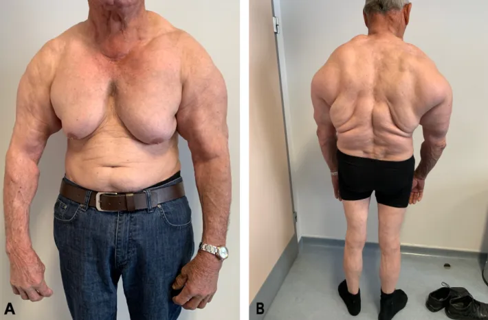
had undergone multiple surgical procedures but found no long-term relief. His past medical and family history were unremarkable, other than a 30-year long history of alcohol consumption. On physical examination, his body mass index was 27.3 kg/m2 and he had a symmetrical enlargement of the superior part of the trunk, including the neck, shoulder girdle, upper arms, chest and upper back, giving the patient a “pseudoathletic appearance”
(Figure 1). The lesions were soft and painless. Both testes were normal. He denied having other symptoms such as dysphagia, hoarseness, muscle weakness, decreased li- bido, or erectile dysfunction. Biochemistry analysis includ- ing liver function tests, lipid profile and renal function was normal. Moreover, endocrine workup (prolactin, luteinizing hormone, follicle-stimulating hormone, estra- diol, testosterone, dehydroepiandrosterone sulfate, in- sulin-like growth factor-I, 17α-hydroxyprogesterone, cor- tisol, haemoglobin A1c, thyroid functions tests and auto- antibodies, thyroglobulin, calcitonin) and testing for he- patitis and HIV, was also unrevealing. Finally, previous pathology results revealed benign lipomas and the dia- gnosis of Madelung’s disease was made.
Discussion
Madelung’s disease (MD), a rare disorder of adipose tis- sue, is characterized by multiple, symmetrical, non-encap- sulated adipose tissue deposition in typical locations, such as the cervical area, shoulders, upper trunk, upper arms, the abdomen, hips and thighs. Enzi distinguishes two types of the disease:
1. deposits in the area of the neck (Madelung’s collar), shoulders, supraclavicular triangle, and proximal up- per limbs giving a “pseudoathletic appearance” and 2. deposits in the thighs and abdomen, resembling
generalized obesity [7], [12].
According to Enzi’s classification system, our patient has type 1 of MD. Another classification system, developed by Donhauser et al. in 1991, categorizes patients in 3 types. Type 1 involves cases with fat distribution in the area of the neck (horsecollar), type 2 in the upper trunk and arms (pseudoathletic phenotype), and type 3 in the thighs (gynecoid type) [17], [18]. Clinical overlap between the aforementioned types may be present [18]. Recently, a novel classification based on a cohort from Germany has been proposed [8]. This comprehensive classification system divides patients with Madelung’s disease into five subtypes (Ia, Ib, Ic, II, and III). More specifically, patients with multiple lipomas in the upper body are described as type I, those with lower body involvement (thighs and legs) as type II. Type I is further subdivided in Ia (lipomas in the area of the neck), Ib (neck, shoulders and arms) and Ic (neck, shoulders, arms and trunk). If fat deposition is generalized, the patient is considered as type III [8].
Therefore, based on Donhauser’s system, our patient is type II MD while type Ic MD, if the more recent classifica- tion system is applied.
Kostopoulos et al.: Madelung’s disease: a man with a phenotype suggestive ...
Figure 1: The characteristic “pseudoathletic appearance” of type 1 Madelung’s disease (Enzi’s classification)
In addition, the symmetrical fat distribution in this case was quite remarkable and profound; the clinical pheno- type was reminiscent of anabolic steroid use as the areas of the upper trunk and arms were involved. Anabolic steroids are in demand among athletes and bodybuilders, leading to muscle hypertrophy, proliferation and strength increase [19]. This characteristic “pseudoathletic” or
“bodybuilder’s” phenotype in combination with history (Mediterranean origin and chronic alcohol abuse) and histopathological examination facilitated the diagnosis of MD.
MD is also widely known as benign symmetrical lipo- matosis [1]. However, MD is not always a “benign” dis- order since associated complications and comorbidities may have a negative impact on prognosis [20], [21], [22].
Patients often present features of metabolic syndrome, such as diabetes mellitus, hyperuricemia and dyslip- idemia, while other cases are related to secondary liver cirrhosis, peripheral (motor or sensory) and autonomy neuropathy [1], [21], [22], [23], [24]. Neuropathy, one of the most frequent chief complaints (85% of the cases) and possibly attributed to chronic alcohol abuse, is linked with increased cardiovascular morbidity and mortality [21], [24], [25]. Hypothyroidism, chronic obstructive pul- monary disease and adrenal dysfunction are also related to MD, whereas fat depositions may occur in HIV positive patients under treatment with protease inhibitors [1], [23], [24]. Furthermore, in some cases, symptoms of compression, such as hoarseness or dysphagia, may be present due to deep infiltration in the larynx and medi-
astinum [1], [21], [23]. On rare occasions, malignant transformation to intramyxoid sarcoma, liposarcoma and squamous cell carcinoma has also been reported [26], [27], [28]. Notably, despite the 30-year-alcohol consump- tion history, our patient did not show any sign of alcohol- induced liver disease or polyneuropathy. Also he did not develop any other MD related condition or complication.
The pathogenetic background of the disease has not been fully elucidated and several hypotheses have been de- veloped. In this case, chronic alcohol intake is believed to be the inducing factor for developing MD, as the asso- ciation between alcohol and impairment of adrenergic lipolysis is well known [1]. Furthermore, most MD cases, as ours, are sporadic, but some familiar cases, inherited in an autosomal dominant manner, are reported [29]. In familiar cases, mutations in mitochondrial DNA, which have maternal origin, have been identified [15], [16], [29].
Due to the existing knowledge gap regarding the etiology of the disease, any therapeutic intervention is character- ized as palliative [1]. To date, surgical resection and liposuction are the only effective therapeutic approaches, yet with high recurrence rates, as complete resection is almost impossible [1], [11]. In fact, the risk of recurrence in lipectomy has been calculated at two-thirds of the cases, while at 95% in liposuction [1]. According to a re- cent systematic review of case series, liposuction was associated with better aesthetic results and higher recur- rence risk, whereas patients subjected to lipectomy were more likely to suffer from post-surgical complications
such as infection and hemorrhage [11]. The reported re- currence rates were lower compared to the study by Szewc et al. (5 out of 95 patients (5.3%) who had under- gone lipectomy; 1 out of 18 (5.6%) who had undergone liposuction and 1 out of 11 (9%) who had undergone both lipectomy and liposuction). The latter, though, might be an underestimation of the real risk, taking into account the limitations of the study. Firstly, on some occasions, patients’ records were incomplete [11]. More importantly, follow-up duration was different in each case, ranging from one week to more than thirty years [11]. In view of the aforementioned complications, the indication for surgical intervention in Madelung’s disease should be taken with care and frequently will be seen in severe cases of compression related symptoms, such as trachea or pharynx obstruction. However, cosmetic deformity can also be seen as an indication for surgery in cases in which the patient presents with a certain level of suffering from the disease [1], [30].
Pre-, peri- and postoperative strategy in cases with MD is of cardinal importance [30], [31], [32]. Endotracheal intubation due to neck immobility or presence of macro- glossia may be strenuous even for experienced anesthesi- ologists [31]. Moreover, the risk of postoperative bleeding or hematoma should be taken into account and patients should be monitored for indicative signs or symptoms [30]. Additionally, potential respiratory depression as a consequence of oversedation should also be considered in these cases [31]. Therefore, different interventions during airway management may be warranted [30], [31], [32]. For instance, awake fiberoptic intubation or awake videolaryngoscopy could be two effective alternatives, when general anesthesia is implemented [30], [31], [32].
Unfortunately, effective pharmacological agents are not available. Apart from surgical therapy, recommendation for alcohol abstinence and lifestyle modifications should also be examined [1], [30]. In our case, the patient was referred to the Plastic Surgery Department, where long- term follow-up was recommended as no symptoms of compression were present.
Conclusion
In conclusion, establishing the diagnosis of MD can be challenging, since clinicians may be unaware of this condition. The presence of comorbidities should be taken into consideration during management of these patients.
Surgical resection and liposuction, the only effective therapeutic approaches, are recognized as palliative treatment due to the lack of efficient pharmacological agents. Future research should focus on the patho- physiology making more treatment options available.
Notes
Competing interests
The authors declare that they have no competing in- terests.
Author contributions
All authors cared for the patient and contributed equally to the preparation of the manuscript. All authors approved the final manuscript.
Informed consent
The patient provided written informed consent for publi- cation of this case.
References
1. Szewc M, Sitarz R, Moroz N, Maciejewski R, Wierzbicki R.
Madelung’s disease – progressive, excessive, and symmetrical deposition of adipose tissue in the subcutaneous layer: case report and literature review. Diabetes Metab Syndr Obes.
2018;11:819-25. DOI: 10.2147/DMSO.S181154
2. Brodie BC. Clinical Lectures on Surgery delivered at St. George’s Hospital. Am J Med Sci. 1846;11(22):437. DOI:
10.1097/00000441-184604000-00028
3. Madelung OW. Ueber den Fetthals. Arch Klin Chir. 1888;37:106- 30.
4. Launois PE, Bensaude R. De l’adenolipomatose symetriqe. Bull Mem Soc Med Hop Paris. 1898;1:298.
5. Chuang CC, Cheng YF, Chang HP, Lin CZ. Madelung’s disease. J Chin Med Assoc. 2004 Nov;67(11):591-4.
6. Feliciani C, Amerio P. Images in clinical medicine. Madelung’s disease: inherited from an ancient Mediterranean population?
N Engl J Med. 1999 May;340(19):1481. DOI:
10.1056/NEJM199905133401906
7. Enzi G. Multiple symmetric lipomatosis: an updated clinical report.
Medicine (Baltimore). 1984 Jan;63(1):56-64. DOI:
10.1097/00005792-198401000-00004
8. Schiltz D, Anker A, Ortner C, Tschernitz S, Koller M, Klein S, Felthaus O, Schreml J, Schreml S, Prantl L. Multiple Symmetric Lipomatosis: New Classification System Based on the Largest German Patient Cohort. Plast Reconstr Surg Glob Open. 2018 Apr;6(4):e1722. DOI: 10.1097/GOX.0000000000001722 9. Wan SC, Huang MH, Perng CK, Liao WC. Madelung Disease:
Analysis of Clinicopathological Experience in Taipei Veterans General Hospital. Ann Plast Surg. 2019 Jan;82(1S Suppl 1):S66- S71. DOI: 10.1097/SAP.0000000000001719
10. Multiple symmetric lipomatosis. In: Orphanet. [last updated 2019 Oct]. Available from: https://www.orpha.net/consor/cgi-bin/OC_
Exp.php?lng=EN&Expert=2398
11. Chen CY, Fang QQ, Wang XF, Zhang MX, Zhao WY, Shi BH, Wu LH, Zhang LY, Tan WQ. Madelung's Disease: Lipectomy or Liposuction? Biomed Res Int. 2018;2018:3975974. DOI:
10.1155/2018/3975974 Kostopoulos et al.: Madelung’s disease: a man with a phenotype suggestive ...
12. Enzi G, Busetto L, Sergi G, Coin A, Inelmen EM, Vindigni V, Bassetto F, Cinti S. Multiple symmetric lipomatosis: a rare disease and its possible links to brown adipose tissue. Nutr Metab Cardiovasc Dis. 2015 Apr;25(4):347-53. DOI:
10.1016/j.numecd.2015.01.010
13. Bergler-Czop B, Wcisło-Dziadecka D, Brzezińska-Wcisło L.
Madelung’s disease in a patient with chronic renal insufficiency:
a case report and review of literature. Postepy Dermatol Alergol.
2014 May;31(2):121-4. DOI: 10.5114/pdia.2014.40922 14. El Ouahabi H, Doubi S, Lahlou K, Boujraf S, Ajdi F. Launois-
bensaude syndrome: A benign symmetric lipomatosis without alcohol association. Ann Afr Med. 2017 Jan-Mar;16(1):33-4. DOI:
10.4103/1596-3519.202082
15. Gámez J, Playán A, Andreu AL, Bruno C, Navarro C, Cervera C, Arbós MA, Schwartz S, Enriquez JA, Montoya J. Familial multiple symmetric lipomatosis associated with the A8344G mutation of mitochondrial DNA. Neurology. 1998 Jul;51(1):258-60. DOI:
10.1212/wnl.51.1.258
16. López-Gallardo E, Cammarata-Scalisi F, Emperador S, Hernández- Ainsa C, Habbane M, Vela-Sebastián A, Bayona-Bafaluy MP, Montoya J, Ruiz-Pesini E. Mitochondrial DNA pathogenic mutations in multiple symmetric lipomatosis. Clin Genet. 2020 May;97(5):731-5. DOI: 10.1111/cge.13701
17. Donhauser G, Vieluf D, Ruzicka T, Braun-Falco O. Benigne symmetrische Lipomatose Launois-Bensaude Typ III und Bureau- Barrière-Syndrom [Benign symmetric Launois-Bensaude type III lipomatosis and Bureau-Barrière syndrome]. Hautarzt. 1991 May;42(5):311-4.
18. Pinto CI, Carvalho PJ, Correia MM. Madelung’s Disease: Revision of 59 Surgical Cases. Aesthetic Plast Surg. 2017 Apr;41(2):359- 68. DOI: 10.1007/s00266-016-0759-x
19. Kicman AT. Pharmacology of anabolic steroids. Br J Pharmacol.
2008 Jun;154(3):502-21. DOI: 10.1038/bjp.2008.165 20. Suito M, Kitazawa T, Takashimizu I, Ikeda T. Madelung’s disease:
long-term follow-up. J Surg Case Rep. 2019 Jan;2019(1):rjy356.
DOI: 10.1093/jscr/rjy356
21. Yeh NC, Yang CY, Chou CW, Yen FC, Lee SY, Tien KJ. Madelung’s disease. J Clin Endocrinol Metab. 2012 Sep;97(9):3012-3. DOI:
10.1210/jc.2012-1649
22. Enzi G, Busetto L, Ceschin E, Coin A, Digito M, Pigozzo S. Multiple symmetric lipomatosis: clinical aspects and outcome in a long- term longitudinal study. Int J Obes Relat Metab Disord. 2002 Feb;26(2):253-61. DOI: 10.1038/sj.ijo.0801867
23. Brea-García B, Cameselle-Teijeiro J, Couto-González I, Taboada- Suárez A, González-Álvarez E. Madelung’s disease: comorbidities, fatty mass distribution, and response to treatment of 22 patients.
Aesthetic Plast Surg. 2013 Apr;37(2):409-16. DOI:
10.1007/s00266-012-9874-5
24. Bucciarelli M, Fan C. Diffuse symmetrical Lipomatosis: a case report depicting the potential for severity. AACE Clinical Case Reports. 2017;3(4):e370-3. DOI: 10.4158/EP161546.CR 25. Fonseca VR, Freitas C, Palmeira M, Ferreira C, Victorino R.
Cardiac noradrenergic denervation in a patient with multiple symmetric lipomatosis. Cardiology. 2012;121(3):160-3. DOI:
10.1159/000336951
26. Tizian C, Berger A, Vykoupil KF. Malignant degeneration in Madelung’s disease (benign lipomatosis of the neck): case report.
Br J Plast Surg. 1983 Apr;36(2):187-9. DOI: 10.1016/0007- 1226(83)90089-9
27. Chan ES, Ahuja AT, King AD, Lau WY. Head and neck cancers associated with Madelung’s disease. Ann Surg Oncol. 1999 Jun;6(4):395-7. DOI: 10.1007/s10434-999-0395-7 28. Borriello M, Lucidi A, Carbone A, Iannone V, Ferrandina G.
Malignant transformation of Madelung’s disease in a patient with a coincidental diagnosis of breast cancer: a case report.
Diagn Pathol. 2012 Sep;7:116. DOI: 10.1186/1746-1596-7- 116
29. Zolotov S, Xing C, Mahamid R, Shalata A, Sheikh-Ahmad M, Garg A. Homozygous LIPE mutation in siblings with multiple symmetric lipomatosis, partial lipodystrophy, and myopathy. Am J Med Genet A. 2017 Jan;173(1):190-4. DOI: 10.1002/ajmg.a.37880 30. Stopar T, Novak Jankovic V, Casati A. Four different airway-
management strategies in patient with Launois-Bensaude syndrome or Madelung’s disease undergoing surgical excision of neck lipomatosis with a complicated postoperative course. J Clin Anesth. 2005 Jun;17(4):300-3. DOI:
10.1016/j.jclinane.2004.07.007
31. Becerra-Bolaños Á, Valencia L, Cabrera-Ramírez L, Rodríguez- Pérez A. Madelung’s Disease and Airway Management.
Anesthesiology. 2019 Feb;130(2):313. DOI:
10.1097/ALN.0000000000002487
32. Stopar-Pintaric T, Markova L, Tomazevic M, Hodzovic I. An awake videolaryngoscope-assisted intubation in a patient with Madelung disease and a critical airway obstruction. Minerva Anestesiol.
2017 Jun;83(6):660-2. DOI: 10.23736/S0375-9393.16.11510- X
Corresponding author:
Georgios N. Kostopoulos, MD
Department of Endocrinology, 424 General Military Hospital, Ring Road, Efkarpia 56429, Thessaloniki, Greece, Phone: +30 2310381000
gnkostop@auth.gr
Please cite as
Kostopoulos GN, Tzikos GG, Kostogloudis NH, Toulis KA. Madelung’s disease: a man with a phenotype suggestive of anabolic steroid use. GMS Ger Plast Reconstr Aesthet Surg. 2020;10:Doc03.
DOI: 10.3205/gpras000054, URN: urn:nbn:de:0183-gpras0000540
This article is freely available from
https://www.egms.de/en/journals/gpras/2020-10/gpras000054.shtml Published:2020-12-08
Copyright
©2020 Kostopoulos et al. This is an Open Access article distributed under the terms of the Creative Commons Attribution 4.0 License. See license information at http://creativecommons.org/licenses/by/4.0/.